Back to Journals » International Journal of Women's Health » Volume 15
Evaluation of Scalp Hydration and pH Values in Hijab-Wearing and Non-Hijab-Wearing Women
Authors Hidayah RMN , Widjaya MRH , Gunawan H , Sutedja E , Dwiyana RF , Sutedja EK
Received 21 July 2023
Accepted for publication 23 October 2023
Published 31 October 2023 Volume 2023:15 Pages 1661—1672
DOI https://doi.org/10.2147/IJWH.S431755
Checked for plagiarism Yes
Review by Single anonymous peer review
Peer reviewer comments 2
Editor who approved publication: Professor Elie Al-Chaer
Risa Miliawati Nurul Hidayah, Muhamad Radyn Haryadi Widjaya, Hendra Gunawan, Endang Sutedja, Reiva Farah Dwiyana, Eva Krishna Sutedja
Department of Dermatology and Venereology, Faculty of Medicine, Universitas Padjadjaran - Dr. Hasan Sadikin Hospital, Bandung, Indonesia
Correspondence: Risa Miliawati Nurul Hidayah, Department of Dermatology and Venereology, Faculty of Medicine, Universitas Padjadjaran - Dr. Hasan Sadikin Hospital, Jl. Pasteur 38, Bandung, West Java, 40161, Indonesia, Tel +62 8122324231, Email [email protected]
Introduction: Indonesia is the most populous Muslim-majority country, where some women wear hijab covering their scalp and neck. Some hijab-wearing women complain of scalp problems eg, itch, dandruff, and hair loss, which might be related to severe and chronic skin barrier impairment due to occlusion. Excessive water accumulation in the occluded stratum corneum might result in increased permeability, followed by increased skin pH values. This study aimed to evaluate scalp hydration and pH values in hijab-wearing and non-hijab-wearing women.
Material and Methods: This was a cross-sectional comparative analytical study using stratified random sampling methods conducted on 63 subjects, who were divided into two groups, consisting of 33 hijab-wearing and 30 non-hijab-wearing women. Both groups underwent physical examination and their medical history recorded. Scalp hydration was measured using a Corneometer (Courage + Khazaka, Koln, Germany), and scalp pH value was measured using a Skin & Scalp pH Tester (Hanna Instruments® HI981037, Rumania). This study was conducted at the Dermatology and Venereology Clinic of Hasan Sadikin General Hospital Bandung.
Results: The mean scalp hydration and pH values were 18.34 ± 2.91 AU and 4.93 ± 0.17, respectively, in hijab-wearing women. Meanwhile, the mean scalp hydration and pH values were 17.71 ± 3.35 AU and 4.91 ± 0.16, respectively, in non-hijab-wearing women. The difference of scalp hydration and pH values between the groups was not statistically significant based on the independent t-test, with p-values of 0.430 and 0.597, respectively.
Conclusion: Scalp hydration and pH values in hijab-wearing and non-hijab-wearing women did not differ significantly. Hijab-wearing women should not worry about scalp barrier impairment as long as they do not have any history of underlying scalp and skin disorders, and do not wear hijab in wet condition.
Keywords: hijab, hydration, pH, scalp, women
Introduction
The scalp is the outermost layer of the human head and serves as a physical barrier to protect the cranial bone1–3 from external exposures and water loss. These functions are served by the epidermis as the outermost layer of the skin and the stratum corneum (SC) as its main component.4–8 The major features of the scalp are large follicles containing terminal hair with a lot of sebaceous glands, sweat glands, and blood vessels compared to other skin areas.1,4,9–11 A well-functioning scalp barrier can maintain the health and integrity of its hair.4,11–13 Similarly, healthy hair not only protects the scalp from sunshine and temperature changes, but also provides an esthetic effect in both men and women.14,15
In some countries, such as Indonesia, Islam is the largest religion in which believers are expected to be around 239 million, or approximately 86.9% of its population by 2021.16–18 In this country, some women wear a headscarf to cover the scalp and neck area, known as a hijab.19,20 However, some hijab-wearing women complain of several scalp health problems. A 2020 study21 in Jakarta reported scalp health problems in some hijab-wearing women that consisted of scalp itches (59.4%), dandruff (56.3%), and hair loss (6.3%).21 Those problems might affect women psychologically by decreasing confidence and quality of life.22–24
Scalp covered with textile garments, such as hijab, might develop skin conditions related to occlusion.25 Skin occlusion may inhibit water diffusion to the outside26 and increase skin hydration values up to 50%,27 which can have substantial effects on the swelling corneocytes,26–29 and promote the uptake of water into the intercellular lipid domains.27,29 This condition increases SC permeability, signified by an elevation in the skin’s pH values.29–32 Inadequately maintained skin hydration and pH values show the impact of occlusion toward the disruption of the skin barrier to prevent water loss, which is mainly served by SC.4,8 Severe and chronic impairment of the skin barrier can induce inflammation and hyperproliferation of the epidermis.6,33,34 These might cause the decrease of epidermal turnover, which could slow keratinization, manifested as itchy and dry scalp, along with irritation and dandruff.6,35
To date, there are limited studies regarding skin conditions in hijab-wearing women, most of which are limited to dandruff and seborrheic dermatitis.36,37 Therefore, we conducted a study that aimed to dig deeper on this topic through evaluation of scalp hydration and pH values in hijab-wearing and non-hijab-wearing women.
Materials and Methods
Study Population
This cross-sectional comparative analytical study was conducted at Dr. Hasan Sadikin Hospital, Bandung, from September 2022 to January 2023. The subjects were visitors, employees, doctors, and students in the hospital who met the inclusion criteria and signed an informed consent form. The inclusion criteria were as follows: women; aged 18 to 50 years; with healthy scalp based on history and physical examination. For the hijab-wearing group, the inclusion criteria were minimum use of hijab of three months, at least six hours a day, five times a week. The exclusion criteria included history of chronic scalp diseases (eg, seborrheic dermatitis, atopic dermatitis, psoriasis, etc.); both scarring and non-scarring alopecia including androgenetic alopecia, alopecia areata, telogen effluvium, anagen effluvium, and trichotillomania; use of topical corticosteroid therapy on the scalp within the last four months; history of smoking (minimum two years or 20 cigarettes per day); as well as menopause. To achieve a statistical power of 90% and significance level of 10%, the minimum sample size was 30 subjects for each study group. Participants were enrolled using stratified random sampling method.
Study Tools
The independent variables in this study were hijab-wearing and non-hijab wearing. The dependent variables were skin hydration and pH values, as well as confounders such as hijab material, number of garment layers, inner hijab, use of other head cover, work environment, shampooing frequency, as well as use of other hair and scalp products. All data were gathered through history taking and physical examination using a predeveloped questionnaire, except for skin hydration and pH values, which were measured using biophysical measurements.
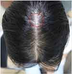
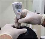
Before performing a one-time biophysical measurement, all subjects were informed to avoid scalp contact with water within three hours and any hair and scalp products including shampoo within 12 hours before the measurements. The measurements were performed in a controlled environment with relative humidity of 40–60% and temperature of 20–22°C. All subjects underwent acclimatization in this environment for 15 minutes before the measurements. The midline of the vertex area (Figure 1) was chosen for measurement in both groups. Scalp hydration values were measured using a Corneometer® CM825 (Courage + Khazaka, Koln, Germany) (Figure 2) and scalp pH values were measured using a Skin & Scalp pH Tester (Hanna Instruments® HI981037, Rumania) (Figure 3). Three readings with sequential waiting time of five seconds were obtained from the same area. The mean values were calculated from these readings as the final result.
 |
Figure 1 Measurement area on the midline of the vertex area. (Red circle). |
 |
Figure 2 Scalp hydration values were measured using a Corneometer® CM825. (a) The probe was placed vertically. (b) The monitor showed the scalp hydration values. |
 |
Figure 3 Scalp pH values were measured using a Skin & Scalp pH Tester Hanna Instruments® HI981037. |
Ethics
This study was conducted in accordance with the principles of the Declaration of Helsinki. The study protocol and ethical clearance were obtained from the Health Research Ethics Committee of Dr. Hasan Sadikin General Hospital, Bandung, Indonesia (ethical clearance number: LB.02.01/x.6.5/164/2022). All subjects were informed of the purpose of the study, and informed consent was obtained prior to their participation in the study. Data confidentiality were strictly maintained and the subjects were informed that they had the right to withdraw from the study at any time.
Statistical Analysis
The obtained data were assessed for normality using the Kolmogorov–Smirnov test, which revealed that scalp hydration (p=0.200) and pH values (p=0.054) had normal data distributions. Descriptive statistics for numerical variables are presented as mean ± standard deviation, median, and minimum-maximum values. For categorical variables, descriptive statistics are presented as frequencies and percentages. Differences in the distribution of proportions related to age, education level, and occupation between the two study groups were assessed using the chi-square test. Confounding factors, including hijab material, number of garment layers, inner hijab, use of other head cover, work environment, shampooing frequency, as well as use of other hair and scalp care products were adjusted using multiple linear regression analysis. An independent t-test was used to compare the scalp hydration and pH values between hijab-wearing and non-hijab-wearing women. All data were analyzed using the Statistical Package for Social Sciences (SPSS) version 24.0 (IBM Statistics, Chicago), with p<0.05 considered statistically significant.
Results
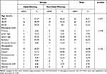
The participants were mostly 30–39 years old (41.27%), with the mean age of hijab-wearing women and non-hijab-wearing women being 35 and 31 years old, respectively. The most common education level was bachelor’s degree (46.03%), and the most common occupation were employee (30.30%) in hijab-wearing women and doctors (40%) in non-hijab-wearing women. The characteristics of the study participants are summarized in Table 1. Chi-square test showed that there was no significant difference between both groups in terms of demographic characteristics, such as age, education level, and occupation.
 |
Table 1 General Characteristics of Study Subjects |
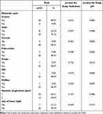
The potential confounding variables considered in this study were hijab material, the number of garment layers, use of inner hijab and other head cover, work environment, shampooing frequency, as well as the use of other hair and scalp care products. Most hijab-wearing women in this study wore cotton-fabric (90.91%) and one-layered (60.61%) hijab without inner hijab (78.79%), as shown in Table 2. Most subjects only used a helmet as another head cover with the duration of 15 (4.76%), 30 (7.94%), and 60 (17.46%) minutes. The use of hat, headcap, and other head covers was not identified. Both hijab-wearing and non-hijab-wearing women in this study worked mostly indoor without air conditioning (AC) (65.08%), rather than in rooms with AC (23.81%) or outdoor (11.11%). The use of other head covers and work environments of the subjects in this study are shown in Table 3. The shampooing frequency of hijab-wearing women was mostly 2–3 times weekly (42.42%), less often than that of non-hijab-wearing women, who washed their hair with shampoo mostly every day (40%). The most common hair and scalp care products used by both groups were hair conditioners (38.10%), followed by hair lotions (11.11%), dyes (11.11%), and tonics (4.76%). Descriptions of the hair care habits in both groups are presented in Table 4.
 |
Table 2 The Effect of Hijab Materials, the Number of Garment Layers, and the Use of Inner Hijab on Scalp Hydration and pH Value Among Hijab-Wearing Women |
 |
Table 3 The Effect of Head Cover Usage and Work Environment on Scalp Hydration and pH Value Among Hijab-Wearing and Non-Hijab-Wearing Women |
 |
Table 4 The Effect of Scalp Care Habit on Scalp Hydration and pH Value Among Hijab-Wearing and Non-Hijab-Wearing Women |
As shown in Tables 2–4, multiple linear regression showed that scalp hydration and pH values in this study were not significantly affected by hijab materials, the number of garment layers, the use of inner hijab and other head cover, work environment, shampooing frequency, or the use of other hair and scalp care products, except hair tonics. Thus, bivariate analysis using an independent t-test was performed to compare scalp hydration and pH values between hair tonic users and non-hair tonic users. Data analysis showed that there was no significant effect of hair tonic usage on scalp hydration or pH values in this study (p=0.259). Adjustment for confounding factors showed that hijab material, the number of garment layers, the use of inner hijab and other head cover, work environment, shampooing frequency, as well as the use of other hair and scalp care products had no significant impact on the results of this study.
The differences of scalp hydration and pH values between both groups are shown in Table 5. Despite the findings of higher scalp hydration (18.34 AU ± 2.91 AU vs 17.71 AU ± 3.35 AU) and pH (4.93 ± 0.17 vs 4.91 ± 0.16) in hijab-wearing women compared to non-hijab-wearing women, there was no significant difference based on the analysis using unpaired t-test (p>0.05).
 |
Table 5 Differences in Scalp Hydration and pH Value Between Hijab-Wearing and Non-Hijab-Wearing Women |
Discussion
There were no significant differences between hijab-wearing and non-hijab-wearing women in this study in terms of age, education level, and occupation. Thus, the confounding factors, including hijab material, the number of garment layers, the use of inner hijab and other head cover, work environment, shampooing frequency, as well as the use of other hair and scalp care products, were adjusted using multiple linear regression analysis. The analysis showed that all of those confounding factors did not have significant impact on scalp hydration and pH values. In this study, the effect of these confounding factors was also minimized by acclimatization and avoiding contact with water within 3 hours as well as hair and scalp care products within 12 hours prior to the biophysical measurements.
Hatch et al38 reported that the upper back skin hydration values among hijab coated with cotton, polyester 1.5 denier, and polyester 3.5 denier fibers were not significantly different in the dry state. In the wet state, the skin hydration values were significantly different, with cotton group having the lowest value and polyester 3.5 denier fibers having the highest.38 The effects of hijab material, the number of garment layers, and the use of inner hijab toward the results of this study were adjusted by ensuring that the subjects’ scalp were in dry state for three hours prior to the biophysical examination.
A study by Park et al36 showed that wearing surgical masks, Korean filter (KF)-anti-droplet (AD), KF80 or KF94 for at least four hours during five working days for three months increased the mean skin hydration values by approximately 9.5% on the cheeks, by 9.08% on the perioral area, and by 3.87% on the chin. Ghane et al39 reported that in most cases the pH values of the hands started to increase significantly within four hours after occlusion with impermeable gloves. The use of other head covers in this study did not significantly affect the scalp hydration and pH values, because the duration of head coverage was no more than an hour a day.
Sato et al40 which studied the effect of relative humidity towards skin barrier function, classified the relative humidity as dry (<10%), normal (40–70%), and humid (>80%). Changes from humid to dry environments increased transepidermal water loss (TEWL) by 6 to 7 folds. However, there was no increase of TEWL in response to the change from normal to dry environments. When TEWL increased significantly, neither skin hydration nor the pH values changed significantly. This study demonstrated that sudden changes from a high- to a low-humidity environment result in abnormal barrier function.40 Thus, acclimatization with humidity of 40–60% and temperature of 20–22°C for 15 minutes reduced the effect of work environment on the results of this study.
Lambers et al41 found that the significantly increased pH values immediately after hand washing with alkaline soap (pH 6) and tap water (pH 8) started to decrease after two hours and returned to the prior values after six hours. The effect of shampooing frequency and the use of other scalp care products, hijab material, the number of garment layers, and the use inner hijab in this study were adjusted by avoiding contact with shampoo as well as other hair and scalp care products for 12 hours prior to the biophysical examination.
The skin is divided into two main structural compartments: the epidermis or epithelial component coating the surface, and the in-depth dermis or connective component of nutrition. Although the whole skin structure actively participates in host defenses, the epidermis is important in preventing the loss of water and other components from the body to the environment,42 by producing a protective sheath, referred to as stratum corneum (SC).43 Skin hydration values reflect the water content of the SC, whereas TEWL represents the diffusion of condensed water through the SC.44 In non-occluded condition, there is an inverse relationship between skin hydration and TEWL values of the epidermis.45 Higher hydration46 and lower TEWL47 values are regarded as indicators of a healthy physical skin barrier,46,47 whereas lower hydration48,49 and higher TEWL47 values seem to be associated with skin barrier dysfunction, as found in some skin diseases, namely psoriasis, atopic dermatitis, and seborrheic dermatitis.47–49 Healthy physical skin barrier might maintain skin acidity, which is indicated by low pH value. Likewise, skin acidity might support the hydrolytic enzyme functions in the formation of SC, particularly the lipophilic components.50–52 High skin hydration values were found to correlate not only with low TEWL but also low pH.53
Skin occlusion may cause cutaneous water diffusion blockade, leading to SC cells swelling and elevation of skin hydration values. Excessive accumulation of water in the SC is often considered to harm the skin barrier function by causing destruction of the lamella/ultrastructure of SC lipids, prevention of lipid organization recovery, and widening of the intercellular space.29 These conditions increase skin permeability and is characterized by both TEWL and skin pH elevation.23,29 A study by Fluhr et al54 which observed volar forearm skin hydration and TEWL elevation after occlusion with a plastic chamber for 24, 48, 72, and 96 hours, concluded that occlusion induced barrier damage without causing skin dryness. Thus, both hydration and TEWL should be considered when assessing the physical skin barrier comprehensively.
This study found no significant difference in terms of either scalp hydration or pH values between hijab-wearing and non-hijab-wearing women. The unique characteristics of the scalp, which contains hair follicles alongside the dense sebaceous glands, best explains why scalp occlusion was not significantly impactful on the skin barrier function.
Scalp hair is useful for thermoregulation in adapting to the increasing skin temperature, which could be caused by occlusion.14,31–33,55 Sweat production is a response of eccrine glands toward the increase of skin temperature, which could increase skin hydration.44,56–60 A study by Lasisi et al57 showed that scalp hair could reduce the number of sweats needed for skin thermoregulation toward the temperature increase. Moreover, this study also found that tightly curled subjects sweated the least while straight-hair types sweated the most. Cabanac et al61 reported that people with bald scalp tended to sweat two to three times more than those with hairy scalp. Kleesz et al62 which studied a normal population, reported that the hairy scalp had the lowest hydration values compared to other anatomical locations, such as the hand, elbow, forehead, cheek, armpits, back, foot, as well as ventral and dorsal surfaces of the lower arm. A study by O’Goshi et al63 found that skin hydration values in alopecia areata and androgenetic patients were significantly higher than those of the volar surface of the lower arm. This phenomenon reflects the vital role of hair in thermoregulation, which explains the non-significant differences in skin hydration values between hijab-wearing and non-hijab-wearing women.
The sebaceous gland is a sebum-producing component of the pilosebaceous unit.63,64 The scalp is a body part with the densest sebaceous and sebum glands, in addition to the face, chest, and back.54,56,65,66 Boelsma et al67 stated that there was a statistically significant negative correlation between sebum level and skin pH on the volar surface of the lower arm. Jung et al68 also stated that there was a statistically significant correlation between improved sebum levels and reduction in pH among Asian women, and vice versa. Etnawati et al69 stated that the sebum level in hijab-wearing women was not significantly different from that in non-hijab-wearing women. Sebum is degraded by lipase into free fatty acids, which serve as regulators of skin acidity with pH level of 4.5–5.5.70–73 The pH level in this study was in similar range, which was 4.73–5.39 in the hijab-wearing group and 4.68–5.18 in the non-hijab-wearing group, with no statistically significant difference. The results of this study, along with those of previous studies, showed that a high sebum level on the hairy scalp was important for maintaining the acidity of the scalp skin, including an occluded, hairy scalp.
Limitations
This study could assess the barrier function more comprehensively if TEWL was included as a parameter in the biophysical measurements. Moreover, data on shampoo and hair type, which could be considered as confounding factors, were not obtained in this study.
Conclusion
There was no statistically significant difference observed in terms of scalp hydration and pH values between hijab-wearing and non-hijab wearing women. The use of hijab, therefore, did not have a significant impact on scalp barrier function in women. As long as the hijab-wearing women do not have any underlying scalp and skin disorders, and do not wear hijab in wet condition, they should not worry about scalp barrier impairment. Adding TEWL as a measurement parameter as well as the subjects’ shampoo and hair type in further study would make the results more convincing.
Consent Statement
The authors certify that they have obtained all appropriate subject consent forms. The subjects signed a consent form for the publication of study details and images.
Acknowledgments
The authors would like to thank the staff of the Department of Dermatology and Venereology, Faculty of Medicine, Universitas Padjadjaran, and Dr. Hasan Sadikin General Hospital, Bandung, West Java, Indonesia.
Disclosure
The authors report no conflicts of interest in this work.
References
1. Kawaguchi M, Kato H, Matsuo M. CT and MRI features of scalp lesions. La Radiol Med. 2019;124(10):1049–1061. doi:10.1007/s11547-019-01060-6
2. Ellis H, Mahadevan V. The surgical anatomy of the scalp. Surgery. 2014;32(1):1–5. doi:10.1016/J.MPSUR.2013.04.024
3. Tse KM, Lim SP, Tan VBC, Lee HP. A review of head injury and finite element head models. Am J Engin Tech Soc. 2014;1(5):28–52.
4. Kim SH, Shin SH, Kim SN, Na YJ. Understanding the characteristics of the scalp for developing scalp care products. J Cosmet Sci App. 2021;11:204–216. doi:10.4236/jcdsa.2021.113018
5. Trotta A, Annaidh AN. Mechanical characterisation of human and porcine scalp tissue at dynamic strain rates. J Mech Behavior Biomed Mater. 2019;100:1–11. doi:10.1016/j.jmbbm.2019.103381
6. Turner GA, Hoptroff M, Harding CR. Stratum corneum dysfunction in dandruff. Int J Cosmet Sci. 2012;34(4):298–306. doi:10.1111/j.1468-2494.2012.00723.x
7. Kim JH, Kim MG, Jeong SH, Kim HJ, Son WS. STAT3 maintains skin barrier integrity by modulating SPINK5 and KLK5 expression in keratinocytes. Exp Dermatol. 2022;31(2):223–232. doi:10.1111/exd.14445
8. Yosipovitch G, Misery L, Proksch E, Metz M, Stander S, Schmelz M. Skin barrier damage and itch: review of mechanisms, topical management, and future directions. Acta Derm Venereol. 2019;99(13):1201–1209. doi:10.2340/00015555-3296
9. Plotczyk M, Higgins CA. Skin biology. In: Gareta EG, editor. Biomaterials for Skin Repair and Regeneration. Philadelphia: Elsevier; 2019:3–25.
10. Takagi Y, Takatoku H, Terazaki H, Nakamura T, Ishida K, Kitahara T. The scalp has a lower stratum corneum function with a lower sensory input than other areas of the skin evaluated by the electrical current perception threshold. Cosmet. 2015;2(4):384–393. doi:10.3390/cosmetics2040384
11. Suchonwanit P, Triyangkulsri K, Ploydaeng M, Leerunyakul K. Assessing biophysical and physiological profiles of scalp seborrheic dermatitis in the Thai population. Bio Med Res Intern. 2019;2019:1–7. doi:10.1155/2019/5128376
12. Schwartz JR, Henry JP, Kerr KM, Mizoguchi H, Li L. The role of oxidative agent in poor Scalp health: ramifications to causality and associated hair growth. Int Journal Cosmet Sci. 2015;37(2):9–15. doi:10.1111/ics.12289
13. Trueb RM, Henry JP, Davis MG, Schwartz JR. Scalp condition impacts hair growth and retention via oxidative stress. Int J Trichol. 2018;10:262–270. doi:10.4103/ijt.ijt_57_18
14. Choi BY. Hair-growth potential of ginseng and its major metabolites: a review on its molecular mechanisms. Int J Mol Sci. 2018;19(9):1–13. doi:10.3390/ijms19092703
15. Gupta AK, Bamimore MA, Foley KA. Efficacy of non-surgical treatment for androgenetic alopecia in men and women: a systematic review with network meta-analysis, an assessment of evidence quality. J Dermatol Treat. 2022;33(1):1–35. doi:10.1080/09546634.2020.1749547
16. Aguh C, Jhaveri M, He A, Okoye GA, Cohen BE, Elbuluk N. Ethnic hair consideration for people of African, South Asian, Muslim, and Sikh origins. In: Aguh C, Okoye GA, editors. Fundamentals of Ethnic Hairs. Cambridge: Springer; 2016:137–149.
17. Boucher NA, Siddiqui EA, Koenig HG. Supporting Muslim patients during advanced illness. Perm J. 2017;21(3):16–19. doi:10.7812/TPP/16-190
18. Direktorat Jenderal Kependudukan dan Pencatatan Sipil. Sebanyak 86,93% penduduk Indonesia beragama Islam pada 31 desember 2021. Kementerian Dalam Negeri; 2021. Available from: https://databoks.katadata.co.id/7fd91eea-e2d0-4f94-a959-62a4517070c8.
19. Arifah L, Sobari N, Usman H. Hijab phenomenon in Indonesia: does religiosity matter? In: Gani L, Gitaharie BY, Husodo ZA, Kuncoro A, editors. Competition and Cooperation in Economics and Business. London: Taylor & Francis Group; 2017:179–186.
20. Yen TS, Lee GP. A conceptual paper on modest wear for Malaysia Muslim women in contemporary hijab fashion. J Islamic Soc Eco Develop. 2018;3(8):41–49.
21. Yarza HN, Mayarni RRF, Elvianasti M, Kurnia LM, Suci AR. Study international conference of education on science, technology. Engi Math. 2020;2020:1–7.
22. Araya M, Kulthanan K, Jiamton S. Clinical characteristics, and quality of life of seborrheic dermatitis patients in a tropical country. Indian J Dermatol. 2015;60(5):519–525. doi:10.4103/0019-5154.164410
23. Misery L, Rahhali N, Duhamel A, Taieb C. Epidemiology of dandruff, scalp pruritus and associated symptoms. Acta Derm Venereol. 2011;93(1):80–81. doi:10.2340/00015555-1315
24. Katoulis AC, Christodoulou C, Liakou AI, et al. Quality of life and psychosocial impact of scarring and non-scarring alopecia in women. J Dtsch Dermatol Ges. 2015;13(2):137–142. doi:10.1111/ddg.12548
25. Zsiko S, Csanyi E, Kovacs A, Szucs MB, Gacsi A, Berko S. Methods to evaluate skin penetration in vivo. Sci Pharm. 2019;87(19):1–21. doi:10.3390/scipharm87030019
26. Hafeez F, Maibach H. Occlusion effect on in vivo percutaneous penetration of chemicals in man and monkey partition coefficient effects. Skin Pharmacol Physiol. 2013;26(2):85–91. doi:10.1159/000346273
27. Chen Y, Feng X, Meng S. Site-specific drug delivery in the skin for the localized treatment of skin diseases. Expert Opin Drug Deliv. 2019;16(8):847–867. doi:10.1080/17425247.2019.1645119
28. Sun Q, Stantchev RI, Wang J, et al. In vivo estimation of water diffusivity in occluded human skin using terahertz reflection spectroscopy. J Biophoton. 2019;12(2):1–9. doi:10.1002/jbio.201800145
29. Choe C, Schleusener J, Choe S, Ri J, Lademann J, Darvin ME. Stratum corneum occlusion induces water transformation towards lower bonding state a molecular level in vivo study by confocal Raman microspectroscopy. Int J Cosmet Sci. 2020;42:482–493. doi:10.1111/ics.12653
30. Safuan H, Towers IN, Jovanosfski Z, Sidhu SH. A simple model for the total microbial biomass under occlusion of healthy human skin. In: 19th International Congress on Modelling and Simulation. Perth: Modelling and Simulation Society of Australia and New Zealand; 2011:733–739.
31. Faergemann J, Aly R, Wilson DR, Maibach HI. Skin occlusion: effect on Pityrosporum orbiculare, skin PCO2, pH, transepidermal water loss, and water content. Arch Dermatol Res. 1983;275(6):383–387. doi:10.1007/BF00417338
32. Aly R, Shirley C, Cunico B, Maibach HI. Effect of prolonged occlusion on the microbial flora, pH, carbon dioxide and transepidermal water loss on human skin. J Invest Dermatol. 1978;71(6):378–381. doi:10.1111/1523-1747.ep12556778
33. Elias PM, Ansel JC, Woods LD, Feingold KR. Signaling networks in barrier homeostasis: the mystery widens. Arch Dermatol. 1996;132(12):1505–1506. doi:10.1001/archderm.1996.03890360097017
34. Elias PM, Woods LD, Feingold KR. Epidermal pathogenesis of inflammatory dermatoses. Am J Contact Dermat. 1999;10(3):119–126.
35. Denda M, Sato J, Tsuchiya T, Elias PM, Feingold KR. Low humidity stimulates epidermal DNA synthesis and amplifies the hyperproliferative response to barrier disruption: implications for seasonal exacerbations of inflammatory dermatoses. J Invest Dermatol. 1998;111:873–878. doi:10.1046/j.1523-1747.1998.00364.x
36. Park SR, Han JY, Yeon YM, Kang NY, Kim EJ, Suh BF. Long-term effects of face masks on skin characteristics during the COVID-19 pandemic. Skin Res Technol. 2022;28(1):153–161. doi:10.1111/srt.13107
37. Martini L. A precious chance for Muslim hijab women of all the world to keep their hair scalp safe and not to incur praecox alopecia. Our Dermatology Online. 2016;7(3):284. doi:10.7241/ourd.20163.76
38. Hatch KL, Markee NL, Maibach HI, Barker RL, Woo SS, Radhakrishnaiah P. In vivo cutaneous and perceived comfort response to fabric. Part III: water content and blood flow in human skin under garments worn by exercising subjects in a hot, humid environment. Textile Res J. 1990;60(9):510–519. doi:10.1177/004051759006000904
39. Ghane Y, Heese A, Martus P, et al. Veränderungen von Haut-pH und TEWL infolge Okklusion durch medizinische Einmalhandschuhe. Hautarzt. 1997;48(1):71.
40. Sato J, Denda M, Chang S, Elias PM, Feingold KR. Abrupt decreases in environmental humidity induce abnormalities in permeability barrier homeostasis. Soc Invest Dermatol. 2022;110(4):900–904. doi:10.1046/j.1523-1747.2002.00589.x
41. Lambers H, Piessens S, Bloem A, Pronk H, Finkel P. Natural skin surface pH is on average below 5, which is beneficial for its resident flora. Int J Cosmet Sci. 2006;28(5):359–370. doi:10.1111/j.1467-2494.2006.00344.x
42. Baroni A, Buommino E, De Gregorio V, Ruocco E, Ruocco V, Wolf R. Structure and function of the epidermis related to barrier properties. Clin Dermatol. 2012;30(3):257–262. doi:10.1016/j.clindermatol.2011.08.007
43. Tagami H, Kanamaru Y, Inoue K, Sueshia S, Inoue F, Iwatsuki K. Water sorption-desorption test of the skin in vivo assessment of stratum corneum. J Invest Dermatol. 1982;78(5):425–428. doi:10.1111/1523-1747.ep12507756
44. Han SH, Shin SH, Park JH, Li K, Kim BJ, Yoo KH. Changes in characteristics after using respiratory protective equipment (medical masks and respirators) in the COVID-19 pandemic among healthcare workers. Contact Dermatitis. 2021;85(2):225–232. doi:10.1111/cod.13855
45. Polanska A, Pazdrowska AD, Silny W, Jenerowitz D, Mankowska AO, Hrab KO. Evaluation of selected skin barrier functions in atopic dermatitis in relation to the disease severity and pruritus. Postep Derm Alergol. 2012;29(5):373–377. doi:10.5114/pdia.2012.31492
46. Danby SG, Chalmers J, Brown K, Williams HC, Cork MJ. A functional mechanistic study of the effect of emollients on the structure and function of the skin barrier. Br J Dermatol. 2016;175(5):1011–1019. doi:10.1111/bjd.14684
47. Akdeniz M, Gabriel S, Lichterfeld-Kottner A, Blume-Peytavi U, Kottner J. Transepidermal water loss in healthy adults: a systematic review and meta-analysis update. Br J Dermatol. 2018;179(5):1049–1055. doi:10.1111/bjd.17025
48. Morales DM, Vilchez TM, Santiago SA. Study of skin barrier function in psoriasis: the impact of emollients. Life. 2021;11(651):1–10. doi:10.3390/life11070651
49. Bravo AG, Vilchez TM, Santiago SA, Eisman AB. The effect of sunscreens on the skin barrier. Life. 2022;12(2083):1–14. doi:10.3390/life12122083
50. Rippke F, Schreiner V, Schwanitz HJ. The acidic milieu of the horny layer–new findings on the physiology and pathophysiology of skin pH. Am J Clin Dermatol. 2002;3(4):261–272. doi:10.2165/00128071-200203040-00004
51. Forestier JP. Les enzymes de l’espace extra-cellulaire du stratum corneum. Int J Cosmet Sci. 1992;14(2):47–63. doi:10.1111/j.1467-2494.1992.tb00039.x
52. Redoules D, Tarroux R, Perie J. Epidermal enzymes: their role in homeostasis and their relationship with dermatoses. Skin Pharmacol Appl Skin Physiol. 1998;11(4–5):183–192. doi:10.1159/000029827
53. Luebberding S, Krueger N, Kerscher M. Age-related changes in skin barrier function–Quantitative evaluation of 150 female subjects. Inter J Cosmet Sci. 2013;35(2):183–190. doi:10.1111/ics.12024
54. Mokos ZB, Krajl M, Juzbasic AB, Jukic IL. Seborrheic dermatitis: an update. Acta Dermatovenereol Croat. 2012;20(2):98–104.
55. Fluhr JW, Lazzerini S, Distante F, Gloor M, Berardesca E. Effects of prolonged occlusion on stratum corneum barrier function and water holding capacity. Skin Pharmacology and Applied Skin Physiology. 1999;12(4):193–198. doi:10.1159/000066243
56. Mustika A, Kusuma M, Nasution LH. The correlation between sebum levels and pityriasis versicolor. Bali Med J. 2021;10(3):1015–1019. doi:10.15562/bmj.v10i3.2859
57. Lasisi T, Smallcombe JW, Kenney WL, et al. Human scalp hair as thermoregulatory adaptation. BioRxiv. 2022;25(1):1–15. doi:10.1101/2023.01.21.524663
58. Kim S, Park JW, Yeon Y, Han JY, Kim E. Influence of exposure to summer environments on skin properties. J Eur Acad Dermatol Venereol. 2019;33(11):2192–2196. doi:10.1111/jdv.15745
59. Baker LB, Wolfe AS. Physiological mechanism determining eccrine sweat composition. Eur J Appl Physiol. 2020;120(4):719–752. doi:10.1007/s00421-020-04323-7
60. du Plessis J, Stefaniak AB, Ellof F, et al. International guidelines for the in vivo assessment of skin properties in non-clinical settings: part 2. transepidermal water loss and skin hydration. Skin Res Tech. 2013;19(3):263–278. doi:10.1111/srt.12037
61. Cabanac M, Brinnel H. Beards, baldness, and sweat secretion. Eur J Appl Physiol. 1988;58(1–2):39–46. doi:10.1007/BF00636601
62. Kleesz P, Darlenski R, Fluhr JW. Full-body skin mapping for six biophysical parameters: baseline values at 16 anatomical sites in 125 human subjects. Skin Pharmacol Physiol. 2012;25(1):25–33. doi:10.1159/000330721
63. O’goshi K, Iguchi M, Tagami H. Functional analysis of the stratum corneum of scalp skin: studies in patient with alopecia areata and androgenetic alopecia. Arch Dermatol Res. 2000;292(12):605–611. doi:10.1007/s004030000185
64. Shamloul G, Khachemoune A. An update review of the sebaceous gland and its role in health and diseases. Part I: embryology, evolution, structure, and function of sebaceous glands. Dermatol Ther. 2021;34(1):1–4. doi:10.1111/dth.14695
65. Shamloul G, Khachemoune A. An update review of the sebaceous gland and its role in health and diseases. Part II: pathophysiological clinical disorders of sebaceous glands. Dermatol Ther. 2021;34(2):1–8. doi:10.1111/dth.14862
66. Tao R, Li R, Wang R. Ski microbiome alterations in seborrheic dermatitis dandruff: a systematic review. Exp Dermatol. 2021;30(10):1546–1553. doi:10.1111/exd.14450
67. Boelsma E, van de Vijver LPL, Goldbohm RA, et al. Human skin condition and its association with nutrient concentration in serum and diet. Am J Clin Nutr. 2003;77(2):348–355. doi:10.1093/ajcn/77.2.348
68. Jung YC, Kim EJ, Cho JC, Suh KD, Nam GW. Effect of skin pH for wrinkle formation on Asian: Korean, Vietnamese, and Singaporean. J Eur Acad Dermatol Venereol. 2012;27(3):1–3. doi:10.1111/j.1468-3083.2012.04516.x
69. Etnawati K, Siswati AS, Pudjiati SR, Susetiati DA, Adwinarni DR, Purbananto A. The role of Malassezia sp., sebum level, and transepidermal water loss toward the dandruff severity between hijab and non hijab wearing subjects. J Med Sci. 2018;50(3):329–334. doi:10.19106/JMedScie/0050032018011
70. Tončić RJ, Kezić S, Hadžavdić SL, Marinović B. Skin barrier and dry skin in the mature patient. Clin Dermatol. 2018;36(2):109–115. doi:10.1016/j.clindermatol.2017.10.002
71. Shetewi T, Finnegan M, Fitzgerald S, Xu S, Duffy E, Morrin A. Investigation of the relationship between skin-emitted volatile fatty acids and skin surface acidity in healthy subjects–a pilot study. J Breath Res. 2021;15(3):1–12. doi:10.1088/1752-7163/abf20a
72. Thaifa SK, Arun Kumar KV. Formulation of anti-dandruff herbal shampoo containing datura metel (Linn) loaded solid lipid nanoparticles. World J Pharmaceut Res. 2017;6(16):599–619. doi:10.20959/wjpr201716-10151
73. Egert M, Simmering R, Riedel CU. The association of the skin microbiota with health, immunity, and disease. Clin Pharmacol Ther. 2017;102(1):62–69. doi:10.1002/cpt.698
 © 2023 The Author(s). This work is published and licensed by Dove Medical Press Limited. The full terms of this license are available at https://www.dovepress.com/terms.php and incorporate the Creative Commons Attribution - Non Commercial (unported, v3.0) License.
By accessing the work you hereby accept the Terms. Non-commercial uses of the work are permitted without any further permission from Dove Medical Press Limited, provided the work is properly attributed. For permission for commercial use of this work, please see paragraphs 4.2 and 5 of our Terms.
