Back to Journals » International Journal of General Medicine » Volume 15
Evaluation of Left Diastolic Function in Dilated Cardiomyopathy According to the 2016 ASE/EACVI Recommendations
Authors Pham QT , Tran TN, Le-Thi TT, Phan AK , Nguyen AV
Received 20 January 2022
Accepted for publication 19 April 2022
Published 30 April 2022 Volume 2022:15 Pages 4527—4533
DOI https://doi.org/10.2147/IJGM.S359248
Checked for plagiarism Yes
Review by Single anonymous peer review
Peer reviewer comments 4
Editor who approved publication: Dr Scott Fraser
Quang Tuan Pham,1 Thua Nguyen Tran,2 Thanh Thuy Le-Thi,1 Anh Khoa Phan,3 Anh Vu Nguyen4
1Cardiology Department, Hue Central Hospital, Hue City, Vietnam; 2Department of General Internal Medicine and Geriatrics, Hue Central Hospital, Hue City, Vietnam; 3Emergency Department of Cardiovascular Intervention, Hue Central Hospital, Hue City, Vietnam; 4Cardiovascular Center, Hospital of University of Medicine and Pharmacy, Hue City, Vietnam
Correspondence: Thua Nguyen Tran, Department of General Internal Medicine and Geriatrics, Hue Central Hospital, Hue City, Vietnam, Tel +84 903597695, Email [email protected]
Objectives: To assess left ventricular diastolic function by using echocardiography in patients with dilated cardiomyopathy, and the relationship between left ventricular diastolic function and left ventricular dilatation, New York Heart Association (NYHA) heart failure index, left ventricular ejection fraction, and left ventricular fractional shortening.
Methods: A descriptive cross-sectional study was conducted on patients with primary dilated cardiomyopathy hospitalized in Hue Central Hospital from April 2018 to August 2020.
Results: The mean end-diastolic left ventricular volume was 133.57± 31.58 mL and the mean end-systolic left ventricular volume was 99.9± 26.03 mL. The mean left atrial volume was 61.63± 27.13 mL. The mean end-diastolic and end-systolic left ventricular diameters were 66.11± 7.3 mm and 57.7± 8.02 mm, respectively. The mean left ventricular ejection fraction was 24.68± 5.97%. The mean left ventricular fractional shortening was 12.91± 4.55%. The highest rate was grade II diastolic dysfunction (44.6%), followed by grade III diastolic dysfunction (35.8%) and grade I diastolic dysfunction at 19.6%. There was a moderate positive correlation between the left ventricular diastolic dysfunction and the NYHA class of heart failure with r=0.445, p< 0.001. All dilated cardiomyopathy patients in the study group had mainly grade II–III severe diastolic dysfunction.
Conclusions: Routine evaluation of diastolic function in patients with heart failure can help in elucidation of pathogenesis and management of patients. This dysfunction was clearly demonstrated by the change in the parameters of the evaluation of left ventricular diastolic function on echocardiography according to the 2016 ASE/EACVI recommendations, a new recommendation introduced to approach the assessment of diastolic function in a more convenient and easier way.
Keywords: dilated cardiomyopathy, left ventricular diastolic dysfunction
Introduction
Dilated cardiomyopathy (DCM) is a disease of the heart muscle characterized by enlargement and dilation of one or both of the ventricles along with impaired contractility defined as left ventricular ejection fraction (LVEF) less than 40%.1 By definition, patients have systolic dysfunction and may or may not have overt symptoms of heart failure. A large number of patients with DCM may have a long latent period where they are clinically asymptomatic. When symptoms do arise, they are the result of LV systolic dysfunction.
Recent studies demonstrate that diastolic impairment predicts mortality in HFrEF independent of LVEF.2,3 The principal focus in congestive heart failure is on the systolic function of the heart, especially in dilated cardiomyopathy patients. The prognosis of patients suffering from diastolic failure is as grave as those with systolic failure.2 In the world, there have been many studies on dilated cardiomyopathy but not many studies on left ventricular diastolic function. Experts around the world have made many recommendations on assessing left ventricular diastolic function, in which most recent recommendation is the 2016 ASE/EACVI guideline.4 Compared with the 2009 EAE/ASE guideline the 2016 ASE/EACVI recommendation on assessing left ventricular diastolic function has fewer parameters. It is, therefore, easier to perform and more convenient in clinical practice.5 The aim of this study is to assess left ventricular diastolic function according to the 2016 ASE/EACVI recommendation in patients with dilated cardiomyopathy by using echocardiography; and determine the relationship between left ventricular diastolic function and left ventricular dilatation, NYHA heart failure, left ventricular ejection fraction, and left ventricular fractional shortening.
Materials and Methods
Study Population
The study was conducted on 56 patients with primary and secondary dilated cardiomyopathy hospitalized and treated at Hue Central Hospital from April 2018 to August 2020.
Inclusion criteria were patients with dilated cardiomyopathy according to European study group criteria: left ventricular systolic dysfunction (ejection fraction <45%, and/or fractional shortening <25%), LV end-diastolic volumes or diameters >2 SD from normal according to normograms (z-scores >2 SD) corrected for age and body surface area.6
Patients with atrial fibrillation, heart valve disease (calcified mitral valve, calcified aortic valve), coronary artery stenosis, congenital heart disease were excluded.
Doppler Echocardiogram Assessment
The patient has a more thorough Doppler echocardiogram to assess diastolic function according to the 2016 ASE/EACVI recommendation in patients with reduced EF and in patients with cardiomyopathy with normal EF:4
(i) If E/A ≤0.8+E ≤50 cm/s: grade I diastolic dysfunction, left atrial pressure or left ventricular filling pressure was normal.
(ii) If E/A ≤0.8+E >50 cm/s or 0.8 <E/A <2, combine three more sub-criteria: (a) mean E/e’ >14; (b) tricuspid regurgitation velocity >2.8 m/s; (c) left atrial volume index > 34 mL/m:2
- If less than two sub-criteria were met, that will be grade I diastolic dysfunction, normal left atrial pressure, or normal left ventricular filling pressure;
- If two or more of the following sub-criteria were met, that will be grade II diastolic dysfunction, increased left atrial pressure, or increased left ventricular filling pressure;
- In case there were only two criteria: if none of the criteria was met, then that will be grade I diastolic dysfunction, normal left atrial pressure or normal left ventricular filling pressure; if 1 criterion was met, the left atrial pressure or left ventricular filling pressure cannot be determined and the diastolic dysfunction cannot be determined; if two criteria were met, then that will be grade II diastolic dysfunction, increased left atrial pressure or increased left ventricular filling pressure.
(iii) If E/A ≥2: grade III diastolic dysfunction, increased left atrial pressure or increased left ventricular filling pressure.
Data Analysis
The data were analyzed using the SPSS software 19.0 version. Independent t-test, Spearman correlation analysis was used wherever appropriate. A p-value was less than 0.05 was considered to indicate statistical significance.
Results
Baseline Characteristics
Of 56 patients, there were 38 males (67.9%), the male to female ratio was 2:1. The mean age was 52.95 years (range: 18–65). Thirty-four patients had heart failure class III according to NYHA accounted for the highest rate (60.8%), followed by NYHA class IV and NYHA class II with the same rate of 19.6%. There were no patients with NYHA class I heart failure. Symptoms of dyspnea on exertion accounted for the highest rate (96.4%), symptoms of jugular vein distention accounted for the lowest rate (16.1%) (Table 1).
 |
Table 1 Clinical Symptoms of the Study Group |
Echocardiographic Parameters
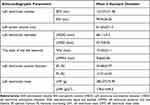
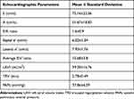
The parameters in Table 2 were measured on 2D echocardiography, showing significant changes in morphology and size of cardiac structures (left atrium, left ventricle) and left ventricular function. Table 3 shows the parameters evaluating left ventricular diastolic function on echocardiography.
 |
Table 2 Morphological Features on Routine Echocardiography (M Mode, 2D) |
 |
Table 3 Parameters Evaluating Left Ventricular Diastolic Function on Echocardiography |
Grades of Left Ventricular Diastolic Dysfunction
According to the 2016 ASE/EACVI recommendation on the assessment of left ventricular diastolic function, all patients with dilated cardiomyopathy in the present study had left ventricular diastolic dysfunction. Of these, grade II diastolic dysfunction (44.6%) and grade III (35.8%) accounted for the majority. Only nearly 1/5 of the patients were classified in grade I diastolic dysfunction.
The Relationship Between the Left Ventricular Diastolic Dysfunction Grade and the Parameters on Echocardiography
There was a significant difference in mean values among EF and FS with left ventricular diastolic dysfunction (p<0.05). There was no significant relationship between the grades of left ventricular diastolic dysfunction and age, left ventricular end-systolic and diastolic diameters, as well as left ventricular mass and left ventricular mass index (p>0.05) (Table 4). There was a moderate positive correlation between the grades of left ventricular diastolic dysfunction and the NYHA classification of heart failure (r=0.445) (Table 5).
 |
Table 4 Relationship of Left Ventricular Diastolic Dysfunction with Age and Parameters on Left Ventricular Echocardiography |
 |
Table 5 Correlation Between Grades of Left Ventricular Diastolic Dysfunction and NYHA Classification of Heart Failure |
Discussion
The present study based on a small cohort of dilated cardiomyopathy and extensively characterized provides insights into categorization and outcome implications of Doppler-derived diastolic measurements.
The left ventricular morphology changed markedly, manifested by increased left ventricular size increased significantly. This is because dilated cardiomyopathy is characterized by four large dilated chambers of the heart, which may be the major cause of left ventricular dilatation.1 The left atrial volume and the left atrial volume index increase in dilated cardiomyopathy, the common sign of dilatation is associated with other cardiac chambers, including the right ventricle, right atrium, and left atrium. Large left atrium in dilated cardiomyopathy reflects left atrial pressure overload caused by chronically elevated left ventricular diastolic filling pressures.7 Contraction of the left atrium plays an important role in left ventricular diastolic activity.8 In our study, we applied the left atrial volume according to the American Society of Echocardiography criteria in 2016.4 According to this criteria, the left atrial volume index significantly increases in diastolic dysfunction when the left atrial volume is ≥34 mL/m.2
On 2D echocardiography, we found reduced amplitude of mobility of all cardiac walls. This is quite a characteristic feature of dilated cardiomyopathy, which is different from ischemic cardiomyopathy. The thickness of the left ventricular walls of DCM population is normal or thin, which is the distinguishing feature from hypertrophic and restrictive cardiomyopathy.9,10
The parameters of ejection fraction (EF) and fractional shortening (FS) both decreased based on the data, the systolic function of the left ventricle is severely reduced, which is due to irreversible myocardial fiber damage. The myocardial fibers are enlarged and elongated, but their contractile forces are very weak, thus greatly affecting the contraction force of the heart.
Evaluation and classification of left ventricular diastolic function based on the 2016 ASE/EACVI recommendations for patients with reduced EF based on the following parameters. Mitral E/A ratio not applicable in AF/atrial flutter patients so we excluded patients with atrial fibrillation from the study for easy and accurate evaluation of the E/A ratio according to the 2016 ASE/EACVI recommendations. However, excluding patients with AF from the study makes the population unrepresentative to general DCM population with such low ejection fraction, where a large proportion of such patients have AF. Mean E/e’ ratio of lateral e’ and the mean value of septal e’ was lower than the values in normal people. The E/e’ ratio helps to assess that left ventricular filling pressure, which is also the left atrial pressure. If the E/e’ ratio was >14 and particularly >20, the mortality of DCM population in the long-term and even in the short-term was high, which is vital to give a high consideration for intensification of medical therapy and for considering new interventions.3
Through this, we found that the increased tricuspid regurgitation velocity indicates the degree of pulmonary hypertension. In the absence of pulmonary disease, increased systolic pulmonary artery pressure suggests increased left atrial pressure, increased blood stasis due to left heart failure. Systolic pulmonary artery pressure could be an auxiliary parameter of mean left atrial pressure. Evidence of pulmonary hypertension shows prognostic significance. Applying 2016 ASE/EACVI recommendations, we obtained the following results: 100% of patients with dilated cardiomyopathy had diastolic dysfunction, of which grade II diastolic dysfunction accounted for the largest proportion of 44.6%, grade I accounted for 19.6%, and grade III accounted for 35.8%.
There was no statistically significant relationship between the grades of left ventricular diastolic dysfunction and left ventricular end-diastolic diameter and end-systolic diameter (p>0.05). The characteristics of dilated cardiomyopathy can explain that the left ventricular dilatation predominates over other cardiac chambers. Although it is assumed that the dilated myocardial injury is diffused, diastolic diameter and systolic diameter are both increased significantly in the study groups; our data sample is not large enough to highlight the difference in left ventricular dilatation between groups of left ventricular diastolic dysfunction. There was a relationship between left ventricular diastolic dysfunction and parameters of ejection fraction (EF) and fractional shortening (FS) (p<0.005). Accordingly, the mean EF decreased gradually with the degree of left ventricular diastolic disorder. The characteristic feature of dilated cardiomyopathy is decreased movement of the heart walls leading to reduced left ventricular systolic function. Diastolic dysfunction also correlates with exercise intolerance.11 SGLT2 inhibitors improve the energetic status of the myocardium, this explains the fact that SGLT2 inhibitors improve diastolic function in dilated cardiomyopathy.12,13
There was a moderate positive correlation between the grades of left ventricular diastolic dysfunction and the NYHA class of heart failure with r=0.445 (p<0.001) (Table 5). Some data showed myocardial energetic impairment exists across a spectrum of heart failure of increasing clinical severity and worsening diastolic function. This highlights that an abnormal resting myocardial energetic state is related to impaired exercise responses of all four cardiac chambers.14 Thus, the more severe the patient’s heart failure was, the more diastolic dysfunction increases. Furthermore, our study shows that the more dilated cardiomyopathy patients are admitted to the hospital in a state of rather severe heart failure (NYHA class III–IV), the more severe the left ventricular diastolic dysfunction.
Our study at the time was a cross-sectional description, not yet eligible for follow-up, which was also the shortcoming of the study. We selected hospitalized patients because we wanted to investigate more closely the clinical symptoms and have better time for echocardiographic assessment of left ventricular diastolic function in these patients. We almost investigated our population with hospitalized scenarios with severe clinical symptoms as NYHA III and IV, which mean the heart decompensation was the main reason take the patient to the hospital. However, we have to bring them back to the recompensation before checking the echocardiography. Bedsides, we still considered this a limited study.
According to the 2016 ASE/EACVI recommendation, mitral E/A ratio are not applicable in AF/atrial flutter patients.4 AF patients are excluded from the study although the rhythm disturbance is highly prevalent in the studied population. Therefore, study patients do not represent general DCM population.
There are many relationships between NT-proBNP (a marker of congestion) and measurements of diastolic function. However, in this study, we only focus on studying the morphological characteristics of dilated cardiomyopathy and the parameters to evaluate diastolic function. A prospective planned study might throw more light on the relationship between diastolic function in DCM and biomarker NT-proBNP.
Conclusions
Our study therefore highlights the importance of often missed finding of diastolic parameter abnormalities in patients with heart failure especially in patients with dilated cardiomyopathy, a disease where people focus so much on systolic function. Routine evaluation of diastolic function in patients with heart failure can help in elucidation of pathogenesis and management of patients. Besides, the application of the 2016 ASE/EACVI recommendation on the assessment of left ventricular diastolic function in patients with fewer parameters, is easier to perform and more convenient in clinical practice. A prospective planned study might throw more light on the relationship between diastolic function in DCM and biomarker NT-proBNP (a marker of congestion).
Ethics Approval and Informed Consent
The study was conducted according to the guidelines of the Declaration of Helsinki and approved by the Institutional Review Board of Hue Central Hospital (Hue city, Vietnam). Written informed consent has been obtained from the patients to publish this paper.
Author Contributions
All authors made a significant contribution to the work reported, whether that is in the conception, study design, execution, acquisition of data, analysis and interpretation, or in all these areas; took part in drafting, revising or critically reviewing the article; gave final approval of the version to be published; have agreed on the journal to which the article has been submitted; and agree to be accountable for all aspects of the work.
Funding
This research received no external funding.
Disclosure
The authors report no conflicts of interest in this work.
References
1. Mahmaljy H, Yelamanchili VS, Singhal M. Dilated Cardiomyopathy. Treasure Island (FL): StatPearls; 2022.
2. Benfari G, Miller WL, Antoine C, Rossi A. Diastolic determinants of excess mortality in heart failure with reduced ejection fraction. JACC Heart Fail. 2019;7(9):808–817. doi:10.1016/j.jchf.2019.04.024
3. Xie GY, Berk MR, Smith MD, Gurley JC, DeMaria AN. Prognostic value of Doppler transmitral flow patterns in patients with congestive heart failure. J Am Coll Cardiol. 1994;24(1):132–139. doi:10.1016/0735-1097(94)90553-3
4. Nagueh SF, Smiseth Otto A, Appleton CP. Recommendations for the evaluation of left ventricular diastolic function by echocardiography: an update from the American Society of Echocardiography and the European Association of Cardiovascular Imaging. J Am Soc Echocardiogr. 2016;17(12):1321–1360.
5. Nagueh Sherif F, Appleton CP. Recommendations for the evaluation of left ventricular diastolic function by echocardiography. J Am Soc Echocardiogr. 2009;22(2):277–314.
6. Pinto YM, Elliott PM, Arbustini E, Adler Y. Proposal for a revised definition of dilated cardiomyopathy, hypokinetic non-dilated cardiomyopathy, and its implications for clinical practice: a position statement of the ESC working group on myocardial and pericardial diseases. Eur Heart J. 2016;37:1850–1858. doi:10.1093/eurheartj/ehv727
7. Parajuli P, Ahmed AA. Left Atrial Enlargement. Treasure Island (FL): StatPearls; 2022.
8. Berman MN, Tupper C, Bhardwaj A. Physiology, Left Ventricular Function. Treasure Island (FL): StatPearls; 2022.
9. Marian AJ, Braunwald E. Hypertrophic cardiomyopathy: genetics, pathogenesis, clinical manifestations, diagnosis, and therapy. Circ Res. 2017;121(7):749–770. doi:10.1161/CIRCRESAHA.117.311059
10. Wood MJ, Picard MH. Utility of echocardiography in the evaluation of individuals with cardiomyopathy. Heart. 2004;90(6):707–712. doi:10.1136/hrt.2003.024778
11. Skaluba SJ, Litwin SE. Mechanisms of exercise intolerance: insights from tissue Doppler imaging. Circulation. 2004;109(8):972–977. doi:10.1161/01.CIR.0000117405.74491.D2
12. García-Ropero A, Vargas-Delgado AP, Santos-Gallego CG, Badimon JJ. Inhibition of sodium glucose cotransporters improves cardiac performance. Int J Mol Sci. 2019;20(13):3289. doi:10.3390/ijms20133289
13. Santos-Gallego CG, Requena-Ibanez JA, Antonio RS. Empagliflozin ameliorates diastolic dysfunction and left ventricular fibrosis/stiffness in nondiabetic heart failure: a Multimodality Study. JACC Cardiovasc Imaging. 2021;14(2):393–407. doi:10.1016/j.jcmg.2020.07.042
14. Burrage MK, Hundertmark M, Valkovic L, Watson WD. Energetic basis for exercise-induced pulmonary congestion in heart failure with preserved ejection fraction. Circulation. 2021;144(21):1664–1678. doi:10.1161/CIRCULATIONAHA.121.054858
 © 2022 The Author(s). This work is published and licensed by Dove Medical Press Limited. The full terms of this license are available at https://www.dovepress.com/terms.php and incorporate the Creative Commons Attribution - Non Commercial (unported, v3.0) License.
By accessing the work you hereby accept the Terms. Non-commercial uses of the work are permitted without any further permission from Dove Medical Press Limited, provided the work is properly attributed. For permission for commercial use of this work, please see paragraphs 4.2 and 5 of our Terms.
