")
Back to Journals » Infection and Drug Resistance » Volume 12
Evaluation of essential oil obtained from Mentha×piperita L. against multidrug-resistant strains
Authors Muntean D, Licker M , Alexa E, Popescu I, Jianu C , Buda V , Dehelean CA, Ghiulai R, Horhat F , Horhat D, Danciu C
Received 3 June 2019
Accepted for publication 6 August 2019
Published 13 September 2019 Volume 2019:12 Pages 2905—2914
DOI https://doi.org/10.2147/IDR.S218141
Checked for plagiarism Yes
Review by Single anonymous peer review
Peer reviewer comments 3
Editor who approved publication: Dr Joachim Wink
Delia Muntean,1 Monica Licker,1 Ersilia Alexa,2 Iuliana Popescu,2 Calin Jianu,3 Valentina Buda,4 Cristina Adriana Dehelean,5 Roxana Ghiulai,6 Florin Horhat,1 Delia Horhat,7 Corina Danciu8
1Department of Microbiology, “Victor Babes” University of Medicine and Pharmacy, Timisoara 300041, Romania; 2Department of Food Control, Faculty of Food Processing Technology, Banat’s University of Agricultural Sciences and Veterinary Medicine “King Michael I of Romania”, Timisoara 300645, Romania; 3Department of Food Tehnologies, Faculty of Food Engineering, Banat’s University of Agricultural Sciences and Veterinary Medicine “King Michael I of Romania”, Timişoara 300645, Romania; 4Department of Pharmacology and Clinical Pharmacy, Faculty of Pharmacy, “Victor Babes” University of Medicine and Pharmacy, Timisoara 300041, Romania; 5Department of Toxicology, Faculty of Pharmacy, “Victor Babes” University of Medicine and Pharmacy, Timisoara 300041, Romania; 6Department of Pharmaceutical Chemistry, Faculty of Pharmacy, “Victor Babes” University of Medicine and Pharmacy, Timisoara 300041, Romania; 7Department of ENT, “Victor Babes” University of Medicine and Pharmacy, Timisoara 300041, Romania; 8Department of Pharmacognosy, “Victor Babes” University of Medicine and Pharmacy, Timisoara 300041, Romania
Correspondence: Valentina Buda; Delia Horhat
“Victor Babes” University of Medicine and Pharmacy, 2nd EftimieMurgu Square, Timisoara 300041, Romania
Tel +40 25 649 4804
Fax +40 25 649 4804
Email [email protected]; [email protected]
Background: Bacterial multidrug resistance currently poses an increasingly serious threat, with important clinical consequences regarding treatment options. In 2017, the WHO released a global list of resistant bacteria, identifying multidrug-resistant (MDR) Gram-negative bacteria such as carbapenem-resistant Enterobacteriaceae, Pseudomonas aeruginosa or Acinetobacter baumannii, extended-spectrum cephalosporin-resistant Enterobacteriaceae as critical priorities for developing new strategies of treatment.
Purpose: The novelty presented in this study refers to the evaluation of the volatile oil obtained from the leaves of Mentha×piperita L., on MDR strains from hospitalized patients.
Material and methods: The essential oil was extracted by steam distillation and tested on six reference bacterial strains and also on the MDR strains collected from patients of the “Pius Brînzeu” Emergency Clinical County Hospital Timișoara. The in vitro antibacterial activity was evaluated by agar disk diffusion method and microdilution method.
Results: Testing the antibacterial activity of peppermint oil on both reference strains and isolated MDR strains from hospitalized patients demonstrated its bactericidal effect. Minimum inhibitory concentration (MIC) was lower (20 mg/mL) for Staphylococcus aureus, Escherichia coli and Proteus mirabilis and higher (40 mg/mL) for Klebsiella pneumoniae, Pseudomonas aeruginosa and Acinetobacter baumannii strains. Minimum bactericidal concentration (MBC) was equal to MIC, with the exception of Pseudomonas aeruginosa strains, where MBC was the double of MIC.
Conclusion: The present study highlights the bactericidal activity of Mentha×piperita L. essential oil on all tested MDR or extensively drug-resistant Gram-positive and Gram-negative strains of Staphylococcus aureus, Escherichia coli, Klebsiellapneumoniae, Proteus mirabilis, Pseudomonas aeruginosa and Acinetobacter baumannii. This oil may be a therapeutic option in the near future for many infectious diseases produced by MDR bacteria.
Keywords: bacterial multidrug resistance, peppermint, essential oil, bactericidal effect
Introduction
Medicinal aromatic plants, defined as plants that contain essential oils with the property of volatilizing at room temperature, have been used since ancient times and are well known by all civilizations and cultures for their nutritional, therapeutic and cosmetic potential.1 Moreover, through the history of mankind, mystic and religious symbols have been assigned to these plant essences.2,3 The formation of volatile phytochemicals takes place in cells, canals, secretory bags or glandular bristles of specialized histological structures.4 From a chemical point of view, essential oils are secondary metabolites formed from tens to hundreds of molecules belonging to the class of terpenoids (bio-generated by the mevalonate pathway) and phenylpropane (bio-generated by the shikimic acid pathway) derivatives.5 The most common constituents in volatile oils are monoterpenes, sesquiterpene and phenolic compounds with oxygenated or non-oxygenated derivatives.6 Stereochemistry is strongly reflected by the odor of the plant’s secondary metabolites.5 Essential oil (EO) can be stored in all organs of the plant (flowers, buds, leaves, fruits, seeds, bark, wood, roots), but in different quantities.4 Commonly plants contain about 1% volatile oil, rarely more than 15%. Some of the most representative families containing essential oil plants are Lamiaceae, Apiaceae, Myrtaceae, Zingiberaceae, Lauraceae, Rutaceae, Asteraceae and Cupressaceae.7,8 The extraction method is chosen depending on the volatile oil content, and the main procedures include: (i) distillation and/or entrainment with water vapor; (ii) extraction with volatile or non-volatile solvents; (iii) extraction with supercritical gases; (iv) mechanical methods – pressing.9,10 The complex chemical composition is reflected by the biological activity, thus, depending on the type of constituents.11,12 In addition to therapeutic uses, volatile oils are widely used in the cosmetics and food industry.
From the different existing mint species, Mentha×piperita L. (MP) is the most known and used in medical as well as industrial and culinary fields. Peppermint is an aromatic perennial herb that belongs to the Lamiaceae family. It has a history as a therapeutic remedy dating back to the ancient Egyptian, Roman and Greek times, being assigned with different therapeutic values.13,14 From the botanical point of view, peppermint is a cross between Mentha spicata L., also known under the common name of spearmint, and Mentha aquatica L., also known under the common name of water mint.15 Europe, North America and Asia are the places where this species is most commonly grown. For therapeutic purpose, dried leaves are employed with at least 1% volatile oil content. The harvest can be done twice a year in June and September, at the full maturity of the flowers. Drying is done to a maximum of 35°C to prevent volatilization.16
From the chemical point of view, the leaves contain essential oil (0.5–4%), flavonoids, tannins, polyphenol carboxylic acids, triterpene, tocopherols, carotenoids and minerals. The major constituents of the volatile oil obtained from this species by distillation are menthol, mentone, isomentone, methyl acetate, menthofuran, limonene, pulegone, eucalyptol and carvone.17 The most well-known therapeutic effects for peppermint leaves conditioned in the form of tea or different types of extracts include choleretic-cholagogue, antispasmodic, stomachic, antidiabetic, antiseptic, antibacterial, antiviral, antifungal, antioxidant, antiallergenic, antitumoural, antipruritic activities.16,18 In case of the volatile oil, the main reported biological effects are anti-infectious (bactericide, antiviral, fungicide, antimalarial, vermicidal), tonic digestive, stimulant, analgesic, local anesthetic, antispasmodic, anti-inflammatory, astringent, decongestant, vasoconstrictor, mucolytic, expectorant and carminative.16,19–24
The novelty presented in this study refers to the evaluation of the volatile oil obtained from the leaves of Mentha×piperita L., collected from the west part of Romania and obtained by distillation, on MDR strains collected from hospitalized patients.
Materials and methods
Ethical approval
This study was approved by the Ethics Committee of the “Pius Branzeu” Timisoara Emergency Clinical County Hospital (ref. no. 130/13 Sep. 2017).
Plant material
The aerial parts of MP were collected manually at the time of the plants’ maximum flowering stage, in Ludeștii de Jos, Hunedoara County (Coordinates: 45°43′5″N 23°10′21″E45°43′5″N 23°10′21″E) in July–August 2017. The plant material was dried under natural conditions and stored in double paper bags at temperatures of 3–5°C. After identification, voucher specimens (VSNH.BUASTM89/5) were deposited in the herbarium of the Department of Agricultural Technologies, Faculty of Agronomy, Banat’s University of Agricultural Sciences and Veterinary Medicine “King Michael I of Romania” at Timișoara.
Isolation of EOs
The EOs were extracted by steam distillation, according to the method previously described by Jianu et al.25 The EOs were separated by decantation, dried (anhydrous sodium sulfate Sigma-Aldrich, Germany) and stored in hermetically sealed vials (−18°C) for subsequent analyses.
GC-MS characterization of Mentha×piperita L. essential oil (MPEO)
Analysis of MPEO was done using the gas-chromatograph equipment with the mass spectrometer (GC-MS) Shimadzu QP 2010 Plus with a capillary column that has the following characteristics: AT WAX 30 m × 0.32 mm × 1 μm. The carrier gas used was Helium with a flow rate of 1 mL/min. The oven temperature was programmed as follows: Initial oven temperature was 40°C for 1 min, then raised to 210°C at a rate of 5°C/min, holding for 5 mins. Temperatures of the injector and ion source were 250°C and 220°C, respectively. The injection volume of 1 μL at a ratio of 1:50 was used to identify volatile compounds. MPEO constituents were identified using the NIST database26 and the calculated linear retention indices (LRI) are presented.
Bacterial strains
The oil was tested on six reference bacterial strains: Escherichia coli ATCC 25922, Escherichia coli ATCC BAA-196, Klebsiella pneumoniae ATCC 1705, Pseudomonas aeruginosa ATCC 27853, Staphylococcus aureus ATCC 25923 and Staphylococcus aureus ATCC 43300 (ThermoScientific, Lenexa, Kansas, USA), and also on the MDR strains collected from patients of the “Pius Brînzeu” Emergency Clinical County Hospital Timișoara (PBECCHT) (Tables 1 and 2).
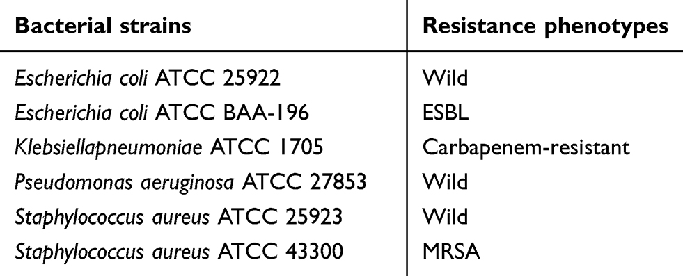 |
Table 1 Reference bacterial strains |
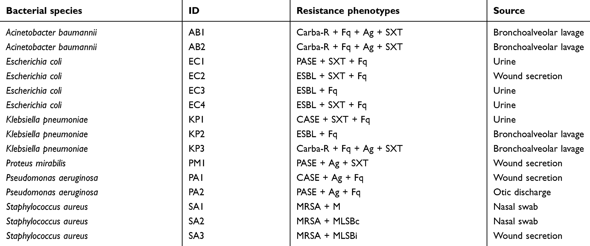 |
Table 2 MDR bacterial clinical isolates |
In our study, the MDR strains came from routine clinical activity. At first, the bacteria were isolated on Columbia 5% sheep blood agar (Sanimed, Bucharest, Romania). Identification of all isolates was performed according to morphological characters of colonies and their biochemical tests obtained using the automated Vitek 2 system (bio-Mérieux, Marcy l’Etoile, France). Sensitivity to antimicrobial agents was tested according to the Clinical Laboratory and Standards Institute (CLSI) criteria, by determining the minimum inhibitory concentration (MIC) with the Vitek 2 system.27
For Gram-negative bacilli (GNB), phenotypic confirmation of ESBL production was done using the synergy test between extended-spectrum cephalosporins and clavulanic acid.27,28
Carbapenemase production was demonstrated by combined disc methods (KPC, MBL and OXA-48 Confirm kit, Rosco Diagnostica, Denmark).27,29,30
For methicillin-resistant Staphylococcus aureus (MRSA) strains, cefoxitin screening was performed. The constitutive macrolide-lincosamide-streptogramin B (MLSBc) phenotype was determined based on its resistance to erythromycin and clindamycin. inducible macrolide-lincosamide-streptogramin B (MLSBi) resistance phenotype was either detected on Vitek AST-P592 cards or by the appearance of a D-region around the clindamycin disk in the vicinity of erythromycin, which was associated with erythromycin resistance (on disk diffusion test).
Inclusion into the MDR group was made according to the definition proposed by Magiorakos (2012). Thus, MDR was defined as acquired resistance to at least one agent in three or more antimicrobial categories, while XDR were considered as bacterial strains that remained susceptible to at most two antimicrobial categories.31
After phenotyping, the MDR strains were stored on a microbank (Pro-Lab Diagnostics, Toronto, Canada) at −80°C. Reconstitution was accomplished by discharging cryobiles on Columbia +5% sheep blood agar medium (Sanimed, Bucharest, Romania) and by incubating for 24 hrs at 37°C. After reconstitution, the susceptibility of strains to peppermint oil was tested.
In vitro antibacterial activity
The antimicrobial activity of this oil was evaluated by agar disk diffusion method and microdilution method as previously described.32,33
Disk diffusion method
The bacterial suspensions were adjusted with physiological saline to a concentration of 0.5 Mac Farland (108bacteria/mL) and 100 µL of these suspensions were placed on the surface of Mueller–Hinton agar (Sanimed, Bucharest, Romania). Ten microliters of oil were added to a blank paper disk (BioMaxima, Lublin, Poland), and then deposited on the surface of the Mueller–Hinton plates inoculated with the microbial suspensions and consequently incubated at 37°C for 24 hrs. The reading of the inhibition zones was done in millimeters. All tests were performed in triplicate for each bacterial strain. Gentamycin 10 μg (BioRad, Marnes la Coquette, France) was used as a positive control, and for negative control, we used a blank paper disk that was not impregnated.
Dilution method-determination of MIC and minimum bactericidal concentration (MBC)
The broth dilution assay was done as recommended by the (CLSI).26 In seven test tubes, serial two-fold dilutions of the oil (80, 40, 20, 10, 5, 2.5, 1.25 mg/mL) in Mueller–Hinton broth (Sanimed, Bucharest, Romania) were done and was added with the bacterial inoculum (5×10⁵ bacteria/mL). After incubating the test tubes at 37°C for 24 hrs, the MIC (the lowest concentration without visible growth) was determined. The MBC was determined by sub-cultivation of 1 µL on Columbia agar +5% sheep blood and was considered as the lowest concentration which killed 99.9% of the initial inoculum.
Statistical analysis
Correlation between parameters (Pearson coefficients) was obtained by using the program “Microsoft Excel 2010”.
Results
The chemical composition of MPEO
Figure 1 and Table 3 present the GC-MS (gas-chromatograph with mass spectrometer) profiles of MPEO and the percentages of volatile components in order of elution. In MPEO, 17 compounds (over 0.08%) were identified comprising 99.896% of the total MPEO composition. Monoterpene hydrocarbons (MH) represent 34.229%, monoterpene oxygenates (MO) 60.826%, sesquiterpene hydrocarbonates (SH) 4.635% and sesquiterpene oxygenate (SO) 0.206% of the total compounds (Table 3).
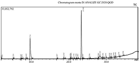 |
Figure 1 Chromatogram of MPEO. |
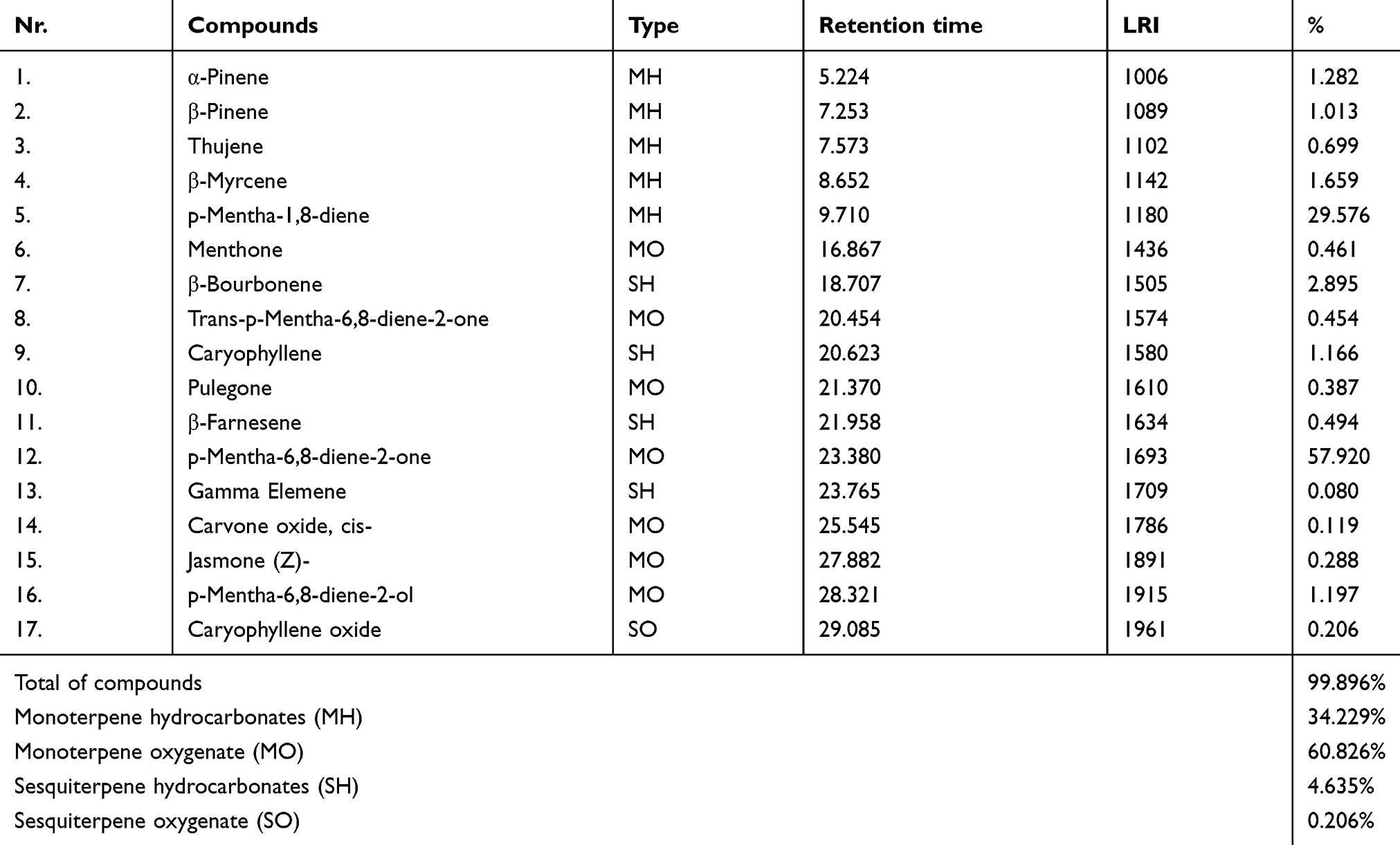 |
Table 3 The chemical composition of MPEO (% of total) |
The GC-MS profile evidenced the major compounds of MPEO: p-Mentha-6,8-diene-2-one (57.920%) and p-Mentha-1,8-diene (29.576%). Also, other characteristic phytocompounds of MPEO could be detected as described in Table 3.
The values of the diameters’ inhibition zones, MIC and MBC for peppermint oil against 21 bacterial strains are listed in Table 4.
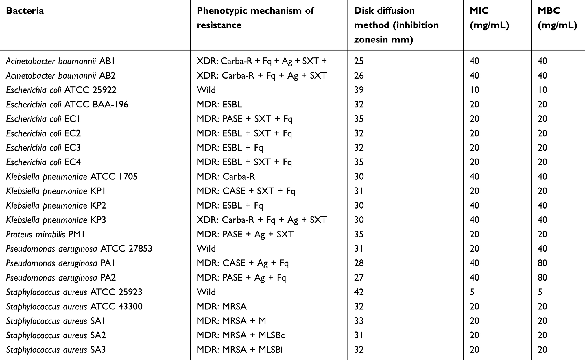 |
Table 4 Antibacterial activity of the essential oil studied |
Testing the antibacterial activity of peppermint oil on both reference strains and isolated MDR strains from hospitalized patients demonstrated its bactericidal effect. Out of the 21 strains tested, 16 were represented by GNB (Acinetobacter baumannii, Escherichia coli, Klebsiella pneumoniae, Proteus mirabilis and Pseudomonas aeruginosa), whereas the remaining five were represented by Staphylococcus aureus. The MIC of the oil tested was lower (20 mg/mL) for Staphylococcus aureus, Escherichia coli and Proteus mirabilis and higher (40 mg/mL) for Klebsiella pneumoniae, Pseudomonas aeruginosa and Acinetobacter baumannii strains (Table 4). MBC was equal to MIC, with the exception of Pseudomonas aeruginosa strains, where MBC was the double of MIC.
In order to evaluate the influence of functional molecules of oil and the possible antibacterial activity of those compounds, Pearson correlation between inhibition zones (mm) of principal bacterial strains (Acinetobacter baumannii, Klebsiella pneumoniae, Escherichia coli, Pseudomonas aeruginosa and Staphylococcus aureus) and the chemical constituent derivates from menthone (p-Mentha-1,8-diene, p-Mentha-6,8-diene-2-one, menthone and trans-p-Mentha-6,8-diene-2-one) were established.
The analysis of correlation (Table 5) highlights a very strong (r=1) positive correlation between the strains of Acinetobacter baumannii and the total compounds which includes menthone in molecule that suggests the effect of these compounds on bacterial cell growth. Also, a strong positive correlation (r=0.867) was obtained between Klebsiella pneumoniae and menthone type compounds. In these cases, we can presume a mutual connection between these chemical compounds and their antibacterial effect. Low positive correlations (r=0.248 and r=0.179, respectively) were identified between pairs: Pseudomonas/menthone type compounds and Staphylococcus/menthone type compounds, while a low negative correlation (r=−0.046) was identified between E. coli and menthone type compounds.
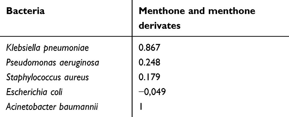 |
Table 5 Pearson coefficients (r) between bacteria strains and chemical composition expressed as menthone and menthone derivates |
Discussion
Regarding the chemical composition of MPEO, in our study, monoterpene hydrocarbons (MH) represent 34.229%, monoterpene oxygenates (MO) 60.826%, sesquiterpene hydrocarbonates (SH) 4.635% and sesquiterpene oxygenate (SO) 0.206% of the total compounds (Table 3). The major detected compounds of MPEO were the following: p-Mentha-6,8-diene-2-one (57.920%) and p-Mentha-1,8-diene (29.576%) (Table 3). Similar composition was reported by Ashrafi et al who recorded a menthol content in MPEO of 45.05% and menthol 17.53%.34 The study published by Yu et al reported that the major constituents are within the phytochemical classes of oxygenated monoterpenes (73.9–94.8%), monoterpenes type hydrocarbons (1.0–21.9%) and sesquiterpenes (0.5–16.6%).35 Reddy et al found menthol (36.02%) as the main chemical compound in MPEO, followed by menthone (24.56%).36 Similar composition was reported in other studies.37,38 In Italian MPEO, menthol (32.4%) and menthone (26.6%) were determined,39 while linalool and linalyl acetate were detected as major compounds of Tunisian MPEO.40
With respect to bacterial multidrug resistance, this currently poses an increasingly serious threat with important clinical consequences regarding the treatment options. Over recent years, infections caused by MDR bacteria have become endemic in many health care units and hospital-acquired outbreaks involving such microorganisms are being reported worldwide.41–43
Because of the impact of rising antimicrobial resistance, since 2001, the World Health Organization (WHO) concluded that high priority should be given to measures that aimed to slow the emergence of MDR, these measures being particularly important given that the development of antimicrobial agents has been reduced over the last years.44 In 2017, the WHO released a global list of resistant bacteria, identifying MDR Gram-negative bacteria such as carbapenem-resistant Enterobacteriaceae, Pseudomonas aeruginosa or Acinetobacter baumannii, extended-spectrum cephalosporin-resistant Enterobacteriaceae as critical priorities for developing new strategies of treatment.
For these reasons, we initiated this multidisciplinary study in order to find a new therapeutic alternative for MDR bacteria-induced infections.
In choosing the bacterial strains obtained from the hospitalized patients, we have tried to show different phenotypes of resistance, either within the same class or in different classes of antibiotics, in order to report the efficacy of the MP oil on several MDR strains. Thus, most of the Enterobacteriaceae and Pseudomonas clinical isolates were MDR, secreting various beta-lactamases, while the strains of Acinetobacter baumannii were XDR.
Different mechanisms could determine the antimicrobial resistance; these include the enzymatic degradation of antimicrobial agents, such as beta-lactamases in the case of beta-lactam resistance or modifying enzymes in aminoglycosides resistance. The alteration of antimicrobial targets in case of MRSA or fluoroquinolones resistance. Moreover, changes in bacterial membrane permeability can lead to resistance to many antimicrobial agents.45
The antibacterial activity of essential oils is incompletely known, but some mechanisms have been proposed in literature over time, such as the alteration of the membrane permeability of pathogens by disrupting transport systems and energy production.
The antimicrobial effect of the essential oil of MP has been studied in various types of microorganisms, both Gram-positive and Gram-negative bacteria.
Mimica-Dukic et al obtained a low MIC (8 μL/mL) on E. coli strains, while Hammer et al reported an MIC of 25 μL/mL for E. coli and 12 μL/mL for S. aureus.46,47 A few groups, including that of Aridogan et al reported antibacterial activity only for S. aureus and not for E. coli.48
These differences could be due to dissimilarity in the oils’ chemical composition collected from different parts of the world.
The antimicrobial action of essential oils has been explained mainly by the presence of terpenes, alcohols, aldehydes and esters. From the terpenes, phenolic compounds, in particular, thymol and carvacrol, appear to be able to increase plasma membrane permeability to cellular metabolites.
Soković et al used volatile oil obtained from Mentha×piperita L., collected from Pančevo, Serbia.20 This geographic region is similar to the area from which the essential mint oil used in our study was obtained. All the mint oils tested showed bacteriostatic activity in concentration of 1 µg/disc. The inhibition zones that were obtained for MP oils were 16.0–25.0 mm and 13.0–25.0 mm. The antibacterial effect was tested for the following human pathogenic bacteria: Micrococcus flavus, Enterobacter cloacae, Escherichia coli O157:H7, Proteus mirabilis, Pseudomonas aeruginosa, Salmonella enteritidis, Salmonella typhimurium, Staphylococcus aureus and Streptococcus epidermidis. Oils from MP exhibited much higher antibacterial activity with the same MIC (1.0–3.0 µg/mL) and MBC (1.5–5.0 µg/mL).20
However, in the literature, there are fewer studies on the antimicrobial activity of essential oils of MP on MDR bacterial strains. Shalayel et al have evaluated the antibacterial activities of ethanol, ethyl acetate, methanol and chloroform peppermint extracts on ten MDR pathogenic bacterial clinical isolates, reporting the bactericidal effect of those extracts, especially ethyl acetate extracts, against MDR S. pyogenes, E. faecalis, MRSA, MRSE and carbapenem-resistant E. coli and K. pneumoniae.49
A potent antibacterial activity of tea tree oil against MDR microorganisms, which presented a high level of synergism with oxacillin against MRSA, was also observed.50
Moreover, dithiodiketopiperazine derivates (isolated from cultures of Trichoderma harzianum and Epicoccum nigrum from Zingiber officinale and Salix sp.) manifested antibacterial, antifungal and cytotoxic activity against several MDR microorganisms.51
For the moment, little is known about the functional molecules responsible for the antibacterial activity of MPEO and their mechanism of action, menthol and menthone being the major constituents responsible for the antibacterial activity and the most described in the literature.52–54 Moreover, association of menthol, menthone, limonene, neomenthol, carvone and 1,8-cineole with other minor constituents could induce a synergistic antibacterial activity, although the part of the plant used (root, leaves, etc.), composition of the used extract, concentration of the active substance and the type of microorganism are crucially important factors for the potency of the antimicrobial action.55–57
In this study, MP oil had a bactericidal effect on all bacterial tested strains, regardless of the resistant phenotypes these strains exhibited against the currently applied anti-infective agents. Different beta-lactam resistant phenotypes of the tested bacteria were associated with minor differences in the diameters of the inhibition zones, but with significant variations in MIC and MBC, thus confirming the importance of determining the MIC/MBC for the determination of antibacterial activity of substances. Resistance to aminoglycosides, fluoroquinolones, sulfamides, macrolides or lincosamides did not influence the antibacterial activity of MP oil. As in other studies, the bacterial species significantly influenced the test results, with the most sensitive strains to MP oil represented by S. aureus, E. coli and Proteus mirabilis, while Pseudomonas aeruginosa strains exhibited the highest MIC and MBC values.58
Regarding the correlation between chemical and microbiological parameters, a strong correlation was recorded between menthone and menthone compounds (p-Mentha-1,8-diene, p-Mentha-6,8-diene-2-one, trans-p-Mentha-6,8-diene-2-one) and the bacterial strains of Klebsiella pneumoniae and Acinetobacter baumannii, that suggest a potential effect of menthone and its derivatives in inhibition of bacterial cell growth. Regarding the development of Escherichia coli, Staphylococcus aureus and Pseudomonas aeruginosa cells in correlation with the chemical composition, low values of Pearson coefficients were observed. These findings lead to the idea that a high level of menthone and menthone derivates is responsible for a pronounced inhibition of Klebsiella pneumoniae and Acinetobacter baumannii strains, without affecting the development of Escherichia coli, Staphylococcus aureus and Pseudomonas aeruginosa strains. The microbiological effect of Mentha piperita essential oil can be explained by the synergism exercised by the minority components of the oil and the compounds derived from menthone. Our studies agree with previous data obtained by our group that reported the influence of synergism of chemical compounds on the microbiological activity of essential oils belonging to Lamiaceae family.59
Due to the bactericidal effect of MP oil demonstrated on MDR bacteria, it could become a new antimicrobial agent. Volatile MPEO could be used locally, by inhalation in the respiratory tract, or in treatment of various skin infections. Further studies are necessary to investigate the potential toxicity of MP oil and standardize the inhibitory effect of MP oil against MDR pathogens.
Conclusion
The present study highlights the bactericidal activity of MPEO on all tested Gram-positive and Gram-negative strains of MDR or XDR: Staphylococcus aureus, Escherichia coli, Klebsiella pneumoniae, Proteus mirabilis, Pseudomonas aeruginosa and Acinetobacter baumannii. This oil may be a therapeutic option for more and more frequent infections caused by MDR bacteria in the future. In this purpose, studies that investigate also the possible adverse effects of this essential oil should be conducted.
Abbreviations
Ag-, aminoglycosides resistance; c, constitutive; Carba-R, carbapenem resistance; CASE, cephalosporinase hyperproduction; CLSI, Clinical Laboratory and Standards Institute; ESBL, extensive spectrum beta-lactamase; EO, sential oil; Fq-, fluoroquinolones resistance; GC-MS, gas-chromatograph with mass spectrometer; i, inducible; ID, identification; LRI, linear retention indices; M-, macrolides resistance; MBC, minimum bactericidal concentration; MDR, multidrug-resistant; MLSB, macrolide-lincosamide-streptogramin B; MIC, minimum inhibitory concentration; MH, monoterpene hydrocarbonates; MO, monoterpene oxygenates; MP, Mentha×piperita L.; MPEO, Mentha×piperita L. essential oil; MRSA, methicillin-resistant S. aureus; PASE, penicillinase hypersecretion; SH, sesquiterpene hydrocarbonates; SO, sesquiterpene oxygenate; SXT, folate pathway inhibitor resistance; WHO, World Health Organization; XDR, extensively drug resistant.
Disclosure
The authors report no conflicts of interest in this work.
References
1. Elshafie HS, Camele I. An overview of the biological effects of some Mediterranean essential oils on human health. Biomed Res Int. 2017;2017:9268468. doi:10.1155/2017/9268468
2. Sharifi-Rad J, Sureda A, Tenore GC, et al. Biological activities of essential oils: from plant chemoecology to traditional healing systems. Molecules. 2017;22:1. doi:10.3390/molecules22010070
3. Alexa E, Sumalan RM, Danciu C, et al. Synergistic antifungal, allelopatic and anti-proliferative potential of Salvia officinalis L., and Thymus vulgaris L. essential oils. Molecules. 2018;23(1):185. doi:10.3390/molecules23010185
4. de Groot A, Schmidt E. Part II: general aspects. Dermatitis. 2016;27(2):43–49. doi:10.1097/DER.0000000000000174
5. de Groot AC, Schmidt E. Essential oils, part IIIEssential oils: chemical composition. Dermatitis. 2016;27(4):161–169. doi:10.1097/DER.0000000000000193
6. Dhifi W, Bellili S, Jazi S, Bahloul N, Mnif W. Essential oils’ chemical characterization and investigation of some biological activities: a critical review. Medicines (Basel). 2016;3(4). Available from: https://www.ncbi.nlm.nih.gov/pmc/articles/PMC5456241/.
7. Samarth RM, Samarth M, Matsumoto Y. Medicinally important aromatic plants with radioprotective activity. Future Sci OA. 2017;3:4. doi:10.4155/fsoa-2017-0061
8. Cocan I, Alexa E, Danciu C, et al. Phytochemical screening and biological activity of Lamiaceae family plant extracts. ExpTher Med. 2018;15(2):1863–1870.
9. Pourmortazavi SM, Hajimirsadeghi SS. Supercritical fluid extraction in plant essential and volatile oil analysis. J Chromatogr A. 2007;1163(1–2):2–24. doi:10.1016/j.chroma.2007.06.021
10. Aziz ZAA, Ahmad A, Setapar SHM, et al. Essential oils: extraction techniques, pharmaceutical and therapeutic potential - a review. Curr Drug Metab. 2018;19(13):1100–1110. doi:10.2174/1389200219666180723144850
11. Nakatsu [xc*] T, Lupo AT, Chinn JW, Kang RKL. Biological Activity of Essential Oils and Their Constituents. In: Atta-ur-Rahman, Editor. Studies in Natural Products Chemistry [internet]. Elsevier; 2000:571–631. (Bioactive Natural Products (Part B); vol. 21). Available from: http://www.sciencedirect.com/science/article/pii/S1572599500800149.
12. Li H-P, Hu Z, Yuan J-L, et al. A novel extracellular protease with fibrinolytic activity from the culture supernatant of Cordyceps sinensis: purification and characterization. Phytother Res. 2007;21(12):1234–1241. doi:10.1002/ptr.2246
13. Alexa E, Danciu C, Radulov I, et al. Phytochemical screening and biological activity of Mentha ×piperita L. and Lavandula angustifolia mill. extracts. Anal Cell Pathol (Amst). 2018;2018:2678924. doi:10.1155/2018/2678924
14. Sustrikova A, Salamon I. Essential oil of peppermint (Mentha ×piperita L.) from fields in Eastern Slovakia. Hortic Sci. 2004;31(1):31–36. doi:10.17221/3789-HORTSCI
15. Rita P, Datta AK. An updated overview on peppermint (Menthapiperita L.). Int Res J Pharm. 2011;2(8):1–10.
16. Istudor V. Pharmacognosy. Phytochemistry. Phytotherapy. Vol. II. Bucharest: Medical Publishing House; 2001.
17. Stanescu U, Hancianu M, Cioanca O, Aprotosoaie CA, Miron A. Medicinal Plants from A to Z.
18. McKay DL, Blumberg JB. A review of the bioactivity and potential health benefits of peppermint tea (Menthapiperita L.). Phytother Res. 2006;20(8):619–633. doi:10.1002/ptr.1936
19. Ali B, Al-Wabel NA, Shams S, Ahamad A, Khan SA, Anwar F. Essential oils used in aromatherapy: a systemic review. Asian Pac J Trop Biomed. 2015;5(8):601–611. doi:10.1016/j.apjtb.2015.05.007
20. Soković M, Glamočlija J, Marin PD, Brkić D, van Griensven LJ. Antibacterial effects of the essential oils of commonly consumed medicinal herbs using an in vitro model. Molecules. 2010;15(11):7532–7546. doi:10.3390/molecules15117532
21. Akdemir Evrendilek G. Empirical prediction and validation of antibacterial inhibitory effects of various plant essential oils on common pathogenic bacteria. Int J Food Microbiol. 2015;202:35–41. doi:10.1016/j.ijfoodmicro.2015.02.030
22. Samie A, Nefefe T, Gundidza M, Mmbengwa V, Magwa M. Antimicrobial activities of essential oils from Southern Africa against selected bacterial and fungal organisms. Afr J Biotechnol. 2012;11:15560–15568. doi:10.5897/AJB
23. Sujana P, Sridhar TM, Josthna P, Naidu CV. Antibacterial activity and phytochemical analysis of menthapiperita L. (Peppermint)—an important multipurpose medicinal plant. Am J Plant Sci. 2013;4:77–83. doi:10.4236/ajps.2013.41012
24. Erdogan O, Celik A, Zeybek A. In vitro antifungal activity of mint, thyme, lavender extracts and essential oils on VerticilliumDahliaeKleb. Fresenius Environ Bull. 2016;25(11):4856–4862.
25. Jianu C, Mihail R, Muntean SG, et al. Composition and antioxidant capacity of essential oils obtained from Thymus vulgaris, Thymus pannonicus and Satureja montana grown in Western Romania. Rev Chim-Bucharest. 2015;66:2157–2160.
26. Atomic spectra Database. Nist Standard Reference Database 78. Version 5.6. 2018. Available from: https://www.nist.gov/pml/atomic-spectra-database.
27. Clinical and Laboratory Standards Institute. Performance Standards for Antimicrobial Susceptibility Testing 2013; Twenty Third Informational Supplement M 100-S23. Wayne PA: CLSI. 2013.
28. Giske CG, Martinez-Martinez L, Cantón R, et al. EUCAST Guidelines for Detection of Resistance Mechanisms and Specific Resistances of Clinical And/or Epidemiological Importance 2013; Version 1.0. European Society of Clinical Microbiology and Infectious Disease. 2013 4–10.
29. Hrabák J, Chudáčková E, Papagiannitsis CC. Detection of carbapenemases in Enterobacteriaceae: a challenge for diagnostic microbiological laboratories. ClinMicrobiol Infect. 2014;20:839–853. doi:10.1111/1469-0691.12678
30. Miriagou V, Tzelepi E, Kotsakis SD, Daikos GL, Bou Casals J, Tzouvelekis LS. Combined disc methods for the detection of KPC-and/or VIM-positive Klebsiellapneumoniae: improving reliability for the double carbapenemase producers. ClinMicrobiol Infect. 2013;19(9):E412–E415. doi:10.1111/1469-0691.12238
31. Magiorakos AP, Srinivasan A, Carey RB, et al. Multidrug-resistant, extensively drug-resistant and pandrug-resistant bacteria: an international expert proposal for interim standard definitions for acquired resistance. ClinMicrobiol Infect. 2012;18:268–281. doi:10.1111/j.1469-0691.2011.03570.x
32. Pavel IZ, Danciu C, Oprean C, et al. In vitro evaluation of the antimicrobial ability and cytotoxicity on two melanoma cell lines of a benzylamide derivative of maslinic acid. Anal Cell Pathol (Amst). 2016;2016:2787623. doi:10.1155/2016/2787623
33. Danciu C, Muntean D, Alexa E, et al. Phytochemical characterization and evaluation of the antimicrobial, antiproliferative and pro-apoptotic potential of ephedra alatadecne. hydroalcoholic extract against the MCF-7 breast cancer cell line. Molecules. 2018;24(1):E13. doi:10.3390/molecules24010013
34. Ashrafi B, Rashidipour M, Marzban A, et al. Mentha piperita essential oils loaded in a chitosan nanogel with inhibitory effect on biofilm formation against S. mutans on the dental surface. Carbohydr Polym. 2019;212:142–149. doi:10.1016/j.carbpol.2019.02.018
35. Yu X, Liang C, Fang H, Weilin XQ, Shang Q. Variation of trichome morphology and essential oil composition of seven Mentha species. Biochem Syst Ecol. 2018;79:30–36.
36. Reddy DN, Al-Rajab AJ, Sharma M, Moses MM, Reddy GR, Albratty M. Chemical constituents, in vitro antibacterial and antifungal activity of Mentha ×Piperita L. (peppermint) essential oils. J King Saud Univ Sci. 2017. doi:10.1016/j.jksus.2017.07.013
37. Rajkumar V, Gunasekaran C, Kanith I, Dharmaraj J, Chinnaraj P, Cheruvathur AP. Toxicity, antifeedant and biochemical efficacy of Mentha piperita L. essential oil and their major constituents against stored grain pest, Pesticide. Biochem Physiol. 2019;156:138–144.
38. Ciobanu A, Mallarda I, Landya D, Brabiec G, Nistor D, Fourmentin S. Retention of aroma compounds from Mentha piperita essential oil by cyclodextrins and crosslinked cyclodextrin polymers. Food Chem. 2013;138(1):291–297. doi:10.1016/j.foodchem.2012.10.106
39. Ebani VV, Nardoni S, Bertelloni F, et al. In vitro antimicrobial activity of essential oils against salmonella enterica serotypes enteritidis and typhimurium strains isolated from poultry. Molecules. 2019;24:900. doi:10.3390/molecules24050900
40. Soilhia Z, Rhimib A, Heuskin S, Fauconnier M, Mekkia M. Essential oil chemical diversity of Tunisian Mentha spp. Collection. Ind Crops Prod. 2019;131:330–340. doi:10.1016/j.indcrop.2019.01.041
41. Spellberg B, Blaser M, Guidos RJ, Boucher HW, Bradley JS. Combating antimicrobial resistance: policy recommendations to save lives. Clin Infect Dis. 2011;52:397–428. doi:10.1093/cid/cir153
42. Gray JW, Mahida N. How do you solve a problem like multidrug-resistant Gram-negative bacteria? J Hosp Infect. 2016;92:1–2. doi:10.1016/j.jhin.2015.11.005
43. Cole J. Antimicrobial resistance – a ‘rising tide’ of national (and international) risk. J Hosp Infect. 2016;92:3–4. doi:10.1016/j.jhin.2015.10.005
44. World Health Organization. WHO Global Strategy for the Containment of Antimicrobial Resistance. Geneva, Switzerland: WHO; 2001. Available from: http://www.who.int/drugresistance/guidance/en/index.html.
45. Ruppé E, Woerther P, Barbier F. Mechanisms of antimicrobial resistance in Gram-negative bacilli. Ann Intensive Care. 2015;5(1):21. doi:10.1186/s13613-015-0061-0
46. Mimica-Dukic N, Bozin B, Sokovic M, Simin N. Antimicrobial and antioxidant activities of Melissa officinalis L. (Lamiaceae) essential oil. J Agric Food Chem. 2004;52(9):2485–2489. doi:10.1021/jf030698a
47. Hammer KA, Carson CF, Riley TV. Antimicrobial activity of essential oils and other plant extracts. J Appl Microbiol. 1999;86:985–990.
48. Aridogan BC, Baydar H, Kaya S, Demirci M, Ozbasar D, Mumcu E. Antimicrobial activity and chemical composition of some essential oils. Arch Pharm Res. 2002;25:860–864.
49. Shalayel MHF, Asaad AM, Elhussein B. Anti-bacterial activity of peppermint (Mentha piperita) extracts against some emerging multi-drug resistant human bacterial pathogens. J Herbal Med. 2017;7:27–30. doi:10.1016/j.hermed.2016.08.003
50. Oliva A, Costantini S, De Angelis M, et al. High potency of melaleuca alternifolia essential oil against multi-drug resistant gram-negative bacteria and methicillin-resistant Staphylococcus aureus. Molecules. 2018;23(10):E2584. doi:10.3390/molecules23102584
51. Harwoko H, Daletos G, Stuhldreier F, et al. Dithiodiketopiperazine derivatives from endophytic fungi Trichoderma harzianum and Epicoccumnigrum. Nat Prod Res. 2019;1–9. doi:10.1080/14786419.2019.1627348
52. Badea ML, Iconaru SL, Groza A, Chifiriuc MC, Beuran M, Predoi D. Peppermint essential oil-doped hydroxyapatite nanoparticles with antimicrobial properties. Molecules. 2019;24(11):E2169. doi:10.3390/molecules24112169
53. Cao L, Si JY, Liu Y, et al. Essential oil composition, antimicrobial and antioxidant of Mosla chinesis Maxim. Food Chem. 2009;115:801–815. doi:10.1016/j.foodchem.2008.12.064
54. Oke F, Aslim B, Ozturk S, Altundag S. Essential oil composition, antimicrobial and antioxidant activities of Satureja cuneifolia ten. Food Chem. 2009;112:874–879. doi:10.1016/j.foodchem.2008.06.061
55. Lopes-Lutz D, Alviano DS, Alviano CS, Kolodziejczyk PP. Screening of chemical composition, antimicrobial and antioxidant activities of Artemisia essential oils. Phytochemistry. 2008;69:1732–1738. doi:10.1016/j.phytochem.2008.02.014
56. Singh R, Shushni MAM, Belkheir A. Antibacterial and antioxidant activities of Mentha piperita L. Arab J Chem. 2015;8:322–328. doi:10.1016/j.arabjc.2011.01.019
57. Antolak H, Czyżowska A, Kręgiel D. Activity of Mentha piperita L. ethanol extract against acetic acid bacteria Asaia spp. Foods. 2018;7(10):E171. doi:10.3390/foods7100171
58. Man A, Santacroce L, Jacob R, Mare A, Man L. Antimicrobial activity of six essential oils against a group of human pathogens: a comparative study. Pathogens. 2019;8(1):E15. doi:10.3390/pathogens8010015
59. Alexa E, Sumalan RM, Danciu C, et al. Synergistic antifungal, allelopatic and anti-proliferative potential of Salvia officinalis L., and Thymus vulgaris L. essential oils. Molecules. 2018;23(1):185–200. ISSN 1420-3049. doi:10.3390/molecules23010185
 © 2019 The Author(s). This work is published and licensed by Dove Medical Press Limited. The full terms of this license are available at https://www.dovepress.com/terms.php and incorporate the Creative Commons Attribution - Non Commercial (unported, v3.0) License.
By accessing the work you hereby accept the Terms. Non-commercial uses of the work are permitted without any further permission from Dove Medical Press Limited, provided the work is properly attributed. For permission for commercial use of this work, please see paragraphs 4.2 and 5 of our Terms.
© 2019 The Author(s). This work is published and licensed by Dove Medical Press Limited. The full terms of this license are available at https://www.dovepress.com/terms.php and incorporate the Creative Commons Attribution - Non Commercial (unported, v3.0) License.
By accessing the work you hereby accept the Terms. Non-commercial uses of the work are permitted without any further permission from Dove Medical Press Limited, provided the work is properly attributed. For permission for commercial use of this work, please see paragraphs 4.2 and 5 of our Terms.