Back to Journals » Infection and Drug Resistance » Volume 15
Evaluation of Antibacterial Activity and Acute Toxicity of Methanol Extracts of Artemisia absinthium, Datura stramonium, and Solanum anguivi
Authors Arage M, Eguale T ![]() , Giday M
, Giday M
Received 20 January 2022
Accepted for publication 17 March 2022
Published 24 March 2022 Volume 2022:15 Pages 1267—1276
DOI https://doi.org/10.2147/IDR.S359280
Checked for plagiarism Yes
Review by Single anonymous peer review
Peer reviewer comments 2
Editor who approved publication: Prof. Dr. Héctor Mora-Montes
Mahlet Arage, Tadesse Eguale, Mirutse Giday
Aklilu Lemma Institute of Pathobiology, Addis Ababa University, Addis Ababa, Ethiopia
Correspondence: Mirutse Giday, Aklilu Lemma Institute of Pathobiology, Addis Ababa University, PO Box 1176, Addis Ababa, Ethiopia, Tel +251-9-1117-1321, Email [email protected]
Background: Bacterial pathogens have evolved numerous defense mechanisms against commercial antimicrobial agents, and their resistance to most of the available antimicrobials is increasing. Medicinal plants are a potential source of antimicrobial agents during the spread and emergence of infectious disease caused by resistant microorganisms. The aim of this study was thus to investigate the antimicrobial activity and acute toxicity of 80% methanol extracts of leaves of Artemisia absinthium, seeds of Datura stramonium, and fruit of Solanum anguivi.
Materials and Methods: The 80% methanol extracts were prepared by cold maceration. Antimicrobial activity was evaluated against five bacterial species using agar-well diffusion at concentrations of 125, 250, and 500 mg/mL in the presence of positive and negative controls. Minimum inhibitory concentration was determined by broth dilution. The acute toxicity study was done following OECD guidelines.
Results: The 80% methanol extract of the fruit of S. anguivi exhibited better activity against most of the bacterial strains, of which Salmonella enterica serovar Typhimurium was found to be the most susceptible, with minimum inhibitory concentration and minimum bactericidal concentration of 1.3 mg/mL and 1.95 mg/mL, respectively, while the seed extract of D. stramonium showed the least activity against most test organisms. The acute toxicity study showed that all three plants had an LD50 > 2,000 mg/kg body weight, and were thus safe.
Conclusion: The results of this study revealed that the methanol extracts of the three plants (A. absinthium, D. stramonium, and S. anguivi) had different degrees of antibacterial activity against the selected pathogenic bacteria and were safe at higher doses, thus being of great potential to be developed as antibacterial agents. The study also provided scientific evidence to support the continued traditional use of these medicinal plants by communities in different parts of Ethiopia to treat infectious diseases.
Keywords: medicinal plants, antimicrobial activity, agar-well diffusion, Artemisia absinthium, Datura stramonium, Solanum anguivi
Introduction
Bacterial infections are responsible for considerable mortality and morbidity worldwide, especially in developing countries, due to poor sanitation and unhygienic and overcrowded living conditions. Antibiotics are powerful medicines that fight bacterial infections by either killing the bacteria or stopping them from reproducing, allowing the body’s natural defenses to easily eliminate them. Drugs used for treating bacterial infections may lose their effectiveness with time because the target organisms keep changing form or hiding themselves from attacking drugs, and hence drug resistance occurs.1 The emergence and spread of antimicrobial resistance have been driven by complex socioeconomic and human behavioral factors, including misuse of antibiotics, unskilled practitioners and laypersons, and use of substandard antimicrobials, particularly in developing countries.2,3 Although antimicrobial resistance is a global threat, its burden is higher in developing countries because of the high prevalence of bacterial diseases and the presence of risk of their emergence and spread.4 Increased prevalence of resistant bacteria, together with lack and high cost of new-generation drugs, has escalated infection-related morbidity and mortality, particularly in developing countries like Ethiopia.5 The increasing failure of chemotherapeutics and the emergence of antibiotic-resistant pathogens have led researchers to look for alternatives in their search for effective and safe therapeutics, including medicinal plants that are used as antimicrobials in traditional medical systems.6
From time immemorial, different communities around the globe have been using medicinal plants to manage various health problems. In Ethiopia, a large portion of the rural population and the poor in urban areas are known to rely on traditional medicine to meet their primary health-care needs.7 Traditional medicine has high acceptability and is an integral part of the local culture in Ethiopia, and people often rely on it even in the presence of demonstrably efficient and less costly alternative health-care in their area. More than 95% of traditional Ethiopian medical preparations are of plant origin.8 A number of ethnobotanical studies conducted on medicinal plants in Ethiopia have demonstrated the use of medicinal plants in the treatment of different infectious diseases,9–15 which shows that plant-derived medicines continue to occupy an important position in the country’s traditional medical practices.16
Although a range of medicinal plants with antimicrobial properties have been widely used by traditional healers, the therapeutic potential of many of these medicinal plants has not been scientifically evaluated.17 In Ethiopia, the plants Artemisia absinthium, Datura stramonium, and Solanum anguivi have been claimed to have antimicrobial efficacy.18–20 However, there is a lack of scientific evidence to prove such traditional claims. As such, this study aimed to investigate antibacterial activity and acute toxicity of the three plants in view of validating the claims of traditional practitioners. In addition, the findings of this study may lead to the discovery of new compounds for the development of novel antimicrobial drugs.
Materials and Methods
Chemicals, Solvents, and Media
Chemicals used for extracting the plant material were distilled water, absolute methanol (Reagent Chemical), dimethyl sulfoxide (DMSO; Loba Chemie), 0.5 McFarland equivalence/standard, and sterile physiological saline (Albert David). All chemicals and solvents were of analytical grade. Bacteriological media used for this study were Mueller–Hinton agar, Mueller–Hinton broth, mannitol salt agar, xylose lysine deoxycholate agar (Oxoid), nutrient agar, and nutrient broth (HiMedia Laboratories). All media were used according to the manufacturers’ guidelines.
Reference Bacterial Test Organisms and Antibiotic Disks
Standard strains of bacteria — Escherichia coli (ATCC 25922), Pseudomonas aeruginosa (ATCC 27853), Staphylococcus aureus (ATCC 25923), Shigella flexneri (ATCC 12022), and Salmonella enterica serovar Typhimurium (S. Typhimurium; ATCC 14028) — were obtained from the Aklilu Lemma Institute of Pathobiology (ALIPB), Addis Ababa University and the Ethiopian Public Health Institute. The bacteria were selected based on availability and by considering the traditional use of the experimental plants. Standard antibacterial disks used for the study were amoxicillin (30 μg/disk, Oxoid) and ciprofloxacin (5 μg/disk, Becton Dickinson).
Collection and Authentication of Plant Materials
Based on reports of traditional use,18–20 leaves of A. absinthium, seeds of D. stramonium, and fruit of S. anguivi were collected from their natural habitats at different locales. Identification and authentication of the plant specimens were conducted by a botanist at ALIPB, and voucher specimens were deposited for reference purposes at the Endod and Other Medicinal Plants Research Unit of ALIPB.
Preparation and Extraction of Plant Materials
The collected plants were washed with tap water to remove any dirt and contaminants, such as insects, microbes, and soils, which could have affected the results, and then thoroughly air-dried at room temperature, avoiding direct exposure to sunlight. The dried plants were ground to powder using a grinding mill. The powdered samples were weighed and stored in airtight containers until extraction commenced. Extraction was carried out using the methods described in Ashebir and Ashenafi.21 Cold maceration was utilized, where 200 g coarsely powdered plant sample was weighed and soaked in 1,000 mL 80% methanol in a flask to get a crude hydroalcoholic extract. The mixture was placed in a shaker (Thermo Forma M 420) at 160 rpm for 72 hours at room temperature and then filtered using gauze. The filtrate was then passed through Whatman number 1 filter paper (GE Healthcare). After filtration, the remaining residue was remacerated twice over 6 days with a fresh solvent of 80% methanol to obtain a better yield. The filtrate from each extraction was mixed and evaporated using a rotary evaporator (Wagtech Projects) to remove the methanol. Finally, the concentrated filtrate was placed in a freezer set at −20°C to get it solidified, then water was removed using a lyophilizer (Wagtech Projects). The extract was kept in a tightly closed bottle under refrigeration at 4°C until required for use.
Acute Toxicity Test
Acute oral toxicity testing for each crude plant extract was carried out as per the Organization for Economic Co-operation and Development (OECD) guidelines.22 Four female Swiss albino mice aged 10–12 weeks weighing 25–35 g were used for each extract in a pilot study and fasted for 4 hours prior to the experiment and 2 hours after the experiment. The mice were administered a single dose of 2,000 mg/kg leaves of A. absinthium, seeds of D. stramonium, and fruit of S. anguivi using oral gavage. Since no death was observed within 24 hours, an additional four mice were used for each extract and administered the same dose. The animals were observed continuously for 4 hours during the experiment and then for 14 consecutive days, with an interval of 24 hours for general toxicity signs like changes in physical appearance, behavioral and feeding changes, hair erection, lacrimation, reduction in motor activity, and other signs of acute toxicity and morbidity.22
Media Preparation and Inoculum Standardization
Both selective and general-purpose media were prepared following the manufacturer’s preparation protocol and placed onto a hot plate with a stirrer until they boiled, then were sterilized in an autoclave at 121°C for 15 minutes. Then, the media were poured into petri dishes under aseptic conditions inside a biosafety cabinet (BioAir) and allowed time to solidify. After that, the standard strains of pathogenic bacteria were inoculated and spread on prepared agar using an inoculating wire loop aseptically in the safety cabinet and incubated for 24 hours at 37°C. The turbidity of each bacterial inoculum was standardized following Clinical and Laboratory Standards Institute (CLSI) guidelines.23 After preparation of the nutrient broth, three to five well-isolated colonies of the same morphological type of each bacterium were picked up by the wire loop from fresh agar plates of bacterial culture and aseptically transferred into test tubes containing 5 mL nutrient broth and incubated for about 6 hours. The turbidity of each inoculum tube was adjusted to 0.5 McFarland standard by either adding bacterial colonies or sterile normal saline solution, which was assumed to contain a bacterial concentration of 108 CFU/mL. In order to assess the visual comparison of 0.5 McFarland standard and tube suspension of the organism, the test and standard were compared with a white background with contrasting black lines in the presence of adequate light. Then, the standardized suspension was used within 15 minutes of its preparation.
Antimicrobial Activity Assays
Agar-Well Diffusion
Antimicrobial susceptibility testing using agar-well diffusion was performed for all bacterial strains following the procedures in the CLSI guidelines.23 Bacterial broth culture was prepared to a density of 108 cells/mL of 0.5 McFarland standard, as previously stated. Aliquots of bacterium inocula were streaked onto sterile Mueller–Hinton agar plates (prepared according to the manufacturer’s guidelines) using a sterile swab to ensure thorough coverage of the plates and a uniform thick lawn of growth following incubation. The plates were rotated approximately 60° each time to ensure even distribution of inocula, and finally the rim of the agar was swabbed. Then, the plated media were allowed to dry at room temperature for 30 minutes. On each plate, four equidistant 6 mm–diameter wells were made using a sterilized cork borer and the wells labeled.24 The corresponding wells were filled with 100 μL of different concentrations (125, 250, and 500 mg/mL) of the solutions of the crude extracts diluted in 5% DMSO using a micropipette. Amoxicillin (30 μg) and ciprofloxacin (5 μg) disks were used as control antimicrobials and 100 μL 5% DMSO used as a negative control. After placement of the plant extracts and negative control into prepared wells and positive control into labeled areas of the agar plates, the plates were placed undisturbed at room temperature for 2 hours25 to allow time for prediffusion on the inoculated agar. Finally, the plates were incubated at 37°C for 24 hours.23 The resulting diameters of zones of inhibition, including the diameter of the well, were measured using a ruler. All tests were performed in triplicate for each bacterial species. The mean zone of inhibition and SEM were calculated for the 80% methanol extracts of plant materials as well as for standard antibacterial disks.
Determination of Minimum Inhibitory Concentration
Extracts that showed antibacterial activity were subjected to determination of minimum inhibitory concentration (MIC). Briefly, ten sterile test tubes were placed in a rack and labeled 1–8 with two controls. Extract (negative) control tubes and positive-control tubes were used for quality control. Freshly prepared Mueller–Hinton broth (1 mL) was added to each tube, sterilized, and cooled. Then, 1 mL extract solution at a concentration of 250 mg/mL was added to test tube 1 and the negative-control tube using sterile micropipette tips. Twofold serial dilutions were performed by transferring 1 mL extract into the second tube with separate sterile micropipette tips, which was then vortexed for homogenization. After through mixing, 1 mL was transferred with another sterile micropipette from tube 2 to tube 3. These procedures continued until the eighth tube with a dilution of 1:128 was reached, and finally 1 mL was taken and discarded from tube 8. The extract solution (250 mg/mL) was serially diluted at 1:2, 1:4, 1:8, 1:16, 1:32, 1:64, and 1:128 ratios for 125 mg/mL, 62.5 mg/mL, 31.25 mg/mL, 15.63 mg/mL, 7.81 mg/mL, 3.95 mg/mL, and 1.95 mg/mL concentrations, respectively. As per the agar-well diffusion, the standardization of bacterial suspension was made in comparison with 0.5 McFarland standard. The bacterial suspension was prepared according to CLSI guidelines23 so that the bacterial concentration was approximately 5×10⁶ CFU/mL by diluting the 0.5 McFarland standard turbidity equivalent bacterial suspensions at a ratio of 1:20 in the broth. Within 15 minutes of standardization of the bacterial suspension, 20 µL of a standard suspension of the test organism was added to each concentration of the extract. For the positive-control tube, only the bacterial suspension was added, while the negative control was filled with all solution, except the bacterial suspension. The tubes were incubated at 37°C for 24 hours and growth evaluated by presence of turbidity or growth of bacteria. The lowest concentration that inhibited the growth of the organisms was taken as the MIC. All experiments were performed in triplicate for each bacterium. The average value was taken as the MIC of test-plant materials.23
Determination of Minimum Bactericidal Concentration
Minimum bactericidal concentration (MBC) is defined as the lowest concentration where no bacteria can survive. To determine the MBC, incubated tubes showing no visible sign of growth/turbidity in MIC were subcultured onto sterile nutrient agar plates by the streak-plate method and incubated at 37°C for 24 hours.26 In this technique, the contents of all tubes containing a concentration of test material above MIC value in the MIC-determination test were streaked using a sterile wire loop on sterile nutrient agar and incubated at 37°C for 24 hours. The lowest concentration of the extract showing no bacterial growth after incubation was noted as the MBC. The entire test was done in triplicate for each bacterial species, and the MBC was taken as the average of these.
Data Analysis
Data are expressed as means ± SEM and were analyzed using SPSS 16.0. Testing for significant differences between zones of inhibition of crude extracts for individual bacteria was carried out using one-way ANOVA followed by Tukey’s post hoc test. P<0.05 was regarded as significant. MIC and MBC were analyzed using descriptive statistics with SPSS.
Results
Acute Toxicity
Mice used in the acute toxicity study were observed for the first 4 hours continuously and for the next 14 days to see if any of the plant extracts was toxic. The LD50 of all of them was estimated to be above the administered dose (2,000 mg/kg), as they had not caused any visible signs of toxicity, gross behavioral changes, or deaths.
Antibacterial Activity
Based on agar well–diffusion assays, the growth of all test bacterial strains was inhibited by the tested concentrations of the crude (80% methanol) extracts of all three plants (leaves of A. absinthium, seeds of D. stramonium, and fruit of S. anguivi). As can be seen in Tables 1 and 2, 80% methanol extracts of A. absinthium and S. anguivi showed a wide spectrum of antimicrobial activity against most of the investigated bacterial species. Among the test bacteria, S. aureus, S. Typhimurium, P. aeruginosa, and S. flexneri were more susceptible than E. coli at the tested concentrations of the crude extracts, especially at 250 mg/mL and 500 mg/mL. The maximum zone of inhibition (20.33 mm) was recorded for S. anguivi extract at 500 mg/mL against S. Typhimurium (Table 2). The minimum zone of inhibition (14.67 mm) was recorded for leaes of A. absinthium at 500 mg/mL against E. coli (Table 1).
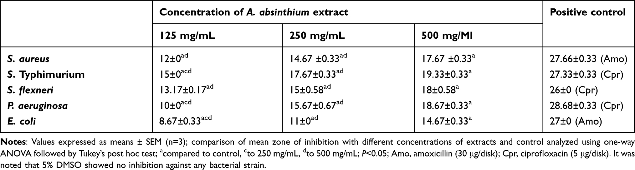 |
Table 1 Antibacterial activity of 80% methanol extract of leaves of A. absinthium against standard bacterial strains using agar-well diffusion, expressed as mean zone of inhibition (mm) |
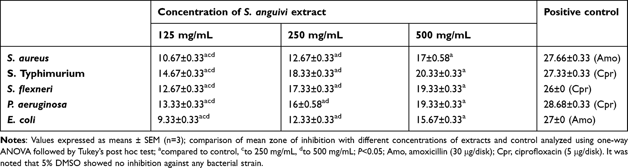 |
Table 2 Antibacterial activity of 80% methanol extract of fruit of S. anguivi against standard bacterial strains using agar-well diffusion, expressed as mean zone of inhibition (mm) |
At a concentration of 500 mg/mL of the fruit extract of S. anguivi, the maximum average zone of inhibition (20.33 mm) was against S. Typhimurium, followed by 19.33 mm against P. aeruginosa and S. flexneri, while the minimum zone of inhibition (15.67 mm) was against E. coli. Similarly, 80% methanol extract of the leaves of A. absinthium induced a maximum zone of inhibition of 19.33 mm against S. Typhimurium at its highest concentration (500 mg/mL), followed by 18.67 mm and 18 mm against P. aeruginosa and S. flexneri, respectively, while the minimum zone of inhibition of 14.67 mm was revealed against E. coli. On the other hand, the maximum average inhibition (17.67 mm) at the highest concentration of the seeds of D. stramonium was against S. flexneri, followed by 15.33 mm against S. aureus, E. coli, and S. Typhimurium, whereas the minimum zone of inhibition of 14.33 mm was recorded against P. aeruginosa (Table 3).
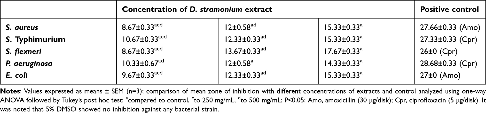 |
Table 3 Antibacterial activity of 80% methanol extract of seeds of D. stramonium against standard bacterial strains using agar-well diffusion, expressed as mean zone of inhibition (mm) |
Comparisons of the mean zone of inhibition of the different concentrations of the extracts of each medicinal plant with one another and with the standard antibacterial disks are presented in Tables 1–3. The average mean zone of inhibition of the extracts of all plants (250 mg/mL) was significantly different (P<0.05) from the average mean zone of inhibition of 500 mg/mL against the growth of each test bacterium. Similarly, the 125 mg/mL extract concentration was significantly different (P<0.05) from the 250 mg/mL one for all bacterial species, except for P. aeruginosa in the case of D. stramonium. Moreover, zones of inhibition of different concentrations of extracts (125, 250, and 500 mg/mL) were significantly different (P<0.05) from their respective positive-control disks. The negative control used here, 5% DMSO, showed no inhibition against any of the bacterial strains. The zone of inhibition of A. absinthium at 500 mg/mL was greater than that of D. stramonium at an equal concentration, with a significant difference (P<0.05) against the tested bacteria, except for E. coli. Similarly greater values of inhibition were observed in S. anguivi extracts than D. stramonium at equal concentrations (500 mg/mL), with significant differences (P<0.05) against S. aureus, S. Typhimurium, S. flexneri, and P. aeruginosa. In contrast, the zone of inhibition of A. absinthium extract was close to S. anguivi at equal concentrations against all tested bacteria. Among the extracts of the three plants tested against the five bacterial pathogens, the fruit extract of S. anguivi showed the highest range of antibacterial activity (9.33–20.33 mm), followed by the extract of the leaves of A. absinthium (8.67–19.33 mm). Among the bacteria tested for susceptibility to the plant extracts, S. Typhimurium was found to be the most susceptible (15.33–20.33 mm), followed by S. flexneri (17.67–19.33 mm). E. coli was the least susceptible (14.67–15.67 mm).
Minimum Inhibitory Concentration
MIC values were determined for extracts of the plants that showed antibacterial properties with ≥7 mm–diameter zone of inhibition in agar well–diffusion tests against all test bacteria species. MIC values of all plant extracts against the tested bacteria ranged from 1.3 mg/mL (S. Typhimurium) to 7.81 mg/mL (P. aeruginosa). MIC values of the leaves of A. absinthium ranged from 1.3 mg/mL (S. Typhimurium) to 6.5 mg/mL (E. coli), whereas for the seeds of D. stramonium it ranged from 1.63 mg/mL (S. flexneri) to 7.81 mg/mL (P. aeruginosa). MIC values of the fruit extracts of S. anguivi varied from 1.3 mg/mL (S. Typhimurium) to 3.25 mg/mL (E. coli), as shown in Table 4. The relatively low MIC values recorded for the S. anguivi and A. absinthium extracts against the test pathogens confirmed the high activity of the extracts at low concentrations. The 5% DMSO showed no inhibition against any of the bacterial strains.
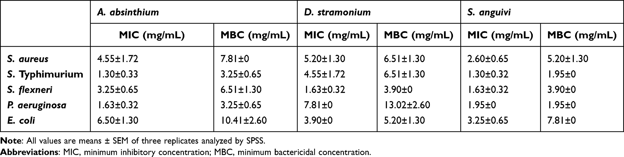 |
Table 4 MIC and MBC of 80% methanol extracts of leaves of A. absinthium, seeds of D. stramonium, and fruit of S. anguivi against tested bacterial species |
Minimum Bactericidal Concentration
For MBC values of extracts, which were determined by subculturing, the contents of wells with concentrations extract greater than or equal to the MIC in the prepared agar plates are presented in Table 4. MBC values of the extracts against the test organisms ranged from 1.95 mg/mL (fruit extracts of S. anguivi against S. Typhimurium and P. aeruginosa) to 13.02 mg/mL (seeds of D. stramonium against P. aeruginosa). Concentrations of 3.25 mg/mL and 1.95 mg/mL (S. Typhimurium and P. aeruginosa) were the minimum values for extracts of the leaves of A. absinthium and fruit of S. anguivi, respectively, and 3.90 mg/mL (S. flexneri) for the seeds of D. stramonium. On the other hand, the highest MBC (13.02 mg/mL for P. aeruginosa) was recorded for D. stramonium, indicating less sensitivity of the bacterium against the extract. The most frequently observed MBC of plant extracts against test bacteria was 6.51 mg/mL. In general, fruit extract of S. anguivi was found to be more potent and killed all tested bacteria at relatively lower concentrations than the other plant extracts. At a concentration of 1.95 mg/mL, unlike the extracts of other plants, 80% methanol extract of S. anguivi induced both bacteriostatic and bactericidal properties (Table 4). The 5% DMSO showed no inhibition against any of the bacterial strains.
Discussion
The present study revealed that all three plant extracts were atoxic, since no treatment-related signs of toxicity were noticed in the mice throughout the observation period at a dose of 2,000 mg/kg body weight. A similar study by Kifleyohannes et al27 also reported that oral administration of 80% methanol extract of A. absinthium produced neither significant toxic signs nor death during an observation period of 14 days after a single administration of 2,000 mg/kg. Likewise, an acute toxicity study by Bouzidi et al28 on D. stramonium administered with a single dose of 100 mg/kg body weight reported that its extract did not show any toxic symptoms, such as paralysis, lacrimation, labored breathing, or death, immediately after administration or at the end of 5 days. Therefore, all the three plant extracts can be considered safe based on the Organization for Economic Co-operation and Development guideline, which recommends a maximum dose for acute toxicity of 2,000 mg/kg.22
The results of this study indicated that all extracts of the three plants had antibacterial effects against both Gram-positive and Gram-negative bacteria with varying degrees of antibacterial activity. This could be due to higher concentrations of bioactive secondary metabolites in the extracts. The zones of inhibition for 80% methanol extracts of A. absinthium and S. anguivi at comparable concentrations were not statistically different for most bacteria against which both extracts were active. This implies that both extracts inhibited the test organisms to a comparable degree. However, there was a slightly lower zone of inhibition for A. absinthium against S. Typhimurium and S. flexneri at equal concentrations. This could be due to differences in composition and concentrations of bioactive secondary metabolites. Unlike A. absinthium and S. anguivi, D. stramonium showed lower antibacterial activities against all the test bacterial strains, regardless of concentration. E. coli was the least susceptible bacteria of the plant extracts at equal concentrations, except for seeds of D. stramonium. A possible reason for this could be differences in mechanisms of action of the bioactive compounds in plant extracts against the different test bacteria, as well as differences in outer cell membrane–permeability barriers of the bacteria.29
The present study revealed that the leaves of A. absinthium had considerable antimicrobial activity against S. Typhimurium, S. aureus, S. flexneri, and P. aeruginosa and moderate inhibitory activity against E. coli, a finding that is in line with a study conducted in Iran by Taherkhani et al30 on essential oil obtained from A. absinthium, which demonstrated significant activity against S. aureus and P. aeruginosa and moderate inhibitory activity against E. coli. Similarly to this finding, methanol leaf extracts of A. absinthium have also been reported to have antimicrobial activity against P. aeruginosa and E. coli, but no significant effect on S. aureus or S. Typhimurium.31 In contrast to our results, Joshi32 reported an absence of activity by essential oil extracted from A. absinthium against S. aureus, E. coli, P. aeruginosa, and S. Typhimurium. This might be due to differences in the geographical location of plant collection, bacterial strains used, extraction methods, and extract concentrations.
The zone of inhibition of 80% methanol extracts of seeds of D. stramonium was lower than that of A. absinthium and S. anguivi at equal concentrations against the individual strains of test bacteria. The reason could be differences in the composition and concentrations of bioactive compounds in the plant extracts. However, the antibacterial property of seeds of D. stramonium extract showed a wide range of zones of inhibition against S. flexneri and moderate activity against P. aeruginosa. In Waza et al,33 the methanol extract of D. stramonium seeds showed the highest zone of inhibition (20 mm) against E. coli, followed by that against S. aureus (17.50 mm) and P. aeruginosa (16 mm). Another study reported that a methanol extract of fruit of D. stramonium inhibited the growth of all tested bacterial strains,34 which is in agreement with the current findings. On the other hand, a study conducted by Julius et al35 revealed poor antibacterial properties of 95% methanol extracts of seeds of D. stramonium against E. coli, P. aeruginosa, and S. aureus, with 7 mm, 6 mm, and 5 mm zones of inhibition at 500 mg/mL, respectively, which might be related to factors like geographic location and extraction method.
The fruit of S. anguivi showed strong activity against S. Typhimurium, S. flexneri, and P. aeruginosa, comparable to the effect of seed extract of D. stramonium, but with less activity against E. coli than the latter. This result suggests that the methanol extract of S. anguivi fruit had a broad spectrum of antimicrobial activity against the tested bacterial strains. A previous study on the fruit of a related plant species (Solanum incanum) found significant antibacterial activity against S. aureus and other pathogenic bacteria.36 The same study reported that S. incanum had several phytochemical constituents, such as alkaloids, flavonoids, phenols, steroids, glycosides, saponins and triterpenoids, which could also be present in S. anguivi.
All plant extracts showed increased dose-dependent inhibitory activity as the concentration increased from 125 to 500 mg/mL. A proportional increase in inhibitory activity (zone of inhibition) as the concentration of extract increases has also been reported in other plants.37 This relationship between the concentrations of extracts and zones of inhibition might be due to the fact that more extracts with active metabolites are able to diffuse into the agar media, inducing more inhibition of microbial growth. Despite the antibacterial activities, the mean zone of inhibition of 80% methanol extracts of all the plants at all test concentrations were not statistically comparable to that of their respective standard control antimicrobials for each of the susceptible bacteria. This might be due to the lower concentration of active principals in all plant extracts against the test bacteria or presence of other chemicals that could also inhibit the effect of the active ingredient. Activity-guided fractionation of the plant extracts could lead to the discovery of more potent compounds in the studied plants.
Antibacterial screening findings in terms of zone of inhibition of the methanol extracts of three plants against susceptible bacteria were inversely proportional to their MIC and MBC values, ie, the more susceptible an organism to an extract in agar-well diffusion, the less its corresponding MIC and MBC values. In other words, the greater inhibition zone of extracts in agar-well diffusion corresponded to lower values of extracts required to inhibit or kill organisms. In the current study, the MIC values of all plant extracts were lower than MBC values, except for the 80% methanol extract of S. anguivi against P. aeruginosa (1.95 mg/mL), which had equal MIC and MBC values, suggesting that the extract had bacteriostatic and bactericidal effects at the same concentration. The higher MIC values indicate that the plant extracts are weaker at killing or inhibiting the tested pathogens, whereas lower MICs reveal that the plant extracts have greater potential of killing the pathogens.
In the current study, most of the plant extracts showed better antimicrobial activity against S. Typhimurium and P. aeruginosa than the other tested organisms, while they were less effective against E. coli. Such differences could be due to selective activity of the bioactive compounds in the plant extracts against different bacterial species due to absence or presence of target sites in the latter. Generally, the methanol extracts of the fruit of S. anguivi exhibited the lowest MIC and MBC, whereas the highest was for D. stramonium. The relatively lower MIC values recorded for the fruit extracts of S. anguivi against the test pathogens confirm the presence of relatively potent antimicrobial agents in its extract. The high activity of antimicrobial agents at low concentrations is essential for the development of compounds for chemotherapeutic purposes, because the lower the dose administered, the lower the chance of its being toxic to patients. The crude extracts of the tested plants had potent antibacterial activity against S. aureus, S. flexneri, S. Typhimurium, P. aeruginosa, and E. coli at all three concentrations tested. These results, together with previous findings from ethnobotanical studies,19,20,34 indicate that these three medicinal plants might have important compounds can be developed and used for the treatment of infectious diseases.
Conclusion
The results of this study revealed that 80% methanol extracts of leaves of A. absinthium, seeds of D. stramonium, and fruit of S. anguivi had antibacterial activities of varying strength and safe at higher doses, and thus have great potential to be developed as antibacterial agents for the treatment of bacterial infections. The antimicrobial activity exhibited by the plant extracts against the pathogenic test organisms used in this study could also be taken as evidence to provide scientific support for continued traditional use of the three medicinal plants by local communities in Ethiopia in the treatment of various diseases caused by bacterial pathogens.
Data Sharing
Research data are stored at the Aklilu Lemma Institute of Pathobiology, Addis Ababa University. Readers may request permission to gain access.
Ethics
Ethics approval for this study was obtained from the Institutional Review Board of the Aklilu Lemma Institute of Pathobiology, Addis Ababa University.
Acknowledgments
First and foremost, we thank the Aklilu Lemma Institute of Pathobiology, Addis Ababa University for financially sponsoring our research work. Our sincere thanks go to Dr Biruhalem Taye, Mr Biruk Zerfu, and Mr Fentaye Kasse for their comments and suggestions during the writing of this paper. We also extend our gratitude to Mrs Yirgalem Gebrehiwot, Mr Haile Alemayehu, Miss Azeb Tekelu, and Mr Jiregna Gemechu for their technical assistance in laboratory work.
Funding
The Office of Research and Technology Transfer, Addis Ababa University financially supported this research (grant TR/036/2016).
Disclosure
The authors declare that they have no competing interests in this work.
References
1. Theuretzbacher U. Resistance drives antibacterial drug development. Curr Opin Pharmacol. 2011;11:433–438. doi:10.1016/j.coph.2011.07.008
2. World Health Organization. Worldwide Country Situation Analysis: Response to Antimicrobial Resistance. Geneva: World Health Organization; 2015.
3. Okeke IN, Lamikanra A, Edelman R. Socioeconomic and behavioral factors leading to acquired bacterial resistance to antibiotics in developing countries. Emerg Infect Dis. 1999;5:18–27. doi:10.3201/eid0501.990103
4. Huynh B, Padget M, Garin B, et al. Burden of bacterial resistance among neonatal infections in low income countries: how convincing is the epidemiological evidence. BMC Infect Dis. 2015;15:127. doi:10.1186/s12879-015-0843-x
5. Mulu A, Moges F, Tessema B, Kassu A. Pattern and multiple drug resistance of bacterial pathogens isolated from wound infection at University of Gondar Teaching Hospital, Northwest Ethiopia. Eth Med J. 2006;44:125–131.
6. Scazzocchio F, Comets MF, Tomassini L, Palmery M. Antibacterial activity of Hydrastis canadensis extract and its major isolated alkaloids. Planta Med. 2001;67:561–563. doi:10.1055/s-2001-16493
7. Mander M, Emana B, Asfaw Z, Busa B. Marketing of Medicinal Plants in Ethiopia: A Survey of the Trade in Medicinal Plants. Addis Ababa: Institute of Biodiversity Conservation; 2006.
8. Petros Z. The need of standardized herbal remedies as alternate sources of antimalarial products in Ethiopia - updated review. Pharmacol Online. 2011;3:1440–1447.
9. Gebre-Mariam T, Neubert R, Schmidt PC, Wutzler P, Schmidtke M. Antiviral activities of some Ethiopian medicinal plants used for the treatment of dermatological disorders. J Ethnopharmacol. 2006;104:182–187. doi:10.1016/j.jep.2005.08.071
10. Teklehaymanot T, Giday M. Ethnobotanical study of medicinal plants used by people in Zegie Peninsula, northwestern Ethiopia. J Ethnobiol Ethnomed. 2007;3:12. doi:10.1186/1746-4269-3-12
11. Wondimu T, Asfaw Z, Kelbessa E. Ethnobotanical study of medicinal plants around ‘Dheeraa’ town, Arsi zone, Ethiopia. J Ethnopharmacol. 2007;112:152–161. doi:10.1016/j.jep.2007.02.014
12. Lulekal E, Asfaw Z, Kelbessa E, Van Damme P. Ethnomedicinal study of plants used for human ailments in Ankober district, North Shewa zone, Amhara region, Ethiopia. J Ethnobiol Ethnomed. 2013;9:63. doi:10.1186/1746-4269-9-63
13. Mesfin F, Demissew S, Teklehaymanot T. An ethnobotanical study of medicinal plants in Wonago district, SNNPR. J Ethnobiol Ethnomed. 2009;5:28. doi:10.1186/1746-4269-5-28
14. Teklehaymanot T. Ethnobotanical study of knowledge and medicinal plants use by people in Dek Island in Ethiopia. J Ethnopharmacol. 2009;124:69–78. doi:10.1016/j.jep.2009.04.005
15. Giday M, Asfaw Z, Woldu Z. Ethnomedicinal study of plants used in Sheko ethnic group of Ethiopia. J Ethnopharmacol. 2010;132:75–85. doi:10.1016/j.jep.2010.07.046
16. Prasad S, Tyagi AK. Traditional medicine: the goldmine for modern drugs. Adv Tech Biol Med. 2015;3:1. doi:10.4172/2379-1764.1000e108
17. Havagiray R, Ramesh C, Sadhna K. Studies on anti-diarrheal activity of Calotropis gigantea R.BR. in experimental animals. J Pharmacol Pharm Sci. 2004;7:70–75.
18. Yineger H, Kelbessa E, Bekele T, Lulekal E. Ethnoveterinary medicinal plants at Bale Mountains National Park. Ethiopia J Ethnopharmacol. 2007;112:55–70. doi:10.1016/j.jep.2007.02.001
19. Yineger H, Kelbessa E, Bekele T, Lulekal E. Plants used in traditional management of human ailments at Bale Mountains National Park, Southeastern Ethiopia. J Med plant Res. 2008;2:132–153.
20. Getaneh S, Girma Z. Ethnobotanical study of medicinal plants in Debre Libanos wereda, central Ethiopia. Afr J Plant Sci. 2014;8(7):366–379. doi:10.5897/AJPS2013.1041
21. Ashebir M, Ashenafi M. Evaluation of the antimicrobial activity of crude preparation of Foeniculum vulgare, Ruta chalepensis and Syzygium guineense on some food borne pathogens. Eth J Pharma. 1999;17:37–43.
22. OECD. Test No. 425: acute oral toxicity: up-and-down procedure. In: OECD Guidelines for the Testing of Chemicals, Section 4. Paris: OECD Publishing; 2008. doi:10.1787/9789264071049-en
23. CLSI. Methods for Dilution Antimicrobial Susceptibility Tests for Bacteria That Grow Aerobically; Approved Standard.
24. Taye B, Giday M, Animut A, Seid J. Antibacterial activities of selected medicinal plants in traditional treatment of human wounds in Ethiopia. Asian Pac J Trop Biomed. 2011;1:370–375. doi:10.1016/S2221-1691(11)60082-8
25. Wasihun Y, Adraro T, Ali S. Evaluation of antibacterial activity and phytochemical constituents of leaf extract of Lippia adoensis. Asia Pac J Energy Environ. 2014;1:45–53. doi:10.18034/apjee.v1i1.209
26. Rouis Z, Abid N, Koudja S, Yangui T, Elaissi A, Cioni PL. Evaluation of the cytotoxic effect and antibacterial, antifungal, and antiviral activities of Hypericum triquetrifolium Turra essential oils from Tunisia. BMC Complement Altern Med. 2013;13:24. doi:10.1186/1472-6882-13-24
27. Kifleyohannes T, Terefe G, Tolossa Y, Giday M, Kebede N. Effect of crude extracts of M. stenopetala and A. absinthium on parasitaemia of mice infected with T. congolense. BMC Res Notes. 2014;7:390. doi:10.1186/1756-0500-7-390
28. Bouzidi A, Mahdeb N, Kara N. Toxicity studies of alkaloids of seeds of Datura stramonium and synthesis alkaloids in male rats. J Med Plants Res. 2011;5:3421–3431.
29. Lopez-Romero JC, González-Ríos H, Borges A, Simões M. Antibacterial effects and mode of action of selected essential oils components against Escherichia coli and Staphylococcus aureus. Evid Based Complement Alternat Med. 2015;9. doi:10.1155/2015/795435
30. Taherkhani M, Rustaiyan A, Rasooli I, Taherkhani T. Chemical composition, antimicrobial activity, antioxidant and total phenolic content within the leaves essential oil of Artemisia absinthium L. growing wild in Iran. Afr J Pharm Pharmacol. 2013;7:30–36. doi:10.5897/AJPP12.945
31. Erel SB, Reznicek G, Konyaliogulu S, Zeybek AU. Antimicrobial and antioxidant properties of Artemisia species from western Anatolia. Turk J Biol. 2012;36:75–84.
32. Joshi RK. Volatile composition and antimicrobial activity of the essential oil of Artemisia absinthium growing in Western Ghats region of North West Karnataka, India. Pharm Biol. 2013;51:888–892. doi:10.3109/13880209.2013.768676
33. Waza SA, Anthony P, Dar S. Phytochemical analysis, antioxidant and antimicrobial activities of methanolic extract of Datura stramonium seeds. Int J Pharm Sci Res. 2015;6:3021–3026.
34. Sharma RA, Sharma P, Yadav A. Antimicrobial screening of sequential extracts of Datura stramonium L. Int J Pharm Pharm Sci. 2013;5:401–404.
35. Julius OO, Oluwasusi VO, Ibyemi MF. Antibacterial and phytochemical screening of crude extracts of leaves and seeds of Datura stramonium. S Asian J Res Microbiol. 2018;2:1–7.
36. Indhumathi T, Mohandass S. Efficacy of ethanolic extract of Solanum incanum fruit extract for its antimicrobial activity. Int J Curr Microbiol App Sci. 2014;3:939–949.
37. Mansoor S, Khan I, Fatima J, Saeed M, Mustafa H. Anti-bacterial, anti-oxidant and cytotoxicity of aqueous and organic extracts of Ricinus communis. Afri J Microbiol Res. 2016;10:260–270. doi:10.5897/AJMR2015.7397
 © 2022 The Author(s). This work is published and licensed by Dove Medical Press Limited. The
full terms of this license are available at https://www.dovepress.com/terms
and incorporate the Creative Commons Attribution
- Non Commercial (unported, 3.0) License.
By accessing the work you hereby accept the Terms. Non-commercial uses of the work are permitted
without any further permission from Dove Medical Press Limited, provided the work is properly
attributed. For permission for commercial use of this work, please see paragraphs 4.2 and 5 of our Terms.
© 2022 The Author(s). This work is published and licensed by Dove Medical Press Limited. The
full terms of this license are available at https://www.dovepress.com/terms
and incorporate the Creative Commons Attribution
- Non Commercial (unported, 3.0) License.
By accessing the work you hereby accept the Terms. Non-commercial uses of the work are permitted
without any further permission from Dove Medical Press Limited, provided the work is properly
attributed. For permission for commercial use of this work, please see paragraphs 4.2 and 5 of our Terms.