Back to Journals » Journal of Pain Research » Volume 16
Evaluating the Extent of Ultrasound-Guided Cervical Selective Nerve Root Block in the Lower Cervical Spine: Evidence Based on Computed Tomography Images
Authors Ma L, Wang Y, Yao M ![]() , Huang B, Deng J, Wen H
, Huang B, Deng J, Wen H
Received 28 November 2022
Accepted for publication 23 February 2023
Published 6 March 2023 Volume 2023:16 Pages 669—676
DOI https://doi.org/10.2147/JPR.S399431
Checked for plagiarism Yes
Review by Single anonymous peer review
Peer reviewer comments 2
Editor who approved publication: Dr Jinlei Li
Ling Ma,1,* Yi Wang,2,* Ming Yao,1 Bing Huang,1 Jiajia Deng,1 Huaichang Wen2
1Department of Anesthesiology and Pain Research Center, The Affiliated Hospital of Jiaxing University, Jiaxing, People’s Republic of China; 2Department of Anesthesiology, First Affiliated Hospital of Wannan Medical College, Wuhu, People’s Republic of China
*These authors contributed equally to this work
Correspondence: Huaichang Wen, No. 2 Zhe shan Street, Wuhu, People’s Republic of China, Email [email protected]
Objective: To verify the injectate dispersal patterns (IDP) and therapeutic outcome of ultrasound-guided cervical selective nerve root block (UG-SCNRB) in treating cervical radiculopathy (CR).
Methods: Overall, 18 CR patients were recruited to undergo UG-SCNRB in the CT room. Following placement of the puncture needle tip between the target nerve root and posterior tubercle, 3 mL of the drug was administered per root (0.33% lidocaine 0.5 mL + Compound betamethasone injection 0.5mL + methylcobalamin injection 1mL + iohexol 1mL). Subsequently, the IDP was assessed on postintervention CT scan images.
Results: In all, 18 participants were analyzed. We injected 21 target cervical nerve roots, namely, 1 C4 nerve, 9 C5 nerves, and 11 C6 nerves. Among the IDPs on postintervention CT scan images, two IDPs were most prevalent, namely, the contrast spread into the extraforaminal spaces (Zone I, the interscalene) in 100% (21/21) of cases, and the foraminal space spread (Zone II) in 61.90% (13/21) of cases. The injectate spread into the epidural spaces (Zone III) in only 2 out of 21 cases (9.52%). The pain relief was significantly improved two hours after surgery, compared to the preoperative VAS pain scores (2 hours, 1.39± 0.50 vs VAS at baseline, P< 0.01). The VAS pain scores during follow-up were significantly lower than preoperation (1 weeks, 1.94± 0.54 vs VAS at baseline; 2 weeks, 2.61± 0.70, P< 0.01 vs VAS at baseline; 4 weeks, 2.67± 0.59, P< 0.01 vs VAS at baseline).
Conclusion: We verified, via CT imaging, that the UG-SCNRB drug diffusion was within safe range (the injectate mainly spread to the extraforaminal spaces), and without any serious complications, such as, intravascular drug injection, extensive diffusion of the epidural space, and general spinal anesthesia.
Keywords: ultrasound-guided, cervical radiculopathy, selective nerve root block, efficacy, tomography
Key Summary Points
Why carry out this study?
We report on / show that limited investigations examined the extent of drug diffusion into the spinal canal after UG-SCNRB.
There is no evidence based on the CT imaging observation in clinic, as US cannot visualize the spinal canal anatomy. This is significant because this study intends to observe the drug diffusion of UG-SCNRB via CT imaging to verify its safety and efficacy.
The drug diffusion of UG-SCNRB was observed by CT imaging for the first time, which further clarified the risk characteristics of ultrasound guided injection, including spinal cord injury, unconfirmed epidural diffusion and inability to exclude intravascular blood flow.
What was learned from the study?
This study shows that the UG-SCNRB drug diffusion was within safe range (the injectate mainly spread to the extraforaminal spaces), and without any serious complications, such as, intravascular drug injection, extensive diffusion of the epidural space, and general spinal anesthesia.
Introduction
Cervical spondylosis (CS) is the fourth leading contributor to disability in the United States, and it severely affects patients’ daily lives. Cervical radiculopathy (CR) is the most common form of CS, and its incidence accounts for about 50–60% of CS.1 The average annual CR incidence is estimated to be approximately 83.2 per 100,000 population.2 Cervical intervertebral disc herniation, articular facet joint, uncinate joint hyperplasia and hypertrophy, intervertebral disc height reduction, ligamentum flavum hypertrophy, and other degenerative stimulation or mechanical compression of cervical nerve root are known to cause upper limb pain in patients.
Most CR patients can relieve pain using conservative treatments, such as, oral non-steroidal drugs and neuropathic pain drugs, transcutaneous electrical nerve stimulation, mechanical traction, and cervical collar. For patients with poor pain control with conservative treatment, minimally invasive treatments are generally provided, such as, selective cervical nerve block. Selective cervical nerve block (SCNRB) is a commonly used nerve block for CR treatment.3 Corticosteroids and local anesthetics can be directly injected around the inflammation-stimulated nerve roots to relieve pain and reduce local inflammation, thereby providing patients with good short-term symptom relief.4
In the past, cervical nerve root blocks were mostly performed under the guidance of X-ray fluoroscopy or computed tomography (CT).5 The aforementioned methods were primarily used to determine the puncture path through bony landmarks. However, they have poor visualization of blood vessels and nerves, hence, these may lead to accidental intravascular injections, and subsequent nerve damage.6–8 Ultrasound (US) not only displays target nerves, blood vessels, and surrounding tissues, but also reveals the positioning of the puncture needle, as well as the diffusion of the medicinal solution around the nerve in real time during the puncture process.9,10 This can markedly enhance the accuracy and safety of the puncture. In addition, US can protect patients and medical staff from radiation exposure. Till date, limited investigations examined the extent of drug diffusion into the spinal canal after UG-SCNRB. Moreover, there is no evidence based on the CT imaging observation in clinic, as US cannot visualize the spinal canal anatomy. Thus, this study intends to observe the drug diffusion of UG-SCNRB via CT imaging to verify its safety and efficacy.
Materials and Methods
Patients
This investigation received ethical approval from China Registered Clinical Trial Ethics Committee (ChiECRCT20200032). All patients read and signed the informed consent, and the patient’s name was not disclosed in the data of this study. The trial was registered prior to patient enrollment at China Clinical Trial Registration Center (ChiCTR2000029859, link to registry page: http://www.chictr.org.cn/showproj.aspx?proj=48984, Principal investigator: Wen Huaichang, Date of registration: 2020/2/15).”
Eighteen patients, aged between 40 and 70 years, who were diagnosed with CR at our hospital between June 2022 and October 2022, and underwent UG-SCNRB, were enrolled in the study.
Inclusion and Exclusion Criteria
The following patients were eligible for analysis: (1) CR, the diagnostic criteria: Clinical symptoms, physical examination, and confirmation of the unilateral disc herniation via cervical CT or magnetic resonance imaging (MRI); (2) Patients aged >18 years; (3) Lower cervical radicular pain lasting ≤3 months; (4) Numerical rating scale, NRS≥ 4.
The following patients were excluded from analysis: (1) Severe heart disease; (2) Severe spinal deformity; (3) Hypersensitivity to local anesthetics or hormones; (4) Coagulation dysfunction; (5) Systemic infection or skin infection at the puncture site; (6) Patients with abnormal mental behavior, severe anxiety, or depression; (7) Lactating and pregnant women; (8) History of cervical surgery; (9) Cervical spondylotic myelopathy; (10) Moderate and severe foraminal stenosis.
Procedures
UG-SCNRB was conducted based on an improved version of the Narouze et al technique.11,16 All procedures were performed by the same experienced pain physician. Prior to puncture, the target nerve root was determined according to the patient’s clinical symptoms, signs and imaging examinations. Patients were placed in a lateral decubitus position prior to US examinations via a standard US device (Sonosite X-PORTE, UJIFILM Sonosite, Inc., Bothell, Washington) and a high-frequency linear array transducer (6–13 MHz). The skin was prepped with povidone-iodine, and meticulous sterile procedures were followed throughout the operation. In order to prevent accidental puncture into vertebral artery, ascending carotid artery, deep carotid artery and V1 segment of vertebral artery were identified by color Doppler. The C4-C7 nerve roots were recognized based on the shape of the transverse process on US; the C7 transverse process contains a rudimentary anterior tubercle and a prominent posterior tubercle, whereas, the C4, C5, and C6 transverse processes have prominent anterior and posterior tubercles. The target transverse process was recognized via slow probe movement in all directions, while maintaining the C7 transverse process as a reference point.11 Subsequently, employing the in-plane technique, a 60-mm, 23-G needle was slowly inserted in the direction of the posterior nerve root edge under real-time US guidance. The needle was introduced just lateral of the transducer, prior to advance from the posterolateral to anteromedial direction, using an in-plane approach (Figure 1). The optimal image of the nerve root, location of the radicular artery, and surrounding vessels near the border of the nerve root were obtained through probe manipulation in the power and color Doppler modes. Once the tip of the puncture needle reached between the target nerve root and posterior tubercle, 3 mL of the drug was administered per root (0.33% lidocaine 0.5 mL + Compound betamethasone injection 0.5mL + methylcobalamin injection 1mL + iohexol 1mL). Following this, the needle tip and IDP positioning was examined in postintervention CT scan images (Figure 2).
 |
Figure 1 (A) Ultrasound-guided selective cervical nerve root block (UG-SCNRB). (B–D) Axial transverse ultrasound image depicting the sharp anterior tubercle (at) of the cervical vertebra transverse process. Solid arrowheads reveal the location of needle placement between the target nerve root and posterior tubercle. ((B) C4; (C) C5; (D) C6). Abbreviations: CA, carotid artery; IJV, internal jugular vein; VA, vertebral artery; sap, superior articular process; ScM, scalenus medius; ScA, scalenus anterior; N represents nerve root; pt, posterior tubercle. |
 |
Figure 2 Cervical nerve root block on CT scan images: The yellow arrow points to the needle in front of the posterior tubercle.((A) C4; (B) C5; (C) C6). |
Review of Clinical Data
Prior to administration, the data regarding the demographic characteristics, such as, pain duration, pain severity, and involved nerve root were extracted. Two radiologists retrospectively analyzed and recorded the IDP on postintervention CT scanning images. The injection spread patterns in the cross-sectional CT images included the following: Zone I: extra-foraminal; Zone II: the foraminal spaces; Zone III: intra-foraminal (Figure 3).
 |
Figure 3 The injection distribution area in the cross section of CT image: Line A is from anterolateral vertebral body to the lateral margin of the facet. Line B is from posterior-lateral vertebral body to the interior margin of the facet. Line C is the axial centerline of the epidural space. Zone I The out space of line A is extra-foraminal; Zone II: Between line A and B is the foraminal spaces; Zone III: Between line B and C is intra-foraminal/epidural spaces. |
An investigator blinded to the patient assignments/treatments performed patient follow-ups, and recorded pain scores, particularly, NRS during hospital visits at 2 hours, 1 week, and 4 weeks after injection.
Safety was assessed as follows: ① Bleeding situation: Prior to drug injection, we recorded whether there was blood upon withdrawal, and verified the presence or absence of hematoma via CT scan. ② Other adverse reactions, including, puncture point pain, shortness of breath, paresthesias, motor deficit, hematoma, dizziness, headache, vomiting, general spinal anesthesia, and so on.
Statistical Analysis
SPSS version 25.0 software (IBM Corp., Armonk, NY, USA) was employed for all data analyses, and data are presented as mean standard deviation for quantitative data and percentages for categorical data. Pre- and post-intervention VAS comparison was performed with paired t-test. A P value of <0.05 was set as the significance threshold.
Results
Overall, 18 participants were recruited for analysis. The mean age was 52.67±9.12 (33–69) years old, and five patients were male (27.78%) and 13 (72.22%) were female. 5 patients exhibited C4/5 disc-related disease, 14 displayed C5/6 disc-related disease, and 3 showed C6/7 disc disease. The patient profiles are summarized in Table 1.
 |
Table 1 Baseline Patient Profile |
In our study, we successfully identified the anterior and posterior tubercles of all target cervical transverse processes, and the hypoechoic nerve roots between them, using US. Once the puncture needle reached the target nerve, we once again confirmed that all the puncture needles were located at the target point on the CT image. Among the 18 patients, 3 underwent 2 nerves root block. So, we injected 21 target cervical nerve roots, including, 1 C4 nerve, 9 C5 nerves, and 11 C6 nerves. The IDP on postintervention CT scanning images revealed two most common IDPs: the contrast spread in the extraforaminal spaces (Zone I, The interscalene) in 100% (21/21) of cases, and in the foraminal spaces (Zone II) in 61.90% (13/21) of cases. The injectate spread to the epidural spaces (Zone III) in only 2 of 21 cases (9.52%)(Table 2). In addition, we observed the injectate spread to the transverse foramen in 80.95% (17/21) of cases.
 |
Table 2 The Injectate Dispersal Patterns (IDP) |
Two hours after the procedure, pain relief was markedly improved, compared to VAS pain scores at baseline (2 hours, 1.39±0.50 vs VAS at baseline, P<0.01). The VAS pain scores throughout the observation period after discharge was significantly lower than preoperation (1 weeks, 1.94±0.54 vs VAS at baseline; 2 weeks, 2.61±0.70, P<0.01 vs VAS at baseline; 4 weeks, 2.67±0.59, P<0.01 vs VAS at baseline) (Table 3). No complications or adverse effects were observed during the trial.
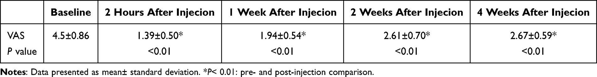 |
Table 3 Comparison of the VAS Scores at Baseline and After US-CSNRB |
Discussion
The primary advantage of US is the direct observation of soft tissue structures, namely, blood vessels, nerves and muscles. It also enhances the visualization of needle tip positioning, as well as IDP around the nerves in real time. Therefore, it is particularly helpful to minimize tissue injury and reduce complication risk, such as, intravascular injection, vascular injury, or nerve injury, and haematomas.12 In addition, this method can prevent medical personnel and patients from ionizing radiation exposure.13 However, US cannot penetrate the bone, thus, the diffusion of medicine within the spinal canal cannot be determined. Therefore, the main objective of this investigation was to verify IDP and therapeutic efficacy of UG-SCNRB based on CT images.
Herein, we successfully identified the anterior and posterior tubercles of all target cervical transverse processes and the hypoechoic nerve roots between them under US. Once the puncture needle reached the target nerve, we confirmed yet again that all puncture needles were located at the target point on the CT image.
The two most common IDPs on the postintervention CT scanning images included the contrast spread into the extraforaminal spaces in 100% (21/21) of cases, and to the foraminal spaces in 61.90% (13/21) of cases. The injectate spread to the epidural spaces in only 2 of 21 cases (9.52%). No complications or adverse effects were observed during the trial. Hence, our results demonstrated that the UG-SCNRB has high accuracy. However, this small study can not guarantee the safety of the UG-SCNRB, and future investigations are warranted involving a large patient population.
At present, CT and fluoroscopy-guided transforaminal epidural injections are still the primary minimally invasive treatments for CS.14 The target area is anterior to the facet joint in the cervical spine,15 otherwise known as the extraforaminal foramen. Using this location, the drug can easily enter the epidural space, thereby alleviating inflammation of the dorsal root ganglia or nerve roots. However, CT and fluoroscopy-guidance has the disadvantage of low soft tissue recognition, such as, blood vessels and nerves, and they are unable to monitor the needle tip position in real time, thereby, resulting in the accidental puncturing of blood vessels and nerve. In this study, we performed UG-SCNRB, whereby the target was located between the anterior and posterior nodules of the transverse cervical processes. US can clearly display the nerve roots associated with the anterior and posterior nodules of the cervical spine and between them. Narouze et al16 proposed that this form of cervical injection be called “cervical selective nerve root block”, instead of cervical foraminal epidural injection (CTFEI), because they “could not monitor the diffusion of the injection through the foramen into the epidural space” under US guidance. The dural sheath of the spinal nerve is fused to the epineurium in or above the intervertebral foramen.17 Therefore, according to the anatomical theory, selective cervical nerve root block between the anterior and posterior nodes will not lead to an unexpected epidural block. In this study, the contrast agent did not enter the spinal canal in most cases, and, in only 2 cases, the contrast agent diffused into the epidural posterior space on the puncture side. However, the drug diffusion range was limited to the corresponding cervical spine segment, which may be due to the limited dosage of the injected drug. Thus, no extensive epidural block occurred. It is necessary to further study the diffusion range of UG-SCNRB with varying injection doses. Therefore, based on the current evidence, we should not use large dose and high concentration of local anesthetics when conducting UG-SCNRB.
Our study showed significant pain relief following UG-SCNRB, consistent with earlier literature.18–20 US-guided transforaminal injection of the cervical spine was similarly precise in injecting the solution near the inflammatory nerve roots or dorsal root ganglia, resulting in significant relief of pain by blocking pain transmission, eliminating radicular inflammation, and regulating dorsal root ganglion excitability.
This investigation had certain limitations. Firstly, the patient population was relatively small, and a conclusion based on multi-center and large sample prospective studies may be more convincing; Secondly the injection dose in our study was single, only 3mL of injection drug was used, so it is necessary to further study the IDP of different injection doses. Thirdly, all blocks were performed by the same experienced physician, which may generate some bias in expertise.
In conclusion, we confirmed using CT images that the drug diffusion of UG-SCNRB was within a safe range (the injectate mainly spread into the extraforaminal spaces), and there were no serious complications, such as, intravascular drug injection, extensive diffusion into the epidural space and general spinal anesthesia.
Funding
This work was supported by the Affiliated Hospital of Jiaxing University. The Rapid Service Fee was funded by the authors.
Disclosure
Ling Ma, Yi Wang are co-first authors for this study. The authors report no conflicts of interest in this work.
References
1. Cohen SP. Epidemiology, diagnosis, and treatment of neck pain. Mayo Clin Proc. 2015;90(2):284–299. doi:10.1016/j.mayocp.2014.09.008
2. Mansfield M, Smith T, Spahr N, et al. Cervical spine radiculopathy epidemiology: a systematic review. Musculoskeletal Care. 2020;18(4):555–567. doi:10.1002/msc.1498
3. Ehsanian R, Schneider BJ, Kennedy DJ, et al. Ultrasound-guided cervical selective nerve root injections: a narrative review of literature. Reg Anesth Pain Med. 2021;46(5):416–421. doi:10.1136/rapm-2020-102325
4. House LM, Barrette K, Mattie R, et al. Cervical epidural steroid injection: techniques and evidence. Phys Med Rehabil Clin N Am. 2018;29(1):1–17.
5. Julien Nathalie J, Bureau TP, Moser AG, et al. CT fluoroscopy-guided transforaminal and intra-articular facet steroid injections for the treatment of cervical radiculopathy: injectate distribution patterns and association with clinical outcome. Eur Radiol. 2020;30(11):5933–5941.
6. Rozin L, Rozin R, Koehler SA, et al. Death during transforaminal epidural steroid nerve root block (C7) due to perforation of the left vertebral artery. Am J Forensic Med Pathol. 2003;24(4):351–355.
7. Wallace MA, Fukui MB, Williams RL, et al. Complications of cervical selective nerve root blocks performed with fluoroscopic guidance. Am J Roentgenol. 2007;188(5):1218–1221.
8. Smuck M, Maxwell MD, Kennedy D, et al. Utility of the anesthetic test dose to avoid catastrophic injury during cervical transforaminal epidural injections. Spine J. 2010;10(10):857–864.
9. Galiano K, Obwegeser AA, Bodner G, et al. Ultrasound-guided periradicular injections in the middle to lower cervical spine: an imaging study of a new approach. Reg Anesth Pain Med. 2005;30:391–396.
10. Gofeld M. Ultrasonography in pain medicine: a critical review. Pain Pract. 2008;8(4):226–240.
11. Hyuk JJ, Yong LW, Woo KJ, et al. Ultrasound-guided selective nerve root block versus fluoroscopy-guided interlaminar epidural block versus fluoroscopy-guided transforaminal epidural block for the treatment of radicular pain in the lower cervical spine: a retrospective comparative study. Pain Res Manag. 2020;2020. doi:10.1155/2020/9103421
12. Brouwers PJ, Kottink EJ, Simon MA, et al. A cervical anterior spinal artery syndrome after diagnostic blockade of the right C6-nerve root[J]. Pain. 2001;91:397–399.
13. Finlayson RJ, Gupta G, Alhujairi M, et al. Cervical medial branch block: a novel technique using ultrasound guidance. Reg Anesth Pain Med. 2012;37:219–223.
14. Chung JY, Yim JH, Seo HY, et al. The efficacy and persistence of selective nerve root block under fluoroscopic guidance for cervical radiculopathy. Asian Spine J. 2012;6(4):227–232.
15. Desai A, Saha S, Sharma N, et al. The short- and medium-term effectiveness of CT-guided selective cervical nerve root injection for pain and disability. Skeletal Radiol. 2014;43(7):973–978.
16. Narouze SN, Vydyanathan A, Kapural L, et al. Ultrasound-guided cervical selective nerve root block: a fluoroscopy- controlled feasibility study. Reg Anesth Pain Med. 2009;34:343–348.
17. Standring S. Gray’s Anatomy. London: Churchill Livingstone; 2008.
18. Yamauchi M, Suzuki D, Niiya T, et al. Ultrasound-guided cervical nerve root block: spread of solution and clinical effect. Pain Med. 2011;12(8):1190–1195.
19. Zhang X, Shi H, Zhou J, et al. The effectiveness of ultrasound-guided cervical transforaminal epidural steroid injections in cervical radiculopathy: a prospective pilot study. J Pain Res. 2018;12:171–177.
20. Dernek B, Ulusoy İ, Aydoğmuş S, et al. Ultrasound-guided cervical selective nerve block: a case series. J Back Musculoskelet Rehabil. 2022;35(5):1013–1019.
 © 2023 The Author(s). This work is published and licensed by Dove Medical Press Limited. The
full terms of this license are available at https://www.dovepress.com/terms
and incorporate the Creative Commons Attribution
- Non Commercial (unported, 3.0) License.
By accessing the work you hereby accept the Terms. Non-commercial uses of the work are permitted
without any further permission from Dove Medical Press Limited, provided the work is properly
attributed. For permission for commercial use of this work, please see paragraphs 4.2 and 5 of our Terms.
© 2023 The Author(s). This work is published and licensed by Dove Medical Press Limited. The
full terms of this license are available at https://www.dovepress.com/terms
and incorporate the Creative Commons Attribution
- Non Commercial (unported, 3.0) License.
By accessing the work you hereby accept the Terms. Non-commercial uses of the work are permitted
without any further permission from Dove Medical Press Limited, provided the work is properly
attributed. For permission for commercial use of this work, please see paragraphs 4.2 and 5 of our Terms.