")
Back to Journals » Open Access Journal of Sports Medicine » Volume 12
Eponyms in Pediatric Sports Medicine: A Historical Review
Authors Bayomy AF , Forrester LA , Crowley SG, Popkin CA
Received 19 October 2020
Accepted for publication 8 December 2020
Published 14 January 2021 Volume 2021:12 Pages 11—22
DOI https://doi.org/10.2147/OAJSM.S287663
Checked for plagiarism Yes
Review by Single anonymous peer review
Peer reviewer comments 3
Editor who approved publication: Prof. Dr. Andreas Imhoff
Ahmad F Bayomy,1 Lynn Ann Forrester,1 Stephen G Crowley,2 Charles A Popkin1
1Department of Orthopedic Surgery, Columbia University Medical Center, New York, NY, USA; 2Department of Orthopedic Surgery, Albany Medical College, Albany, NY, USA
Correspondence: Charles A Popkin
Department of Orthopedic Surgery, Columbia University Medical Center, 3959 Broadway 8 th Floor, New York, NY 10032, USA
Tel +1 (212) 305-4787
Email [email protected]
Abstract: The use of eponyms in the orthopedics literature has come under scrutiny, and there is a growing body of literature evaluating the utility of these terms in modern healthcare delivery. Although the field of pediatric orthopedic sports medicine is a relatively modern subspecialty, it is built on a foundation of over 100 years of pediatric musculoskeletal medicine. As a result, eponyms account for a significant portion of the vernacular used in the field. The purpose of this review is to summarize and describe the history of common eponyms relevant to pediatric sports pathology, examination maneuvers, classification systems, and surgical procedures. Use of eponyms in medicine is flawed. However, an improved understanding of these terms allows for informed use in future scientific discourse, patient care and medical education and may encourage future innovation and research into understanding pediatric orthopedic pathologies.
Keywords: eponyms, orthopedics, sports medicine, pediatrics, Galeazzi, Greulich, Pyle, Beighton, Horan, apophysitis
Background
The field of pediatric orthopedic sports medicine is a relatively modern subspecialty built on a foundation of over 100 years of pediatric musculoskeletal medicine. In a recent study investigating the completion of multiple fellowships in orthopedic subspecialties, the only fellowship pairing that continues to increase is pediatric orthopedics and orthopedic sports medicine.19 As the field continues to grow, the terminology used to describe physical exam maneuvers, pathologic states, classification systems and treatment options for common pediatric orthopedic sports medicine pathologies is likely to evolve. A commonly used form of terminology used in the practice of pediatric sports medicine is the eponym, or an entity that has been named after a physician that has usually played a significant role in describing said entity.24 Recently, orthopedic surgeons have debated the clinical utility of eponyms, citing their purported limitations, inappropriate use and historical inaccuracy.39,57,82,86 However, given that eponyms serve as a convenient form of short hand, they are likely here to stay.71 Additionally, eponyms serve as a reminder that the field of orthopedic surgery was built on a foundation of innovative scientific inquiry and devotion to improving the lives of others. Several previously published articles have sought to preserve and study the history of eponyms in orthopedic surgery.9,57,70,78 However, none have examined the emerging field of pediatric sports medicine. The purpose of the review is to describe the history of the most commonly used eponyms in pediatric sports medicine, characterize the current use of these eponyms, and describe current research regarding the clinical application of these eponyms.
Physical Exam Maneuvers
Wilson’s Test
The Wilson’s Test is a provocative knee maneuver used in the clinical evaluation of osteochondritis dissecans of the medial femoral condyle. The test is considered positive if pain is produced with internal rotation of the tibia during knee extension, and then relieved with external rotation of the tibia. This maneuver is attributed to Dr. James N. Wilson (1919–2006), and was first described in his 1967 case series, “A Diagnostic Sign in Osteochondritis Dissecans of the Knee.”83 There is debate concerning the specificity of the Wilson’s test – in a 2003 retrospective review of 32 patients, 75% had osteochondritis dissecans on radiographs, yet had a negative test.17 Nevertheless, this exam maneuver is still frequently used today in the clinical setting as it was originally described. Dr. Wilson was a British orthopedic surgeon who completed his medical training at Birmingham University. In addition to first describing the eponymous Wilson’s test, his many accomplishments include developing the Stanmore total hip arthroplasty and devising an osteotomy for the correction of adolescent hallux valgus. He also established multiple British bone tumor registries, and served as an editorial board member of the Journal of Bone and Joint Surgery. For his domestic and international contributions to orthopedic surgery, he was appointed Order of the British Empire in 1995.63
Lachman Test
The Lachman Test is one of several physical exam maneuvers used in the clinical evaluation of anterior cruciate ligament (ACL) injuries, and is performed by translating the tibia anteriorly relative to the femur with the knee in 20–30 degrees of flexion.51 When the test is positive, suggestive of a torn ACL, there is a soft endpoint to this anterior translation. The Lachman test is attributed to Dr. John W. Lachman (1919–2007), and was first described in the literature in 1976 by Torg et al.76 The grading of the test with respect to excursion and firmness of endpoint (eg, Grade II) was later described in 1986 by Gurtler et al.35 Today, the Lachman test is still used as it was originally described, and has been shown to be the most sensitive physical diagnostic test for the diagnosis of ACL tear.58 Dr. Lachman was a Philadelphia native who served as a Professor of Orthopedic Surgery and Chairman of the Department of Orthopedics at Temple University. He retired from medical practice in 1998.48
Pathologic States
Sinding-Larsen-Johansson Disease
Sinding-Larsen-Johansson (SLJ) disease is defined as traction apophysitis at the insertion of the patellar tendon into the inferior aspect of the patella.21 This disease was first characterized in the 1920’s by two Scandinavian surgeons, Dr. Christian M. F. Sinding-Larsen (1866–1930) and Dr. Sven C. Johansson (1880–1959) (Figure 1). In 1921, Dr. Sinding-Larsen published a case series describing 10 girls who experienced pain at the inferior pole of the patella after dancing and jumping. These children were found to have radiographic abnormalities in the same region, and were given the presumptive diagnosis of epiphysitis secondary to overstrain.68 Three years later, Dr. Johansson published a comparable case series describing 4 children with similar presentation.43 In 1932, Dr. Paul W. Greeley contended that Dr. Johansson was unaware of Larsen’s paper, and thus recommended that osteochondrosis of the inferior pole of the patella be named “Larsen-Johansson’s disease.”32 Dr. Johansson was a Swedish physician who contributed substantially to the research literature for various surgical fields. He also served as head physician of the Gothenburg hospital from 1914 to his retirement.87 Dr. Sinding-Larsen was a Norwegian physician with expertise in the surgical management of tuberculosis. He served as physician-in-chief of the Kysthospitalet Hospital for Children with Tuberculosis early in his career, and later served 19 years as the director of the Rikshospitalet, the national research hospital.88
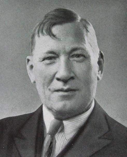 |
Figure 1 Dr. Sven Christian Johansson. |
Osgood-Schlatter Disease
Osgood-Schlatter Disease has a similar pathophysiology to SLJ disease, and is defined as traction apophysitis of the tibial tubercle.14 This pathology was characterized in a rigorous fashion for the first time by Dr. Robert Bayley Osgood (1873–1956) (Figure 2) and Dr. Carl Schlatter (1864–1934) (Figure 3) in 1903.52,65 Although several physicians had previously described similar cases in the late 1800s, Osgood and Schlatter each independently described the condition in more discrete terms than their predecessors, and thus, apophysitis of the tibial tubercle was named after Osgood and Schlatter.72 Additionally, the two 1903 publications occurred after the introduction of the “Roentgen ray” and thus were the first to provide radiographic evidence of the disease. Dr. Osgood grew up in New England and trained at Massachusetts General Hospital. After serving as an army orthopedic surgeon in England and France during World War I, Dr. Osgood returned to Boston to serve as Chief of Orthopedics at Massachusetts General Hospital. He was later appointed Professor of Orthopedic Surgery at Harvard Medical School and Chief of Orthopedics at Boston Children’s Hospital.53 Dr. Schlatter was a Swiss general and trauma surgeon who reportedly performed the first total gastrectomy in 1897. He later served as a military surgeon during World War I, and at the completion of his service, was appointed Professor of Surgery at the University Clinic in Zurich.20,47
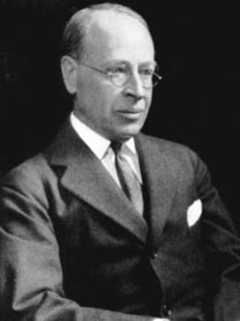 |
Figure 2 Dr. Robert Bayley Osgood. |
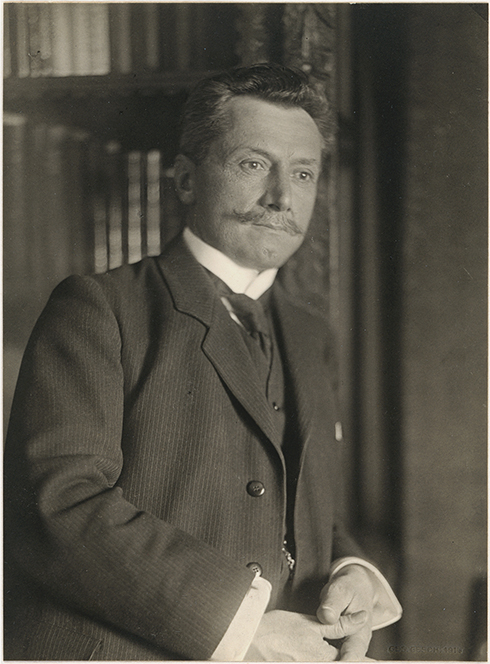 |
Figure 3 Dr. Carl Schlatter. |
Sever’s Disease
An additional form of traction apophysitis with an eponymous name is Sever’s disease, defined as apophysitis at the Achilles tendon insertion into the calcaneus.42 Dr. James W. Sever (1878–1964) first identified patients with this disease entity in his 1912 case series, “Apophysitis of os calcis.”67 In this series, he described children with painful heels, persistent limps, and radiographic changes in the calcaneal apophysis.61 Dr. Sever was a New England native who trained and practiced at Harvard Medical School and Boston Children’s Hospital until his retirement in 1951.85
Panner’s Disease
An additional pathologic state commonly observed in active children is Panner’s disease, defined as osteochondrosis of the humeral capitellum.16 This entity was first described by the radiologist Dr. Hans Jessen Panner (1871–1930) at the 1927 meeting of the Northern Association of Medical Radiology in Copenhagen. He later published his findings in his 1929 paper, “A Peculiar Affect of the Capitulum Humeri, Resembling Calve-Perthes Disease of the Hip,” where he described a disease of the humeral capitellum comparable to “Calve-Perthes disease of the hip.”54 Although a 1959 review by Haraldsson identified clinical descriptions of osteochondrosis of the humeral capitellum that appeared as early as 1916, Panner was recognized as the first to provide a clinical description of this disease entity with sufficient detail.36 Dr. Panner was born in Denmark and completed his medical training at the University of Copenhagen. Over the course of his career in radiology, Panner served as president of the Danish Radiological Society and co-editor of Acta Radiologica. In addition to characterizing osteochondrosis of the humeral capitellum, Panner also significantly contributed to the field of orthopedic surgery by contributing to the understanding of avascular necrosis of the navicular bone, referred to today as Kohler’s disease.6
Hegemann’s Disease
Hegemann’s disease has a pathogenesis similar to Panner’s disease, and is defined as osteochondrosis of the humeral trochlea.15 Avascular necrosis of the trochlea was first reported in 1933. However, in his comprehensive 1951 analysis of 1200 elbow radiographs, Dr. Gerd Hegemann (1912–1999) identified 3 cases of aseptic osteonecrosis of the humeral trochlea.37 Thus, osteochondrosis of the trochlea was subsequently referred to as Hegemann’s disease. Dr. Hegemann was a Professor of Orthopedic Surgery and Chair of the Department of Surgery at Erlangen Hospital, Germany. He was a prolific researcher and innovative surgeon who most notably presided over a fourfold expansion in surgical volume at Erlangen Hospital, and performed one of the first open heart surgeries in Germany.38
Classification Systems
Watanabe Classification of the Discoid Meniscus
The Watanabe classification system of the discoid meniscus describes the arthroscopic appearance of a patient’s meniscus according to three types: complete (Type I), incomplete (Type II), and Wrisberg ligament (Type III). This classification system was first described in the second edition of the Atlas of Arthroscopy (1969) by the Tokyo-based surgeons Dr. Masaki Watanabe (1911–1994), Dr. Sakae Takeda, and Dr. Hiroshi Ikeuchi.84 The authors credited Amako with first classifying lateral discoid meniscus into types I–IV, and based their classification system on his work. Although there is some debate concerning the clinical utility of this system, it is still in frequent use in the clinical setting.79 However, the classification system is rarely used in the research setting, based on review of the literature. Dr. Watanabe studied medicine at the University of Tokyo and served as Chief of the Tokyo Teishin Hospital.41 Dr. Watanabe has been described as the “father of modern arthroscopy” and is credited with transitioning the arthroscope from diagnostic to therapeutic tool. He performed the first known arthroscopic intervention on May 4, 1962, a partial medial meniscectomy in a 17-year-old boy who twisted his knee playing basketball.18
Meyers and McKeever Classification for Tibial Spine Avulsions
The Meyers and McKeever classification system characterizes tibial spine fractures on plain radiographs. This system classifies Type I fractures as nondisplaced, Type II as hinged, Type III as completely displaced without rotation, and Type III+ as completely displaced with rotation.33 Although this system has been used in clinical practice for decades, recent research suggests that there may be unacceptably low intraobserver and interobserver reliability when using this classification.22 Additionally, a 2018 study comparing the standard Meyers and McKeever classification to an MRI-based classification system found that use of MRI led to an increase in fracture grade and subsequent changes in treatment in 32.5% of cases.33 Therefore, additional research is needed to characterize the utility of the Meyer and McKeever in the practice of modern orthopedic surgery. Dr. Marvin H Meyers (1918–2004) and Dr. Francis M McKeever (1901–1973) first described this classification system in 1959,49 though subsequently elaborated on their findings in 1970.50 Dr. Meyers (1918–2004) was born in Connecticut, and grew up blind in one eye after a childhood injury. He completed his medical training at the University of California Berkeley and then moved to southern California to practice orthopedic surgery.5 He was later appointed Associate Professor of Orthopedic Surgery at the University of Southern California, and Residency Program Director of the County General Hospital in Los Angeles. He was also committed to research in orthopedic surgery and served on the editorial board for JBJS.5 Dr. McKeever (1901–1973) was born in California and completed his medical training at the University of Southern California. After further training in orthopedic surgery at the Los Angeles County General Hospital and Massachusetts General Hospital, he returned to California to enter private practice. During World War II, Dr. McKeever initially entered the Medical Corps of the United States Army, and was subsequently appointed Chief of the Surgical Service and the Percy Jones General Hospital in Michigan. At the completion of his service, he returned to California where he became a Professor of Orthopedic Surgery and Chair of the Orthopedics Department at the University of Southern California.80
Caton-Deschamps Index
Although there have been myriad methods used to assess patellar height on lateral radiographs of the knee, the Caton-Deschamps Index (CDI) has been shown to have the highest interobserver reliability, even when compared to the commonly used Insall-Salvati index.4 This method was first reported by Dr. Jacques H. Caton (Figure 4) and Dr. Gerard P. Deschamps (Figure 5) in their 1982 study of “patella infera,” or low-riding patella.12 The measurement can be performed on a radiograph with the knee in any position from 10 to 80 degrees of flexion. The index is then calculated by dividing the distance from the inferior edge of the patella articular surface to the anterosuperior angle of the tibial plateau by the cranial-caudal length of the patella articular surface.55 Although the CDI has high interobserver reliability, the index can be influenced by a patient’s age, because patella ossification begins at the proximal portion of the patella and progresses in an age dependent manner.73 Therefore, the use of the CDI in the clinical setting may be strengthened by established an age-based CDI for reference. Dr. Deschamps and Dr. Caton currently practice orthopedic surgery in France, and Dr. Caton recently modified the original CDI for use in total knee arthroplasty.59
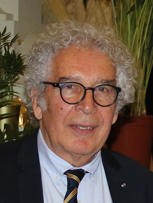 |
Figure 4 Dr. Jacques H. Caton. Photo provided by Dr. Caton. |
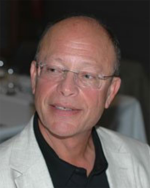 |
Figure 5 Dr. Gerard Deschamps. Photo provided by Dr. Caton. |
Beighton-Horan Score
The Beighton-Horan score is a screening tool used in the assessment of hypermobility. The tool evaluates 5 maneuvers, four passive bilateral and one active unilateral, using a 9-point scale.69 Dr. Cedric Carter and Dr. John Wilkinson described the first known hypermobility scoring tool in 1964. In their paper, “Persistent Joint Laxity and Congenital Dislocation of the Hip,” they described five exam maneuvers: (1) passive apposition of the thumb to the flexor aspect of the forearm, (2) passive hyperextension of the fingers so that they lie parallel with the extensor aspect of the forearm, (3) ability to hyperextend the elbow more than 10 degrees, (4) ability to hyperextend the knee more than 10 degrees, and (5) an excess range of passive dorsiflexion of the ankle and eversion of the foot.11 Dr. Peter Beighton (1934-) (Figure 6) and Dr. Frank T. Horan (1933–2015) (Figure 7) proposed a modified version of this scoring system in their 1969 paper, “Orthopaedic aspects of the Ehlers-Danlos syndrome.” In this case series, Beighton and Horan evaluated 100 patients with Ehlers-Danlos syndrome, and substituted passive dorsiflexion of the small finger beyond 90 degrees with the forearm flat on the table for exam maneuver (2), and forward flexion of the trunk to rest the palms on the floor for exam maneuver (5). This proposed scoring system awarded patients a score of 0–5 points.9 However, Beighton subsequently modified the score to a 9-point system by assigning 1 point per extremity for each relevant maneuver.9 Although at times referred to as the “Beighton Score,” this iteration of the hypermobility assessment is still used today in both research and clinical settings. Dr. Beighton completed his medical education at the University of London before he served as a Medical Officer with the United Nations. He was subsequently appointed Professor of Human Genetics at the University of Cape Town where he researched inherited disorders of the skeleton and connective tissue. He later served as Vice-President of the Royal Society of South Africa and Chairman of the South African Human Genetics Society, and for his achievements in medical genetics, he received the Order of Mapungubwe.77 Dr. Horan was a British orthopedic surgeon who devoted his career to the study of skeletal dysplasias. He served as Medical Director of the Princess Royal Hospital at Haywards Health, and later served as an editor of the British volume of JBJS.74
 |
Figure 6 Dr. Peter Beighton. Photo provided by Dr. Beighton. |
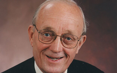 |
Figure 7 Dr. Frank Horan. Photo provided by Dr Beighton and Dr. Horan’s family. |
Greulich and Pyle Atlas
The Greulich and Pyle Atlas is one of the oldest and most frequently cited resources used to assess bone age in children. This method relies on the fact that hand and wrist ossification centers appear on radiographs in a fixed order, and thus allow for calculation of a patient’s bone age. Dr. William W. Greulich (1899–1986) and Dr. Sarah Idell Pyle first described this method in their 1950 publication, “Radiographic Atlas of Skeletal Development of the Hand and Wrist.”34 The atlas is still regularly used in the daily practice of pediatric orthopedics. Dr. Greulich was born in Ohio and completed his PhD at Stanford University. Later in his career, he was appointed Professor and Executive Head of the Department of Anatomy at Stanford University. Additionally, Dr. Greulich served as Science Advisor to the US High Commissioner for Germany, Chairman of the US Educational Commission in the Federal Republic of Germany, and Scientific Attaché at the US Embassy in England following World War II. He also served as President of the American Association of Physical Anthropologists and the Society for Research in Child Development, and was named a Scientific Fellow of the Zoological Society of London.59,60 Dr. Pyle was born in Iowa in 1895, and received her Masters Degree in Kinesiology and Physical Growth in 1925 from the State University of Iowa. After 33 years of graduate level research and teaching, she was finally awarded her PhD in Anatomy from Western Reserve University in 1950 for her thesis “Radiographic Atlas of Skeletal Development of the Knee.” She is the author of over 35 publications, provided substantial unnamed contributions to numerous other publications, and dedicated her life to the collection of data establishing normal development of growing bone in children from infancy to adulthood. The Sarah Idell Pyle Professorship in Anthropology was established in her honor at Case Western Reserve University.64
Surgical Procedures
MacIntosh ACL Reconstruction
The MacIntosh ACL reconstruction was originally described as a “lateral substitution reconstruction” that requires mobilization of a strip of the fascia lata while preserving its attachment to Gerdy’s tubercle, and then passage of the strip over the top of the femoral condyle before securing it to the tibia.1 At a 1972 meeting of the Canadian Orthopaedic Association, Dr. H.R. Galway and Dr. David L. MacIntosh (1914–2013) described the lateral pivot shift as a sign of ACL insufficiency.26 Using this work as a foundation, Dr. MacIntosh developed what is now referred to as the MacIntosh ACL reconstruction. The MacIntosh procedure gained greater notoriety when Dr. Lyle Micheli and Dr. Mininder Kocher described a modified version of this technique in their 2006 paper, “Physeal sparing reconstruction of the anterior cruciate ligament in skeletally immature prepubescent children and adolescents,” and has become a principal tool used in the treatment of skeletally immature patients with ACL injuries.44 Dr. MacIntosh was born in Nova Scotia, and completed his undergraduate and medical studies at Dalhousie University. Shortly after graduating from medical school, Dr. MacIntosh volunteered as a surgeon-lieutenant in the Royal Navy. At the conclusion of World War II, Dr. MacIntosh continued his medical studies at the University of Toronto, where he was subsequently appointed Professor of Orthopedic Surgery. The University of Toronto’s David L. MacIntosh Sport Medicine Clinic, thought to be the oldest dedicated sports medicine facility in the world, is named in his honor.27
Anderson All-Epiphyseal ACL Reconstruction
The Anderson all-epiphyseal ACL reconstruction is performed by first securing a quadrupled hamstring autograft within the femoral epiphysis using a suspensory button, and then passing the graft obliquely through the tibial epiphysis before fixing the graft to the tibial metaphysis using a screw and post. There is relatively limited long-term data on this technique given that it was described relatively recently, however, one 2017 case series observed that 48% of patients had a complication and 37% of patients had a secondary procedure after having an all-epiphyseal ACL reconstruction.81 This technique was first developed by Dr. Allen F. Anderson (1949–2017) (Figure 8) in the 1990s.2 Dr. Anderson completed his medical education at the University of Tennessee and his orthopedics residency at Vanderbilt University. His myriad professional achievements include serving on the American Journal of Sports Medicine editorial board, and serving as Chair of the International Knee Documentation Committee and President of the American Orthopedic Society for Sports Medicine.75 He was dedicated to the production of high-quality research in orthopedic surgery, and for his efforts, he was awarded the Hughston Award in 2002 and the O’Donoghue Award in 2014.3
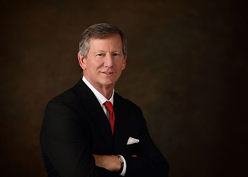 |
Figure 8 Dr. Allen F. Anderson. Photo provided by the Tennessee Orthopedic Alliance. |
Roux-Goldthwait Transfer
The Roux-Goldthwait transfer is used in the treatment of pediatric patellar instability. This procedure requires detachment of the lateral portion of the patellar tendon, passing of the detached tendon under the medial portion of the patellar tendon, and finally reattachment of the lateral portion of the tendon to the medial fibrous plate.13 Dr. Cesar Roux (1857–1934) (Figure 9) first described this procedure in his 1888 case report, “Recurrent Dislocation of the Patella: Operative Treatment.”62 Several years later at the 1896 meeting of the American Orthopaedic Association in Buffalo, Dr. Joel E. Goldthwait (1866–1961) (Figure 10) described a case of “permanent” patellar dislocation. Three years later, he published a description of his treatment for this patient, which consisted of transferring a portion of the lateral patellar tendon to the anteromedial tibia.28 Although the Roux-Goldthwait procedure has long been included in discussions regarding treatment of patellar instability, there is relatively limited data on outcomes following the procedure, and it is rarely performed in isolation from other procedures. Therefore, the clinical utility of this procedure remains uncharacterized.23 Dr. Cesar Roux (1857–1934) was a Swiss-born general surgeon who completed his medical training at the University of Berne before a long tenure as chief of surgery at the Hospital of Lausanne. Dr. Roux also lends his name to the Roux-en-Y gastric bypass, a procedure he first described in his 1893 report in Revue de Chirurgie.40 Dr. Goldthwait was born in Massachusetts and completed his medical training at Harvard Medical School. He practiced orthopedic surgery at Massachusetts General Hospital and was chosen as the first chief of the hospital’s new orthopedic service. Later in his career, he assisted in the development of the Brigham Hospital, and subsequently served as the hospital’s Chairman of the Medical Committee and President of the Board of Trustees. Dr. Goldthwait also assisted in the organization of orthopedic care for wounded soldiers in the United States and in England during World War I, and due to his efforts, he concluded his service having achieved the rank of Brigadier General.46
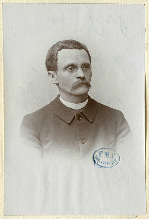 |
Figure 9 Dr. Cesar Roux. |
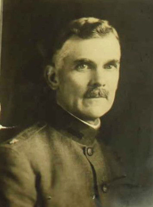 |
Figure 10 Dr. Joel E. Goldthwait. Photo provided by the Goldthwait Reservation and Dr. Goldthwait’s family. |
Galeazzi Tenodesis
The Galeazzi tenodesis is a soft tissue reconstruction technique used in the surgical management of pediatric patellofemoral instability. This semitendinosus tenodesis is performed by isolating and dividing the semitendinosus at the myotendinous junction, exposing the anterior and lateral patella, drilling an oblique tunnel, passing the tendon from medial to lateral, and sewing the tendon back onto itself on tension while manually realigning the patella.31 This technique is attributed to Dr. Ricardo Galeazzi (1866–1952), who first described his methods in his 1922 paper, “New applications of muscle and tendon transplant.”25 Although Galeazzi’s original manuscript is difficult to find, fifty years after Galeazzi’s paper was published, Baker et al shared their 10-year, 58 patient experience with semitendinosus tenodesis in Toronto. This group described Galeazzi’s technique and proposed that this surgical intervention was less injurious to the tibial tubercle apophysis than alternatives such as the Roux-Goldthwait and Hauser procedures.7 The shortcoming of this technique is that it there has been report of 82% recurrence of subluxation or dislocation after surgical intervention. Nevertheless, patients may benefit from the Galeazzi as a temporizing procedure before pursuing a bony realignment procedure as an adult.31 Dr. Galeazzi was an Italian orthopedic surgeon who made numerous contributions to the field as a patient advocate and scientist. He was largely responsible for the development of rehabilitation centers for injured and disable patients, and developed a systematic method of treating and classifying occupational disorders. In addition to his health advocacy work, Galeazzi contributed to the understanding and treatment of multiple pediatric orthopedic pathologies, including the radial shaft fracture with concurrent distal radioulnar joint disruption (Galeazzi fracture) and developmental dysplasia of the hip (Galeazzi test).66
Grammont Procedure
More recently, Dr. Paul M. Grammont (1940–2013) (Figure 11) developed the periosteal sleeve transfer as an alternative surgical option for patellar instability. In his 1985 paper, Grammont described (1) a lateral retinacular release with (2) a distal patellar dissection from the apophysis with preservation of the periosteal attachment and elevation of a portion of the periosteum distally, that concluded with (3) fixation of the medial patella to the tibia.30 Kraus et al recently described a modified version of the Grammont procedure that excludes fixation of the medial patella. In recent years, use of the modified Grammont procedure has been favored in skeletally immature patients because it allows for dynamic positioning of the patella, reduced deforming forces on the knee, and thus lower long-term rates of osteoarthritis.45 This technique has been used more frequently in patients where there is concern that an MPFL reconstruction or adductor sling may fail.56 Dr. Grammont was born in France and completed his medical training in Lyon. After expanding his surgical knowledge during his military service in French Guiana, he first became a Professor of Orthopedic Surgery and Traumatology in Lyon, and later served as Chairman of Orthopedic Surgery at the University Hospital in Dijon.10 Although Dr. Grammont’s name is often cited in reference to pediatric patellar instability, he is also responsible for the development of the reverse total shoulder arthroplasty.29
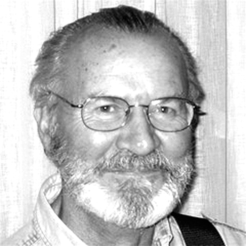 |
Figure 11 Dr. Paul M. Grammont. Photo provided by Dr. Pascal Boileau. |
Conclusions
Multiple scientific disciplines over the course of over 100 years have contributed eponyms to pediatric sports medicine. This phenomenon is consistent with our modern understanding of pediatric sports medicine as inherently interdisciplinary. The use of eponyms in the practice of medicine is flawed. They may have limited sensitivity or specificity, limited clinical applicability, or low intra- and/or interobserver reliability. They also may be easy to confuse or hard to remember. Nevertheless, improved understanding of eponyms allows for modern orthopedic surgeons to appreciate the field’s legacy of scientific exploration and commitment to improving the lives of children. This understanding will also facilitate further examination of the utility of these eponyms, and may act as a foundation for future research and clinical innovation. In sum, this paper serves as a description of eponyms commonly used in pediatric sports medicine. Although this review is by no means exhaustive, this discussion may inform future use of the above-described eponyms in scientific discourse, patient care and medical education, and may encourage future innovation and research into understanding pediatric orthopedic pathologies.
Abbreviations
ACL, anterior cruciate ligament; CDI, Caton-Deschamps index; SLJ disease, Sinding-Larsen-Johansson disease.
Data Sharing Statement
All information and images included in this review are publicly available.
Author Contributions
All authors contributed to data analysis, drafting or revising the article, have agreed on the journal to which the article will be submitted, gave final approval of the version to be published, and agree to be accountable for all aspects of the work.
Funding
No funding was used to complete any portion of this review.
Disclosure
Ahmad F Bayomy reports personal fees from the Journal of Bone & Joint Surgery, outside the submitted work. Charles A Popkin reports research support from Arthrex and educational support from Smith and nephew, outside the submitted work. The authors report no other potential conflicts of interest for this work.
References
1. Amirault JD, Cameron JC, MacIntosh DL, Marks P. Chronic anterior cruciate ligament deficiency. Long-term results of MacIntosh’s lateral substitution reconstruction. J Bone Joint Surg Br. 1988;70-B(4):622–624. doi:10.1302/0301-620X.70B4.3403611
2. Anderson AF. Transepiphyseal replacement of the anterior cruciate ligament in skeletally immature patients. A preliminary report. J Bone Joint Surg Am. 2003;85(7):1255–1263. doi:10.2106/00004623-200307000-00011
3. Anderson AF. In Memoriam. Am J Sports Med. 2017;46(1):250.
4. Aparicio G, Abril JC, Albiñana J, Rodríguez-Salvanés F. Patellar height ratios in children: an interobserver study of three methods. J Pediatr Orthop B. 1999;8(1):29–32.
5. The Arizona Republic. Marvin H. Meyers. Available from: https://www.legacy.com/obituaries/azcentral/obituary.aspx?n=Marvin-H.Meyers&pid=19810010&fhid=1.
6. Baastrup CI. Hans Jessen Panner in Memoriam. Acta Radiol. 2010;11(4):347–349.
7. Baker RH, Carroll N, Dewar FP, Hall JE. The semitendinosus tenodesis for recurrent dislocation of the patella. J Bone Joint Surg Br. 1972;54(1):103–109.
9. Beighton P, Horan F. Orthopaedic aspects of the Ehlers-Danlos syndrome. J Bone Joint Surg Br. 1969;51(3):444–453.
10. Boileau P. Biographical Sketch: paul M. Grammont, MD (1940). Clin Orthop Relat Res. 2011;469(9):2422–2423. doi:10.1007/s11999-011-1959-y
11. Carter C, Wilkinson J. Persistent Joint Laxity and Congenital Dislocation of the Hip. J Bone Joint Surg Br. 1964;46:40–45. doi:10.1302/0301-620X.46B1.40
12. Caton J, Deschamps G, Chambat P, Lerat JL, Dejour H. Patella infera. Apropos of 128 cases. Rev Chir Orthop Reparatrice Appar Mot. 1982;68(5):317–325.
13. Chotel F, Raux JBS. Patellar instability in children and adolescents. Orthop Traumatol. 2014;100(1 Suppl):S125–S137.
14. Circi E, Atalay Y, Beyzadeoglu T. Treatment of Osgood-Schlatter disease: a review of the literature. Musculoskelet Surg. 2017;101(3):195–200.
15. Claessen FMAP, Louwerens JKG, Doornberg JN, van Dijk CN, van den Bekerom MPJ, Eygendaal D. Hegemann’s disease and fishtail deformity: aetiopathogenesis, radiographic appearance and clinical outcome. J Child Orthop. 2015;9(1):1–8. doi:10.1007/s11832-014-0630-z
16. Claessen FMAP, Louwerens JKG, Doornberg JN, van Dijk CN, Eygendaal D. van den Bekerom MPJ. Panner’s disease: literature review and treatment recommendations. Journal of Children’s Orthopaedics. 1988;70-B(4):9–17. doi:10.1007/s11832-015-0635-2
17. Conrad JM, Stanitski CL. Osteochondritis Dissecans: Wilson’s Sign Revisited. Am J Sports Med. 1964;46-B(1):777–778. doi:10.1177/03635465030310052301
18. DeMaio M. Giants of Orthopaedic Surgery: masaki Watanabe MD. Clin Orthop Relat Res. 2015;9(1):2443–2448. doi:10.1007/s11999-013-3052-1
19. DePasse JM, Daniels AH, Durand W, Kingrey B, Prodromo J, Mulcahey MK. Completion of Multiple Fellowships by Orthopedic Surgeons: analysis of the American Board of Orthopaedic Surgery Certification Database. Orthopedics. 2018;41(1):e33–e37. doi:10.3928/01477447-20171106-05
20. Dictionnaire historique de la Suisse. Schlatter. Available from: http://www.hls-dhs-dss.ch/textes/f/F14628.php.
21. Draghi F, Danesino GM, Coscia D, Precerutti M, Pagani C. Overload syndromes of the knee in adolescents: sonographic findings. Journal of Ultrasound. 2008;11(4):151–157. doi:10.1016/j.jus.2008.09.001
22. Ellis HB, Zynda AJ, Cruz AI, et al. Classification and Treatment of Pediatric Tibial Spine Fractures Assessing Reliability Among a Tibial Spine Research Interest Group. Journal of Pediatric Orthopaedics. 2020;Publish Ahead of Print. doi:10.1097/BPO.0000000000001654
23. Felli L, Capello AG, Lovisolo S, Chiarlone F, Allessio-Mazzola M. Goldthwait technique for patellar instability: surgery of the past or here to stay procedure? A systematic review of the literature. Musculoskel Surg. 2019;103:107–113.
24. Ferguson RP, Thomas D. Medical eponyms. J Community Hosp Intern Med Perspect. 2014;4(3):10.
25. Galeazzi R. Nuove applicazioni del trapianto muscular e tendineo. Ard Di Orthop Milano. 1922;38:315–323.
26. Galway HR, MacIntosh DL. The lateral pivot shift: a symptom and sign of anterior cruciate ligament insufficiency. Clin Orthop Relat Res. 1980;147:45–50.
27. The Globe and Mail. Surgeon David MacIntosh, 98, was a pioneer in sports medicine. Available from: https://www.theglobeandmail.com/news/enness/surgeon-david-macintosh-98-was-a-pioneer-in-sports-medicine/article7996871/.
28. Goldthwait JEV. Permanent Dislocation of the Patella. The Report of a Case of Twenty Years’ Duration, successfully treated by Transplantation of the Patella Tendons with the Tubercle of the Tibia. Ann Surg. 1899;29(1):62–68.
29. Grammont PM, Baulot E. Delta shoulder prosthesis for rotator cuff rupture. Orthopedics. 1993. 16(1):65–68.
30. Grammont PM, Latune D, Lammaire IP. Treatment of subluxation and dislocation of the patella in the child. Elmslie technic with movable soft tissue pedicle (8 year review). Orthopade. 1985;14(4):229–238.
31. Grannatt K, Heyworth BE, Ogunwole O, Micheli LJ, Kocher MS. Galeazzi Semitendinosus Tenodesis for Patellofemoral Instability in Skeletally Immature Patients. J Pediatr Orthop. 2012;32(6):621–625.
32. Greeley PW. Multiple ossification centers of the patella. Am J Surg. 1932;18(3):456–459.
33. Green D, Tuca M, Luderowkski E, Gausden E, Goodbody C, Konin G. A new MRI-based classification system for tibial spine fractures changes clinical treatment recommendations when compared to Myers and Mckeever. Knee Surg Sports Traumatol Arthros. 2018. doi:10.1007/s00167-018-5039-7
34. Greulich WW, Pyle SI. Radiographic Atlas of Skeletal Development of the Hand and Wrist. Stanford, CA: Stanford University Press; 1950.
35. Gurtler RA, Stine R, Torg JS. Lachman test evaluated. Quantification of a clinical observation. Clin Orthop Rel Res. 1987;216:141–150.
36. Haraldsson S. On Osteochondrosis Deformans Juvenilis Capituli Humeri Including Investigation of Intra-Osseous Vasculature in Distal Humerus. Acta Orthop Scand. 2014;30(sup38):5–232.
37. Die HG. „spontanen“, aseptischen Knochennekrosen des Ellenbogengelenkes. Fortschr Röntgenstr. 2009;75(07):89–92.
38. Heidelberg University, Public Relations Department. The death of Prof. Dr. med. Gerd Hegemann. Available from: http://www.presse.uni-erlangen.de/Aktuelles/Hegemann-.html.
39. Hunter TB, Peltier LF, Lund PJ. Radiologic history exhibit: musculoskeletal eponyms: who are those guys? Radiographics. 2000;20(3):819–836.
40. Hutchison RL, Hutchison AL. Cesar Roux and His Original 1893 Paper. Acta Orthop. 2010;20(7):953–956.
41. Jackson RW. In Memoriam Masaki Watanabe. M D Arthroscopy. 1995;11(4):385.
42. James AM, Williams CM, Haines TP. Effectiveness of interventions in reducing pain and maintaining physical activity in children and adolescents with calcaneal apophysitis (Sever’s disease): a systematic review. J Foot Ankle Res. 2013;6(1):16.
43. Johansson SC. Eine bisher anscheinend unbekannte Erkrankung der Patella. Ztschr f Orthop Chir. 1924;43:82.
44. Kocher MS, Garg S, Micheli LJ. Physeal sparing reconstruction of the anterior cruciate ligament in skeletally immature prepubescent children and adolescents. Surgical technique. J Bone Joint Surg Am. 2006;88(1_suppl_2):283–293.
45. Kraus T, Lidder S, Svehlik M, et al. Patella re-alignment in children with a modified Grammont technique. Outcome in 65 knees after mean 8 years. Acta Orthop. 2012;83(5):504–510.
46. Kuhns JG. Joel Ernest Goldthwait. Clin Orthop Relat Res. 1955;5:3.
47. Life in the Fastlane. Carl Schlatter. Available from: https://litfl.com/carl-schlatter/.
48. Legacy.com. John W. Lachman. Available from: https://www.legacy.com/obituaries/name/john-lachman-obituary?pid=95753109.
49. Meyers MH, McKeever FM. Fracture of the intercondylar eminence of the tibia. J Bone Joint Surg. 1959;41(2):209–222.
50. Meyers MH, McKeever FM. Fracture of the intercondylar eminence of the tibia. J Bone Joint Surg. 1970;52(8):1677–1684.
51. Mulligan EP, Harwell JL, Robertson WJ. Reliability and Diagnostic Accuracy of the Lachman Test Performed in a Prone Position. J Orthop Sports Phys Ther. 2011;41(10):749–757.
52. Osgood RB. Lesions of the Tibial Tubercle Occurring During Adolescence. Boston Med Surg J. 1903;148:114–117.
53. Osgood RB, Mankin HJ. Lesions of the Tibial Tubercle Occurring During Adolescence. Clin Orthop Rel Res. 1993:286:4–9.
54. Panner HJ, Peculiar A. Affection of the Capitulum Humeri, Resembling Calvé-Perthes’ Disease of the Hip. Acta Radiol. 2013;10(3):234–242.
55. Phillips CL, Silver DAT, Schranz PJ, Mandalia V. The measurement of patellar height: a review of the methods of imaging. J Bone Joint Surg Br. 2010;92(8):1045–1053.
56. Popkin CA, Bayomy AF, Trupia EP, Chan CM, Redler LH. Patellar Instability in the Skeletally Immature. Curr Rev Musculoskelet Med. 2018;11(2):172–181.
57. Popkin CA, Gundry CR, Larson CM, Murnaghan ML. Remembering our Roots: eponyms in Sports Medicine. Am J Sports Med. 2012;41(7):1703–1711.
58. The PM. Lachman test is the most sensitive and the pivot shift the most specific test for the diagnosis of ACL rupture. Aust J Physiother. 2006;52(1):66.
59. Prudhon JL, Caton JH, Aslanian T, Verdier R. How is patella height modified after total knee arthroplasty? Int Orthop. 2018;42(2):311–316.
60. Roche AF. Obituary: william Walter Greulich, 1899-1986. Am J of Phys Anthropol. 1987;72:273–276.
61. Roth PB. Apophysitis of Os Calcis. Proc R Soc Med. 1919;12:99–103.
62. Recurrent RC. Dislocation of the Patella: operative Treatment. 1888. Clinc Orthop Rel Res. 2006;452:17–20.
63. Royal College of Surgeons. Wilson, James Noël Chalmers Barclay (1919 – 2006). Available from: https://livesonline.rcseng.ac.uk/biogs/E000283b.htm.
64. Pyle SI. Case Western Reserve University Archives, Digital. Cleveland, Ohio: United States of America. 2014
65. Schlatter C. Verletzungen des Schnabel-formigen Fortsatzes der aberen Tibiaepiphyse. Bruns Beit Klin Chir. 1903;38:874.
66. Sebastin SJ, Chung KC. A Historical Report on Riccardo Galeazzi and the Management of Galeazzi Fractures. Acta Orthop. 2010;35(11):1870–1877.
67. Sever JW. Apophysitis of os calcis. N Y Med J. 1912;95:1025–1029.
68. Sinding-Larsen CM. A hitherto unknown affection of the patella in children. Acta Radiol. 2016;57(4):e42.
69. Smits-Engelsman B, Klerks M, Beighton Score: KA, Valid A. Measure for Generalized Hypermobility in Children. J Pediatr. 2011;158(1):119–123.
70. Somford NP, Nieuwe Weme RA, Hoornenborg D, et al. Biographical background and origin of common eponymous terms in orthopedic surgery: anatomy and fractures in knee surgery. Eur J Orthop Surg Tramatol. 2018;28(1):79–84.
71. Somford MP, Nieuwe Weme RA, Sierevelt I, et al. Orthopeaedic Eponymous Terms Study Group. The Reliability of Orthopaedic Eponymous Terms. J Bone Joint Surg Am. 2017;99(13):e70–e76.
72. Soule RE. Sprain-fracture of the Tubercle of Tibia in Adolescence. J Orthop Surg. 1921;19:550.
73. Thevenin-Lemoine C, Ferrand M, Courvoisier A, Damsin JP. Is the Caton-Deschamps index a valuable ratio to investigate patellar height in children? J Bone Joint Surg Am. 2011;93(8):e35.
74. The Telegraph. Frank Horan, orthopaedic surgeon – obituary. Available from: https://www.telegraph.co.uk/news/obituaries/12012128/Frank-Horan-orthopaedic-surgeon-obituary.html.
75. Tennessean. D, Allen F. Anderson, award-winning Tennessee Orthopedic Alliance surgeon, dies on Maury County farm. Available from: https://www.tennessean.com/story/news/2017/11/14/dr-allen-f-anderson-tennessee-orthopedic-alliance-surgeon-dies-maury-county-obituary/861532001/.
76. Torg JS, Conrad W, Kalen V. Clinical diagnosis of anterior cruciate ligament instability in the athlete. Am J Sports Med. 1976;4(2):84–93.
77. U.C.T. Faculty of Health Sciences, Division of Human Genetics. Emer Prof Peter Beighton. Available from: http://www.humangenetics.uct.ac.za/hg/division/staff/principal/peter_beighton.
78. Visotsky JL, Benson LS. Eponyms in orthopaedics. J Bone Joint Surg Am. 2001;83(suppl 2 pt 2):123–127.
79. Yaniv M, Blumberg N. The discoid meniscus. J Child Orthop. 2007;1(2):89–96.
80. Jr WJC. Francis Michael McKeever 1901-1973. J Bone Joint Surg Am. 1974;56(3):643–644.
81. Wall EJ, Eismann EA, Myer GD, Outcomes CP. Complications After All-Epiphyseal Anterior Cruciate Ligament Reconstruction in Skeletally Immature Patients. Ortho J of Sports Med. 2017;5(3):2325967117693604.
82. Whitworth JA. Should eponyms be abandoned? No. BMJ. 2007;335(7617):425.
83. Wilson J. A Diagnostic Sign in Osteochondritis Dissecans of the Knee. J Bone Joint Surg Am. 1967;49(3):477.
84. Watanabe M, Takeda S, Ikeuchi H. Atlas of Arthroscopy.
85. Relate W. James Warren Sever. Available from: https://www.werelate.org/wiki/Person.
86. Woywodt A. Should eponyms be abandoned? Yes. BMJ. 2007;335(7617):424.
87. Whonamedit Sven Christian Johansson. Available from: http://www.whonamedit.com/doctor.cfm/2635.html.
88. Whonamedit Christian Magnus Falsen Sinding-Larsen. Available from: http://www.whonamedit.com/doctor.cfm/2634.html.
 © 2021 The Author(s). This work is published and licensed by Dove Medical Press Limited. The full terms of this license are available at https://www.dovepress.com/terms.php and incorporate the Creative Commons Attribution - Non Commercial (unported, v3.0) License.
By accessing the work you hereby accept the Terms. Non-commercial uses of the work are permitted without any further permission from Dove Medical Press Limited, provided the work is properly attributed. For permission for commercial use of this work, please see paragraphs 4.2 and 5 of our Terms.
© 2021 The Author(s). This work is published and licensed by Dove Medical Press Limited. The full terms of this license are available at https://www.dovepress.com/terms.php and incorporate the Creative Commons Attribution - Non Commercial (unported, v3.0) License.
By accessing the work you hereby accept the Terms. Non-commercial uses of the work are permitted without any further permission from Dove Medical Press Limited, provided the work is properly attributed. For permission for commercial use of this work, please see paragraphs 4.2 and 5 of our Terms.