
Back to Journals » Diabetes, Metabolic Syndrome and Obesity » Volume 19
Endothelial Metabolic Reprogramming Links Diabetes to Atherosclerosis
Received 11 September 2025
Accepted for publication 9 January 2026
Published 11 February 2026 Volume 2026:19 565805
DOI https://doi.org/10.2147/DMSO.S565805
Checked for plagiarism Yes
Review by Single anonymous peer review
Peer reviewer comments 2
Editor who approved publication: Dr Rebecca Baqiyyah Conway
Yangze Pan, Jin Si
Department Endocrinology, Liyuan Hospital, Tongji Medical College, Huazhong University of Science and Technology, Wuhan, Hubei Province, 430077, People’s Republic of China
Correspondence: Jin Si, Department Endocrinology, Liyuan Hospital, Tongji Medical College, Huazhong University of Science and Technology, Wuhan, Hubei Province, 430077, People’s Republic of China, Email [email protected]
Abstract: Diabetic vascular complications are common and severe, worsening quality of life and long-term outcomes. In diabetes, chronic hyperglycemia together with dyslipidemia and hypertension reshapes endothelial metabolism and homeostasis. Endothelial cells shift the balance of glucose utilization, fatty acid oxidation, and mitochondrial function, and these metabolic changes bias endothelial behavior toward reduced nitric oxide bioavailability, oxidative stress, and a pro-inflammatory, pro-thrombotic state. Over time, maladaptive stress responses promote senescence, cell loss, and endothelial-to-mesenchymal transition, accelerating the initiation and progression of atherosclerotic plaques. This review summarizes how diabetic cues drive endothelial metabolic reprogramming and how this, in turn, links endothelial dysfunction to plaque formation, growth, and instability. We highlight key metabolic pathways and discuss how local hemodynamic forces at athero-prone regions further shape endothelial phenotypes and inflammatory signaling. Finally, we outline therapeutic opportunities that target endothelial metabolism and stress responses—including modulation of glycolytic flux, mitochondrial and redox-directed strategies, and pathway-level interventions that curb hypoxia and inflammatory programs. We emphasize translational priorities such as endothelium-selective delivery, biomarkers of endothelial metabolic state, and rigorous clinical testing to enable earlier and more effective prevention of diabetes-associated atherosclerotic disease.
Keywords: atherosclerosis, metabolic reprogramming, diabetes, endothelium
Introduction
Diabetes mellitus is a chronic metabolic disease marked by persistent hyperglycemia and a growing global health burden. The International Diabetes Federation estimates ~530 million affected adults in 2024, rising to 783 million by 2045.1 Much of its morbidity and mortality is driven by vascular complications across macrovascular and microvascular beds.2 Macrovascular complications include coronary artery disease, cerebrovascular disease, and peripheral artery disease, whereas microvascular complications include diabetic nephropathy, diabetic retinopathy, diabetic peripheral neuropathy, and diabetic cardiomyopathy. These complications are the main drivers of the excess morbidity and mortality associated with diabetes. Cardiovascular disease remains the leading cause of death in diabetes, with atherosclerotic plaque formation and progression providing the central pathological substrate for most events.3 Hyperglycemia, insulin resistance, and dyslipidemia converge to reduce the bioavailability of endothelial nitric oxide (NO), amplify oxidative stress, and activate inflammatory pathways, thereby promoting vascular dysfunction and plaque vulnerability.4 Epidemiologic studies indicate that individuals with type 2 diabetes have a risk of atherosclerotic events that is two to four times higher than that of individuals without diabetes, and earlier disease onset is associated with accelerated trajectories.5
The vascular endothelium maintains vascular tone, regulates angiogenesis and hemostasis, and provides key antioxidant, anti-inflammatory, and antithrombotic functions. Endothelial dysfunction is considered a precursor of atherosclerosis and cardiovascular disease, and evidence also links it to plaque progression and clinical events.6 It is characterized by impaired endothelium-dependent vasodilation together with endothelial activation, which creates a pro-inflammatory, pro proliferative, and prothrombotic milieu that drives atherogenesis and its complications.7 Endothelial dysfunction is an early hallmark of atherosclerosis that can identify patients at elevated vascular risk and has important value in vascular health assessment.8 Notably, endothelial dysfunction associated with insulin resistance appears to precede overt hyperglycemia in type 2 diabetes.9 These observations position endothelial dysfunction as a promising early target for preventing atherosclerosis and cardiovascular disease in patients with diabetes or insulin resistance.
Despite strong clinical and experimental links between diabetes, endothelial dysfunction, and accelerated atherosclerosis, the mechanistic chain that connects diabetic cues to specific endothelial behaviors that drive plaque initiation, progression, and instability remains incompletely resolved. Prior reviews have extensively discussed diabetic endothelial dysfunction and inflammation/oxidative stress, as well as individual metabolic pathways in endothelial cells.10,11 However, an integrated and stage-aware framework that connects diabetes-driven endothelial metabolic reprogramming to phenotypic switching such as EndMT, to the amplification of inflammatory signaling, and to the influence of local hemodynamic forces at atherosclerosis-prone sites has not yet been clearly articulated. To address this gap, this review synthesizes current evidence on endothelial metabolic reprogramming in diabetes and its roles in endothelial-to-mesenchymal transition (EndMT), inflammation, and atherosclerosis. We further highlight how disturbed flow modulates metabolic state and inflammatory signaling to create regional vulnerability, and we outline therapeutic opportunities that target endothelial metabolism and stress-response circuits, with emphasis on translational challenges such as endothelium-selective delivery and rigorous clinical evaluation.
Methods
PubMed (https://pubmed.ncbi.nlm.nih.gov/) and Web of Science (https://clarivate.com/academia-government/scientific-and-academic-research/research-discovery-and-referencing/web-of-science/) databases were searched from their inception to August 2025, with the language restricted to English. The search strategy focused on the association between diabetes mellitus, endothelial dysfunction, endothelial metabolic reprogramming, endothelial-to-mesenchymal transition (EndMT), hemodynamic forces, and atherosclerosis. Key words and their combinations included “diabetes mellitus,” “hyperglycemia,” “insulin resistance,” “endothelial dysfunction,” “endothelial metabolism,” “glycolysis,” “fatty acid oxidation,” “mitochondrial dysfunction,” “EndMT,” “endothelial-to-mesenchymal transition,” “disturbed flow,” “shear stress,” “inflammation,” and “atherosclerosis,” as well as mechanism-related terms such as “PFKFB3,” “HIF-1α,” “TGF-β,” “acetyl-CoA,” “NRF2,” and “mitophagy.”
This review considered original studies, including in vitro and in vivo experiments, animal models, and clinical or epidemiologic investigations, that examined the relationship between diabetes, insulin resistance, or hyperglycemia and endothelial dysfunction; characterized metabolic pathways in endothelial cells such as glucose metabolism, fatty acid oxidation, and mitochondrial biology and their regulation by hemodynamic forces; explored how metabolic reprogramming contributes to EndMT, vascular inflammation, and atherosclerotic plaque initiation, progression, and instability; or evaluated therapeutic interventions targeting endothelial metabolism, mitochondrial function, or related inflammatory pathways in the context of atherosclerosis or diabetic vascular complications. High-quality review articles and position papers were additionally used to supplement background information, provide theoretical frameworks, and contextualize emerging mechanistic findings.
Studies were excluded when endothelial cells or vascular endothelium were not clearly involved, when the primary focus was on vascular diseases unrelated to diabetes, atherosclerosis, or metabolic/endothelial mechanisms, or when the work did not address metabolic, inflammatory, or hemodynamic mechanisms relevant to endothelial dysfunction or EndMT. Non-English publications, conference abstracts, non–peer-reviewed preprints, incomplete case reports, and commentaries or editorials with limited relevance to the review topic were also excluded.
Diabetes and Endothelial Dysfunction
The vascular endothelium is central to vascular health, and “endothelial dysfunction” denotes a reduced capacity of the endothelium to maintain vascular homeostasis.12 This phenotype reflects not only a deficiency of vasodilators but also an excess of vasoconstrictors together with disordered regulation of inflammation, thrombosis, and vascular cell proliferation.13 Substantial evidence links endothelial dysfunction and downstream atherosclerosis to insulin-resistant states, including obesity and diabetes.14,15 Cross-sectional studies show blunted endothelium-dependent vasodilation in both the coronary and peripheral circulations of patients with type 1 diabetes16 and type 2 diabetes.17 Endothelial dysfunction is likewise observed with obesity accompanying type 2 diabetes,18 sedentary behavior, and the metabolic syndrome.19 Beyond impaired vasodilatory responses, circulating levels of endothelial adhesion molecules and plasminogen activator inhibitor-1 are elevated in diabetes, indicating a pro-inflammatory and prothrombotic state.20,21 Insulin resistance, the core pathophysiologic feature of type 2 diabetes, has been closely examined in relation to endothelial function. Insulin sensitivity quantified by the hyperinsulinemic–euglycemic clamp correlates positively with acetylcholine-induced increases in skin blood flow in obese women22 and with brachial artery flow-mediated dilation in nondiabetic individuals.23 Greater insulin secretion during an oral glucose tolerance test is associated with worse endothelial function in the coronary circulation24 and with attenuated forearm blood-flow responses.25 Higher plasma insulin levels also track with lower flow-mediated dilation.26 Consistently, the homeostasis model assessment of insulin resistance is inversely associated with methacholine-induced changes in lower-limb blood flow27 and with acetylcholine-induced skin perfusion in both diabetic and nondiabetic participants.27 In the Framingham cohort, flow-mediated dilation was inversely related to insulin resistance, although this association was attenuated after adjustment for metabolic-syndrome components.19 Notably, endothelial dysfunction may precede the onset of diabetes. Nondiabetic first-degree relatives of individuals with type 2 diabetes exhibit reduced endothelium-dependent vasodilation and higher circulating markers of endothelial activation,28 and similar abnormalities are seen in insulin-resistant nondiabetic offspring identified by oral glucose tolerance testing.28 Prospective data further indicate that circulating markers of endothelial activation predict incident type 2 diabetes after adjustment for body mass index, physical activity, lipids, family history, and glucose tolerance.29 Impaired flow-mediated dilation30 and genetic polymorphisms in endothelial NO synthase (eNOS)31 have also been reported as independent predictors of type 2 diabetes. Taken together, these observations suggest shared pathobiology and a potential causal link between insulin resistance and endothelial dysfunction. Intervention studies strengthen this connection. Treatments that improve insulin sensitivity often produce parallel improvements in endothelial function. Rosiglitazone32 and troglitazone33 enhance forearm microvascular dilation in patients with type 2 diabetes. Metformin improves endothelium-dependent dilation in type 2 diabetes34 and in the metabolic syndrome.35 Pioglitazone augments endothelial function in hypertensive or hypercholesterolemic nondiabetic subjects with or without insulin resistance.36 Rosiglitazone has also been shown to increase flow-mediated dilation and lower inflammatory markers in healthy, non-obese, low-risk volunteers.37 In addition, weight loss, regular physical activity, and inhibition of the renin–angiotensin system contribute to endothelial recovery, in part through enhanced insulin sensitivity or reduced diabetes risk.38
In summary, diabetes coexists with marked endothelial dysfunction, which likely contributes to the excess cardiovascular risk in this population. Defining the underlying molecular mechanisms may open new avenues for clinical management.
Endothelial Metabolic Rewiring in the Diabetic Milieu
Glucose Metabolism Oxidative Stress AGEs and Signaling Imbalance
Hyperglycemia, a defining metabolic hallmark of diabetes, is closely linked to disordered metabolism, functional impairment, and secondary vascular pathology in vascular endothelial cells (VECs). A prominent feature is the increased production of reactive oxygen species and reactive nitrogen species (ROS and RNS).39 In VECs, ROS elevation arises primarily from three sources. First, expression and activity of NADPH oxidase subunits (p22phox, p67phox, p47phox) are upregulated; this pathway is accompanied by downregulation of 8-oxoguanine glycosylase and activation of protein kinase C (PKC), as shown in high glucose–treated human umbilical vein endothelial cells (HUVECs).40,41 Second, xanthine oxidase contributes substantially, supported by the ability of the xanthine oxidase inhibitor allopurinol to block high glucose–induced ROS generation.42 Third, eNOS uncoupling augments oxidative stress in diabetic mice.43 Hyperglycemia also elevates intracellular cAMP, which activates protein kinase A and induces inhibitory phosphorylation of glucose-6-phosphate dehydrogenase (Figure 1). This reduces flux through the pentose phosphate pathway, lowers NADPH availability, and further amplifies ROS.44 In parallel, high glucose upregulates the mitochondrial fission proteins Fis1 and Drp1, promoting excessive fission and impaired autophagy, which increases ROS, blunts eNOS activation, and reduces NO bioavailability.45 Excess ROS can activate poly(ADP-ribose) polymerase-1 (PARP1), leading to poly-ADP-ribosylation and inactivation of glyceraldehyde-3-phosphate dehydrogenase (GAPDH), thereby restricting glycolytic flux.46 When glycolysis is bottlenecked, glucose is diverted into alternative pathways. In the polyol pathway, aldose reductase converts glucose to sorbitol at the expense of NADPH, exacerbating oxidative stress; sorbitol is subsequently converted to fructose and 3-deoxyglucosone, which promote the formation of advanced glycation end products (AGEs).39 Accumulated glycolytic intermediates also undergo maladaptive shunting. Fructose-6-phosphate enters the hexosamine biosynthetic pathway via glutamine: fructose-6-phosphate amidotransferase, generating UDP-N-acetylglucosamine. Although protein O-GlcNAcylation is essential for endothelial physiology, hyperglycemia-driven excess O-GlcNAcylation can impair angiogenesis.47,48 Glyceraldehyde-3-phosphate and dihydroxyacetone phosphate feed the methylglyoxal pathway, enhancing AGE formation, and support de novo diacylglycerol synthesis, which activates PKC and contributes to vascular dysfunction.49 AGEs bind the endothelial surface receptor RAGE. This interaction promotes heterodimerization of Toll-like receptor 4, activates NF-κB signaling, and increases adhesion molecule expression; as inflammatory cells and cytokines infiltrate local tissue, the associated oxidative stress further raises vascular permeability.50 Engagement of RAGE by AGEs also induces aberrant overexpression of vascular endothelial growth factor, driving pathological neovascularization.51,52 Together, these processes directly injure endothelial cells (ECs) through lipid peroxidation, structural damage, and increased apoptosis, while amplifying oxidative stress and accelerating further endothelial injury.53 It has also been proposed that glucose-6-phosphate may enter the glucuronic acid pathway, although evidence for this route in diabetic VECs remains limited.54
 |
Figure 1 Hyperglycemia-induced metabolic reprogramming and ROS-mediated vascular dysfunction in endothelial cells. In VECs, the expression and activity of NAD(P)H oxidase subunits (p22phox, p67phox, p47phox) are upregulated, leading to increased ROS generation, which promotes OGG1 downregulation and PKC activation. In addition, the XO pathway and eNOS uncoupling also contribute to ROS production. Hyperglycemia elevates intracellular cAMP, which activates PKA and induces inhibitory phosphorylation of G6PD, thereby reducing glucose entry into the PPP, lowering NADPH levels, and further amplifying ROS. Meanwhile, high glucose upregulates mitochondrial fission–related proteins Fis1 and Drp1, resulting in enhanced mitochondrial fission and impaired autophagy, which aggravates ROS production, suppresses eNOS activation, and reduces NO bioavailability. ROS activate PARP-1, leading to GAPDH inactivation and blockade of glycolytic flux. Glucose not metabolized through glycolysis is shunted into the polyol pathway, where AR catalyzes its conversion into sorbitol at the expense of NADPH consumption, further exacerbating oxidative stress. Sorbitol is subsequently metabolized to fructose and the highly reactive 3DG, promoting AGE formation. Moreover, accumulated glycolytic intermediates undergo abnormal diversion: F6P enters the HBP via GFAT, producing UDP-GlcNAc. In parallel, G3P can enter the methylglyoxal pathway to enhance AGE formation and contribute to de novo DAG synthesis, thereby triggering PKC activation and vascular abnormalities. Large amounts of AGEs bind to their receptor RAGE on ECs. On one hand, this interaction promotes TLR-4 heterodimerization and NF-κB pathway activation, thereby upregulating adhesion molecule expression and increasing vascular permeability. On the other hand, AGEs–RAGE binding induces aberrant VEGF overexpression, resulting in excessive angiogenesis. Abbreviations: VECs, Vascular endothelial cells; ROS, Reactive oxygen species; OGG1, 8-oxoguanine DNA glycosylase; PKC, Protein kinase C; XO, Xanthine oxidase; eNOS, Endothelial nitric oxide synthase; NO, Nitric oxide; cAMP, Cyclic adenosine monophosphate; PKA, Protein kinase A; G6PD, Glucose-6-phosphate dehydrogenase; PPP, Pentose phosphate pathway; Fis1, Mitochondrial fission 1 protein; Drp1, Dynamin-related protein 1; PARP-1, Poly(ADP-ribose) polymerase-1; GAPDH, Glyceraldehyde-3-phosphate dehydrogenase; AR, Aldose reductase; 3DG, 3-deoxyglucosone; AGEs, Advanced glycation end products; F6P, Fructose-6-phosphate; HBP, Hexosamine biosynthesis pathway; GFAT, Glutamine: fructose-6-phosphate amidotransferase; UDP-GlcNAc, Uridine diphosphate N-acetylglucosamine; G3P, Glyceraldehyde-3-phosphate; DAG, Diacylglycerol; ECs, Endothelial cells; RAGE, Receptor for advanced glycation end products; TLR-4, Toll-like receptor 4; NF-κB, Nuclear factor kappa-light-chain-enhancer of activated B cells; VEGF, Vascular endothelial growth factor. |
Fatty Acid Oxidation Maintaining Endothelial Identity and Vascular Homeostasis
Fatty acid oxidation (FAO) is an alternative pathway that converts long-chain fatty acids into acetyl-CoA while bypassing the pyruvate dehydrogenase complex. Acetyl-CoA then condenses with oxaloacetate to form citrate and enters the tricarboxylic acid cycle for energy production. In ECs, FAO is regulated primarily by hypoxia activated AMP-activated protein kinase (AMPK).55 AMPK indirectly activates carnitine palmitoyltransferase 1A (CPT1A), which transports long chain fatty acyl groups into mitochondria and constitutes the rate limiting step of FAO.56 Although FAO can fuel ATP synthesis through mitochondrial respiration, ECs derive most of their energy from glycolysis. Recent work shows that FAO also serves crucial non energetic functions in endothelial biology. Kalucka et al profiled metabolism in quiescent ECs and found FAO activity to be approximately threefold higher than in proliferating ECs, mainly to regenerate nicotinamide adenine dinucleotide phosphate in its reduced form and thereby maintain redox homeostasis. Inhibition of FAO provoked marked oxidative stress, whereas acetate supplementation reversed this injury, indicating an essential role for FAO in redox balance in quiescent endothelium.57 By contrast, Schoors et al examined proliferating ECs during sprouting angiogenesis and focused on functions of FAO beyond energy supply. Knockdown of CPT1A markedly impaired spheroid sprouting because of reduced cell proliferation. CPT1A knockdown did not lower ATP levels and increased ROS by only about 20%, a degree that may even favor proliferation. Mechanistic studies showed that FAO derived acetyl-CoA feeds the tricarboxylic acid cycle and supports de novo deoxynucleotide synthesis. Inhibition of CPT1A reduced nucleotide synthesis, impaired DNA synthesis, and consequently limited endothelial proliferation and sprouting.58 These findings differ from the role of FAO in preserving redox balance in quiescent cells yet converge on the conclusion that FAO is essential for normal endothelial function and angiogenesis. In diabetes, the role of FAO is complex and tissue specific. Notably, FAO supports endothelial proliferation and biomass accumulation during angiogenesis.58
Although diabetes is defined by hyperglycemia, it is frequently accompanied by elevated circulating free fatty acids (FFA). Multiple studies have examined the impact of increased FFA on endothelial function. In healthy volunteers, experimental elevation of FFA was associated with endothelial dysfunction,59 and follow up work showed that higher FFA levels blunt insulin mediated vasodilation and NO production.60 Vigili et al similarly observed impaired endothelium-dependent vasodilation with increased FFA.61 Related studies indicate that obesity related insulin resistance stimulates vascular smooth muscle cell proliferation and promotes excessive FFA release from adipose tissue, which in turn induces oxidative stress and activates PKC, creating a vicious cycle.62 When endothelial FAO is impaired, intracellular calcium oscillations are enhanced, NADPH content falls, and the NADP to NADPH ratio rises. The result is vascular leakage, reduced antioxidant capacity, and exacerbation of lipopolysaccharide mediated endothelial activation.57,63 In a hyperinsulinemic milieu, hepatic lipoprotein synthesis increases, and AGEs promote glycation of lipoproteins. This accelerates lipid dysregulation, yielding higher levels of triglyceride rich lipoproteins and low-density lipoprotein with a concomitant decrease in high density lipoprotein.64 Small dense low-density lipoprotein exhibits weaker binding to the low-density lipoprotein receptor, impairing clearance.65 Owing to their small particle size, small dense low-density lipoproteins deposit more readily in the subendothelial space and bind proteoglycans, and the resulting complexes are prone to oxidative modification. Activated macrophages in blood and tissues engulf oxidized low-density lipoprotein, transform into foam cells, and, within an inflammatory milieu, accelerate the development and progression of diabetic vascular complications.65,66 Insulin resistance also suppresses eNOS activity through PI3K and MAPK signaling, reducing NO levels, which markedly impairs endothelium-dependent vasodilation and increases permeability.67
In quiescent endothelial cells, fatty acid β-oxidation (FAO) is not the primary energy-producing pathway; however, it can support NADPH regeneration by sustaining tricarboxylic acid (TCA) cycle activity, thereby preserving redox homeostasis. Endothelial-specific deletion of CPT1A increases oxidative stress and is accompanied by barrier disruption and inflammation-associated endothelial dysfunction, and acetate supplementation can partially ameliorate these phenotypes.57 At the level of endothelial barrier regulation, acute inhibition of CPT1A increases intracellular Ca2⁺ oscillations and leads to elevated permeability in vitro and vascular leakage in vivo; these changes can be reversed by blocking Ca2⁺ influx or by supplementing TCA cycle intermediates, indicating a functional coupling between FAO, Ca2⁺ signaling, and barrier stability.63 In early atherosclerosis, endothelial barrier function and transendothelial transport are closely linked to LDL entry into the arterial wall.68 Mechanistic studies further show that endothelial SR-B1 mediates LDL translocation into the arterial wall and promotes atherosclerosis.69 The inflammatory cytokine tumor necrosis factor α (TNF-α) can also enhance LDL transendothelial transport and increase LDL retention within the vessel wall.70 In addition, EPAS1 has been reported to attenuate atherosclerosis initiation at sites of disturbed flow by promoting endothelial fatty acid uptake to support proliferative repair.71
Beyond its impact on barrier function, FAO deficiency is also linked to plaque vulnerability through EndMT and extracellular matrix remodeling. Xiong et al showed that endothelial FAO restrains TGF-β–driven EndMT by maintaining intracellular acetyl-CoA levels; in endothelial-specific Cpt2-deficient mice, enhanced EndMT and increased permeability across multiple vascular beds were observed, suggesting that reduced FAO can bias endothelial fate toward a mesenchymal-like phenotype and thereby cause structural barrier defects.72 In atherosclerotic lesions, Evrard et al used endothelial lineage tracing in Apoe−/− mice to detect abundant EndMT-derived fibroblast-like cells within plaques, and found that the extent of EndMT was associated with vulnerable plaque features; these cells were closely linked to altered expression programs of collagen and matrix metalloproteinases (MMPs), providing direct evidence that EndMT contributes to regulation of fibrous-cap structure and stability.73 Mechanistically, Chen et al, using human coronary artery specimens and high-fat diet–fed Apoe−/− mice, reported that loss of endothelial FGFR1 protective signaling enhances TGF-β pathway activity, promotes EndMT, and correlates with disease progression, indicating that EndMT in the atherosclerotic microenvironment is not merely a passive byproduct but a regulated pathological process driven by specific signaling pathways.74 In subsequent work, the same group further demonstrated in hyperlipidemic models that endothelial TGF-β signaling is a key driver of vascular wall inflammation and atherogenesis; inhibition of this pathway reduces vascular inflammation and permeability and can delay or even partially reverse disease progression, supporting a reversible causal relationship between the EndMT-associated TGF-β axis and plaque burden as well as intramural inflammation.75 Moreover, Zhu et al proposed that an atypical metabolic route converting glucose to acetate can downregulate PDK4 and enhance ACSS2-dependent acetyl-CoA synthesis, thereby promoting acetylation of ALK5 and SMAD2/4 to stabilize TGF-β signaling and sustain the EndMT state. This work links acetyl-CoA availability to persistent EndMT activation and provides experimental support for the concept that metabolic reprogramming can drive EndMT and thereby influence plaque evolution.76 Finally, Cao et al reported that endothelial cells in atherosclerosis-prone regions are enriched for genes related to polyunsaturated fatty acid metabolism, and that upregulation of soluble epoxide hydrolase (sEH) in endothelium promotes oxidative stress and disease progression. In an AAV-PCSK9 plus high-fat diet–induced atherosclerosis model, inducible endothelial-specific deletion of sEH reduced plaque area, suggesting that endothelial lipid-metabolic enzyme axes represent key regulatory nodes in atherosclerosis progression with potential therapeutic relevance.77
Mitochondrial Biogenesis and Dynamics in Endothelial Injury
Emerging evidence identifies mitochondrial dysfunction as a direct driver of vascular oxidative stress and excessive PKC activation in diabetes. Beyond generating adenosine triphosphate, mitochondria participate in intracellular signaling through the production of ROS.78 Under physiological conditions, most of the oxygen consumed is coupled to ATP synthesis, whereas approximately 1–2% is converted to superoxide anion.79 Mitochondria-derived ROS modulate cell growth, differentiation, and apoptosis.80 In diabetes and insulin resistance, ROS can also promote activation of AMP-activated protein kinase, a central regulator of cellular energy balance.81 In selected vascular beds, mitochondrial hydrogen peroxide contributes to shear stress–induced, endothelium-dependent vasodilation.82 When ROS are excessive, as in diabetes, the effects are pathologic.47 Mitochondria-targeted antioxidants such as lipoic acid reduce free radical generation, improve insulin sensitivity, enhance Akt activation, and restore NO–mediated vasodilation.83 Mitochondrial biogenesis and dynamics have gained attention for their roles in energy metabolism and ROS regulation in diabetes.84 The generation of new mitochondria is governed by peroxisome proliferator–activated receptor gamma coactivator 1 alpha and nuclear respiratory factor 1 and depends on eNOS activity and NO bioavailability.85 Mitochondria have lifespans of hours to days and undergo iterative cycles of fusion and fission. Fusion facilitates the distribution of metabolites and mitochondrial DNA within the reticulum; fission yields daughter organelles and enables mitophagy to remove damaged components, which is protective under normal conditions.86 Under pathological conditions, a shift toward predominant fission with impaired autophagy disrupts network integrity, promotes the accumulation of dysfunctional mitochondria, increases ROS production, and lowers ATP output.84 Diabetes and insulin resistance are closely associated with impaired mitochondrial function.87 Reduced capacity for mitochondrial FAO and or decreased mitochondrial number promotes diacylglycerol accumulation and PKC activation, which impairs insulin signaling and favors NF-κB activation.88 Hyperglycemia elevates the mitochondrial membrane potential and augments free radical generation.89 ROS damage mitochondrial DNA, which lacks histone protection, leading to defective expression of oxidative phosphorylation enzymes and diminished substrate utilization.90 Uncoupling proteins help prevent excessive membrane polarization and limit superoxide formation;91 overexpression in skeletal muscle can protect against diet-induced obesity and diabetes.92 In diabetes, mitochondrial biogenesis, fusion, and mitophagy are suppressed, yielding fewer mitochondria with fragmented, dysfunctional morphology.93 Conversely, interventions that promote biogenesis, including resveratrol and other SIRT1 activators, improve insulin sensitivity.94 Clinical studies corroborate mitochondrial defects in diabetes. Patients with diabetes and their offspring exhibit reduced skeletal muscle oxidative phosphorylation capacity87 together with downregulation of genes involved in mitochondrial function and biogenesis.95 Skeletal muscle shows decreased mitochondrial volume and number and reduced expression of biogenesis-related genes.96 Lifestyle interventions such as exercise and caloric restriction stimulate mitochondrial biogenesis.97 More recently, isolated resistance arterioles from patients with diabetes demonstrated endothelial dysfunction accompanied by impaired mitochondrial biogenesis and increased superoxide production.98 Collectively, these findings link mitochondrial dysfunction tightly to diabetes and implicate it as a key mechanism underlying endothelial dysfunction in this setting.
Flow-Dependent Metabolic Rewiring at Atheroprone Sites
Mechanical forces are key determinants in the pathogenesis of diabetic vascular complications. Compared with straight arterial segments exposed to laminar flow, regions subjected to disturbed flow are more prone to endothelial dysfunction and atherosclerotic plaque formation.99 EC metabolism under flow differs markedly from that observed under static culture conditions.100 HUVECs exposed to laminar shear exhibit enhanced mitochondrial metabolism together with remodeling of the mitochondrial network toward elongated, tubular morphologies, largely due to increased mitochondrial fusion.101 Laminar shear also augments mitophagy;102 removal of damaged mitochondria through autophagy helps create a cellular environment that favors mitochondrial metabolism.103 Notably, induction of mitophagy is required for differentiation of induced pluripotent stem cells into ECs. PINK1-dependent mitophagy precedes the increase in mitochondrial biogenesis and is accompanied by upregulation of PGC-1α.104 PGC-1α itself is flow responsive. Relative to oscillatory shear, exposure of human aortic ECs to steady, undisturbed flow elicits higher PGC-1α levels, consistent with enhanced mitochondrial biogenesis and function.101 In general, complex and mature mitochondrial networks are characteristic of cells that rely more on oxidative phosphorylation. FAO, which is critical for EC identity, supplies reducing equivalents to support OXPHOS72 and thereby sustains mitochondrial metabolism. Consistent with this, laminar shear upregulates KLF2 and has been shown to lower glucose uptake and limit glycolysis.105 Together, these observations indicate that mitochondrial metabolism likely plays a more prominent role in EC energetics and homeostasis under physiological flow than suggested by studies performed under static conditions.106 By contrast, ECs in regions of disturbed flow commonly display enhanced glycolysis.107 Whether this shift is an adaptive, atheroprotective response or increases vulnerability remains unresolved.107 Low shear activates the mechanotransducers YAP and TAZ, which promote glycolysis and induce a pro-inflammatory phenotype.108 In porcine aorta, ECs from athero-prone regions show higher HIF-1α, which activates a suite of glycolytic genes including PFKFB3, HK2, ENO2, and GLUT1/3.109 Mechanistically, disturbed flow elevates NOX4-derived ROS, which stabilizes HIF-1α.110 In models of disturbed flow using human aortic ECs, HIF-1α is likewise increased, with concomitant enhancement of glycolysis and a reduction in mitochondrial respiratory capacity.110 Under disturbed flow, LDHA is upregulated, which can limit pyruvate entry into the tricarboxylic acid cycle and thereby reduce OXPHOS.110 Relative to steady flow, human aortic ECs under disturbed flow also exhibit impaired mitophagy, a highly fragmented mitochondrial network, and increased mitochondrial ROS.102 Such fragmented networks, composed of rounded and unfused mitochondria, are typical of cells that depend primarily on glycolysis, in line with HIF-1α-driven aerobic glycolysis under low shear. Disturbed flow further upregulates ENO1, reinforcing the glycolytic pathway.111 These disturbed flow–induced phenotypes are pro-inflammatory and are accompanied by heightened TGF-β signaling and increased activity of EndMT.111 Accordingly, ECs treated with TGF-β exhibit reduced FAO.72 Recent work also indicates that EPAS1 (endothelial PAS domain protein 1, that is HIF-2α or HIF2A) promotes fatty acid uptake and oxidation by upregulating CD36 and LIPG, thereby sustaining EC proliferation at sites of disturbed flow. Obesity downregulates EPAS1, predisposing these regions to atherogenesis.71 These findings further underscore a central role for FAO in maintaining EC identity and homeostasis.71
In the context of hemodynamic regulation of endothelial metabolism, stable laminar shear stress suppresses endothelial glycolysis by upregulating KLF2, which downregulates key glycolytic enzymes such as PFKFB3, PFK1, and HK2. This results in reduced glucose uptake and glycolytic flux, accompanied by attenuated endothelial sprouting capacity. These findings suggest that, under an atheroprotective flow milieu, laminar shear–mediated moderate inhibition of glycolysis contributes to maintaining a quiescent endothelial phenotype characterized by low proliferative activity and anti-inflammatory properties.99,105 In contrast, in the disturbed-flow model induced by partial carotid ligation in mice and in vitro oscillatory shear experiments, endothelial PRKAA1/AMPKα1 is activated and upregulated, in parallel with increased expression of GLUT1 (Slc2a1) and PFKFB3 and elevated glycolytic flux. On an Apoe−/− background, endothelial-specific Prkaa1 deletion leads to downregulation of glycolysis-related genes, restricted endothelial proliferation, increased apoptosis, and an augmented atherosclerotic burden. Notably, restoration of glycolysis by endothelial overexpression of GLUT1 improves endothelial survival and barrier integrity and reduces lesion burden, supporting the notion that AMPK-dependent enhancement of glycolysis in disturbed-flow regions exerts endothelial-protective and anti-atherosclerotic effects.107 During vascular repair, shear stress can promote the production of the glycolytic intermediate dihydroxyacetone via the VEGFR2–PKCε–PFKFB3 signaling axis, thereby facilitating endothelial lumen formation and post-injury vascular regeneration in zebrafish. Inhibition of PKCε or PFKFB3 compromises this reparative response, whereas exogenous dihydroxyacetone partially rescues the vascular regeneration defect.112 Evidence from non-vascular systems further indicates that a moderate increase in glycolysis under stress conditions can be cytoprotective. In osteoarthritic cartilage and chondrocytes, downregulation of PFKFB3 is associated with reduced glycolysis, enhanced endoplasmic reticulum stress, and increased apoptosis, whereas PFKFB3 overexpression enhances glycolysis, alleviates endoplasmic reticulum stress, and improves cell survival.113 Taken together, along with reviews highlighting phenotypic differences between laminar and disturbed flow, these data support a model in which moderate upregulation of AMPK-dependent glycolysis under disturbed flow may represent a compensatory metabolic strategy that sustains energy supply and supports proliferative repair under high mechanical stress, whereas in stable laminar-flow regions, KLF2-mediated glycolytic repression helps preserve an anti-inflammatory quiescent state.99,105,107
However, when glycolysis is chronically maintained at high flux, PFKFB3-driven metabolic reprogramming may also contribute to inflammation and disease progression. De Bock et al reported that PFKFB3 localizes to F-actin–enriched endothelial filopodia and lamellipodia, thereby enhancing local glycolysis to support migration and tip-like endothelial sprouting, indicating that high PFKFB3 expression tightly couples high-flux glycolysis with endothelial activation and morphological remodeling.114 In atherosclerosis, PFKFB3 expression is elevated in vulnerable plaques from human carotid and coronary arteries, predominantly in CD68⁺ macrophages but also detectable in PECAM⁺ endothelial and other cell types. In Ldlr−/− mice fed a high-fat diet, pharmacological inhibition of PFKFB3 with PFK158 reduces the incidence of advanced fibrous-cap atherosclerotic plaques, decreases necrotic core size and intraplaque apoptosis, and increases fibrous-cap thickness and the plaque stability index, suggesting that suppressing inducible glycolysis can improve plaque architecture and stability.115 Moreover, in an LPS-induced acute lung injury model, endothelial-specific Pfkfb3 deletion mitigates alveolar edema and inflammatory cell infiltration, reduces pulmonary vascular permeability, and improves survival, indicating that excessive endothelial PFKFB3-dependent glycolysis under systemic inflammatory stress exacerbates barrier disruption and organ injury, whereas moderate inhibition of this pathway is protective.116
Taken together, current evidence supports a model in which hyperglycemia, dyslipidemia, mitochondrial dysfunction, and disturbed flow jointly reshape endothelial metabolism and thereby promote oxidative stress, EndMT, and atherogenesis. Nevertheless, several aspects of this framework remain speculative or internally inconsistent. In particular, the boundary between adaptive and maladaptive glycolysis and FAO under disturbed flow is unclear, and it is not known how these pathways are quantitatively integrated in different vascular beds and disease stages in human diabetes. The temporal order linking mitochondrial injury, metabolic rerouting, EndMT, and inflammatory activation also remains unresolved. Clarifying these knowledge gaps will require longitudinal, hemodynamically informed models and human single-cell or spatial multi-omics studies that can distinguish causal metabolic drivers from secondary or compensatory changes.
Metabolic Control of EndMT in Atherosclerosis
Metabolic Drivers of EndMT During Atherogenesis
Glycolytic Rewiring Loss of Endothelial Identity and Plaque Progression
Atherosclerosis is the most common and clinically important macrovascular complication of diabetes. Its pathogenesis is thought to arise from chronic inflammation and injury within the arterial wall, which drive progressive plaque accumulation, luminal narrowing, and flow limitation.74 The core lesion is endothelial imbalance. As in the microvasculature, sustained hyperglycemia perturbs signaling in large-vessel endothelium and, through ROS and inflammatory cytokines, promotes EndMT.117 ROS activate NF-κB, amplify inflammation, and facilitate lipid retention and fatty-streak formation, an early event in plaque development.118 Mesenchymal-like cells derived from EndMT play pivotal roles as disease progresses. They secrete pro-inflammatory mediators and produce and deposit extracellular matrix, providing a structural scaffold for plaques.73 Lineage tracing indicates that a substantial fraction of intimal mesenchymal cells originates from endothelium, reaching approximately 30% in mice.74 Hyperglycemia activates multiple pathways that cooperate to enhance TGF-β signaling, which directly drives EndMT.119 TGF-β signals through ALK5 to activate SMAD2 and SMAD3, upregulate the transcription factors Snail, Slug, and Twist, induce EndMT, and promote atherogenesis (Figure 2).120 Type 2 diabetes frequently coexists with obesity and hypertension. The latter can further amplify TGF-β signaling via SMAD pathways. TGF-β suppresses PDK4 and redirects glucose carbon toward acetate. Acetate is converted by ACSS2 to acetyl-CoA, which acetylates ALK5 and SMAD2 or SMAD4, thereby augmenting and stabilizing TGF-β signaling and establishing a metabolic to epigenetic positive feedback loop for EndMT.76 In type 1 diabetes, endothelial PFKFB3 is upregulated. The resulting increase in glycolysis enhances lactate production, promotes Nox1-dependent oxidative stress, and activates methylglyoxal–HIF-1α signaling, culminating in impaired endothelium-dependent relaxation.121 Mechanistically, PFKFB3-driven glycolysis diverts glucose flux away from the pentose phosphate pathway. Cytosolic NADPH falls, and cells compensate by exporting mitochondrial reducing power through the isocitrate to alpha-ketoglutarate shuttle. This compensation impairs mitochondrial iron–sulfur cluster biogenesis and weakens mitochondrial respiration.122 The resulting imbalance in energy and redox homeostasis erodes endothelial identity, favors acquisition of mesenchymal traits, drives EndMT, and exacerbates fibrosis.122 At the epigenetic level, EndMT-related alterations are also observed in atherosclerosis. Polycomb repressive complex 2, which catalyzes the repressive H3K27me3 mark, is upregulated in endothelium at athero-prone sites,123 and H3K27me3 is higher in plaque endothelium than in plaque-free regions in humans.124 High glucose increases H3K4me3 in rat aortic endothelium, accompanied by enrichment of Notch signaling and acquisition of mesenchymal features.125 EZH2 is upregulated in atherosclerotic lesions and in high-glucose–treated ECs.126 The NF-κB-regulated microRNA miR-10a is downregulated in endothelium from vulnerable regions, and low serum levels associate with human atherosclerosis.127 Although direct epigenetic proof linking EndMT to atherosclerosis remains incomplete, these changes align closely with EndMT pathways. For example, miR-126, which regulates EndMT in cardiac fibrosis, also modulates atherogenesis in coronary and aortic endothelium. miR-126 suppresses vascular cell adhesion molecule 1 (VCAM-1), a key adhesion molecule that cooperates with inflammatory mediators to drive lesion formation.128 It is important to note that atherosclerosis is not confined to diabetes, yet clarifying the roles of EndMT may yield new therapeutic avenues for this prevalent disease.
 |
Figure 2 High Glucose Induced Metabolic Regulation of EndMT in Atherosclerosis. High glucose enhances TGF-β signaling, leading to SMAD activation and subsequent upregulation of transcription factors such as Snail, Slug, and Twist, which induce EndMT and promote atherosclerosis progression. TGF-β suppresses PDK4, redirecting glucose flux toward acetate production. Acetate is converted to Ac-CoA by ACSS2, which acetylates ALK5 and SMAD, amplifying and stabilizing TGF-β signaling and forming a metabolic-epigenetic positive feedback loop for EndMT. Upregulation of PFKFB3 enhances glycolysis, diverting glucose away from the PPP, thereby reducing cytosolic NADPH production. To compensate, cells export mitochondrial NADPH via the isocitrate/α-KG shuttle, which in turn inhibits mitochondrial Fe–S cluster biogenesis and impairs mitochondrial respiration. This metabolic disturbance promotes the loss of endothelial identity and acquisition of mesenchymal features, driving EndoMT. High glucose exposure also increases H3K4me3 in endothelial cells, accompanied by Notch signaling enrichment and mesenchymal phenotypic transition. Furthermore, EZH2 is upregulated in atherosclerotic lesions and in endothelial cells under high glucose conditions. Abbreviations: TGF-β, transforming growth factor-beta; SMAD, small mothers against decapentaplegic; EndMT, endothelial-to-mesenchymal transition; PDK4, pyruvate dehydrogenase kinase 4; ACSS2, acyl-CoA synthetase short-chain family member 2; Ac-CoA, acetyl-CoA; ALK5, activin receptor-like kinase 5; PFKFB3, 6-phosphofructo-2-kinase/fructose-2,6-bisphosphatase 3; PPP, pentose phosphate pathway; NADPH, nicotinamide adenine dinucleotide phosphate (reduced form); α-KG, alpha-ketoglutarate; Fe–S, iron–sulfur; H3K4me3, trimethylation of histone H3 at lysine 4; EZH2, enhancer of zeste homolog 2. |
FAO and Acetyl-CoA Signaling Restraining TGF-β Driven EndMT
An increasing body of work links cell fate to intracellular metabolism, although the precise connections remain to be fully defined. EndMT appears to have a metabolic basis.72 In vitro, TGF-β1 induction of EndMT decreases mitochondrial FAO. A defining early feature is rapid downregulation of CPT1A. CPT1A resides on the outer mitochondrial membrane and is the rate-limiting enzyme for long-chain fatty acid β-oxidation.129 Cytokine-induced EndMT lowers acetyl-CoA levels. Modulating acetyl-CoA is causal. Acetate supplementation suppresses TGF-β-induced EndMT, whereas pharmacologic depletion promotes it. Acetyl-CoA influences EndMT susceptibility through post-translational regulation of SMAD7, a potent inhibitor of TGF-β signaling.129 FAO-derived acetyl-CoA is required for SMAD7 acetylation and stability, thereby restraining TGF-β signaling. Notably, although FAO inhibition would be expected to primarily affect mitochondrial acetyl-CoA, it is cytosolic acetyl-CoA that determines cell-fate transitions. These acetyl-CoA pools are not in equilibrium, and growing evidence indicates tight spatial and temporal control of acetyl-CoA actions.130 In mice, genetic disruption of endothelial FAO increases the contribution of EndMT during mitral valve development, suggesting that targeting endothelial metabolism may offer a therapeutic strategy to modulate EndMT.72 Overall, plastic changes in endothelial identity are accompanied by profound metabolic rewiring. Acquisition of mesenchymal features appears to align with a low-energy state or with a shift in which high-energy metabolism is no longer required and may be compensated by alternative pathways. Fatty acid β-oxidation is the most energy-yielding catabolic route and is a dominant source of ATP in high-demand cells such as cardiomyocytes and renal tubular epithelium.131
Crosstalk Between Glucose Metabolism, FAO and Hemodynamic Cues in Endothelial Metabolic Reprogramming and EndMT
Current evidence indicates that in diabetes and related vascular pathologies, endothelial cell metabolism is jointly shaped by hyperglycemia and hemodynamic forces. High glucose can induce mitochondrial fission and increase ROS production through ROCK1-mediated mitochondrial translocation of Drp1, whereas inhibition of mitochondrial fission attenuates excessive ROS under high-glucose conditions.132,133 Steady laminar shear stress upregulates endothelial KLF2, suppresses the expression of key glycolytic enzymes such as PFKFB3, reduces glucose uptake and glycolytic flux, and is accompanied by a decrease in mitochondrial content, thereby promoting a quiescent phenotype characterized by low glycolysis.105 In contrast, disturbed flow stabilizes HIF-1α via NOX4-derived ROS, upregulates genes such as GLUT1, HK2 and PDK1, shifts endothelial cells towards a state of high glycolysis with suppressed mitochondrial respiration, and is accompanied by increased expression of inflammation-related genes.110 In quiescent microvascular endothelial cells, fatty acid β-oxidation (FAO) flux is markedly higher than in the proliferative state, and its main function is to maintain redox homeostasis by supporting the tricarboxylic acid (TCA) cycle and NADPH regeneration; endothelial-specific Cpt1a deletion or pharmacological inhibition of FAO leads to increased ROS and barrier disruption, whereas supplementation with acetate (to generate acetyl-CoA) can partially restore endothelial function.57 On the other hand, PFKFB3-driven glycolysis can hijack the pentose phosphate pathway to lower cytosolic NADPH and export mitochondrial NADPH through the isocitrate/α-ketoglutarate shuttle, thereby impairing mitochondrial iron–sulfur clusters and respiratory chain function and promoting EndMT and cardiac fibrosis.122 Preclinical studies have shown that in type 1 diabetes (T1D) mice, endothelial PFKFB3 and Nox1 expression is increased and glycolysis is enhanced, which is associated with impaired endothelium-dependent vasorelaxation; inhibition of PFKFB3 or Nox1 improves vasorelaxation.121 In peripheral artery disease and ischemia models, miR-93 enhances G6PD-mediated pentose phosphate pathway activity, increases NADPH and reduces ROS, and, compared with VEGF treatment that solely augments PFKFB3-dependent glycolysis, is more effective at improving local blood flow and vascular permeability.134
In addition, TGFβ-induced EndMT can downregulate PDK4 and increase ACSS2-dependent acetyl-CoA production, which promotes acetylation of ALK5 and SMAD2/4 and sustains TGFβ signaling, thereby exacerbating vascular fibrosis.76 In mice with endothelial-specific Cpt2 deletion, FAO is impaired and acetyl-CoA levels are reduced, accompanied by excessive valvular EndMT during embryogenesis and increased microvascular permeability in multiple adult organs, indicating that the endothelial FAO–acetyl-CoA axis is critical for maintaining endothelial lineage and barrier function.72 Overall, these studies at multiple levels support the important roles of hemodynamic status, glucose metabolism, FAO and acetyl-CoA regulation in EndMT and vascular inflammation, but their specific contributions to diabetic vascular complications still need to be further validated in dedicated disease models.
EndMT in Plaque Growth Instability and Remodeling
EndMT is a highly dynamic and complex process in which ECs acquire mesenchymal-like features, such as those of myofibroblasts and vascular smooth muscle cells, accompanied by marked molecular and phenotypic reprogramming.135 Single-cell RNA sequencing has identified endothelial subpopulations expressing mesenchymal markers within human and murine atherosclerotic plaques.136 These studies indicate that EndMT does not occur as a binary switch but spans a continuum, with a proportion of cells occupying intermediate states that co-express endothelial and mesenchymal markers.137 Although EndMT is frequently observed in diabetes-related complications, its mechanistic roles in diabetes-associated atherosclerosis remain underexplored.1 In type 2 diabetes models, including db/db mice and aortas from patients with diabetes, co-expression of CD31 and α-SMA has been observed together with upregulation of the transcription factors SLUG, TWIST, and SNAIL.138 Krüppel-like factor 7 (KLF7) has been validated as a target of miR-132-3p and contributes to high-glucose-induced EndMT; overexpression of miR-132-3p in human ECs lowers KLF7 and mitigates EndMT.139 Additional transcriptional regulators, including SMAD2, SMAD3, Rho-associated kinase 1, and serum response factor, modulate mesenchymal marker expression during EndMT.140 In vitro high-glucose models using human ECs further implicate multiple signaling pathways and transcription factors in driving EndMT, including SMAD signaling, angiotensin II, endothelin-1, PARP1, and extracellular signal-regulated kinase.141 These findings underscore the therapeutic potential of targeting EndMT-related pathways in diabetic atherosclerosis. For example, inhibition of TGF-β signaling, a central driver of EndMT, using endothelial-specific small interfering RNA delivered by lipid nanoparticles suppresses EndMT and reverses atherosclerosis in preclinical models.142 Epigenetic mechanisms, such as DNA methylation and histone modifications, are also closely linked to EndMT.143 In mice, endothelial-specific deletion of Hdac9 reduces EndMT, decreases lesion area, and enhances plaque stability.144
In vascular pathologies such as diabetes and atherosclerosis, endothelial cells undergo not only metabolic reprogramming but also EndMT. Accumulating evidence indicates that the metabolic milieu and inflammatory signaling are tightly intertwined during this process.145–147 In diabetes-related settings, human aortic endothelial cells and retinal microvascular endothelial cells exposed to hyperglycemia or advanced glycation end products (AGEs) exhibit downregulation of endothelial markers, upregulation of mesenchymal markers, and a morphological shift toward a fibroblast-like phenotype. Mechanistically, hyperglycemia-induced changes appear to be partially dependent on local angiotensin II signaling, and glycated bovine serum albumin (AGE-BSA) can directly trigger EndMT in vitro.147–149 These observations suggest that, within a diabetic microenvironment, metabolic and stress-associated cues are sufficient to elicit EndMT-like phenotypic remodeling before overt structural fibrosis becomes apparent; however, the precise temporal ordering and causal hierarchy of these events along disease trajectories remain to be defined with higher resolution.
From a metabolic standpoint, endothelial FAO and acetyl-CoA metabolism have been identified as key modulators of TGF-β–driven EndMT. Suppression of endothelial FAO reduces intracellular acetyl-CoA availability and histone acetylation, thereby potentiating TGF-β–induced EndMT, whereas restoration of FAO or supplementation with acetate/acetyl-CoA can partially reverse this program.72 In models of diabetic nephropathy, upregulation of ACSS2-mediated acetyl-CoA production correlates with enhanced EndMT and aggravated renal interstitial fibrosis; pharmacological inhibition of ACSS2 (including treatment with asiatic acid) reduces EndMT marker expression and ameliorates renal fibrotic remodeling, providing experimental support for a causal link between discrete metabolic nodes and EndMT outputs.150 Collectively, these findings support the concept that diabetes-associated metabolic reprogramming can tune the intensity of profibrotic signaling (eg, TGF-β) by reshaping acetyl-CoA supply and the histone acetylation landscape, although current evidence remains insufficient to conclude that this axis is universally required across all diabetic vascular injury contexts.72,145,147,150
In atherosclerosis, histological analyses of human coronary plaques and lineage-tracing studies in Apoe−/− mice have identified lesion-resident cells of endothelial origin that co-express endothelial and mesenchymal markers. The abundance of these transitional populations correlates with features of plaque vulnerability, including expansion of the necrotic core and reduced fibrous-cap collagen content.73 In high-fat diet–challenged Apoe−/− mice, endothelial downregulation of FGFR1 and concomitant enhancement of TGF-β signaling increase the fraction of endothelial-derived mesenchymal-like cells and exacerbate plaque burden and inflammation. Conversely, preservation of FGF signaling or endothelial inhibition of the TGF-β pathway attenuates EndMT and slows disease progression; notably, endothelial TGF-β blockade has also been associated with partial regression of established lesions.74,75 In addition, the gut microbiota–derived metabolite trimethylamine N-oxide (TMAO) has been shown to activate the endoplasmic reticulum stress PERK pathway in atherosclerotic models and in cultured endothelial cells, thereby promoting EndMT and endothelial apoptosis; PERK inhibition mitigates EndMT and reduces plaque burden, implying the existence of an actionable positive-feedback loop linking metabolic signaling, ER stress, and phenotypic transition.151
Current studies indicate that diabetes related alterations in glycolysis, FAO and acetyl-CoA metabolism appear to reshape endothelial sensitivity to TGF-β and other stress signals, thereby influencing plaque growth and stability. At the same time, several key issues remain unclear. The actual proportion of EndMT derived cells at different stages and in different plaque phenotypes of diabetic atherosclerosis, and whether their role is closer to that of a driver or a bystander, still depends strongly on context and requires systematic quantification. Most evidence for the FAO/acetyl-CoA/TGF-β axis comes from developmental and renal disease models, and its general relevance to diabetic large artery disease in humans has not yet been rigorously established. Studies on glycolytic rewiring also show some tension. In certain disturbed flow settings, enhanced endothelial glycolysis appears to support repair, whereas in the setting of sustained high flux driven by PFKFB3 it is associated with aggravated inflammation and fibrosis. The temporal order and reversibility of metabolic changes and EndMT within human plaques, as well as the feasibility of safely and effectively targeting these pathways in vivo, remain important knowledge gaps for future work.
Inflammatory Metabolic Reprogramming in Diabetic Atherosclerosis
Chronic low-grade inflammation is a major driver of the onset and progression of diabetes. Hyperglycemia activates multiple signaling pathways and pro-inflammatory mediators, culminating in endothelial dysfunction. First, hyperglycemia can trigger the noncanonical NF-κB pathway and stimulate the production of cytokines and chemokines, which both amplify inflammation and impair pancreatic beta cell function.152 Early studies established central roles for NF-κB, TNF-α, and interleukin 6 (IL-6) in diabetic endothelial inflammation.153 TNF-α activates key nodes in inflammatory signaling such as c Jun N terminal kinase (JNK) and IκB kinase β (IKKβ).154 Intracellularly, high glucose promotes the formation of AGEs, which engage their receptor RAGE (AGER) and initiate downstream signaling.155 AGE–RAGE signaling upregulates VCAM-1, macrophage inflammatory protein 1 (MIP-1), matrix metalloproteinase 9 (MMP9), interleukin 1β (IL-1β), and TNF-α, thereby fostering leukocyte adhesion and vascular inflammation.156 In patients with diabetes, hyperglycemia induces glycation of serum albumin to form AGE-HSA, which drives macrophage secretion of C-C chemokine ligand 5 (CCL5) and IL-8, worsening inflammation. Inflammatory mediators in turn raise AGE-HSA levels, creating a vicious cycle.157 Hyperglycemia also promotes inflammation at the transcriptional level. Partial inhibition of the transcription factor STAT1 increases expression of pro-inflammatory genes such as CCL5, CXCL10, and ICAM-1 and biases tumor necrosis factor like weak inducer of apoptosis (TWEAK) toward pro atherogenic effects.158 Zhu et al showed that high glucose activates the mechanosensitive ion channel Piezo1, which upregulates inflammatory genes including IL-1β, enhances the procoagulant actions of platelets, erythrocytes, and neutrophils, increases thrombosis under mechanical stress, and under disturbed flow promotes NF-κB activation and atherosclerosis.159,160 ECs can also regulate a nonclassical monocyte subset that is considered atheroprotective in early atherogenesis through a CD36 independent mechanism.161 In diabetes, hyperglycemia and hyperlipidemia downregulate the aortic endothelial transcription factor TFEB, weakening its anti-inflammatory capacity to restrain NF-κB through IKK inhibition and IκBα upregulation.162 Transcriptomic analyses indicate that lipid related damage associated molecular patterns (DAMPs), such as lysophospholipids, can drive human aortic ECs toward an innate immune like state. Lysophosphatidylcholine (LPC) upregulates genes involved in cholesterol biosynthesis, likely via sterol regulatory element binding protein 2 (SREBP2), and activates the NLRP3 inflammasome.163 Lysophosphatidylinositol (LPI) promotes transcriptional programs linked to carbohydrate, lipid, and amino acid metabolism. Both LPC and LPI induce endothelial activation markers, including adhesion molecules, cytokines, and chemokines, and promote endothelial transdifferentiation accompanied by increased expression of potent DAMP receptors such as CD36, immune checkpoint molecules, and MHC class II proteins.
Targeting Endothelial Metabolism in Atherosclerosis
Accumulating evidence indicates that cell fate and metabolic reprogramming are tightly coupled during atherogenesis. ECs deploy specific metabolic pathways to adapt to microenvironmental change and to meet functional demands in quiescent or activated states. These adaptations influence disease trajectory, suggesting that pharmacologic modulators of cellular metabolism may confer benefit under defined pathologic conditions. Table 1 summarizes recent agents that target endothelial metabolism and their effects on atherosclerosis.
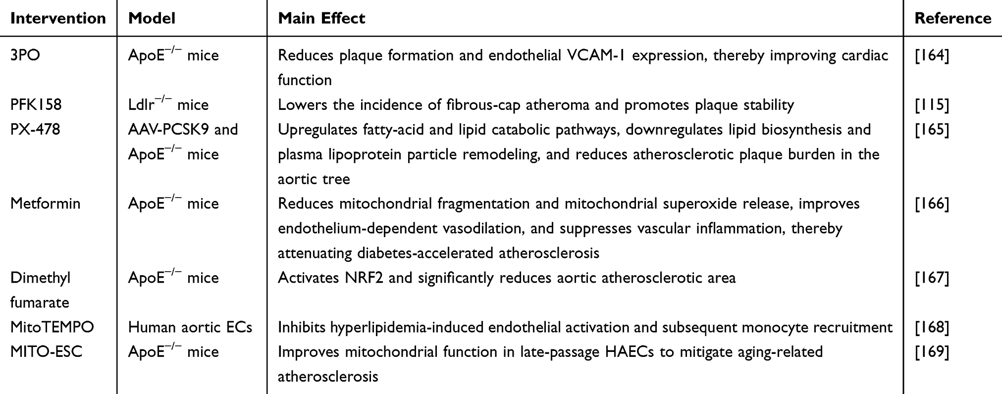 |
Table 1 Therapeutic Interventions Targeting Mitochondrial Function and Metabolic Pathways to Attenuate Atherosclerosis |
Metabolic Drugs and Endothelial Reprogramming in Atherosclerosis
Suppressing endothelial glycolytic flux improves atherosclerotic outcomes. Perrotta et al showed that the glycolytic flux inhibitor 3PO (3-[3-pyridinyl]-1-[4-pyridinyl]-2-propen-1-one) reduced plaque burden and endothelial VCAM-1 expression in ApoE−/− mice and improved cardiac function.164 Glycolysis driven by PFKFB3 is a key driver of endothelial activation and inflammation. Compared with stable fibrous plaques, vulnerable human carotid plaques exhibit higher PFKFB3 expression that localizes predominantly to endothelium, and the glycolysis inhibitor PFK158 lowered the incidence of fibrous cap atheroma in Ldlr−/− mice.115 Endothelial specific deletion of PFKFB3 reduced vein graft lesion area and thickness by 35% and 32%, respectively, and decreased stenosis by 23%, supporting endothelial glycolysis inhibition as a potential strategy to slow plaque progression.170 Targeting HIF-1α signaling is also promising. In hyperlipidemia, unsaturated lysophosphatidic acid derived from mildly oxidized LDL increases endothelial HIF-1α, which triggers miR-19a dependent CXCL1 expression and monocyte adhesion, thereby promoting atherosclerosis.171 Endothelial Hif1a deletion in ApoE−/− mice reduced lesion formation, lesional macrophage accumulation, and endothelial CXCL1 expression.171 Consistently, the selective HIF-1α inhibitor PX-478 decreased aortic plaque burden in AAV-PCSK9 treated C57BL or ApoE−/− mice. RNA sequencing indicated upregulation of fatty acid and lipid catabolic pathways and downregulation of lipid biosynthesis and lipoprotein particle remodeling.165 Activation of the NRF2 antioxidant program counteracts endothelial inflammation. Endothelial Nrf2 knockdown increases endothelial activation and lipid peroxidation and accelerates atherosclerosis, whereas endothelial Nrf2 activation restrains disease.172 Dimethyl fumarate, an NRF2 activator, significantly reduced aortic atherosclerotic area.167 Mitochondria targeted approaches improve endothelial function and limit disease progression. Metformin, a widely used antidiabetic agent, reduced mitochondrial fragmentation, decreased mitochondrial superoxide production, improved endothelium-dependent vasodilation, and suppressed vascular inflammation, thereby attenuating diabetes accelerated atherosclerosis.166 MitoTEMPO inhibited endothelial activation and monocyte recruitment, demonstrating therapeutic potential in atherosclerosis models.168 MITO-ESC therapy increased oxygen consumption rate and improved mitochondrial function in late passage human aortic ECs, mitigating features of vascular aging and atherosclerosis.169 Regarding SGLT2 inhibitors, several studies have directly linked endothelial energy-metabolic reprogramming to plaque amelioration in atherosclerosis models. For example, in ApoE−/− mice, dapagliflozin was reported to activate NRF2 and GPX4 via RAP1B, thereby promoting mitochondrial biogenesis and enhancing oxidative phosphorylation in endothelial cells; this attenuated ferroptosis-associated endothelial phenotypes and reduced plaque formation.173 In male ApoE−/− mice fed a high-fat Western diet, hematoxylin–eosin staining-based assessments showed that empagliflozin treatment decreased both lesion area and lesion size.174 Mechanistically, empagliflozin induced autophagy through AMPK signaling, evidenced by increased Beclin1 expression, an elevated LC3B-II/I ratio, and higher p-AMPK levels, alongside reduced P62 expression and lower levels of inflammatory cytokine proteins; it also suppressed foam-cell formation in RAW264.7 macrophages and HASMCs.174
The principal anti-atherosclerotic benefit of PCSK9 inhibitors is generally attributed to their potent LDL-lowering effect. In individuals at high cardiovascular risk, short-term evolocumab therapy has been shown to improve endothelial function, with the magnitude of improvement proportional to the reduction in LDL.175 In patients with type 2 diabetes, adding evolocumab on top of background empagliflozin therapy further enhanced endothelial function.176 Moreover, in an oxLDL-induced endothelial pyroptosis model, PCSK9 overexpression promoted mitochondrial dysfunction, increased ROS production, and exacerbated pyroptosis by suppressing UQCRC1, whereas PCSK9 silencing reversed mitochondrial membrane potential loss and mitochondrial injury.177
Endothelium-Selective Delivery Platforms for Metabolic and Inflammatory Targets
An HA-modified, dual-targeting nanodrug delivery system (HA-ML@ES NPs) was engineered to co-encapsulate shikonin and evolocumab. Leveraging the specific interaction between HA and CD44, the nanoparticles can concurrently home to endothelial cells with dysfunction and inflammatory macrophages, enabling coordinated intervention across key pathogenic cellular subsets within lesions.178 In a human HUVEC–macrophage co-culture model, this formulation exhibited clear dual-cell targeted uptake, and in vivo it showed prolonged blood circulation and robust enrichment within atherosclerotic plaques—providing a favorable pharmacokinetic and biodistribution basis for localized therapeutic activity.178 In a hyperhomocysteinemia-associated early atherosclerosis model, the strategy mitigated endothelial metabolic imbalance and dysfunction by suppressing endothelial glycolysis, while simultaneously promoting cholesterol efflux and restoring lipid homeostasis through macrophage phenotypic modulation, thereby collectively slowing atherosclerosis progression.178
Separately, cationic liposomes functionalized with a VCAM1-binding peptide were developed to deliver methylated NLRP3 siRNA. Under inflammatory conditions, this platform preferentially targets VCAM1-high endothelial cells, improving cell-type specificity and reducing off-target effects.179 In a rat carotid partial ligation model, local administration markedly reduced endothelial NLRP3 levels and attenuated TNFα-induced pro-atherogenic LDL deposition within the endothelial layer, suggesting disruption of endothelial inflammation–linked pathways that facilitate early transendothelial lipid entry into the vessel wall.179 Moreover, intravenous delivery in ApoE−/− mice decreased plaque formation, supporting a direct anti-atherosclerotic benefit of endothelial-targeted inhibition of the NLRP3 axis in limiting lipid infiltration and lesion expansion.179
Conclusion and Future Direction
Diabetes is a common metabolic disease with a markedly elevated risk of cardiovascular complications, among which atherosclerosis is the principal pathological substrate. Metabolic derangements including hyperglycemia, insulin resistance, and dyslipidemia drive endothelial dysfunction characterized by impaired endothelium-dependent vasodilation and heightened pro-inflammatory and pro-thrombotic states. These abnormalities not only mark the early stages of atherogenesis but also contribute to plaque progression and clinical events. Within the diabetic milieu, ECs undergo multifaceted metabolic reprogramming. Disordered glucose metabolism increases oxidative stress and disrupts signaling, promotes the accumulation of AGEs, and activates inflammatory pathways. In lipid metabolism, FAO is essential for endothelial homeostasis and angiogenesis; its impairment reduces antioxidant capacity and destabilizes endothelial phenotype. Mitochondrial dysfunction is widespread in diabetes, with reduced biogenesis, fragmented networks, and disturbed redox balance that further exacerbate endothelial injury. Hemodynamic forces also shape metabolic programs. Steady laminar shear favors mitochondrial metabolism and preserves function, whereas disturbed flow enhances glycolysis, suppresses oxidative phosphorylation, and promotes a pro-inflammatory phenotype. Metabolic reprogramming is closely intertwined with EndMT. Hyperglycemia, oxidative stress, and multiple signaling axes promote loss of endothelial identity and acquisition of mesenchymal traits, accelerating plaque formation and growth. Shifts in glycolysis and FAO alter cellular energy and metabolite availability, thereby modulating key signaling nodes and epigenetic states. In parallel, inflammation is both a driver and a consequence of these changes, creating positive feedback between metabolic imbalance and endothelial activation. Therapeutically, targeting endothelial metabolism shows promise in experimental models. Strategies that inhibit glycolysis, modulate HIF-1α, activate NRF2, or restore mitochondrial function improve endothelial performance, dampen vascular inflammation, and reduce atherosclerotic burden in animals. From a translational perspective, approaches that leverage endothelial metabolic reprogramming while building on drugs already used in cardiometabolic care, such as metformin, SGLT2 inhibitors, and PCSK9 inhibitors, are likely to be the most immediately applicable in clinical practice. In contrast, more direct targeting of endothelial metabolism, for example by inhibiting inducible glycolysis at nodes such as PFKFB3, modulating the FAO–acetyl-CoA–TGF-β axis that links metabolism to EndMT, or strengthening mitochondrial redox and quality-control programs via NRF2 and mitophagy, remains at an earlier stage but points to particularly attractive targets for future drug development. Important knowledge gaps include the lack of robust in vivo biomarkers of endothelial metabolic and EndMT states, limited insight into how sex, age, vascular bed, and cardiometabolic comorbidities shape endothelial metabolic responses to diabetes, and uncertainty about the reversibility and therapeutic window of manipulating these pathways in established human disease. Progress will also depend on endothelium-focused delivery platforms that can concentrate small molecules, nucleic acids, or biologics within the vascular lining, for example ligand- or antibody-guided nanoparticles directed to adhesion molecules or matrix components enriched at high-risk sites, while minimizing off-target exposure. Meanwhile, future studies could leverage single-cell sequencing to systematically delineate endothelial subpopulations and state transitions within diabetic and atherosclerotic lesions, and to track differences in metabolism-related pathways within a lineage framework, with particular attention to alterations in lipid metabolism and FAO accompanying phenotypic transitions such as EndMT, thereby providing a basis for subpopulation-specific prioritization of metabolic targets. Spatial transcriptomics is expected to precisely map these endothelial states to distinct plaque structural units or the local microenvironments of specific vascular beds, helping to explain the spatial coupling between metabolic reprogramming and inflammation, barrier dysfunction, and lesion instability. Furthermore, integration with spatial proteomics or spatial metabolomics could enable in situ validation of metabolic alterations and facilitate assessment of regional and organ-specific differences in the efficacy of metabolic interventions.
Author Contributions
Panyang Ze: Conceptualization; Data curation; Formal analysis; Investigation; Methodology; Project administration; Writing – original draft; Writing – review and editing. Jin Si: Data curation; Investigation; Resources; Writing – review and editing. Both authors read and approved the final version of the paper. All authors made substantial contributions to conception and design, acquisition of data, or analysis and interpretation of data; took part in drafting the article or revising it critically for important intellectual content; gave final approval of the version to be published; have agreed on the journal to which the article has been submitted; and agree to be accountable for all aspects of the work.
Funding
This work was supported by the National Natural Science Foundation of China (Grant No.82370840).
Disclosure
The authors declare that they have no competing interests.
References
1. Yang D-R, Wang M-Y, Zhang C-L. et al. Endothelial dysfunction in vascular complications of diabetes: a comprehensive review of mechanisms and implications. Front Endocrinol. 2024;15:1359255. doi:10.3389/fendo.2024.1359255
2. Tomic D, Shaw JE, Magliano DJ. The burden and risks of emerging complications of diabetes mellitus. Nat Rev Endocrinol. 2022;18(9):525–22. doi:10.1038/s41574-022-00690-7
3. ElSayed NA, Aleppo G, Aroda VR, et al. Classification and Diagnosis of Diabetes: standards of Care in Diabetes—2023. Diabetes Care. 2023;46(Suppl 1):S19–s40. doi:10.2337/dc23-S002
4. Atar AI, Altuner TK, Bozbas H, et al. Endothelial dysfunction in prediabetes and diabetes mellitus in patients with normal coronary arteries. BMC Cardiovasc Disord. 2024;24(1):622. doi:10.1186/s12872-024-04314-y
5. Zhao N, Yu X, Zhu X, et al. Diabetes Mellitus to Accelerated Atherosclerosis: shared Cellular and Molecular Mechanisms in Glucose and Lipid Metabolism. J Cardiovasc Transl Res. 2024;17(1):133–152. doi:10.1007/s12265-023-10470-x
6. Alexander Y, Osto E, Schmidt-Trucksäss A, et al. Endothelial function in cardiovascular medicine: a consensus paper of the European Society of Cardiology Working Groups on Atherosclerosis and Vascular Biology, Aorta and Peripheral Vascular Diseases, Coronary Pathophysiology and Microcirculation, and Thrombosis. Cardiovasc Res. 2021;117(1):29–42. doi:10.1093/cvr/cvaa085
7. Brunner H, Cockcroft JR, Deanfield J, et al. Endothelial function and dysfunction. Part II: association with cardiovascular risk factors and diseases. A statement by the Working Group on Endothelins and Endothelial Factors of the European Society of Hypertension. J Hypertens. 2005;23(2):233–246. doi:10.1097/00004872-200502000-00001
8. Xu J, Zou MH. Molecular insights and therapeutic targets for diabetic endothelial dysfunction. Circulation. 2009;120(13):1266–1286. doi:10.1161/CIRCULATIONAHA.108.835223
9. Caballero AE. Endothelial dysfunction, inflammation, and insulin resistance: a focus on subjects at risk for type 2 diabetes. Curr Diab Rep. 2004;4(4):237–246. doi:10.1007/s11892-004-0074-9
10. Karakasis P, Theofilis P, Patoulias D, et al. Diabetes-Driven Atherosclerosis: updated Mechanistic Insights and Novel Therapeutic Strategies. Int J Mol Sci. 2025;26(5):2196. doi:10.3390/ijms26052196
11. Xue C, Chen K, Gao Z, et al. Common mechanisms underlying diabetic vascular complications: focus on the interaction of metabolic disorders, immuno-inflammation, and endothelial dysfunction. Cell Commun Signal. 2023;21(1):298. doi:10.1186/s12964-022-01016-w
12. Widlansky ME, Gokce N, Keaney JF, et al. The clinical implications of endothelial dysfunction. J Am Coll Cardiol. 2003;42(7):1149–1160. doi:10.1016/S0735-1097(03)00994-X
13. Vita JA, Keaney Jr JF. Endothelial function: a barometer for cardiovascular risk? Circulation. 2002;106(6):640–642. doi:10.1161/01.CIR.0000028581.07992.56
14. Bakker W, Eringa EC, Sipkema P, et al. Endothelial dysfunction and diabetes: roles of hyperglycemia, impaired insulin signaling and obesity. Cell Tissue Res. 2009;335(1):165–189. doi:10.1007/s00441-008-0685-6
15. Zhang H, Dellsperger KC, Zhang C. The link between metabolic abnormalities and endothelial dysfunction in type 2 diabetes: an update. Basic Res Cardiol. 2012;107(1):237. doi:10.1007/s00395-011-0237-1
16. Nicolls MR, Haskins K, Flores SC. Oxidant stress, immune dysregulation, and vascular function in type I diabetes. Antioxid Redox Signal. 2007;9(7):879–889. doi:10.1089/ars.2007.1631
17. Tabit CE, Chung WB, Hamburg NM, et al. Endothelial dysfunction in diabetes mellitus: molecular mechanisms and clinical implications. Rev Endocr Metab Disord. 2010;11(1):61–74. doi:10.1007/s11154-010-9134-4
18. Gokce N, Vita JA, McDonnell M, et al. Effect of medical and surgical weight loss on endothelial vasomotor function in obese patients. Am J Cardiol. 2005;95(2):266–268. doi:10.1016/j.amjcard.2004.09.016
19. Hamburg NM, Larson MG, Vita JA, et al. Metabolic syndrome, insulin resistance, and brachial artery vasodilator function in Framingham Offspring participants without clinical evidence of cardiovascular disease. Am J Cardiol. 2008;101(1):82–88. doi:10.1016/j.amjcard.2007.07.053
20. Keaney Jr JF, Massaro JM, Larson MG, et al. Heritability and correlates of intercellular adhesion molecule-1 in the Framingham Offspring Study. J Am Coll Cardiol. 2004;44(1):168–173. doi:10.1016/j.jacc.2004.03.048
21. Meigs JB. Biomarkers of endothelial dysfunction and risk of type 2 diabetes mellitus. JAMA. 2004;291(16):1978–1986. doi:10.1001/jama.291.16.1978
22. de Jongh RT, Serne EH, IJzerman RG, et al. Impaired microvascular function in obesity: implications for obesity-associated microangiopathy, hypertension, and insulin resistance. Circulation. 2004;109(21):2529–2535. doi:10.1161/01.CIR.0000129772.26647.6F
23. Campia U, Sullivan G, Bryant MB, et al. Insulin impairs endothelium-dependent vasodilation independent of insulin sensitivity or lipid profile. Am J Physiol Heart Circ Physiol. 2004;286(1):H76–82. doi:10.1152/ajpheart.00539.2003
24. Shinozaki K, Hirayama A, Nishio Y, et al. Coronary endothelial dysfunction in the insulin-resistant state is linked to abnormal pteridine metabolism and vascular oxidative stress. J Am Coll Cardiol. 2001;38(7):1821–1828. doi:10.1016/S0735-1097(01)01659-X
25. Lampinen KH, Ronnback M, Groop P-H, et al. A relationship between insulin sensitivity and vasodilation in women with a history of preeclamptic pregnancy. Hypertension. 2008;52(2):394–401. doi:10.1161/HYPERTENSIONAHA.108.113423
26. Ardigo D, Franzini L, Valtuena S, et al. Relation of plasma insulin levels to forearm flow-mediated dilatation in healthy volunteers. Am J Cardiol. 2006;97(8):1250–1254. doi:10.1016/j.amjcard.2005.11.047
27. Lteif AA, Han K, Mather KJ. Obesity, insulin resistance, and the metabolic syndrome: determinants of endothelial dysfunction in whites and blacks. Circulation. 2005;112(1):32–38. doi:10.1161/CIRCULATIONAHA.104.520130
28. Tesauro M, Rizza S, Iantorno M, et al. Vascular, metabolic, and inflammatory abnormalities in normoglycemic offspring of patients with type 2 diabetes mellitus. Metabolism. 2007;56(3):413–419. doi:10.1016/j.metabol.2006.10.026
29. Meigs JB, O’Donnell CJ, Tofler GH, et al. Hemostatic markers of endothelial dysfunction and risk of incident type 2 diabetes: the Framingham Offspring Study. Diabetes. 2006;55(2):530–537. doi:10.2337/diabetes.55.02.06.db05-1041
30. Rossi R, Cioni E, Nuzzo A, et al. Endothelial-dependent vasodilation and incidence of type 2 diabetes in a population of healthy postmenopausal women. Diabetes Care. 2005;28(3):702–707. doi:10.2337/diacare.28.3.702
31. Monti LD, Barlassina C, Citterio L, et al. Endothelial nitric oxide synthase polymorphisms are associated with type 2 diabetes and the insulin resistance syndrome. Diabetes. 2003;52(5):1270–1275. doi:10.2337/diabetes.52.5.1270
32. Pistrosch F, Passauer J, Fischer S, et al. In type 2 diabetes, rosiglitazone therapy for insulin resistance ameliorates endothelial dysfunction independent of glucose control. Diabetes Care. 2004;27(2):484–490. doi:10.2337/diacare.27.2.484
33. Caballero AE, Saouaf R, Lim SC, et al. The effects of troglitazone, an insulin-sensitizing agent, on the endothelial function in early and late type 2 diabetes: a placebo-controlled randomized clinical trial. Metabolism. 2003;52(2):173–180. doi:10.1053/meta.2003.50023
34. Mather KJ, Verma S, Anderson TJ. Improved endothelial function with metformin in type 2 diabetes mellitus. J Am Coll Cardiol. 2001;37(5):1344–1350. doi:10.1016/S0735-1097(01)01129-9
35. de Aguiar LGK, Bahia LR, Villela N, et al. Metformin improves endothelial vascular reactivity in first-degree relatives of type 2 diabetic patients with metabolic syndrome and normal glucose tolerance. Diabetes Care. 2006;29(5):1083–1089. doi:10.2337/dc05-2146
36. Campia U, Matuskey LA, Panza JA. Peroxisome Proliferator–Activated Receptor-γ Activation With Pioglitazone Improves Endothelium-Dependent Dilation in Nondiabetic Patients With Major Cardiovascular Risk Factors. Circulation. 2006;113(6):867–875. doi:10.1161/CIRCULATIONAHA.105.549618
37. Hetzel J, Balletshofer B, Rittig K, et al. Rapid effects of rosiglitazone treatment on endothelial function and inflammatory biomarkers. Arterioscler Thromb Vasc Biol. 2005;25(9):1804–1809. doi:10.1161/01.ATV.0000176192.16951.9a
38. Cheetham C, Collis J, O’Driscoll G, et al. Losartan, an angiotensin type 1 receptor antagonist, improves endothelial function in non-insulin-dependent diabetes. J Am Coll Cardiol. 2000;36(5):1461–1466. doi:10.1016/S0735-1097(00)00933-5
39. Goveia J, Stapor P, Carmeliet P. Principles of targeting endothelial cell metabolism to treat angiogenesis and endothelial cell dysfunction in disease. EMBO Mol Med. 2014;6(9):1105–1120. doi:10.15252/emmm.201404156
40. Guzik TJ, Mussa S, Gastaldi D, et al. Mechanisms of increased vascular superoxide production in human diabetes mellitus: role of NAD(P)H oxidase and endothelial nitric oxide synthase. Circulation. 2002;105(14):1656–1662. doi:10.1161/01.CIR.0000012748.58444.08
41. Xie X, Chen Y, Liu J, et al. High glucose induced endothelial cell reactive oxygen species via OGG1/PKC/NADPH oxidase pathway. Life Sci. 2020;256:117886. doi:10.1016/j.lfs.2020.117886
42. Eleftheriadis T, Pissas G, Antoniadi G, et al. Allopurinol protects human glomerular endothelial cells from high glucose-induced reactive oxygen species generation, p53 overexpression and endothelial dysfunction. Int Urol Nephrol. 2018;50(1):179–186. doi:10.1007/s11255-017-1733-5
43. Sasaki N, Yamashita T, Takaya T, et al. Augmentation of vascular remodeling by uncoupled endothelial nitric oxide synthase in a mouse model of diabetes mellitus. Arterioscler Thromb Vasc Biol. 2008;28(6):1068–1076. doi:10.1161/ATVBAHA.107.160754
44. Zhang Z, Apse K, Pang J, et al. High glucose inhibits glucose-6-phosphate dehydrogenase via cAMP in aortic endothelial cells. J Biol Chem. 2000;275(51):40042–40047. doi:10.1074/jbc.M007505200
45. Shenouda SM, Widlansky ME, Chen K, et al. Altered mitochondrial dynamics contributes to endothelial dysfunction in diabetes mellitus. Circulation. 2011;124(4):444–453. doi:10.1161/CIRCULATIONAHA.110.014506
46. Du X, Matsumura T, Edelstein D, et al. Inhibition of GAPDH activity by poly(ADP-ribose) polymerase activates three major pathways of hyperglycemic damage in endothelial cells. J Clin Invest. 2003;112(7):1049–1057. doi:10.1172/JCI18127
47. Brownlee M. Biochemistry and molecular cell biology of diabetic complications. Nature. 2001;414(6865):813–820. doi:10.1038/414813a
48. Luo B, Soesanto Y, McClain DA. Protein modification by O-linked GlcNAc reduces angiogenesis by inhibiting Akt activity in endothelial cells. Arterioscler Thromb Vasc Biol. 2008;28(4):651–657. doi:10.1161/ATVBAHA.107.159533
49. Das Evcimen N, King GL. The role of protein kinase C activation and the vascular complications of diabetes. Pharmacol Res. 2007;55(6):498–510. doi:10.1016/j.phrs.2007.04.016
50. Tang G, Li S, Zhang C, et al. Clinical efficacies, underlying mechanisms and molecular targets of Chinese medicines for diabetic nephropathy treatment and management. Acta Pharm Sin B. 2021;11(9):2749–2767. doi:10.1016/j.apsb.2020.12.020
51. Shen CY, Lu C-H, Wu C-H, et al. The Development of Maillard Reaction, and Advanced Glycation End Product (AGE)-Receptor for AGE (RAGE) Signaling Inhibitors as Novel Therapeutic Strategies for Patients with AGE-Related Diseases. Molecules. 2020;25(23):5591. doi:10.3390/molecules25235591
52. Fujii EY, Nakayama M. The measurements of RAGE, VEGF, and AGEs in the plasma and follicular fluid of reproductive women: the influence of aging. Fertil Steril. 2010;94(2):694–700. doi:10.1016/j.fertnstert.2009.03.029
53. Gong DJ, Wang L, Yang -Y-Y, et al. Diabetes aggravates renal ischemia and reperfusion injury in rats by exacerbating oxidative stress, inflammation, and apoptosis. Ren Fail. 2019;41(1):750–761. doi:10.1080/0886022X.2019.1643737
54. Eelen G, de Zeeuw P, Treps L, et al. Endothelial Cell Metabolism. Physiol Rev. 2018;98(1):3–58. doi:10.1152/physrev.00001.2017
55. Fisslthaler B, Fleming I. Activation and signaling by the AMP-activated protein kinase in endothelial cells. Circ Res. 2009;105(2):114–127. doi:10.1161/CIRCRESAHA.109.201590
56. Dagher Z, Ruderman N, Tornheim K, et al. Acute regulation of fatty acid oxidation and amp-activated protein kinase in human umbilical vein endothelial cells. Circ Res. 2001;88(12):1276–1282. doi:10.1161/hh1201.092998
57. Kalucka J, Bierhansl L, Conchinha NV, et al. Quiescent Endothelial Cells Upregulate Fatty Acid β-Oxidation for Vasculoprotection via Redox Homeostasis. Cell Metab. 2018;28(6):881–894.e13. doi:10.1016/j.cmet.2018.07.016
58. Schoors S, Bruning U, Missiaen R, et al. Fatty acid carbon is essential for dNTP synthesis in endothelial cells. Nature. 2015;520(7546):192–197. doi:10.1038/nature14362
59. Steinberg HO, Tarshoby M, Monestel R, et al. Elevated circulating free fatty acid levels impair endothelium-dependent vasodilation. J Clin Invest. 1997;100(5):1230–1239. doi:10.1172/JCI119636
60. Steinberg HO, Paradisi G, Hook G, et al. Free fatty acid elevation impairs insulin-mediated vasodilation and nitric oxide production. Diabetes. 2000;49(7):1231–1238. doi:10.2337/diabetes.49.7.1231
61. de Kreutzenberg SV, Crepaldi C, Marchetto S, et al. Plasma free fatty acids and endothelium-dependent vasodilation: effect of chain-length and cyclooxygenase inhibition. J Clin Endocrinol Metab. 2000;85(2):793–798. doi:10.1210/jcem.85.2.6352
62. Cheng C-I, Lee Y-H, Chen P-H, et al. Free Fatty Acids Induce Autophagy and LOX-1 Upregulation in Cultured Aortic Vascular Smooth Muscle Cells. J Cell Biochem. 2017;118(5):1249–1261. doi:10.1002/jcb.25784
63. Patella F, Schug ZT, Persi E, et al. Proteomics-based metabolic modeling reveals that fatty acid oxidation (FAO) controls endothelial cell (EC) permeability. Mol Cell Proteomics. 2015;14(3):621–634. doi:10.1074/mcp.M114.045575
64. Akhter F, Khan MS, Alatar AA, et al. Antigenic role of the adaptive immune response to d-ribose glycated LDL in diabetes, atherosclerosis and diabetes atherosclerotic patients. Life Sci. 2016;151:139–146. doi:10.1016/j.lfs.2016.02.013
65. Jin JL, Zhang H-W, Cao Y-X, et al. Association of small dense low-density lipoprotein with cardiovascular outcome in patients with coronary artery disease and diabetes: a prospective, observational cohort study. Cardiovasc Diabetol. 2020;19(1):45. doi:10.1186/s12933-020-01015-6
66. Zhang Q, Liu J, Duan H, et al. Activation of Nrf2/HO-1 signaling: an important molecular mechanism of herbal medicine in the treatment of atherosclerosis via the protection of vascular endothelial cells from oxidative stress. J Adv Res. 2021;34:43–63. doi:10.1016/j.jare.2021.06.023
67. Wu F, Wang H, Li J, et al. Homoplantaginin modulates insulin sensitivity in endothelial cells by inhibiting inflammation. Biol Pharm Bull. 2012;35(7):1171–1177. doi:10.1248/bpb.b110586
68. Mundi S, Massaro M, Scoditti E, et al. Endothelial permeability, LDL deposition, and cardiovascular risk factors-a review. Cardiovasc Res. 2018;114(1):35–52. doi:10.1093/cvr/cvx226
69. Huang L, Chambliss KL, Gao X, et al. SR-B1 drives endothelial cell LDL transcytosis via DOCK4 to promote atherosclerosis. Nature. 2019;569(7757):565–569. doi:10.1038/s41586-019-1140-4
70. Zhang Y, Yang X, Bian F, et al. TNF-α promotes early atherosclerosis by increasing transcytosis of LDL across endothelial cells: crosstalk between NF-κB and PPAR-γ. J Mol Cell Cardiol. 2014;72:85–94. doi:10.1016/j.yjmcc.2014.02.012
71. Pirri D, Tian S, Tardajos-Ayllon B, et al. EPAS1 Attenuates Atherosclerosis Initiation at Disturbed Flow Sites Through Endothelial Fatty Acid Uptake. Circ Res. 2024;135(8):822–837. doi:10.1161/CIRCRESAHA.123.324054
72. Xiong J, Kawagishi H, Yan Y, et al. A Metabolic Basis for Endothelial-to-Mesenchymal Transition. Mol Cell. 2018;69(4):689–698.e7. doi:10.1016/j.molcel.2018.01.010
73. Evrard SM, Lecce L, Michelis KC, et al. Endothelial to mesenchymal transition is common in atherosclerotic lesions and is associated with plaque instability. Nat Commun. 2016;7(1):11853. doi:10.1038/ncomms11853
74. Chen PY, Qin L, Baeyens N, et al. Endothelial-to-mesenchymal transition drives atherosclerosis progression. J Clin Invest. 2015;125(12):4514–4528. doi:10.1172/JCI82719
75. Chen P-Y, Qin L, Li G, et al. Endothelial TGF-β signalling drives vascular inflammation and atherosclerosis. Nat Metab. 2019;1(9):912–926. doi:10.1038/s42255-019-0102-3
76. Zhu X, Y Wang, I Soaita, et al. Acetate controls endothelial-to-mesenchymal transition. Cell Metab. 2023;35(7):1163–1178.e10. doi:10.1016/j.cmet.2023.05.010
77. Cao M, Li M, Li X, et al. Endothelial soluble epoxide hydrolase links polyunsaturated fatty acid metabolism to oxidative stress and atherosclerosis progression. Redox Biol. 2025;85:103730. doi:10.1016/j.redox.2025.103730
78. Darley-Usmar V. The powerhouse takes control of the cell; the role of mitochondria in signal transduction. Free Radic Biol Med. 2004;37(6):753–754. doi:10.1016/j.freeradbiomed.2004.05.026
79. Hamanaka RB, Chandel NS. Mitochondrial reactive oxygen species regulate cellular signaling and dictate biological outcomes. Trends Biochem Sci. 2010;35(9):505–513. doi:10.1016/j.tibs.2010.04.002
80. Dan Dunn J, Alvarez LA, Zhang X, et al. Reactive oxygen species and mitochondria: a nexus of cellular homeostasis. Redox Biol. 2015;6:472–485. doi:10.1016/j.redox.2015.09.005
81. Quintero M, Colombo SL, Godfrey A, et al. Mitochondria as signaling organelles in the vascular endothelium. Proc Natl Acad Sci U S A. 2006;103(14):5379–5384. doi:10.1073/pnas.0601026103
82. Liu Y, Zhao H, Li H, et al. Mitochondrial Sources of H2O2 Generation Play a Key Role in Flow-Mediated Dilation in Human Coronary Resistance Arteries. Circ Res. 2003;93(6):573–580. doi:10.1161/01.RES.0000091261.19387.AE
83. Heitzer T, B Finckh, S Albers, et al. Beneficial effects of alpha-lipoic acid and ascorbic acid on endothelium-dependent, nitric oxide-mediated vasodilation in diabetic patients: relation to parameters of oxidative stress. Free Radic Biol Med. 2001;31(1):53–61. doi:10.1016/S0891-5849(01)00551-2
84. Widlansky ME, Hill RB. Mitochondrial regulation of diabetic vascular disease: an emerging opportunity. Transl Res. 2018;202:83–98. doi:10.1016/j.trsl.2018.07.015
85. Scarpulla RC. Metabolic control of mitochondrial biogenesis through the PGC-1 family regulatory network. Biochim Biophys Acta. 2011;1813(7):1269–1278. doi:10.1016/j.bbamcr.2010.09.019
86. Twig G, Elorza A, Molina AJA, et al. Fission and selective fusion govern mitochondrial segregation and elimination by autophagy. EMBO j. 2008;27(2):433–446. doi:10.1038/sj.emboj.7601963
87. Petersen KF, Dufour S, Befroy D, et al. Impaired mitochondrial activity in the insulin-resistant offspring of patients with type 2 diabetes. N Engl J Med. 2004;350(7):664–671. doi:10.1056/NEJMoa031314
88. Lowell BB, Shulman GI. Mitochondrial dysfunction and type 2 diabetes. Science. 2005;307(5708):384–387. doi:10.1126/science.1104343
89. Yu T, Jhun BS, Yoon Y. High-glucose stimulation increases reactive oxygen species production through the calcium and mitogen-activated protein kinase-mediated activation of mitochondrial fission. Antioxid Redox Signal. 2011;14(3):425–437. doi:10.1089/ars.2010.3284
90. Sangwung P, Petersen KF, Shulman GI, et al. Mitochondrial Dysfunction, Insulin Resistance, and Potential Genetic Implications. Endocrinology. 2020;161(4). doi:10.1210/endocr/bqaa017.
91. Liu J, Li J, Li W-J, et al. The role of uncoupling proteins in diabetes mellitus. J Diabetes Res. 2013;2013:585897. doi:10.1155/2013/585897
92. Li B, Nolte LA, Ju J-S, et al. Skeletal muscle respiratory uncoupling prevents diet-induced obesity and insulin resistance in mice. Nat Med. 2000;6(10):1115–1120. doi:10.1038/80450
93. Zorzano A, Liesa M, Palacín M. Role of mitochondrial dynamics proteins in the pathophysiology of obesity and type 2 diabetes. Int J Biochem Cell Biol. 2009;41(10):1846–1854. doi:10.1016/j.biocel.2009.02.004
94. Milne JC, Lambert PD, Schenk S, et al. Small molecule activators of SIRT1 as therapeutics for the treatment of type 2 diabetes. Nature. 2007;450(7170):712–716. doi:10.1038/nature06261
95. Mootha VK, Lindgren CM, Eriksson K-F, et al. PGC-1alpha-responsive genes involved in oxidative phosphorylation are coordinately downregulated in human diabetes. Nat Genet. 2003;34(3):267–273. doi:10.1038/ng1180
96. Ritov VB, Menshikova EV, He J, et al. Deficiency of subsarcolemmal mitochondria in obesity and type 2 diabetes. Diabetes. 2005;54(1):8–14. doi:10.2337/diabetes.54.1.8
97. Pilegaard H, Saltin B, Neufer PD. Exercise induces transient transcriptional activation of the PGC-1alpha gene in human skeletal muscle. J Physiol. 2003;546(Pt 3):851–858. doi:10.1113/jphysiol.2002.034850
98. Kizhakekuttu TJ, Wang J, Dharmashankar K, et al. Adverse alterations in mitochondrial function contribute to type 2 diabetes mellitus-related endothelial dysfunction in humans. Arterioscler Thromb Vasc Biol. 2012;32(10):2531–2539. doi:10.1161/ATVBAHA.112.256024
99. Hahn C, Schwartz MA. Mechanotransduction in vascular physiology and atherogenesis. Nat Rev Mol Cell Biol. 2009;10(1):53–62. doi:10.1038/nrm2596
100. Rohlenova K, Veys K, Miranda-Santos I, et al. Endothelial Cell Metabolism in Health and Disease. Trends Cell Biol. 2018;28(3):224–236. doi:10.1016/j.tcb.2017.10.010
101. Kant S, Tran K-V, Kvandova M, et al. PGC1α Regulates the Endothelial Response to Fluid Shear Stress via Telomerase Reverse Transcriptase Control of Heme Oxygenase-1. Arterioscler Thromb Vasc Biol. 2022;42(1):19–34. doi:10.1161/ATVBAHA.121.317066
102. Coon BG, Timalsina S, Astone M, et al. A mitochondrial contribution to anti-inflammatory shear stress signaling in vascular endothelial cells. J Cell Biol. 2022;221(7). doi:10.1083/jcb.202109144.
103. Gong G, Song M, Csordas G, et al. Parkin-mediated mitophagy directs perinatal cardiac metabolic maturation in mice. Science. 2015;350(6265):aad2459. doi:10.1126/science.aad2459
104. Krantz S, Kim Y-M, Srivastava S, et al. Mitophagy mediates metabolic reprogramming of induced pluripotent stem cells undergoing endothelial differentiation. J Biol Chem. 2021;297(6):101410. doi:10.1016/j.jbc.2021.101410
105. Doddaballapur A, Michalik KM, Manavski Y, et al. Laminar shear stress inhibits endothelial cell metabolism via KLF2-mediated repression of PFKFB3. Arterioscler Thromb Vasc Biol. 2015;35(1):137–145. doi:10.1161/ATVBAHA.114.304277
106. Falkenberg KD, Rohlenova K, Luo Y, et al. The metabolic engine of endothelial cells. Nat Metab. 2019;1(10):937–946. doi:10.1038/s42255-019-0117-9
107. Yang Q, Xu J, Ma Q, et al. PRKAA1/AMPKα1-driven glycolysis in endothelial cells exposed to disturbed flow protects against atherosclerosis. Nat Commun. 2018;9(1):4667. doi:10.1038/s41467-018-07132-x
108. Kim J, Kim YH, Kim J, et al. YAP/TAZ regulates sprouting angiogenesis and vascular barrier maturation. J Clin Invest. 2017;127(9):3441–3461. doi:10.1172/JCI93825
109. Feng S, Bowden N, Fragiadaki M, et al. Mechanical Activation of Hypoxia-Inducible Factor 1α Drives Endothelial Dysfunction at Atheroprone Sites. Arterioscler Thromb Vasc Biol. 2017;37(11):2087–2101. doi:10.1161/ATVBAHA.117.309249
110. Wu D, Huang R-T, Hamanaka RB, et al. HIF-1α is required for disturbed flow-induced metabolic reprogramming in human and porcine vascular endothelium. Elife. 2017;6. doi:10.7554/eLife.25217
111. Chen L-J, Li JY-S, Nguyen P, et al. Single-cell RNA sequencing unveils unique transcriptomic signatures of endothelial cells and role of ENO1 in response to disturbed flow. Proc Natl Acad Sci U S A. 2024;121(5):e2318904121. doi:10.1073/pnas.2318904121
112. Baek KI, Li R, Jen N, et al. Flow-Responsive Vascular Endothelial Growth Factor Receptor-Protein Kinase C Isoform Epsilon Signaling Mediates Glycolytic Metabolites for Vascular Repair. Antioxid Redox Signal. 2018;28(1):31–43. doi:10.1089/ars.2017.7044
113. Qu J, Lu D, Guo H, et al. PFKFB 3 modulates glycolytic metabolism and alleviates endoplasmic reticulum stress in human osteoarthritis cartilage. Clin Exp Pharmacol Physiol. 2016;43(3):312–318. doi:10.1111/1440-1681.12537
114. De Bock K, Georgiadou M, Schoors S, et al. Role of PFKFB3-driven glycolysis in vessel sprouting. Cell. 2013;154(3):651–663. doi:10.1016/j.cell.2013.06.037
115. Poels K, Schnitzler JG, Waissi F, et al. Inhibition of PFKFB3 Hampers the Progression of Atherosclerosis and Promotes Plaque Stability. Front Cell Dev Biol. 2020;8:581641. doi:10.3389/fcell.2020.581641
116. Wang L, Cao Y, Gorshkov B, et al. Ablation of endothelial Pfkfb3 protects mice from acute lung injury in LPS-induced endotoxemia. Pharmacol Res. 2019;146:104292. doi:10.1016/j.phrs.2019.104292
117. Giordo R, Ahmed YMA, Allam H, et al. EndMT Regulation by Small RNAs in Diabetes-Associated Fibrotic Conditions: potential Link With Oxidative Stress. Front Cell Dev Biol. 2021;9:683594. doi:10.3389/fcell.2021.683594
118. Chang JC, SJ Kou, WT Lin, et al. Regulatory role of mitochondria in oxidative stress and atherosclerosis. World J Cardiol. 2010;2(6):150–159. doi:10.4330/wjc.v2.i6.150
119. Chen PY, Schwartz MA, Simons M. Endothelial-to-Mesenchymal Transition, Vascular Inflammation, and Atherosclerosis. Front Cardiovasc Med. 2020;7:53. doi:10.3389/fcvm.2020.00053
120. Kokudo T, Suzuki Y, Yoshimatsu Y, et al. Snail is required for TGFbeta-induced endothelial-mesenchymal transition of embryonic stem cell-derived endothelial cells. J Cell Sci. 2008;121(Pt 20):3317–3324. doi:10.1242/jcs.028282
121. Atawia RT, Batori RK, Jordan CR, et al. Type 1 Diabetes Impairs Endothelium-Dependent Relaxation Via Increasing Endothelial Cell Glycolysis Through Advanced Glycation End Products, PFKFB3, and Nox1-Mediated Mechanisms. Hypertension. 2023;80(10):2059–2071. doi:10.1161/HYPERTENSIONAHA.123.21341
122. Zeng H, Pan T, Zhan M, et al. Suppression of PFKFB3-driven glycolysis restrains endothelial-to-mesenchymal transition and fibrotic response. Signal Transduct Target Ther. 2022;7(1):303. doi:10.1038/s41392-022-01097-6
123. Xu S, Xu Y, Yin M, et al. Flow-dependent epigenetic regulation of IGFBP5 expression by H3K27me3 contributes to endothelial anti-inflammatory effects. Theranostics. 2018;8(11):3007–3021. doi:10.7150/thno.21966
124. Greißel A, Culmes M, Burgkart R, et al. Histone acetylation and methylation significantly change with severity of atherosclerosis in human carotid plaques. Cardiovasc Pathol. 2016;25(2):79–86. doi:10.1016/j.carpath.2015.11.001
125. Pandya Thakkar N, Pereira BMV, Katakia YT, et al. Elevated H3K4me3 Through MLL2-WDR82 upon Hyperglycemia Causes Jagged Ligand Dependent Notch Activation to Interplay with Differentiation State of Endothelial Cells. Front Cell Dev Biol. 2022;10:839109. doi:10.3389/fcell.2022.839109
126. Maleszewska M, Vanchin B, Harmsen MC, et al. The decrease in histone methyltransferase EZH2 in response to fluid shear stress alters endothelial gene expression and promotes quiescence. Angiogenesis. 2016;19(1):9–24. doi:10.1007/s10456-015-9485-2
127. Fang Y, Shi C, Manduchi E, et al. MicroRNA-10a regulation of proinflammatory phenotype in athero-susceptible endothelium in vivo and in vitro. Proc Natl Acad Sci U S A. 2010;107(30):13450–13455. doi:10.1073/pnas.1002120107
128. Santovito D, Egea V, Bidzhekov K, et al. Noncanonical inhibition of caspase-3 by a nuclear microRNA confers endothelial protection by autophagy in atherosclerosis. Sci Transl Med. 2020;12(546). doi:10.1126/scitranslmed.aaz2294.
129. Lovisa S, Kalluri R. Fatty Acid Oxidation Regulates the Activation of Endothelial-to-Mesenchymal Transition. Trends Mol Med. 2018;24(5):432–434. doi:10.1016/j.molmed.2018.03.003
130. Sivanand S, Viney I, Wellen KE. Spatiotemporal Control of Acetyl-CoA Metabolism in Chromatin Regulation. Trends Biochem Sci. 2018;43(1):61–74. doi:10.1016/j.tibs.2017.11.004
131. Houten SM, Violante S, Ventura FV, et al. The Biochemistry and Physiology of Mitochondrial Fatty Acid β-Oxidation and Its Genetic Disorders. Annu Rev Physiol. 2016;78(1):23–44. doi:10.1146/annurev-physiol-021115-105045
132. Wang W, Wang Y, Long J, et al. Mitochondrial fission triggered by hyperglycemia is mediated by ROCK1 activation in podocytes and endothelial cells. Cell Metab. 2012;15(2):186–200. doi:10.1016/j.cmet.2012.01.009
133. Yu T, Robotham JL, Yoon Y. Increased production of reactive oxygen species in hyperglycemic conditions requires dynamic change of mitochondrial morphology. Proc Natl Acad Sci U S A. 2006;103(8):2653–2658. doi:10.1073/pnas.0511154103
134. Zaied AA, Ushio‐Fukai M, Fukai T, et al. Pentose Pathway Activation Is Superior to Increased Glycolysis for Therapeutic Angiogenesis in Peripheral Arterial Disease. J Am Heart Assoc. 2023;12(7):e027986. doi:10.1161/JAHA.122.027986
135. Moonen J-RAJ, Krenning G, Brinker MGL, et al. Endothelial progenitor cells give rise to pro-angiogenic smooth muscle-like progeny. Cardiovasc Res. 2010;86(3):506–515. doi:10.1093/cvr/cvq012
136. Zhao G, Lu H, Liu Y, et al. Single-Cell Transcriptomics Reveals Endothelial Plasticity During Diabetic Atherogenesis. Front Cell Dev Biol. 2021;9:689469. doi:10.3389/fcell.2021.689469
137. Hall IF, Kishta F, Xu Y, et al. Endothelial to mesenchymal transition: at the axis of cardiovascular health and disease. Cardiovasc Res. 2024;120(3):223–236. doi:10.1093/cvr/cvae021
138. Tian J, Zhang M, Suo M, et al. Dapagliflozin alleviates cardiac fibrosis through suppressing EndMT and fibroblast activation via AMPKα/TGF-β/Smad signalling in type 2 diabetic rats. J Cell Mol Med. 2021;25(16):7642–7659. doi:10.1111/jcmm.16601
139. Hulshoff MS, Schellinger IN, Xu X, et al. miR-132-3p and KLF7 as novel regulators of aortic stiffening-associated EndMT in type 2 diabetes mellitus. Diabetol Metab Syndr. 2023;15(1):11. doi:10.1186/s13098-022-00966-y
140. Yu CH, Gong M. High glucose induced endothelial to mesenchymal transition in human umbilical vein endothelial cell. Exp Mol Pathol. 2017;102(3):377–383. doi:10.1016/j.yexmp.2017.03.007
141. Liu X, Mujahid H, Rong B, et al. Irisin inhibits high glucose-induced endothelial-to-mesenchymal transition and exerts a dose-dependent bidirectional effect on diabetic cardiomyopathy. J Cell Mol Med. 2018;22(2):808–822. doi:10.1111/jcmm.13360
142. Chen PY, Qin L, Li G, et al. Smooth Muscle Cell Reprogramming in Aortic Aneurysms. Cell Stem Cell. 2020;26(4):542–557.e11. doi:10.1016/j.stem.2020.02.013
143. Glaser SF, Heumüller AW, Tombor L, et al. The histone demethylase JMJD2B regulates endothelial-to-mesenchymal transition. Proc Natl Acad Sci U S A. 2020;117(8):4180–4187. doi:10.1073/pnas.1913481117
144. Lecce L, Xu Y, V’Gangula B, et al. Histone deacetylase 9 promotes endothelial-mesenchymal transition and an unfavorable atherosclerotic plaque phenotype. J Clin Invest. 2021;131(15). doi:10.1172/JCI131178.
145. Li X, Sun X, Carmeliet P. Hallmarks of Endothelial Cell Metabolism in Health and Disease. Cell Metab. 2019;30(3):414–433. doi:10.1016/j.cmet.2019.08.011
146. Peng H, Wang X, Du J, et al. Metabolic Reprogramming of Vascular Endothelial Cells: basic Research and Clinical Applications. Front Cell Dev Biol. 2021;9:626047. doi:10.3389/fcell.2021.626047
147. Vu GH, Jeon BH, Kim CS. Endothelial to mesenchymal transition: a central mechanism in diabetes-induced vascular pathology. Korean J Physiol Pharmacol. 2025;29(6):699–711. doi:10.4196/kjpp.25.236
148. Tang R, Li Q, Lv L, et al. Angiotensin II mediates the high-glucose-induced endothelial-to-mesenchymal transition in human aortic endothelial cells. Cardiovasc Diabetol. 2010;9(1):31. doi:10.1186/1475-2840-9-31
149. Ma J, Liu T, Dong X. Advanced glycation end products of bovine serum albumin-induced endothelial-to-mesenchymal transition in cultured human and monkey endothelial cells via protein kinase B signaling cascades. Mol Vis. 2010;16:2669–2679.
150. Lin RH, Chen J-C, Xu -S-S, et al. Asiatic acid inhibits endothelial-to-mesenchymal transition in diabetic kidney disease by reducing acetyl-CoA production via targeting ACSS2. Biochem Pharmacol. 2025;242(Pt 2):117311. doi:10.1016/j.bcp.2025.117311
151. Bingyu W, Jun Q, Bingyang L, et al. Trimethylamine N-oxide promotes PERK-mediated endothelial-mesenchymal transition and apoptosis thereby aggravates atherosclerosis. Int Immunopharmacol. 2024;142(Pt B):113209. doi:10.1016/j.intimp.2024.113209
152. Meyerovich K, Ortis F, Cardozo AK. The non-canonical NF-κB pathway and its contribution to β-cell failure in diabetes. J Mol Endocrinol. 2018;61(2):F1–f6. doi:10.1530/JME-16-0183
153. Xu S, Ilyas I, Little PJ, et al. Endothelial Dysfunction in Atherosclerotic Cardiovascular Diseases and Beyond: from Mechanism to Pharmacotherapies. Pharmacol Rev. 2021;73(3):924–967. doi:10.1124/pharmrev.120.000096
154. Arkan MC, Hevener AL, Greten FR, et al. IKK-beta links inflammation to obesity-induced insulin resistance. Nat Med. 2005;11(2):191–198. doi:10.1038/nm1185
155. Byun K, Yoo Y, Son M, et al. Advanced glycation end-products produced systemically and by macrophages: a common contributor to inflammation and degenerative diseases. Pharmacol Ther. 2017;177:44–55. doi:10.1016/j.pharmthera.2017.02.030
156. Bongarzone S, Savickas V, Luzi F, et al. Targeting the Receptor for Advanced Glycation Endproducts (RAGE): a Medicinal Chemistry Perspective. J Med Chem. 2017;60(17):7213–7232. doi:10.1021/acs.jmedchem.7b00058
157. Shen CY, Li K-J, Wu C-H, et al. Unveiling the molecular basis of inflamm-aging induced by advanced glycation end products (AGEs)-modified human serum albumin (AGE-HSA) in patients with different immune-mediated diseases. Clin Immunol. 2023;252:109655. doi:10.1016/j.clim.2023.109655
158. Ratajczak W, Atkinson SD, Kelly C. The TWEAK/Fn14/CD163 axis-implications for metabolic disease. Rev Endocr Metab Disord. 2022;23(3):449–462. doi:10.1007/s11154-021-09688-4
159. Zhu W, Guo S, Homilius M, et al. PIEZO1 mediates a mechanothrombotic pathway in diabetes. Sci Transl Med. 2022;14(626):eabk1707. doi:10.1126/scitranslmed.abk1707
160. Albarrán-Juárez J, Iring A, Wang S, et al. Piezo1 and G(q)/G(11) promote endothelial inflammation depending on flow pattern and integrin activation. J Exp Med. 2018;215(10):2655–2672. doi:10.1084/jem.20180483
161. Marcovecchio PM, Thomas GD, Mikulski Z, et al. Scavenger Receptor CD36 Directs Nonclassical Monocyte Patrolling Along the Endothelium During Early Atherogenesis. Arterioscler Thromb Vasc Biol. 2017;37(11):2043–2052. doi:10.1161/ATVBAHA.117.309123
162. Song W, Zhang C-L, Gou L, et al. Endothelial TFEB (Transcription Factor EB) Restrains IKK (IκB Kinase)-p65 Pathway to Attenuate Vascular Inflammation in Diabetic db/db Mice. Arterioscler Thromb Vasc Biol. 2019;39(4):719–730. doi:10.1161/ATVBAHA.119.312316
163. Xiao H, Lu M, Lin TY, et al. Sterol regulatory element binding protein 2 activation of NLRP3 inflammasome in endothelium mediates hemodynamic-induced atherosclerosis susceptibility. Circulation. 2013;128(6):632–642. doi:10.1161/CIRCULATIONAHA.113.002714
164. Perrotta P, Van der Veken B, Van Der Veken P, et al. Partial Inhibition of Glycolysis Reduces Atherogenesis Independent of Intraplaque Neovascularization in Mice. Arterioscler Thromb Vasc Biol. 2020;40(5):1168–1181. doi:10.1161/ATVBAHA.119.313692
165. Villa-Roel N, Ryu K, Gu L, et al. Hypoxia inducible factor 1α inhibitor PX-478 reduces atherosclerosis in mice. Atherosclerosis. 2022;344:20–30. doi:10.1016/j.atherosclerosis.2022.01.002
166. Wang Q, Zhang M, Torres G, et al. Metformin Suppresses Diabetes-Accelerated Atherosclerosis via the Inhibition of Drp1-Mediated Mitochondrial Fission. Diabetes. 2017;66(1):193–205. doi:10.2337/db16-0915
167. Luo M, Sun Q, Zhao H, et al. The Effects of Dimethyl Fumarate on Atherosclerosis in the Apolipoprotein E-Deficient Mouse Model with Streptozotocin-Induced Hyperglycemia Mediated By the Nuclear Factor Erythroid 2-Related Factor 2/Antioxidant Response Element (Nrf2/ARE) Signaling Pathway. Med Sci Monit. 2019;25:7966–7975. doi:10.12659/MSM.918951
168. Li X, Fang P, Li Y, et al. Mitochondrial Reactive Oxygen Species Mediate Lysophosphatidylcholine-Induced Endothelial Cell Activation. Arterioscler Thromb Vasc Biol. 2016;36(6):1090–1100. doi:10.1161/ATVBAHA.115.306964
169. Karnewar S, Pulipaka S, Katta S, et al. Mitochondria-targeted esculetin mitigates atherosclerosis in the setting of aging via the modulation of SIRT1-mediated vascular cell senescence and mitochondrial function in Apoe(-/-) mice. Atherosclerosis. 2022;356:28–40. doi:10.1016/j.atherosclerosis.2022.07.012
170. Perrotta P, de Vries MR, Peeters B, et al. PFKFB3 gene deletion in endothelial cells inhibits intraplaque angiogenesis and lesion formation in a murine model of venous bypass grafting. Angiogenesis. 2022;25(1):129–143. doi:10.1007/s10456-021-09816-3
171. Akhtar S, Hartmann P, Karshovska E, et al. Endothelial Hypoxia-Inducible Factor-1α Promotes Atherosclerosis and Monocyte Recruitment by Upregulating MicroRNA-19a. Hypertension. 2015;66(6):1220–1226. doi:10.1161/HYPERTENSIONAHA.115.05886
172. He L, Chen Q, Wang L, et al. Activation of Nrf2 inhibits atherosclerosis in ApoE(-/-) mice through suppressing endothelial cell inflammation and lipid peroxidation. Redox Biol. 2024;74:103229. doi:10.1016/j.redox.2024.103229
173. Zhu Y, Yang J, Zhang J-L, et al. Dapagliflozin activates the RAP1B/NRF2/GPX4 signaling and promotes mitochondrial biogenesis to alleviate vascular endothelial ferroptosis. Cell Signal. 2025;132:111824. doi:10.1016/j.cellsig.2025.111824
174. Xu H, Fu J, Tu Q, et al. The SGLT2 inhibitor empagliflozin attenuates atherosclerosis progression by inducing autophagy. J Physiol Biochem. 2024;80(1):27–39. doi:10.1007/s13105-023-00974-0
175. Maulucci G, Cipriani F, Russo D, et al. Improved endothelial function after short-term therapy with evolocumab. J Clin Lipidol. 2018;12(3):669–673. doi:10.1016/j.jacl.2018.02.004
176. Sposito AC, Breder I, Barreto J, et al. Evolocumab on top of empagliflozin improves endothelial function of individuals with diabetes: randomized active-controlled trial. Cardiovasc Diabetol. 2022;21(1):147. doi:10.1186/s12933-022-01584-8
177. Zeng J, Tao J, Xi L, et al. PCSK9 mediates the oxidative low‑density lipoprotein‑induced pyroptosis of vascular endothelial cells via the UQCRC1/ROS pathway. Int J Mol Med. 2021;47(4). doi:10.3892/ijmm.2021.4886.
178. Zhang Q, Ma S, Kang X, et al. A dual-targeting bio-liposomes nanodrug repair endothelial cell dysfunction and restore macrophage cholesterol flow homeostasis to treat early atherosclerosis. J Nanobiotechnology. 2025;23(1):365. doi:10.1186/s12951-025-03436-5
179. Jia X, Bai X, Yang X, et al. VCAM-1-binding peptide targeted cationic liposomes containing NLRP3 siRNA to modulate LDL transcytosis as a novel therapy for experimental atherosclerosis. Metabolism. 2022;135:155274. doi:10.1016/j.metabol.2022.155274
 © 2026 The Author(s). This work is published and licensed by Dove Medical Press Limited. The
full terms of this license are available at https://www.dovepress.com/terms
and incorporate the Creative Commons Attribution
- Non Commercial (unported, 4.0) License.
By accessing the work you hereby accept the Terms. Non-commercial uses of the work are permitted
without any further permission from Dove Medical Press Limited, provided the work is properly
attributed. For permission for commercial use of this work, please see paragraphs 4.2 and 5 of our Terms.
© 2026 The Author(s). This work is published and licensed by Dove Medical Press Limited. The
full terms of this license are available at https://www.dovepress.com/terms
and incorporate the Creative Commons Attribution
- Non Commercial (unported, 4.0) License.
By accessing the work you hereby accept the Terms. Non-commercial uses of the work are permitted
without any further permission from Dove Medical Press Limited, provided the work is properly
attributed. For permission for commercial use of this work, please see paragraphs 4.2 and 5 of our Terms.
Recommended articles

Loss of OTUD6B Stimulates Angiogenesis and Promotes Diabetic Atherosclerosis
Wang Z, Zhang L, Li L, Zhou M
Diabetes, Metabolic Syndrome and Obesity 2022, 15:3027-3038
Published Date: 28 September 2022