
Back to Journals » Journal of Pain Research » Volume 19
Efficacy and Spinal Noradrenergic Mechanisms of Contralateral Melittin Acupuncture Against Paclitaxel-Induced Peripheral Neuropathic Pain in Rats
Authors Li D  , Xue F, Wu Y, Chang L , Song Y, Du X, Wu J, Liu Q, Wang T
Received 27 November 2025
Accepted for publication 1 February 2026
Published 11 February 2026 Volume 2026:19 584909
DOI https://doi.org/10.2147/JPR.S584909
Checked for plagiarism Yes
Review by Single anonymous peer review
Peer reviewer comments 2
Editor who approved publication: Dr Alaa Abd-Elsayed
Daxian Li,1 Fenqin Xue,2 Yan Wu,3,4 Lirong Chang,3 Yizhi Song,3 Xiaoqiang Du,3,5 Jie Wu,1 Qing Liu,1 Tianlong Wang1
1Department of Anesthesiology, Xuanwu Hospital, Capital Medical University, Beijing, People’s Republic of China; 2Core Facilities Center, Capital Medical University, Beijing, People’s Republic of China; 3Beijing Key Laboratory of Neural Regeneration and Repair, Department of Anatomy, School of Basic Medical Sciences, Beijing Institute of Brain Disorders, Capital Medical University, Beijing, People’s Republic of China; 4College of Veterinary Medicine, Beijing University of Agriculture, Beijing, People’s Republic of China; 5School of Basic Medical Sciences, Shanxi Medical University, Taiyuan, Shanxi Province, People’s Republic of China
Correspondence: Tianlong Wang, Department of Anesthesiology, Xuanwu Hospital, Capital Medical University, 45 Changchun Street, Xicheng District, Beijing, 100053, People’s Republic of China, Email [email protected]
Background: Paclitaxel-induced peripheral neuropathy is a leading cause of premature discontinuation of taxane-based chemotherapeutic regimens. Studies have affirmed the analgesic properties of bee venom-containing pharmacoacupuncture, demonstrating that anti-nociceptive effects occur following melittin treatment at the ipsilateral ST36 (Zusanli acupoint). However, current understanding of the therapeutic potential of melittin-based approaches for the contralateral side is limited.
Objective: This study comprehensively explored the analgesic potential and central mechanisms of melittin pharmacoacupuncture using behavioral, in vivo electrophysiological, and neuropharmacological techniques in rats with paclitaxel-induced peripheral neuropathy, focusing on the contralateral limb.
Methods: Neuropathic signs following intraperitoneal paclitaxel regimens were quantified on the right-hind paw of rats using acetone drop and von Frey filament experiments. In vivo electrophysiological single-cell recordings of spinal wide-dynamic-range (WDR) neurons were made from the right-dorsal horn extracellularly (n=9– 10/group). Melittin was administered subcutaneously at the ST36 acupoint on the left-hind limb (n=7/group). For neuropharmacological analysis, prazosin (an α 1-adrenoceptor antagonist) or idazoxan (an α 2-adrenoceptor antagonist) was administered before apitherapy (n=6/group).
Results: Following contralateral melittin treatments, a marked attenuation of peripheral cold and mechanical hypersensitivities, along with a sustained reversal of central sensitization in WDR neurons in response to peripheral cutaneous stimuli, was observed in neuropathic rodents after apitherapy. Melittin-induced analgesia involved central noradrenergic mechanisms: its effects on mechanical allodynia and hyperalgesia were counteracted by spinal α 2-adrenoceptor antagonism, and its effects on cold allodynia were dependent on activation of spinal α 1- and α 2-adrenoceptors.
Conclusion: Applications of melittin to ST36 modulated spinal α 1- and α 2-adrenoceptors. This modulation induced a significant reorganization of nociceptive processing within dorsal horn pain-transmitting neurons, leading to attenuated neuropathic signs in the contralateral limb of rats. Collectively, our behavioral and neurophysiological findings provide pre-clinical evidence supporting melittin-based pharmacoacupuncture as a potential therapy for paclitaxel-induced neuropathic pain.
Keywords: chemotherapy-induced peripheral neuropathy, apitherapy, analgesia, dorsal horn, in vivo recording, adrenoceptor
Introduction
Paclitaxel, a natural derivative of Taxus brevifolia, is a life-saving key countermeasure for non-small cell lung cancer, breast cancer, and gynecologic cancers.1 Nonetheless, like vinca alkaloids and platinum-based antineoplastic (eg, vincristine and oxaliplatin), paclitaxel frequently causes symmetrical neuropathic allodynia or hyperalgesia in the digits, which can ultimately compromise limb function.2,3 In clinical settings, these dose-limiting peripheral toxicities induce premature chemotherapy discontinuation, which eventually compromises the survival prognoses of patients already experiencing immense mental stress due to a cancer diagnosis.4,5 The American Society of Clinical Oncology (ASCO) has issued guidelines stating that serotonin and noradrenaline reuptake inhibitors (SNRIs), such as duloxetine, are the only class of medication that has been proven to provide modest relief for those experiencing paclitaxel-induced neuropathic pain.6–8 Conventional pharmaceuticals against neuropathic pain, such as anticonvulsants and tricyclic antidepressants (TCAs), are hampered by adverse effects, such as confusion, dizziness, and tremors, and by limited analgesic properties, which prevent their wide use against chemotherapy-induced peripheral neuropathy (CIPN).6,9,10 Undoubtedly, implementing optimal avenues to address these neuropathic challenges is paramount in oncological practice.
Bee venom acupuncture (BVA, also known as apipuncture), which is administered by stimulating specific acupoints with venom extract obtained from honey bees, has been used as a classic mainstay of pain relief in traditional medicine for millennia.11–13 Recently, we reported that subcutaneous administration of melittin (a bioactive peptide constituting 50% of BV’s weight, s.c., 0.5 mg/kg) at ST36 could alleviate paclitaxel-induced cold and mechanical hypersensitivity in the treated hind limb via restoring the noradrenergic inhibitory tone.14 In the central nervous system (CNS), pain signals are inhibited by the endogenous noradrenergic pathway, mediated by the activation of spinal α-adrenoceptors by noradrenaline (NA).15,16 In vivo neurophysiological recordings revealed that after melittin treatment, this analgesia was prominently accompanied by a marked decrement in the spinal sensitization of wide-dynamic-range (WDR) neurons in the dorsal horn.14 While previous studies have shed light on the analgesic properties of apitherapy on the treated side in CIPN rodent models,14,17,18 direct research on the potential and suppressive mechanisms underlying melittin administration at acupoints contralateral to the pain assessment site is still lacking.
In clinical practice, contralateral acupuncture treatment is often performed, which has been reported to be more clinically effective in a number of conditions.19,20 Optimal contralateral analgesic strategies may open up new avenues for patients with contraindications to ipsilateral apitherapy, such as severe epidermal infection in the ipsilateral acupoint area and ipsilateral sciatic nerve injury.21,22 These contraindications may stem from chemotherapy-induced immunosuppression as well as direct action on normal neurons within the peripheral nervous system (PNS).23 In addition to its location inferior to the knee joint, ST36 lies adjacent to the ascending peripheral nerve pathways that innervate the extremities.24 Anatomically, NA-containing nuclei within the pons and midbrain project bilaterally to the spinal cord through the dorsolateral funiculus.25,26 Within this context, we hypothesize that melittin acupuncture at ST36 may alleviate neuropathic pain in the contralateral limb by activating the spinal α-adrenoceptors.
To further develop melittin-based therapeutic strategies for CIPN, an in-depth understanding of the following is required: (1) the curative potential of melittin on the contralateral side and (2) its potential to modulate spinal pain-transmitting neurons and central anti-nociceptive mechanisms. In this study, we first demonstrated whether administration of melittin at ST36 could attenuate neuropathic signs in the contralateral hind limb of paclitaxel-treated rats. Afterwards, we performed in vivo single-cell recordings to examine the suppressive potential of melittin on paclitaxel-induced spinal sensitization of pain-transmitting neurons in the contralateral dorsal horn. Finally, using spinal pharmacological antagonism of adrenoceptors, we aimed to probe the involvement of specific adrenergic mechanisms underlying the analgesic activity of melittin on paclitaxel-induced neuropathic manifestations.
Methods
Animal Preparation
In this study, the analgesic potential and mechanisms of melittin apitherapy on the contralateral side were investigated in a series of randomized double-blind trials involving 78 male Sprague-Dawley (SD) rats. Male rodents were selected to minimize differences in pain responses resulting from sex hormones.14,27,28 Animals (5 weeks old, 160–180g, purchased from Vital River Laboratory Animal Technology Co., Ltd, Beijing, China) were housed in a specific pathogen-free (SPF) environment with water and chow available ad libitum (n=2–3/cage).14 The animal facility was maintained at 22 ± 1 °C, with a humidity of 60–70%, and with a fixed artificial light-dark cycle (lights off from 20:00 to 08:00). Animal research was reviewed and approved by the Ethical Committee of Capital Medical University (Nos. AEEI-2024-119; approved in May 2024, and AEEI-2025-511; approved in September 2025) and was performed following the ethical guidelines of the International Association for the Study of Pain (IASP).29
Study Design and Groups
In this study, n refers to the number of spinal neurons recorded during the in vivo electrophysiological experiments. For behavioral assays (ie, assessing analgesia and neuropharmacology), n denotes the number of rodents. Based on published studies using chemotherapy-induced neuropathic pain (CIPN) models in rodents, a sample size of 6–7 rats per group was used for the behavioral assays.17,18,30–32 For extracellular single-unit recordings, a sample size of 9–10 neurons per group was set, with each recording yielding one single-cell waveform trace.17,18,31,33
Animals were randomly assigned to each treatment group using an online program (https://www.graphpad.com/quickcalcs/randomize1/). This randomization was performed by an independent researcher. Experimenters who conducted behavioral testing or in vivo recording remained blinded to group assignment as well as the specific drugs (eg, intrathecal antagonisms) and interventions administered.14 The order of apitherapy treatment was randomized across all groups.
In the analgesic behavioral investigation (n=7/group): Control (saline) group: Rats with paclitaxel-induced neuropathic pain were treated with saline at the left ST36. Melittin group: Rats with paclitaxel-induced neuropathic pain were treated with melittin apitherapy at the left ST36.
In the in vivo spinal recording (to confirm spinal desensitization): Control (saline, n=9) group: Rats with paclitaxel-induced neuropathic pain were treated with saline at the left ST36 during recording. Melittin (n=10) group: Rats with paclitaxel-induced neuropathic pain were treated with melittin apitherapy at the left ST36 during recording. In the subsequent in vivo spinal recording (to determine central noradrenergic mechanisms; n=9/group): Control (saline) group: Rats with paclitaxel-induced neuropathic pain were treated with saline at the spinal cord during recording. Prazosin group: Rats with paclitaxel-induced neuropathic pain were treated with prazosin at the spinal cord during recording. Idazoxan group: Rats with paclitaxel-induced neuropathic pain were treated with idazoxan at the spinal cord during recording.
In the neuropharmacological behavioral trial (n=6/group): Control (saline) group: Rats with paclitaxel-induced neuropathic pain were treated with intrathecal saline. Prazosin group: Rats with paclitaxel-induced neuropathic pain were treated with intrathecal prazosin. Idazoxan group: Rats with paclitaxel-induced neuropathic pain were treated with intrathecal idazoxan.
Behavioral Evaluation
The rats were habituated to the experimental environment and handled by the experimenters to establish stable familiarity 7 days before behavioral testing (days 1 to 7, Figure 1A).10,17,18 On each evaluation day, the animals were placed in an inverted, transparent acrylic chamber (15 × 19×28 cm) over a metal mesh floor and allowed 30 min to acclimate to the environment. After acclimation, peripheral hypersensitivity was assessed using the acetone drop test and the von Frey filament (VFF, RWD Life Science Co., Ltd, Sugar Land, TX, USA) test, following established methods.14,18
 |
Figure 1 The reporting of this study follows the recommendations in the animal research: reporting of in vivo experiments (ARRIVE) guidelines. Animals were habituated to the testing environment for 7 days prior to behavioral evaluations. Paclitaxel was administered on days 8, 10, 12, and 14. From days 15 to 23—the period when all three types of CIPN symptoms were most pronounced—we conducted spinal extracellular electrophysiological and neuropharmacological studies, as well as behavioral experiments involving melittin-based pharmacoacupuncture (A). (B) illustrates that in vivo single-unit recordings were obtained from the right dorsal horn WDR neurons. Acetone and von Frey filament tests were performed on the right hind limb before and 30 min after apitherapy (melittin, 0.5 mg/kg) at the left ST36. In mechanical allodynia and hyperalgesia, the spinal α2-adrenoceptor antagonism, but not the α1-adrenoceptor antagonism, abolished melittin-induced analgesia in the contralateral limb. Furthermore, the therapeutic property of contralateral melittin apitherapy in cold allodynia requires the activation of spinal α1- and α2-adrenoceptors (B). Abbreviations: CIPN, chemotherapy-induced peripheral neuropathy; WDR neuron, wide-dynamic-range neuron; ST36, Zusanli acupoint. |
Neuropathic signs were evaluated by stimulating the right-hind paw in the following order: VFFs with bending forces of 4 g (for innocuous stimuli) and 15 g (for noxious stimuli), and acetone cooling stimuli.14 To quantify peripheral mechanical hypersensitivity, VFF stimuli were applied perpendicularly to the mid-plantar surface of the hind limb 10 times at 10-s intervals.18 The frequencies of hind paw brisk withdrawal responses elicited by calibrated filaments with bending forces of 4 g and 15 g were calculated as the response percentage and used for statistical analysis.34 These responses represented mechanical allodynia and hyperalgesia, respectively.14,35
Rapid withdrawal frequencies of the right-hind paw in response to evaporative acetone drops were used to assess cold allodynia. In brief, 50 µL of acetone was applied topically to the ventral surface of the hind paw using a micropipette three times, once every 10 min.36 Characteristic withdrawal behaviors, including shaking, flicking, and licking of the hind limb, were monitored over 30s.14 The total responses were averaged over the three trials and used as read-outs for the severity of cold allodynia.10,18
Recently, we affirmed that neuropathic symptoms were evident one day after the paclitaxel regimen and persisted for up to nine days (ie, from day 15 to day 23).14 Melittin apitherapy (0.5 mg/kg) demonstrated limited efficacy in alleviating mechanical allodynia 60 min post-intervention.14 However, at 30 min post-treatment, melittin apitherapy ameliorated all three types of neuropathic signs (cold allodynia, mechanical allodynia, and mechanical hyperalgesia).14 Based on these previous findings, behavioral evaluations were performed at three time points: (1) before the initiation of chemotherapy on day 8; (2) pre-treatment (at a time point when both cold and mechanical hypersensitivity were significant); and (3) 30 min after melittin treatment.
Chemotherapeutic Regimen
To elicit tri-modal peripheral neuropathy, rats received paclitaxel treatment (i.p., 2 mg/kg/day; Macklin Biochemical Technology Co., Ltd., Shanghai, China) as described in detail elsewhere.37–39 Paclitaxel was dissolved to a concentration of 6 mg/mL with a mixture of Cremophor EL (Macklin Biochemical Technology Co., Ltd., Shanghai, China) and absolute ethanol, and then further diluted with sterile saline (SAL; China Resources Double-Crane Pharmaceutical Co., Ltd., Beijing, China) to a final concentration of 2 mg/mL (ie, vehicle formula was Cremophor EL: ethanol: saline, 1:1:4, v/v/v).14 Paclitaxel was administered 4 times, once every other day (days 8, 10, 12, and 14).14,38
Melittin Treatment
Melittin (Macklin Biochemical Technology Co., Ltd., Shanghai, China) was dissolved in 50 µL of sterile SAL and administered subcutaneously (s.c.) using a 30-gauge insulin syringe to a depth of approximately 5 mm at ST36 of the left-hind limb (ie, contralateral to the locations of the behavioral and recording investigations), anatomically defined as in the tibialis anterior muscle, 5 mm lateral and distal to the anterior tibial tubercle, at 0.5 mg/kg.17,18 This optimum dose was chosen from previous dose-response studies showing therapeutic action against chemotherapy-induced neuropathic hypersensitivity in rodent models.14,17,38,40 Post-apitherapy, the treated limbs were smoothly massaged before returning the animals to their cage.27
In vivo Spinal Extraneural Recording
In vivo electrophysiological recordings of WDR neurons were made extracellularly in the right-dorsal horn of the spinal cord.33,41 Briefly, before thoracolumbar laminectomy (T13-L2), rats were anesthetized with urethane (1.2–1.5 g/kg, i.p). The dorsal surface of the spinal L3 to L5 regions (ie, the lumbar enlargement) was exposed, and the dura mater was then removed.42 The rats were secured in a stereotaxic apparatus (MA-1, Narishige, Tokyo, Japan) and maintained in a horizontal prone position on a warm plate to allow superfusion of the exposed spinal cord with Krebs solution.18,43 Following this, the pia-arachnoid membranes over the target insertion site were carefully incised to make a window.33 An insulated tungsten electrode (impedance 10 MΩ; 243860, FHC, ME, USA) was then inserted at a 30-degree angle.31 A series of cutaneous stimuli was sequentially applied to the peripheral receptive field restricted to the plantar surface of the right-hind paw corresponding to the identified WDR neuron, as follows: (i) A 4-s brush stimulus was made by stroking the surface five times using a camel brush;17,31 (ii) A pinch stimulus was delivered by slightly pinching the skin for 4 s with toothed forceps (Fine Science Tools, Heidelberg, Germany);8,31 (iii) An innocuous evaporative cold stimulus was applied by spraying a 50-µL drop of acetone.14 The action potentials of isolated WDR neurons were high-pass filtered (250 to 7500 Hz) and digitized at 30 kHz using a Digital Headstage Processor (Plexon, Dallas, TX, USA).14 Recorded extracellular signals were stored using OmniPlex Software and further sorted with Offline Sorter V4.0 (both from Plexon, Dallas, TX, USA).44 Each basic analog trace of firings was obtained using the NeuroExplorer program (Nex Technologies, Colorado Springs, CO, USA).14
Spinal Administration of Adrenoceptor Antagonist
To clarify which spinal α-adrenoceptor subtype mediates the analgesia of melittin, either prazosin (α1-adrenoceptor antagonist; 30 µg) or idazoxan (α2-adrenoceptor antagonist; 50 µg) was administered before apitherapy.17,18 Both antagonists were diluted in 10 µL of SAL.30 In the in vivo neurophysiological recordings, the Krebs solution superfusing the spinal cord was aspirated, and the respective antagonist was topically applied to the site of electrode insertion using a Hamilton syringe. Melittin (0.5 mg/kg) was then injected into ST36 of the left-hind limb 1 min after spinal application. The exposed tissue was perfused again with Krebs solution 1 min after antagonist administration. Neuronal spikes from extraneural recordings of the WDR neuron were monitored before spinal application and 30 min after apitherapy.14,33 In the behavioral measurements, antagonists were administered intrathecally (i.t.) 1 min before melittin treatment.17,33 In a prone position and under brief isoflurane anesthesia, rats were subjected to insertion of a Hamilton syringe needle into the subarachnoid space at the L3–L4 intervertebral level.17,18 Peripheral sensitivities were measured twice: immediately before the administration of antagonists and 30 min after apitherapy.17,18,33 At the study endpoint, animals were humanely euthanized via CO2 inhalation using the gradual displacement method, in accordance with the AVMA Guidelines for the Euthanasia of Animals (2020 Edition).27
Statistics
Rats were excluded if they developed paclitaxel-induced toxicity (eg, alopecia, motor dysfunction), severe subcutaneous infection or swelling at the ST36 following apitherapy, or if they died due to urethane overdose or substantial spinal hemorrhage during the in vivo recording of WDR neurons.14,18 No animals met these exclusion criteria for the subsequent statistical analysis.
We present data as mean ± standard deviation (SD). Data were analyzed by a statistician blinded to all treatments. Normality was assessed using the Shapiro–Wilk test, and a parametric method was applied. Statistical analysis was performed using GraphPad Prism version 10.0 (GraphPad Software, La Jolla, USA), with two-way ANOVA, followed by Bonferroni’s multiple-comparison test. Effect sizes (η2) and 95% confidence intervals (CI) were calculated. A p-value < 0.05 was considered statistically significant. Figure 1 illustrates the study flowchart and schematic.
Results
Melittin at ST36 Dampened Paclitaxel-Induced Cold and Mechanical Hypersensitivities in the Contralateral Hind Limb of Rats
First, we investigated the ameliorative properties of melittin therapy on the contralateral side. Using our established neuropathic models,14 we arbitrarily assigned rats with confirmed paclitaxel-induced peripheral hypersensitivity (i.p., total dose 8 mg/kg; assessed on days 15–23, Figure 1A) to two groups, receiving either a subcutaneous (s.c.) injection of melittin (0.5 mg/kg) or saline (SAL, 50 µL) at ST36 of the left-hind limb. Both baseline sensitivity before chemotherapy (BL; mechanical allodynia, 95% CI: −16.18 to 14.75; mechanical hyperalgesia, 95% CI: −16.00 to 18.86; cold allodynia, 95% CI: −0.6970 to 0.9827, all p > 0.05, Figure 2A–C) and the degree of neuropathic responses measured immediately prior to apitherapy (T0; mechanical allodynia, 95% CI: −10.46 to 20.46; mechanical hyperalgesia, 95% CI: −18.86 to 16.00; cold allodynia, 95% CI: −0.6018 to 1.078, all p > 0.05, Figure 2A–C) did not differ markedly between the groups. In the mechanical sensitivity assessments, withdrawal frequencies in response to 4 or 15 g VFF stimulation were noticeably eased 30 min after apitherapy (mechanical allodynia, 95% CI: 6.679 to 37.61, p < 0.01; mechanical hyperalgesia, 95% CI: 1.855 to 36.72, p < 0.05, Figure 2A and B). Likewise, in the acetone drop trial, the worsening of cold abnormalities in the right-hind paw was substantially impeded by melittin treatment compared to the controls (95% CI: 0.7316 to 2.411, p < 0.001, Figure 2C). In summary, melittin apitherapy markedly reduced cold and mechanical hypersensitivities in the contralateral limbs. Based on these results, we subsequently sought to explore alterations in neuronal hyperexcitability in spinal neurons following melittin application at the left ST36, 30 min post-application.
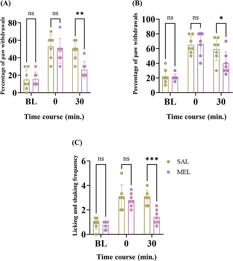 |
Figure 2 Neuropathic rats were randomly assigned to receive either melittin or SAL at ST36 (n = 7/group). Mechanical allodynia (A), mechanical hyperalgesia (B) and cold allodynia (C) were determined at three time points: baseline, before treatment, and 30 min after dosing at ST36 (timeline labels: BL, 0, and 30). Bars represent the mean ± SD; * p < 0.05, ** p < 0.01, *** p < 0.001, vs SAL ((A), η2 =0.7127, F (2, 24) = 60.76, p < 0.0001; (B), η2 =0.7285, F (2, 24) = 42.99, p < 0.0001; (C), η2 =0.5800, F (2, 24) = 52.06, p < 0.0001). ns, no significance (p > 0.05). Abbreviations: SAL, saline; MEL, melittin. |
Melittin at ST36 Suppressed the Spinal Neuronal Hyperexcitation of the Contralateral Dorsal Horn Following Systemic Paclitaxel Regimens
In our recent work,14 following paclitaxel regimens, in addition to intense behavioral hypersensitivities in the hind limb, the excitability of spinal WDR neurons was markedly heightened. Since contralateral melittin therapy substantially dampened cold and mechanical disturbances in the right-hind limbs (Figure 2), we further recorded stimulus-elicited acute spikes of dorsal horn WDR neurons in neuropathic rodents to examine whether and how melittin injections at the left ST36 could constrain paclitaxel-induced spinal sensitization. Melittin treatment (s.c., 0.5 mg/kg) conspicuously reduced mechanical stimuli-evoked discharges of WDR neurons located in the right-dorsal horn in response to stimulation of their peripheral receptive fields (specifically, the ventral surface of the right-hind paw) (brush, 95% CI: 3.397 to 21.57, p < 0.01; pinch, 95% CI: 5.979 to 21.91, p < 0.001, Figure 3A and B). Additionally, melittin potently reduced firing in WDR neurons in response to cutaneous application of evaporative acetone (50 µL) 30 min after treatment (95% CI: 0.7862 to 10.21, p < 0.05, Figure 3C). In rats with neuropathic signs, the neuronal discharge frequency elicited by mechanical and cold stimuli remained unchanged by SAL administration (brush, 95% CI: −8.192 to 10.96; pinch, 95% CI: −2.505 to 14.28; acetone, 95% CI: −4.302 to 5.635, all p > 0.05, Figure 3A–C). A representative raw trace illustrating the decline in isolated WDR neurons’ responses to the peripheral brush, pinch, and acetone cooling stimulation 30 min after melittin treatment at ST36 is shown in Figure 3D–F. These in vivo electrophysiology results are consistent with the ability of melittin to counteract peripheral allodynia and hyperalgesia in the contralateral hind limb (Figure 2), indicating its significant analgesic properties following intervention at ST36. Furthermore, the specificity of the contralateral acupoint location and the normalization of spinal sensitization underscore that central mechanisms may predominantly mediate its analgesic action against paclitaxel-induced neuropathic conditions.
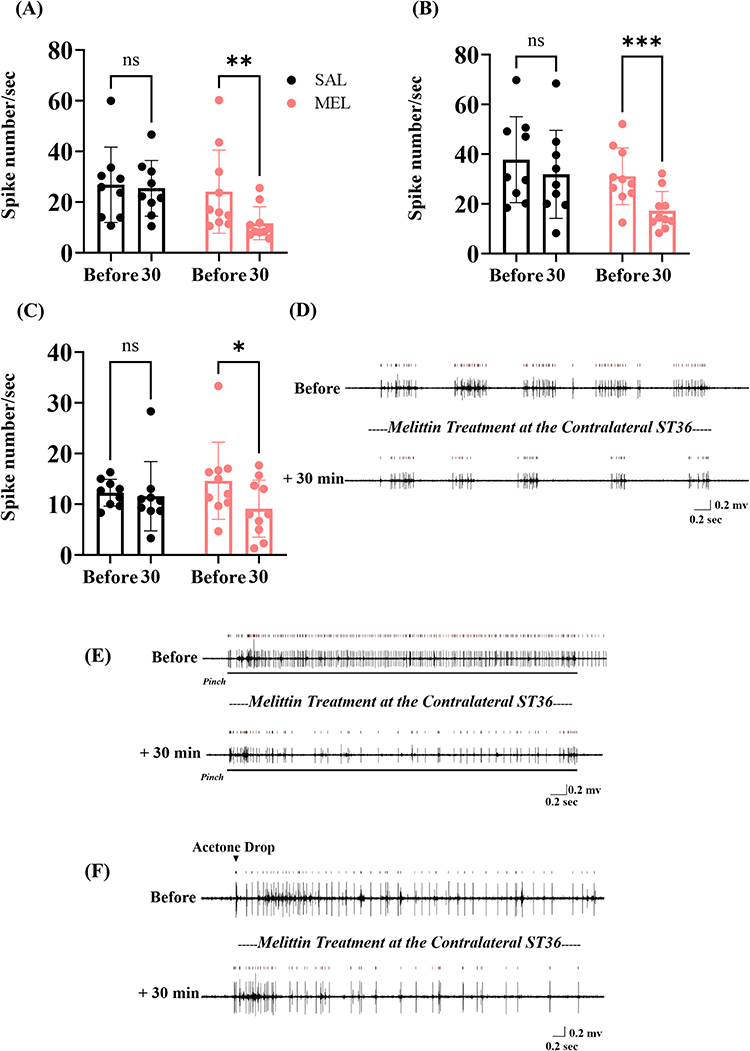 |
Figure 3 Extracellular recordings in response to cutaneous brush (A), pinch (B), and acetone (C) stimuli were obtained from WDR neurons before and 30 min after melittin (n=10 neurons) or SAL (n=9 neurons) treatment (time points labels: Before and 30). Typical discharge profiles show neuronal responses to brush (D), pinch (E), and acetone (F) stimulation were attenuated 30 min after apitherapy. Bars represent the mean ± SD; * p < 0.05, ** p < 0.01, *** p < 0.001, vs Before ((A), η2 =0.1784, F (1, 17) = 6.667, p = 0.0194; (B), η2 =0.3049, F (1, 17) = 17.75, p = 0.0006; (C), η2 =0.1967, F (1, 17) = 4.898, p = 0.0409). ns, no significance (p > 0.05). Abbreviations: SAL, saline; MEL, melittin. |
Effects of Antagonism of Spinal Adrenoceptors on the Melittin-Induced Dorsal Horn Neuronal Suppressive Properties
Subsequent neurophysiological analyses aimed to gauge the specific role of α-adrenoceptors underlying the analgesic features of contralateral melittin therapy. To achieve this, we pretreated rats exhibiting pain behaviors by microinjecting prazosin (30 µg) or idazoxan (50 µg) at the recording site in the right-dorsal horn before administering melittin. Neurophysiological single-cell recordings of WDR neurons were done twice: first, prior to the spinal microinjection, and second, 30 min after melittin intervention at ST36 of the left-hind limb. Pretreatment with prazosin did not affect the inhibitory effects of melittin on peripheral mechanical stimulus-induced neuronal spikes (brush, 95% CI: 3.262 to 19.27, p < 0.01; pinch, 95% CI: 9.275 to 28.00, p < 0.0001, Figure 4A and B). Conversely, melittin-induced neuronal inhibitions were conspicuously reversed by spinal α2-adrenoceptor blockade (ie, microinjections of idazoxan; brush, 95% CI: −7.941 to 8.065; pinch, 95% CI: −8.084 to 10.64, all p > 0.05, Figure 4A and B). For acetone stimulation, neuronal activity exhibited no considerable decrease at 30 min after melittin therapy compared to baseline (detected prior to antagonist microinjection), in rodents pretreated with either prazosin (95% CI: −4.298 to 6.965, p > 0.05, Figure 4C) or idazoxan (95% CI: −5.668 to 5.595, p > 0.05, Figure 4C). In the control group, spinal microinjection of SAL failed to diminish the inhibitory effects of melittin on neuronal firings 30 min post-intervention at the left ST36 (brush, 95% CI: 6.735 to 22.74, p < 0.001; pinch, 95% CI: 7.911 to 26.64, p < 0.001; acetone, 95% CI: 0.6647 to 11.93, p < 0.05, Figure 4A–C). Together, microinjections of prazosin and idazoxan counteracted the suppressive effect of melittin on dorsal horn neuronal sensitization in neuropathic rats. Figure 4D–F illustrate representative analog waveforms of the WDR neuronal firings to peripheral brush, pinch, and acetone stimuli before spinal microinjection and 30 min after melittin-based pharmacoacupuncture.
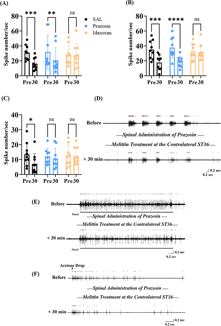 |
Figure 4 In vivo recordings in response to cutaneous brush (A), pinch (B), and acetone (C) stimuli were conducted before microinjection and 30 min after apitherapy at ST36 (n=9 neurons/group, time points labeled: Pre and 30). Typical discharge profiles detected from WDR neurons in response to brush (D), pinch (E), and acetone (F) stimulation, both before antagonism of spinal α1-adrenoreceptors and 30 min after apitherapy. Bars represent the mean ± SD; * p < 0.05, ** p < 0.01, *** p < 0.001, **** p < 0.0001, vs Pre ((A), η2 =0.3711, F (1, 24) = 23.42, p < 0.0001; (B), η2 =0.4476, F (1, 24) = 34.84, p < 0.0001; (C), η2 =0.1164, F (1, 24) = 4.014, p = 0.0565). ns, no significance (p > 0.05). Abbreviation: SAL, saline. |
Spinal Blockade of α-Adrenorceptors Reversed Melittin-Induced Analgesic Action on the Contralateral Peripheral Neuropathy
To examine the central noradrenergic mechanism of melittin treatment in the contralateral limb, either 30 µg of prazosin or 50 µg of idazoxan was administered intrathecally before s.c. injection of melittin (0.5 mg/kg at the left ST36) in neuropathic rats. Data revealed that pretreatment with idazoxan (mechanical allodynia, 95% CI: −9.209 to 19.21; hyperalgesia, 95% CI: −8.624 to 25.29, all p > 0.05, Figure 5A and B), but not prazosin (mechanical allodynia, 95% CI: 2.458 to 30.88; hyperalgesia, 95% CI: 1.376 to 35.29, all p < 0.05, Figure 5A and B) or vehicle (SAL, allodynia, 95% CI: 4.958 to 33.38, p < 0.01; hyperalgesia, 95% CI: 3.042 to 36.96, p < 0.05, Figure 5A and B), abolished the melittin-induced decrease in withdrawal frequency of the right-hind paw in response to VFFs. In contrast, melittin treatment failed to result in substantial mitigation of cold hypersensitivity in both the prazosin-injected and idazoxan-injected rats (prazosin, 95% CI: −0.7403 to 1.074; idazoxan, 95% CI: −0.7403 to 1.074, all p > 0.05, Figure 5C). Our data indicated that melittin-induced analgesia involved central noradrenergic mechanisms. Specifically, its effects on mechanical allodynia and hyperalgesia were counteracted by spinal α2-adrenoceptor antagonism, while its effects on cold allodynia were dependent on the activation of both spinal α1- and α2-adrenoceptors.
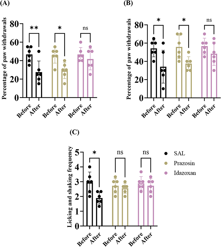 |
Figure 5 Neuropathic rodents were randomly allocated into three groups (n = 6/group). Mechanical allodynia (A), mechanical hyperalgesia (B), and cold allodynia (C) were determined in the right hind limb twice: just before the antagonist administration (Before) and 30 min after apitherapy (After). Bars represent the mean ± SD; * p < 0.05, ** p < 0.01, vs Before ((A), η2 =0.4578, F (1, 15) = 19.98, p = 0.0004; (B), η2 =0.4695, F (1, 15) = 18.32, p = 0.0007; (C), η2 =0.2025, F (1, 15) = 5.672, p = 0.0309). ns, no significance (p > 0.05). Abbreviation: SAL, saline. |
Discussion
Paclitaxel-induced peripheral neuropathy, a dose-dependent side effect with considerable medical and socio-economic costs, arguably remains a leading cause of premature non-adherence to scheduled chemotherapy.6,23 Known risk factors include older age, higher total dosage, and chronic alcohol abuse, among others.45,46 This painful disorder persists as a long-lasting and prominent challenge, affecting up to 90% of survivors even years after completing paclitaxel-containing cytostatic prescriptions.1,4 Consequently, intensified research into reliable methods for managing disabling paclitaxel-induced neuropathic comorbidities is strongly warranted worldwide. Pure bioactive ingredients derived from flora and fauna serve as a natural source of analgesics.13,47,48 Modern processing methods for natural BV allow the production of its primary bioactive elements, such as melittin, with enhanced purity.49 As expected, these technical breakthroughs have further driven pharmaceutical research on melittin-based apitherapy for various pain conditions.11,47 As reported in previous studies, melittin has been demonstrated to exert anti-viral, anti-cancer, anti-arthritis, and anti-nociceptive properties.50,51 Our team and other researchers have investigated the therapeutic activities of varying paradigms of apitherapy using 1.0 mg/kg of BV and 0.5 mg/kg of melittin.14,17,18,38,40 These optimum doses, chosen based on dose–response evaluation, were administered at ST36 in rodents with chemotherapy (paclitaxel, oxaliplatin, or vincristine)-induced neuropathic pain.18,38,40 Among these approaches, we recently demonstrated that ipsilateral melittin intervention (s.c., ST36, 0.5 mg/kg) apparently dampened paclitaxel-induced neuropathic pain, with its underlying mechanisms specifically involving the noradrenergic inhibitory process.14 Nevertheless, the current understanding of whether central mechanisms underlie the analgesic potential of melittin-based apitherapy on the contralateral side remains limited.
ST36 is certainly one of the most frequently utilized acupoints in research on peripheral neuropathic disorders.14,17,18,21,38,40,52 It has been proven that BVA at this acupoint exerts a particularly curative effect against paclitaxel-induced allodynia, in contrast to interventions at non-acupoint sites.38 In conventional East Asian medicine, acupuncture can be applied both proximally to the affected area and, at times, to more remote localizations (eg, on the extremities), or even on the contralateral side.22,52,53 Emerging evidence supports the potential of contralateral acupuncture in ameliorating shoulder pain, post-stroke pain, and acute lumbar sprain; this evidence encompasses diverse assessments, including the severity of nociception, mobility of the affected limbs, and other relevant parameters.54–59 This pre-clinical study is the first to establish the analgesic potential of melittin intervention at ST36 in reducing paclitaxel-induced neuropathic pain on the contralateral side. We observed that melittin (s.c., 0.5 mg/kg) administered at the left ST36 largely impeded three modalities of neuropathic signs in the right-hind paw of rats (Figure 2). Indeed, treating the contralateral ST36 with moxibustion or electrostimulation in models with inflammatory or neuropathic conditions has been shown to exert potent suppressive action.21,52,60 Our previous data and current observations have highlighted that therapeutic intervention applied both ipsilaterally and contralaterally, specifically at ST36 in rodents, alleviates hind limb painful hypersensitivity, a finding consistent with earlier outcomes in several pain conditions.14,52,53 While another practice revealed somewhat different results, experimental apitherapy generated negligible improvements in the mechanical allodynia in the contralateral limbs.61 We speculated that distinctions in chemotherapeutic agents and intervention protocols could partially account for this (paclitaxel 8 mg/kg vs oxaliplatin 10 mg/kg; 0.5 mg/kg of melittin vs 0.1 mg/kg of BV).
There is a body of electrophysiological data suggesting that chemotherapy exacerbates evoked discharges in dorsal horn WDR neurons, where somatosensory inputs, as well as ascending and descending signals, converge and are heavily integrated.14,18,31,33 This spinal sensitization of pain-transmitting neurons can serve as a measurable read-out of neuropathic progressions within the CNS.62–66 It is accepted that somatosensory stimulation-based interventions, such as electro-acupuncture or BV-containing acupuncture, can alleviate pathological nociception by reorganizing and normalizing central nociceptive circuitry in pain states.17,62,64 Reflecting the behavioral outcomes observed peripherally, marked hypoactivity of stimulus-evoked neuronal discharges in the dorsal horn was recorded 30 min following the peripheral ST36 melittin intervention in vivo (Figure 3). Our electrophysiological finding is consistent with a previous study showing that duloxetine markedly attenuated oxaliplatin-induced spinal sensitization via noradrenergic mechanisms in a rodent model of neuropathy.8 The integrity of the functional ascending tracts from the periphery that transmit acupuncture-induced input to the CNS is proposed to be a vital prerequisite for pain-relieving outcomes.22 It has been reported that electro-acupuncture was ineffective on the ipsilateral side, while interventions at the contralateral side protected against allodynic signs resulting from pre-clinical chronic constriction injury (CCI) procedures.67 Furthermore, after acute transection of the thoracic spinal cord, the anti-nociceptive effects of acupuncture applied at the contralateral ST36 were totally reversed.21 These previous findings, together with the fact that the locations of pain evaluations and apitherapy interventions are contralateral in this validation (ie, right-hind paw vs left ST36), suggest that rearrangements in nociceptive processing within the CNS likely account for the analgesic feature observed with contralateral apitherapy.
Using an in vivo spinal extraneural recording approach, we gauged, for the first time, the effects of pretreating the right-dorsal horn recording area with prazosin or idazoxan before apitherapy. Our neuropharmacological results (Figures 4 and 5) demonstrated that: (1) the dorsal horn α1- and α2-adrenoceptors are activated following acupuncture, and these noradrenergic tones are responsible for mitigating cold allodynia and (2) activation of the dorsal horn α2-adrenoceptors is essential for the analgesic properties of melittin in mechanical neuropathy. Although our current findings do not allow us to definitively explain this difference, we speculate that it may stem from differences in anatomical underpinnings: peripheral cold sensation is primarily mediated by C fibers, whereas mechanical sensory input is transmitted to the CNS by both A and C fibers.16,32,68 Further fiber-type-specific validations are needed to elucidate the detailed mechanisms. A similar anti-nociceptive pattern was observed: the suppressive effects of activating spinal α1-adrenoceptors by phenylephrine (an α1 agonist) and spinal α2-adrenoceptors by clonidine (an α2 agonist) on peripheral cold allodynia and central sensitization in oxaliplatin-induced neuropathic rats were simultaneous and reliable.33 As previously revealed, specific antagonism of α2-adrenoceptors rekindled pre-apitherapy mechanical hypersensitivity in behavioral trials.18,61 Established anti-nociceptive mechanisms within the spinal noradrenergic pathway include engagement of α2-adrenoceptors at the central terminals of primary afferent neurons (presynaptic inhibition) and at spinal pain-relay neurons (postsynaptic inhibition).15,16,69 Additionally, noradrenergic signaling via spinal α1-adrenoceptors on interneurons contributes to pain relief by further releasing inhibitory substances (eg, glycine).16,70,71 This study sought to determine which adrenoceptor subtype predominates in mediating melittin-induced analgesia on the contralateral side and observed that spinal α2-adrenoceptors play a more tonic role than α1-adrenoceptors in both cold and mechanical neuropathy (compared to α1-adrenoceptors, which only influence cold sensitivity; Figures 4 and 5). In part, this aligns with clinical evidence in volunteers with reflex sympathetic dystrophy, where clonidine provided considerable analgesia, whereas administration of α1-adrenoceptor agonists even exerted an opposing pro-nociceptive action.72 Apitherapy at ST36 may act as a noxious stimulus. In fact, diffuse noxious inhibitory control (DNIC), a fundamental analgesic paradigm induced by heterotopic conditioning stimuli, was completely absent when spinal α2-adrenoceptors were antagonized.69 It will be interesting to further investigate the detailed connections between apipuncture-induced analgesia and DNIC facilitation in the CIPN condition.
A thorough understanding of the efficacy and central mechanisms of melittin administered to the contralateral limb is crucial for developing melittin-based analgesic therapies for CIPN. Our recent study demonstrated that depletion of NA with the neurotoxin N-(2-Chloroethyl)-N-ethyl-2-bromobenzylamine (DSP-4) abolished melittin-induced analgesia in the ipsilateral limb.14 Furthermore, melittin’s analgesic effects in the contralateral limb were mediated by spinal α1- and α2-adrenoceptors (Figures 4 and 5). Collectively, these findings indicate that endogenous noradrenergic pathways critically mediate the suppression of paclitaxel-induced neuropathic pain by melittin treatment at ST36.
Limitations
One limitation of the current study is that it focused solely on the analgesic properties following a single treatment of melittin. In the future, the longer-lasting, and potentially prophylactic, effects of repetitive melittin pharmacoacupuncture should be comprehensively studied. Another limitation is the use of male animals only, which precludes the assessment of potential sex-specific effects. Therefore, the generalizability of our findings to females requires further investigation.
Conclusion
At present, CIPN cannot be cured. Our findings demonstrated that melittin applied at ST36 activates spinal α1- and α2-adrenoceptors. This activation modulates nociceptive processing in WDR neurons of the dorsal horn, resulting in significant analgesia in the contralateral hind limb of rats with paclitaxel-induced neuropathic pain. The potential value of our results for the future management of CIPN is considerable. Clinically, CIPN symptoms often present in a symmetrical “glove-and-stocking” distribution.9,45 Previous articles have shed light on the ipsilateral analgesic properties of melittin treatments in CIPN rodents. In this context, our current findings address a significant gap in the literature. They provide valuable insights for decision-making in integrative pain management and support theoretical evidence for using apitherapy against paclitaxel-induced neuropathy.
Abbreviations
CIPN, chemotherapy-induced peripheral neuropathy; WDR neuron, wide-dynamic-range neuron; ST36, Zusanli acupoint; BVA, bee venom acupuncture; SNRIs, serotonin and noradrenaline reuptake inhibitors; ASCO, American Society of Clinical Oncology; TCAs, tricyclic antidepressants; SPF, specific pathogen-free; IASP, International Association for the Study of Pain; VFF, von Frey filament; CNS, central nervous system; DSP-4, N-(2-Chloroethyl)-N-ethyl-2-bromobenzylamine; PNS, peripheral nervous system; NA, noradrenaline.
Data Sharing Statement
The data generated for the present study are available from the corresponding author, Tianlong Wang: [email protected], upon reasonable request.
Ethical Statement
This study was ratified by the Ethical Committee of Capital Medical University (Nos. AEEI-2024-119; approved in May 2024, and AEEI-2025-511; approved in September 2025).
Acknowledgments
We thank Professor Sun Kwang Kim and Seunghwan Choi (Kyung Hee University, Korea) for their technical guidance and encouragement.
Author Contributions
All authors made a significant contribution to the work reported, whether that is in the conception, study design, execution, acquisition of data, analysis and interpretation, or in all these areas; took part in drafting, revising or critically reviewing the article; gave final approval of the version to be published; have agreed on the journal to which the article has been submitted; and agree to be accountable for all aspects of the work.
Funding
This research was supported by a grant from the National Natural Science Foundation of China (82204932).
Disclosure
The authors declare that they have no conflicts of interest in this work.
References
1. Tanabe Y, Hashimoto K, Shimizu C, et al. Paclitaxel-induced peripheral neuropathy in patients receiving adjuvant chemotherapy for breast cancer. Int J Clin Oncol. 2013;18(1):132–16. doi:10.1007/s10147-011-0352-x
2. Krishna SS, Shaji N, Kumar NV, et al. Emerging targets and translational challenges in treating paclitaxel-induced peripheral neuropathy. Mol Biol Rep. 2025;52(1):833. doi:10.1007/s11033-025-10938-w
3. Sawa A, Sato R, Matsuo T, et al. Paclitaxel-induced peripheral neuropathy and quality of life: sub-analysis of the MICHEL study. Anticancer Res. 2025;45(9):3827–3833. doi:10.21873/anticanres.17742
4. Starobova H, Vetter I. Pathophysiology of chemotherapy-induced peripheral neuropathy. Front Mol Neurosci. 2017;10:174. doi:10.3389/fnmol.2017.00174
5. Carozzi VA, Canta A, Chiorazzi A. Chemotherapy-induced peripheral neuropathy: what do we know about mechanisms? Neurosci Lett. 2015;596:90–107. doi:10.1016/j.neulet.2014.10.014
6. Hershman DL, Lacchetti C, Loprinzi CL. Prevention and management of chemotherapy-induced peripheral neuropathy in survivors of adult cancers: American society of clinical oncology clinical practice guideline summary. J Oncol Pract. 2014;10(6):e421–e424. doi:10.1200/JOP.2014.001776
7. Chow R, Novosel M, So OW, et al. Duloxetine for prevention and treatment of chemotherapy-induced peripheral neuropathy (CIPN): systematic review and meta-analysis. BMJ Support Palliat Care. 2023;13(1):27–34. doi:10.1136/spcare-2022-003815
8. Kim W, Chung Y, Choi S, et al. Duloxetine protects against oxaliplatin-induced neuropathic pain and spinal neuron hyperexcitability in rodents. Int J Mol Sci. 2017;18(12):2626. doi:10.3390/ijms18122626
9. Maihofner C, Diel I, Tesch H, et al. Chemotherapy-induced peripheral neuropathy (CIPN): current therapies and topical treatment option with high-concentration capsaicin. Support Care Cancer. 2021;29(8):4223–4238. doi:10.1007/s00520-021-06042-x
10. Li D, Park S, Lee K, et al. 5-HT 1A receptors mediate the analgesic effect of rosavin in a mouse model of oxaliplatin-induced peripheral neuropathic pain. Korean J Physiol Pharmacol. 2021;25(5):489–494. doi:10.4196/kjpp.2021.25.5.489
11. Sung SH, Lee G. Bee venom acupuncture effects on pain and its mechanisms: an updated review. Toxins (Basel). 2021;13(9):608. doi:10.3390/toxins13090608
12. Cherniack EP, Govorushko S. To bee or not to bee: the potential efficacy and safety of bee venom acupuncture in humans. Toxicon. 2018;154:74–78. doi:10.1016/j.toxicon.2018.09.013
13. Lee B, Seo BK, Kwon OJ, et al. Effect of combined bee venom acupuncture and NSAID treatment for non-specific chronic neck pain: a randomized, assessor-blinded, pilot clinical trial. Toxins (Basel). 2021;13(7):436. doi:10.3390/toxins13070436
14. Li D, Wu Y, Chang L, et al. Therapeutic effects of melittin on paclitaxel-induced peripheral neuropathic pain and spinal neuronal hyperactivity in male rats. Front Neurosci. 2025;19:1622553. doi:10.3389/fnins.2025.1622553
15. Yoshimura M, Furue H. Mechanisms for the anti-nociceptive actions of the descending noradrenergic and serotonergic systems in the spinal cord. J Pharmacol Sci. 2006;101(2):107–117. doi:10.1254/jphs.crj06008x
16. Millan MJ. Descending control of pain. Prog Neurobiol. 2002;66(6):355–474. doi:10.1016/s0301-0082(02)00009-6
17. Choi S, Chae HK, Heo H, et al. analgesic effect of melittin on oxaliplatin-induced peripheral neuropathy in rats. Toxins. 2019;11(7):396. doi:10.3390/toxins11070396
18. Li D, Chung G, Kim SK. The involvement of central noradrenergic pathway in the analgesic effect of bee venom acupuncture on vincristine-induced peripheral neuropathy in rats. Toxins. 2020;12(12):775. doi:10.3390/toxins12120775
19. Dimitrova A, Harrington A, Memmott T, et al. Acupuncture-induced changes in nociception, measured by pain-related evoked potentials: a pilot mechanistic study. J Integr Complement Med. 2022;28(11):862–869. doi:10.1089/jicm.2021.0369
20. Park S, Lee D, Kim GW, et al. Effect of contralateral acupuncture (Geoja Technique) in musculoskeletal disorders stratified by pulse strength differences: a retrospective study. J Pain Res. 2025;18:6149–6163. doi:10.2147/JPR.S559379
21. Zhu B, Xu WD, Rong PJ, et al. A C-fiber reflex inhibition induced by electroacupuncture with different intensities applied at homotopic and heterotopic acupoints in rats selectively destructive effects on myelinated and unmyelinated afferent fibers. Brain Res. 2004;1011(2):228–237. doi:10.1016/j.brainres.2004.03.034
22. Zhang H, Sun J, Xin X, et al. Contralateral electroacupuncture relieves chronic neuropathic pain in rats with spared nerve injury. Med Sci Monit. 2018;24:2970–2974. doi:10.12659/MSM.909741
23. Boyette-Davis JA, Walters ET, Dougherty PM. Mechanisms involved in the development of chemotherapy-induced neuropathy. Pain Manag. 2015;5(4):285–296. doi:10.2217/pmt.15.19
24. Yin CS, Jeong HS, Park HJ, et al. A proposed transpositional acupoint system in a mouse and rat model. Res Vet Sci. 2008;84(2):159–165. doi:10.1016/j.rvsc.2007.04.004
25. Westlund KN, Coulter JD. Descending projections of the locus coeruleus and subcoeruleus/medial parabrachial nuclei in monkey: axonal transport studies and dopamine-beta-hydroxylase immunocytochemistry. Brain Res. 1980;2(3):235–264. doi:10.1016/0165-0173(80)90009-0
26. Westlund KN, Bowker RM, Ziegler MG, et al. Noradrenergic projections to the spinal cord of the rat. Brain Res. 1983;263(1):15–31. doi:10.1016/0006-8993(83)91196-4
27. Kamau VN, Yang S, Lee G, et al. Acupuncture modalities differentially modulate pain and joint damage in a rat model of knee osteoarthritis. J Pain Res. 2025;18:5377–5384. doi:10.2147/JPR.S555223
28. Ro JY, Zhang Y, Tricou C, et al. Age and sex differences in acute and osteoarthritis-like pain responses in rats. J Gerontol a Biol Sci Med Sci. 2020;75(8):1465–1472. doi:10.1093/gerona/glz186
29. Zimmermann M. Ethical guidelines for investigations of experimental pain in conscious animals. Pain. 1983;16(2):109–110. doi:10.1016/0304-3959(83)90201-4
30. Li D, Lee Y, Kim W, et al. Analgesic effects of bee venom derived phospholipase A(2) in a mouse model of oxaliplatin-induced neuropathic pain. Toxins. 2015;7(7):2422–2434. doi:10.3390/toxins7072422
31. Chae HK, Kim W, Kim SK. Phytochemicals of cinnamomi cortex: cinnamic acid, but not cinnamaldehyde, attenuates oxaliplatin-induced cold and mechanical hypersensitivity in rats. Nutrients. 2019;11(2):432. doi:10.3390/nu11020432
32. Li D, Lee JH, Choi CW, et al. The analgesic effect of venlafaxine and its mechanism on oxaliplatin-induced neuropathic pain in mice. Int J Mol Sci. 2019;20(7):1652. doi:10.3390/ijms20071652
33. Choi S, Yamada A, Kim W, et al. Noradrenergic inhibition of spinal hyperexcitation elicited by cutaneous cold stimuli in rats with oxaliplatin-induced allodynia: electrophysiological and behavioral assessments. J Physiol Sci. 2017;67(3):431–438. doi:10.1007/s12576-016-0505-9
34. Zheng FY, Xiao WH, Bennett GJ. The response of spinal microglia to chemotherapy-evoked painful peripheral neuropathies is distinct from that evoked by traumatic nerve injuries. Neuroscience. 2011;176:447–454. doi:10.1016/j.neuroscience.2010.12.052
35. Flatters SJ, Bennett GJ. Ethosuximide reverses paclitaxel- and vincristine-induced painful peripheral neuropathy. Pain. 2004;109(1–2):150–161. doi:10.1016/j.pain.2004.01.029
36. Polomano RC, Mannes AJ, Clark US, et al. A painful peripheral neuropathy in the rat produced by the chemotherapeutic drug, paclitaxel. Pain. 2001;94(3):293–304. doi:10.1016/S0304-3959(01)00363-3
37. Li D, Yoo JH, Kim SK. Long-lasting and additive analgesic effects of combined treatment of bee venom acupuncture and venlafaxine on paclitaxel-induced allodynia in mice. Toxins. 2020;12(10):620. doi:10.3390/toxins12100620
38. Costa-Pereira JT, Ribeiro J, Martins I, et al. Role of spinal cord alpha(2)-adrenoreceptors in noradrenergic inhibition of nociceptive transmission during chemotherapy-induced peripheral neuropathy. Front Neurosci. 2019;13:1413. doi:10.3389/fnins.2019.01413
39. Kim W, Kim MJ, Go D, et al. Combined effects of bee venom acupuncture and morphine on oxaliplatin-induced neuropathic pain in mice. Toxins (Basel). 2016;8(2):33. doi:10.3390/toxins8020033
40. Yamada A, Koga K, Kume K, et al. Ethanol-induced enhancement of inhibitory synaptic transmission in the rat spinal substantia gelatinosa. Mol Pain. 2018;14:1744806918817969. doi:10.1177/1744806918817969
41. Sikandar S, West SJ, McMahon SB, et al. Sensory processing of deep tissue nociception in the rat spinal cord and thalamic ventrobasal complex. Physiol Rep. 2017;5(14):e13323. doi:10.14814/phy2.13323
42. Hickey L, Li Y, Fyson SJ, et al. Optoactivation of locus ceruleus neurons evokes bidirectional changes in thermal nociception in rats. J Neurosci. 2014;34(12):4148–4160. doi:10.1523/JNEUROSCI.4835-13.2014
43. Sun X, Xue F, Wen J, et al. Longitudinal analysis of sleep-wake states in neonatal rats subjected to hypoxia-ischemia. Nat Sci Sleep. 2022;14:335–346. doi:10.2147/NSS.S352035
44. Boyette-Davis JA, Hou S, Abdi S, et al. An updated understanding of the mechanisms involved in chemotherapy-induced neuropathy. Pain Manag. 2018;8(5):363–375. doi:10.2217/pmt-2018-0020
45. Argyriou AA, Koltzenburg M, Polychronopoulos P, et al. Peripheral nerve damage associated with administration of taxanes in patients with cancer. Crit Rev Oncol Hematol. 2008;66(3):218–228. doi:10.1016/j.critrevonc.2008.01.008
46. Lin TY, Hsieh CL. Clinical applications of bee venom acupoint injection. Toxins. 2020;12(10):618. doi:10.3390/toxins12100618
47. Jeong H, Jang S, Park JK, et al. Bee venom acupuncture for shoulder pain: a literature review of clinical studies. Toxins. 2024;16(11):501. doi:10.3390/toxins16110501
48. Park M, Shin S. Bee venom acupuncture in traditional korean medicine: a review of clinical practice guidelines. Toxins. 2025;17(4):158. doi:10.3390/toxins17040158
49. Memariani H, Memariani M, Moravvej H, et al. Melittin: a venom-derived peptide with promising anti-viral properties. Eur J Clin Microbiol Infect Dis. 2020;39(1):5–17. doi:10.1007/s10096-019-03674-0
50. Ullah A, Aldakheel FM, Anjum SI, et al. Pharmacological properties and therapeutic potential of honey bee venom. Saudi Pharm J. 2023;31(1):96–109. doi:10.1016/j.jsps.2022.11.008
51. Lu KW, Hsu CK, Hsieh CL, et al. Probing the effects and mechanisms of electroacupuncture at ipsilateral or contralateral ST36-ST37 acupoints on CFA-induced inflammatory pain. Sci Rep. 2016;6(1):22123. doi:10.1038/srep22123
52. Zeng YJ, Lin YH, Wang YC, et al. Laser acupuncture-induced analgesic effect and molecular alterations in an incision pain model: a comparison with electroacupuncture-induced effects. Lasers Med Sci. 2018;33(2):295–304. doi:10.1007/s10103-017-2367-7
53. Zhang S, Wang X, Yan CQ, et al. Different mechanisms of contralateral- or ipsilateral-acupuncture to modulate the brain activity in patients with unilateral chronic shoulder pain: a pilot fMRI study. J Pain Res. 2018;11:505–514. doi:10.2147/JPR.S152550
54. Guo Q, Di Z, Tian HF, et al. Contralateral acupuncture for the treatment of phantom limb pain and phantom limb sensation in oncologic lower limb amputee: a case report. Front Neurosci. 2021;15:713548. doi:10.3389/fnins.2021.713548
55. Zhang H, Sun J, Wang C, et al. Randomised controlled trial of contralateral manual acupuncture for the relief of chronic shoulder pain. Acupunct Med. 2016;34(3):164–170. doi:10.1136/acupmed-2015-010947
56. Kim MK, Choi TY, Lee MS, et al. Contralateral acupuncture versus ipsilateral acupuncture in the rehabilitation of post-stroke hemiplegic patients: a systematic review. BMC Complement Altern Med. 2010;10(1):41. doi:10.1186/1472-6882-10-41
57. Tang X, Li Q, Huang G, et al. Immediate efficacy of contralateral acupuncture on SI3 combined with active exercise for acute lumbar sprains: protocol for a randomized controlled trial. J Pain Res. 2024;17:2099–2110. doi:10.2147/JPR.S475839
58. Huang H, Tang K, Song X, et al. Effects of contralateral versus ipsilateral electroacupuncture for analgesia and rehabilitation after unilateral total knee arthroplasty: a randomized controlled trial. Acupunct Med. 2024;42(4):183–193. doi:10.1177/09645284231211601
59. Zuo CY, Lv P, Zhang CS, et al. Ipsi- and contralateral moxibustion generate similar analgesic effect on inflammatory pain. Evid Based Complement Alternat Med. 2019;2019:1807287. doi:10.1155/2019/1807287
60. Yoon SY, Yeo JH, Han SD, et al. Diluted bee venom injection reduces ipsilateral mechanical allodynia in oxaliplatin-induced neuropathic mice. Biol Pharm Bull. 2013;36(11):1787–1793. doi:10.1248/bpb.b13-00469
61. Zhang Z, Zheng H, Yu Q, et al. Understanding of spinal wide dynamic range neurons and their modulation on pathological pain. J Pain Res. 2024;17:441–457. doi:10.2147/JPR.S446803
62. McGaraughty S, Chu KL, Xu J. Characterization and pharmacological modulation of noci-responsive deep dorsal horn neurons across diverse rat models of pathological pain. J Neurophysiol. 2018;120(4):1893–1905. doi:10.1152/jn.00325.2018
63. Qu Z, Liu L, Yang Y, et al. Electro-acupuncture inhibits C-fiber-evoked WDR neuronal activity of the trigeminocervical complex: neurophysiological hypothesis of a complementary therapy for acute migraine modeled rats. Brain Res. 2020;1730:146670. doi:10.1016/j.brainres.2020.146670
64. Shimazu Y, Shibuya E, Takehana S, et al. Local administration of resveratrol inhibits excitability of nociceptive wide-dynamic range neurons in rat trigeminal spinal nucleus caudalis. Brain Res Bull. 2016;124:262–268. doi:10.1016/j.brainresbull.2016.06.001
65. Latremoliere A, Woolf CJ. Central sensitization: a generator of pain hypersensitivity by central neural plasticity. J Pain. 2009;10(9):895–926. doi:10.1016/j.jpain.2009.06.012
66. Somers DL, Clemente FR. Transcutaneous electrical nerve stimulation for the management of neuropathic pain: the effects of frequency and electrode position on prevention of allodynia in a rat model of complex regional pain syndrome type II. Phys Ther. 2006;86(5):698–709. doi:10.1093/ptj/86.5.698
67. Pertovaara A. Antinociception induced by alpha-2-adrenoceptor agonists, with special emphasis on medetomidine studies. Prog Neurobiol. 1993;40(6):691–709. doi:10.1016/0301-0082(93)90011-g
68. Bannister K, Lockwood S, Goncalves L, et al. An investigation into the inhibitory function of serotonin in diffuse noxious inhibitory controls in the neuropathic rat. Eur J Pain. 2017;21(4):750–760. doi:10.1002/ejp.979
69. Baliki MN, Apkarian AV. Nociception, pain, negative moods, and behavior selection. Neuron. 2015;87(3):474–491. doi:10.1016/j.neuron.2015.06.005
70. Ossipov MH, Morimura K, Porreca F. Descending pain modulation and chronification of pain. Curr Opin Support Palliat Care. 2014;8(2):143–151. doi:10.1097/SPC.0000000000000055
71. Davis KD, Treede RD, Raja SN, et al. Topical application of clonidine relieves hyperalgesia in patients with sympathetically maintained pain. Pain. 1991;47(3):309–317. doi:10.1016/0304-3959(91)90221-I
72. Yoon C, Wook YY, Sik NH, et al. Behavioral signs of ongoing pain and cold allodynia in a rat model of neuropathic pain. Pain. 1994;59(3):369–376. doi:10.1016/0304-3959(94)90023-X
 © 2026 The Author(s). This work is published and licensed by Dove Medical Press Limited. The
full terms of this license are available at https://www.dovepress.com/terms
and incorporate the Creative Commons Attribution
- Non Commercial (unported, 4.0) License.
By accessing the work you hereby accept the Terms. Non-commercial uses of the work are permitted
without any further permission from Dove Medical Press Limited, provided the work is properly
attributed. For permission for commercial use of this work, please see paragraphs 4.2 and 5 of our Terms.
© 2026 The Author(s). This work is published and licensed by Dove Medical Press Limited. The
full terms of this license are available at https://www.dovepress.com/terms
and incorporate the Creative Commons Attribution
- Non Commercial (unported, 4.0) License.
By accessing the work you hereby accept the Terms. Non-commercial uses of the work are permitted
without any further permission from Dove Medical Press Limited, provided the work is properly
attributed. For permission for commercial use of this work, please see paragraphs 4.2 and 5 of our Terms.