")
Back to Journals » Clinical, Cosmetic and Investigational Dermatology » Volume 13
Effects of Skin Lightening Cream Agents – Hydroquinone and Kojic Acid, on the Skin of Adult Female Experimental Rats
Authors Owolabi JO , Fabiyi OS , Adelakin LA , Ekwerike MC
Received 2 October 2019
Accepted for publication 29 March 2020
Published 6 April 2020 Volume 2020:13 Pages 283—289
DOI https://doi.org/10.2147/CCID.S233185
Checked for plagiarism Yes
Review by Single anonymous peer review
Peer reviewer comments 3
Editor who approved publication: Dr Jeffrey Weinberg
Joshua Oladele Owolabi,1,2 Oluseyi Sunday Fabiyi,1 Lola Adeola Adelakin,1 Miriammillicent Chinenyenwa Ekwerike1
1Department of Anatomy, Ben Carson School of Medicine, Babcock University, Ilishan-Remo, Nigeria; 2Department of Anatomy, University of Global Health Equity, Butaro, Rwanda
Correspondence: Joshua Oladele Owolabi
Department of Anatomy, Ben Carson School of Medicine, Babcock University Ilishan-Remo, Nigeria
Tel +2348064884305
Email [email protected]
Introduction: Skin bleaching is the act of using steroid- and chemical-containing products to lighten the skin. Hydroquinone and kojic acid are often used in skin bleaching creams. Hydroquinone was suspected to be harmful. This study investigated the effects of kojic acid and hydroquinone on the skin of adult female Wistar rats and the potential use of aloe vera for amelioration.
Materials and Methods: Eighty [n=80] adult female Wistar rats with an average weight of 120 g were randomly divided into eight groups, marked A, B, C, D, E, F, G, and H. Group A animals served as the control group; group B was treated with 2% hydroquinone, group C was treated with 2% kojic acid, group D was treated with 4% hydroquinone, group E was treated with 4% kojic acid, group F was treated with 2% hydroquinone and 2% kojic acid, group G was treated with 4% hydroquinone and 4% kojic acid, and group H was treated with 4% hydroquinone, 4% kojic acid, and aloe vera. The preparations were applied to the tail skin and treatment lasted 28 days. Skin samples were excised and processed using H&E, Masson’s trichrome, and p65 immunohistochemical assays on tissue sections.
Results: Hydroquinone caused structural disruptions of the stratum corneum of the epidermis and the overlying keratin. p65 was also prominently expressed in the treated groups. Hydroquinone reduced skin thickness and caused epidermis disruption.
Discussion: The prominent expression of p65 in the sections indicated deleterious effects of hydroquinone. Kojic acid was not found to have deleterious effects. Aloe vera prevented extensive disruption of stratum corneum by hydroquinone. The use of hydroquinone in skin lightening creams might raise health concerns. Aloe vera could be protective against hydroquinone.
Keywords: hydroquinone, kojic acid, aloe vera, bleaching products, skin
Introduction
Skin tone is branded as beauty, grace, and high social status in many parts of the world, and this perception encourages, people, especially women to indulge in skin lightening products.1,2 Harmful products such as Hydroquinone, Kojic acid, and Mercury are present in many skin lightening products.2,3 Hydroquinone is considered as a primary topical ingredient for inhibiting melanin production because it reduces the skin’s production of melanin which is responsible for skin color.4 However, because of the carcinogenic nature of hydroquinone, it has been banned in some countries in a bid to reduce the risks of skin cancer.1 Apart from its effects on the skin, hydroquinone has been found to expose users to acute toxicity from oral exposure and it can also cause diseases such as thyroid disorder, leukemia, and liver damage.1,3 Chronic occupational exposure to hydroquinone dust had reportedly resulted in eye injuries, which varied from mild irritation and staining of conjunctivae and cornea to changes in the thickness and curvature of the cornea, loss of corneal luster, and impaired vision.5 Prolonged exposure could lead to the development of severe ocular effects.4 Higher concentrations frequently irritate the skin, and if used for prolonged periods, it could cause disfiguring effects including epidermal thickening.2 Oral ingestion of doses between 5 g and 15 g doses caused convulsions and hemolytic anemia.6
Kojic acid on the other hand, known as Koji in Japan, is a fungal metabolic product which has the advantage of not being oxidized in skin lotions.1 It is a chelation agent that is produced by several fungi including Aspergillus oryzae. It inhibits and prevents the formation of tyrosine and it contains some antimicrobial properties against several common bacterial stains even in small dilutions.7 A study8 had reported that 4% hydroquinone and 0.75% Kojic when combined for typical treatment were effective in treating facial melasma. What might not have been adequately investigated would the possible effects of its prolonged or chronic use on the skin.
Skin, the organ of study, is the largest organ of the human body which accounts for about 15% of the human body weight and its health could be affected by topical agents.9,10 The vital functions of the skin include protection, prevention of excess water loss from the body and thermoregulation. The skin is composed of the superficial epidermis and the underlying dermis. The hypodermis lies beneath the dermis. Aloe vera, the plant that was used for the intervention is also known as Barbadensis miller. It is a shrubby or arborescent, perennial, xerophytic succulent, pea-green color plant that has been known and used in centuries for its health, beauty, medicinal, and skin care properties.11 The high incidences of skin lightening using creams, which is otherwise called bleaching and the attendant potential consequences necessitated this study. Hence, this study also used aloe vera as a possible remedy for the effects of kojic acid and hydroquinone which are found in skin lightening creams.6
Materials and Methods
Animal Treatment and Tissue Processing
Eighty adult [n=80] female Wistar rats, with the average body weight of 120 g, were procured for the study from the institutional animal holding facility. The standard procedures for animal use and handling were followed according to Babcock University’s Health Research Ethical Committee which approved the project (BUHREC 2018). The adult female Wistar rats were divided into eight groups of ten each after the period of acclimatization as follows:
Group A: the control group; animals were treated with only olive oil as a placebo.
Group B: This group was treated with 2% hydroquinone and 98% olive oil during the period of the experiment.
Group C: This group was treated with 2% kojic acid and 98% olive oil.
Group D: This group was treated with 4% hydroquinone and 96% olive oil.
Group E: This group was treated with 4% kojic and 96% olive oil.
Group F: This group was treated 2% hydroquinone, 2% kojic acid, and 96% olive oil.
Group G: This group was treated with 4% hydroquinone, 4% kojic acid, and 92% olive oil.
Group H: This group was treated with 4% hydroquinone, 4% kojic acid, aloe vera gel 4%, and 88% olive oil.
The required quantities of hydroquinone and kojic acid were dissolved in measured quantities of olive oil under mild heat [˂ 50°C]. The treatments lasted 28 days. The animals were sacrificed through the cervical dislocation. The tail skins were excised and preserved in 10% formal saline for immunohistochemical and histological procedures. The fixed tissues were processed following specific histological and histochemical protocols. Basic tissue processing included dehydration [using graded concentration of alcohol], clearing [using xylene], impregnation, and embedding [using molten wax]. The tissue samples were sectioned with a rotary microtome (~20 microns). The sections were mounted on glass slides for staining.
Staining Techniques
The H&E staining technique was done following the methods of Ref. 12 The slides, after dewaxing and rehydration, were placed in hematoxylin for 8–15 minutes for staining. The slides were then rinsed in tap water. The slides were further dipped in a bluing agent for 3–5 long dips. They were stained with Eosin for a period of 30 seconds and hematoxylin for ~2 minutes. They were also dehydrated in 95% and 100% alcohol, with 3 changes each for 2 minutes. They were then cleared in 3 changes of xylene for 2 minutes each and after which the cover glass was mounted.
The Masson’s Trichrome staining procedure for collagen fiber was done following the methods of.13 Tissues were deparaffinized and rehydrated through 100% alcohol, 95% alcohol, and 70% alcohol, then washed in distilled water. They were re-fixed in Bouin’s solution for 1 hour at 56°C and rinsed under running tap water for 5–10 minutes to remove the yellow color. They were then stained in Weigert’s iron hematoxylin working solution for 10 minutes and rinsed in running warm tap water for 10 minutes. Furthermore, they were washed in distilled water and stained again, in Biebrich scarlet-acid Fuchsin solution for 10–15 minutes. They were washed again in distilled water and differentiated in phosphomolybdic-phosphotungstic acid solution for 10–15 minutes. Sections were transferred directly (without rinsing) to aniline blue solution and stained for 5–10 minutes, then rinsed briefly in distilled water and differentiated in 1% acetic acid solution for 2–5 minutes. They were again washed in distilled water, then dehydrated very quickly through 95% ethyl alcohol, absolute ethyl alcohol to wipe off Biebrich scarlet-acid Fuchsin staining, and cleared in xylene, after which they were mounted with resinous mounting medium. The collagen fibers stained blue, the nuclei stained black, and the background stained red.
The p65 immunohistochemistry technique protocol was carried out based on Abcam® specifications. Paraffin sections were used for the immunohistochemistry study. Antigen retrieval was done though boiling before commencing with immunostaining. The slides were washed 2 x 5 min in TBS plus 0.025% Triton X-100 with gentle agitation. The sections were blocked in 10% normal serum with 1% BSA in TBS for 2 hours at room temperature. Slides were drained for a few seconds. The primary antibody was diluted in TBS with 1% BSA and applied. Sections were incubated overnight at 4°C. Thereafter, sections were rinsed 2 x 5 min TBS 0.025% Triton with gentle agitation; and incubated in 0.3% H2O2 in TBS for 15 minutes. The slides were mounted using albumin and observed under the microscope. Suitably representative photomicrographs were captured.
Results
Results of the study are presented (below) as photomicrographs of the skin sections as demonstrated using the H and E (Figure 1), Masson’s trichrome technique (Figure 2) and the p65 immunohistochemistry technique (Figure 3).
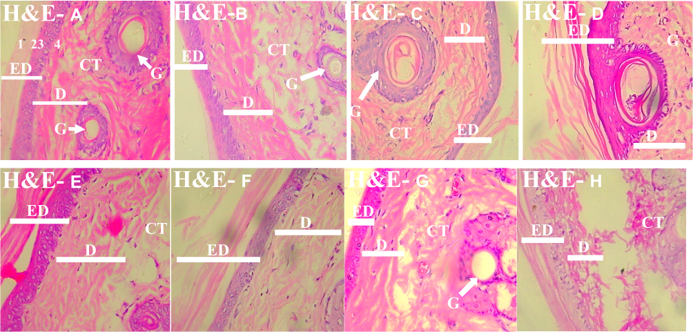 |
Figure 1 Photomicrograph of the skin of the experimental animals in Groups A–H, demonstrating the skin using H and E (H&E A-H; X400). The stratum corneum was disrupted in Groups B, D, and G (H&E B, D, and G). Note: Arrows point to specific features that are denoted by letters. Abbreviations: ED, Epidermis - (1) Stratum corneum (2) Stratum Granulosum (3) Stratum Spinosum (4) Stratum Basale; D, Dermis; G, Gland; C, Connective tissue; K, Keratin. |
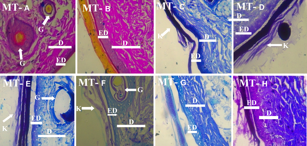 |
Figure 2 Photomicrograph of the skin of the experimental animals in Groups A–H demonstrating the skin using the Masson’s trichrome technique (MT A-H; X400). The connective tissue was relatively generally preserved in the skin layers (MT A-H). Note: Arrows point to specific features that are denoted by letters. Abbreviations: ED, Epidermis - (1) Stratum corneum (2) Stratum Granulosum (3) Stratum Spinosum (4) Stratum Basale; D, Dermis; G, Gland; C, Connective tissue; K, Keratin. |
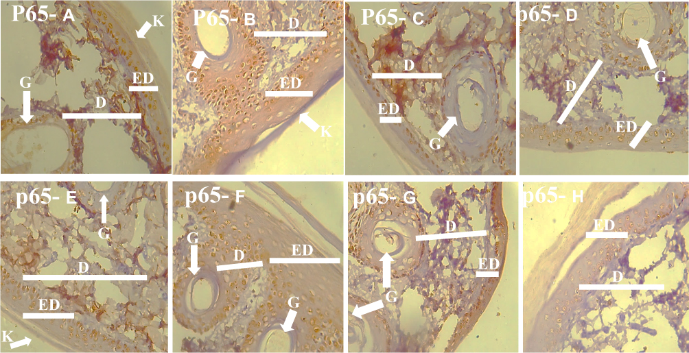 |
Figure 3 Photomicrograph of the skin of the experimental animals in Groups A–H demonstrating the skin using the p65 immunohistochemistry technique (p65 A-H). More cells in the groups B, D, F, and G expressed p65 which was used as an inflammation marker. Note: Arrows point to specific features that are denoted by letters. Abbreviations: ED, Epidermis - (1) Stratum corneum (2) Stratum Granulosum (3) Stratum Spinosum (4) Stratum Basale; D, Dermis; G, Gland; C, Connective tissue; K, Keratin. |
Disruption of the Stratum Corneum Is Attributable to Hydroquinone Effects
A major effect of the application of hydroquinone to the skin of the experimental animals is the disruption of the more superficial layers of the epidermis, the stratum corneum, which observable in Figure 1 photomicrographs. This further showed that the cells in this layer were affected. While the mechanisms of the disruption could either be due to the removal of superficial cells as a result of the hydroquinone effects, it might also be due to retarded formation of the corneum layer cells due to alterations in the migration of cells from the deeper layers of the epidermis. Whichever of the two possible mechanisms was responsible largely for this observation, the implications are numerous and might include the lesions of the skin, potential fragility of the skin and induced susceptibility to the ultraviolet (UV) rays and other environmental hazards. It is also known that increased UV exposure is a major risk factor for skin cancer.14 By implication, reduced superficial protection means increased exposure of the deeper cell layers including the melanocytes to UV radiation15 This has serious implications because there are reports that this might increase the risk of skin cancer.16
Loss of the superficial cells would mean that the treated skin was relatively thinner. This means a significant loss of the protective stratum corneum. This skin layer is very important to skin protection because it is selectively permeable to chemicals and other agents.17 This also would imply that the mechanical strength of the skin to protect the body from mechanical trauma was reduced or compromised. One implication of this might be more frequent or serious skin trauma due to mechanical assaults. Hence, the skin that had been affected in such a manner by hydroquinone use might have reduced integrity against mechanical or physical assaults. Another consequence of such thinness of the skin might be compromised ability to perform its function of homeostasis. There might include reduced ability to conserve heat, because of the loss in the cells that account partly for the thickness of the skin. This might also be extended to the reduced ability to conserve water as reduced superficial covering might lead to relatively increased skin vulnerability to losing water, especially when the environment is hot. A fragile skin is not only susceptible to mechanical trauma but also susceptible to penetrations that reach to the deeper structures of the skin such as bites from insects for instance. This might also make it easier for pathogens and toxins to access the body through the skin.
Susceptibility to the UV rays and other environmental hazards is a major complication that might result from this effect. The skin ordinarily has the right thickness to protect the body from the effects of the sun’s ultraviolet [UV] rays. A significant loss of the stratum corneum might result in a major compromise of this ability. In addition to this, the inability of the skin to replace the loss stratum corneum cells might also be extended to the melanocytes that are also largely required to protect the skin against the UV radiations. To this end, the skin might be vulnerable to the UV radiation effects. The complications that can arise from this might therefore include cancer of the skin.15
It is worthy of note however that Kojic acid did not cause any observable disruption to the stratum corneum as evident in the photomicrographs in Figure 1 and 2. On the other hand, its effects and interactions with the hydroquinone when both were applied appear to be beneficial, by mildly ameliorating the effects of hydroquinone. This might explain why it might often be a co-ingredient of certain creams with hydroquinone. Its actions, however, were not potent enough to totally ameliorate the effects of hydroquinone. Thus, this study partly aligns with certain previous studies that kojic acid at prescribed doses might not have deleterious effects when used for skin lightening or bleaching because there is no histological evidence of a potentially deleterious effect of kojic acid on the treated skins; however, it would not suffice to support the acclaimed multiple health benefits of its uses in certain reports (18; 19; 20).
Connective Tissue of the Skin
The agents that were used did not produce any extensive observable effects on the connective tissues of the skin. Thus, the underlying connective tissues were largely preserved and relatively undisrupted by all the agents that were administered [Figure 2].
Prominent Expression of P56 Is Attributable to Hydroquinone Effects
The immunohistochemistry method demonstrated p65 which is a REL-associated protein that served as a marker for cell proliferation, degeneration, and inflammation. The prominent demonstration or expression of p56 in the skin of the experimental animals was attributable to the effects of hydroquinone [Figure 3]. This also strongly suggests inflammations or a chemical irritation or assault on the cells or the basal layers of the epidermis as well as cells surrounding the sebaceous glands and the hair follicles. This also implies that the effects of hydroquinone as associated with the expression of p56 were not limited to the superficial layers of the skin. This might have a number of implications including the inflammation of the skin as well as links to certain cancers of the skin and other disorders. The expression of p65 in the study skins has significant implications on skin health, especially, relative to inflammations and skin cancers. The affected skins, from existing evidences, have increased levels of risk. From previous studies, epidermal p65/NF-κB signalling was essential for skin carcinogenesis.21 Its normal function is also associated to the maintenance of skin immune homeostasis; hence, it is protective against spontaneous dermatitis22. The complications that might arise from its anomalies are therefore of serious skin health consequences. A previous study7 had also stated that increased NF-κB activity caused hyper-proliferation and dysplasia of the mouse epidermis.
Aloe Vera Showed Potentials to Ameliorate Hydroquinone Deleterious Effects
The application of aloe vera gel to the already treated skin had observable effects as Aloe vera gel application showed potentials to ameliorate hydroquinone deleterious effects by persevering the stratum corneum which were generally disrupted in other groups that were administered hydroquinone [to the skin] [Figures 1 and 2]. Furthermore, the expression of p56 in these groups was relatively reduced [Figure 3]. Thus, aloe-vera gel as used had protective effects and by extension might be protecting the skin from the potential consequences associated with the effects of hydroquinone. Aloe vera has been reported to have significant protective effects on the skin.11 Such protective effects also reportedly include anti-inflammatory effects23 and ameliorative effects against skin damages and traumas.24
The current findings are similar to a previous work,25 in which it was reported that the epidermis of hydroquinone-treated rabbits showed inflammatory cells, infiltration mainlylymphocytes and eosinophils. It was also reported to have potential carcinogenic effects. Furthermore, researchers had, in their previous report, labelled hydroquinone and its analogs – in dermatology – as potential health risks.26 Previous reports also showed that hydroquinone caused histological alterations in the liver; mainly hydrophobic degeneration. It has also been recommended that hydroquinone should be used with moderation (27; 28).
Conclusions and Recommendations
The results obtained from this research showed that hydroquinone disrupted the epidermis and caused inflammation to the cells of the deeper skin layers and other cells surrounding sebaceous glands and hair follicles. The application of aloe vera gel ameliorated the effects of hydroquinone on the experimental animals’ skin. The existing notion that the use of hydroquinone and kojic acid might be safe is not in agreement with the results of this work; hence, further work is recommended especially on the chronic and systemic effects of hydroquinone and kojic acid on the skin and body.
Disclosure
The authors report no conflicts of interest in this work.
References
1. Al-Saleh IA. Mercury contenting skin lightening creams and potential hazards to the Health of Saudi Women. J Toxical Environ Health. 2010;5(1):123–130.
2. Dyall-Smith DA, Scurry JP. Mercury pigmentation and mercury levels from the use of cosmetic creams. Med J Aust. 2014;153:409–415. doi:10.5694/j.1326-5377.1990.tb125501.x
3. Ferreira AO, Freire ES, Polonini HC, Candido da Silva PJL, Brandaio MAF, Raposo NRB. Anti-ageing effects of monomethylsilanetriol and maltodextrin-stabilized orthosilicic acid on nails, skin and hair. Cosmet J. 2018;5(4):12–15.
4. Brigs AP. Principles of Biological Chemistry.
5. Considine GD. Van Nostrand’s encyclopaedia of Chemistry, Fifth Edition. New York: John Wiley & Sons; 2006.
6. Li Q, Geiselhart L, Mittler JN, Mudzinski SP, Lawrence DA, Freed BM. Inhibition of human T lymphoblast proliferation by hydroquinone. Toxicol Appl Pharmacol. 1996;139(2):317–323.
7. Poligone B, Hayden MS, Chen L, Pentland AP, Jimi E, Ghosh S. A role for NF-κB activity in skin hyperplasia and the development o keratoacanthomata in mice. PLoS One. 2013;8(8):e71887. doi:10.1371/journal.pone.0071887
8. Rochelle MBN, Kishore B, Bhat R, Sukumar D, Martis J, Ganesh H. A comparative study of the efficacy of 4% hydroquinone vs 0.75% kojic acid cream in the treatment of facial melasma. Indian J Dermatol. 2013;58(2):157–161. doi:10.4103/0019-5154.108070
9. Yetunde MA. Complications of Chronic use of Skin Lightening Cosmetics. Int J Dermatol. 2008;7:344–353.
10. Seyyed MA, Rashid FA, Rahul EI, Quadri KI. The review on properties of aloe vera in healing of cutaneous wounds. PMC. 2015;2015:714216.
11. Surjushe A, Vasani R, Saple DG. Aloe vera: a short review. Indian J Dermatol. 2008;53(4):163–166. doi:10.4103/0019-5154.44785
12. Sheehan DC, Hrapchak BB. Theory and Practice of Histotechnology.
13. Masson LE. Verhoeff; 2009. Stain.vetmed.vt.edu. Available from: http://education.vetmed.vt.edu/Curriculum/VM8054/Labs/.
14. Gilchrest BA, Eller MS, Geller AC, Yaar M. The pathogenesis of melanoma induced by ultraviolet radiation. N Engl J Med. 1999;340(17):1341–1348. doi:10.1056/NEJM199904293401707
15. Watson M, Holman DM, Maguire-Eisen M. Ultraviolet radiation exposure and its impact on skin cancer risk. Semin Oncol Nurs. 2016;32(3):241–254. doi:10.1016/j.soncn.2016.05.005
16. Sheehan JM, Potten CS, Young AR. Tanning in human skin types ii and iii offers modest photo-protection against erythema. Photochem Photobiol. 1998;68(4):588–592. doi:10.1111/j.1751-1097.1998.tb02518.x
17. Bos D, Meinardi MM. The 500 dalton rule for the skin penetration of chemical compounds and drugs. Exp Dermatol. 2000;9(3):165–169. doi:10.1034/j.1600-0625.2000.009003165.x
18. Moon KY, Ahn KS, Lee J, Kim YS. Kojic acid, a potential inhibitor of nf-κb activation in transfectant human HaCaT and SCC-13 cells. Arch Pharm Res. 2001;24(4):307–311.
19. Ma DL, Xu T, Chan DS, Man BY, Fong WF, Leung CH. A highly selective, label-free, homogenous luminescent switch-on probe for the detection of nanomolar transcription factor NF-kappaB. Nucleic Acids Res. 2011;39(10):e67. doi:10.1093/nar/gkr106
20. Saeedi M, Eslamifar M, Khezri K. Kojic acid applications in cosmetic and pharmaceutical preparations. Biomed Pharmacother. 2019;110:582–593. doi:10.1016/j.biopha.2018.12.006
21. Kim C, Pasparakis M. Epidermal p65/NF-κBsignalling is essential for skin carcinogenesis. EMBO Mol Med. 2014;6(7):970–983. doi:10.15252/emmm.201303541
22. Grinberg-Bleyer Y, Dainichi T, Oh H, et al. Genetic toxicology testing of 41 industrial chemicals, research. J Dermatol Sci. 2015;15:357–377.
23. Hutter JA, Salmon M, Stavinoha WB, Satsangi N, Williams RF, Streeper RT. Anti-Inflammatory C-glucosyl Chromone from Aloe Barbadensis. J National Prod. 1996;59:541–543.
24. Feily A, Namazi MR. Aloe vera in dermatology: a brief review. G Ital Dermatol Venereol. 2009;144(1):85–91.
25. Kooyers TJ, Westerhof W. Toxicology and health risks of hydroquinone in skin lightening formulations. J Eur Acad Dermatol Venereology. 2006;20(7):777–780. doi:10.1111/j.1468-3083.2005.01218.x
26. Westerhof W, Kooyers TJ. Hydroquinone and its analogues in dermatology - a potential health risk. J Cosmet Dermatol. 2005;4(2):55–59. doi:10.1111/j.1473-2165.2005.40202.x
27. Tse TW. Hydroquinone for skin lightening: safety profile, duration of use and when should we stop?. J Dermatol Treat. 2010;21(5):272–275. doi:10.3109/09546630903341945
28. Al-Saleh I, Elkhatib R, Al-Rouqi R, Al-Enazi S, Shinwari N. The dangers of skin-lightening creams. Toxicol Environ Chem. 2012;94(1):195–219. doi:10.1080/02772248.2011.631925
 © 2020 The Author(s). This work is published and licensed by Dove Medical Press Limited. The
full terms of this license are available at https://www.dovepress.com/terms.php
and incorporate the Creative Commons Attribution
- Non Commercial (unported, v3.0) License.
By accessing the work you hereby accept the Terms. Non-commercial uses of the work are permitted
without any further permission from Dove Medical Press Limited, provided the work is properly
attributed. For permission for commercial use of this work, please see paragraphs 4.2 and 5 of our Terms.
© 2020 The Author(s). This work is published and licensed by Dove Medical Press Limited. The
full terms of this license are available at https://www.dovepress.com/terms.php
and incorporate the Creative Commons Attribution
- Non Commercial (unported, v3.0) License.
By accessing the work you hereby accept the Terms. Non-commercial uses of the work are permitted
without any further permission from Dove Medical Press Limited, provided the work is properly
attributed. For permission for commercial use of this work, please see paragraphs 4.2 and 5 of our Terms.