Back to Journals » Infection and Drug Resistance » Volume 15
Effect of Mixed Infections with Mycobacterium tuberculosis and Nontuberculous Mycobacteria on Diagnosis of Multidrug-Resistant Tuberculosis: A Retrospective Multicentre Study in China
Authors Huang M, Tan Y, Zhang X, Wang Y, Su B, Xue Z, Wang J, Pang Y
Received 29 October 2021
Accepted for publication 30 December 2021
Published 20 January 2022 Volume 2022:15 Pages 157—166
DOI https://doi.org/10.2147/IDR.S341817
Checked for plagiarism Yes
Review by Single anonymous peer review
Peer reviewer comments 2
Editor who approved publication: Prof. Dr. Héctor Mora-Montes
Mingxiang Huang,1,* Yaoju Tan,2,* Xuxia Zhang,3,* Yufeng Wang,3 Biyi Su,2 Zhongtan Xue,3 Jingping Wang,4 Yu Pang3
1Department of Clinical Laboratory, Fuzhou Pulmonary Hospital and Fujian Medical University Clinical Teaching Hospital, Fuzhou, People’s Republic of China; 2Department of Clinical Laboratory, Guangzhou Chest Hospital, State Key Laboratory of Respiratory Disease, Guangzhou, People’s Republic of China; 3Department of Bacteriology and Immunology, Beijing Chest Hospital, Capital Medical University/Beijing Tuberculosis & Thoracic Tumor Research Institute, Beijing, People’s Republic of China; 4Clinical Department, Beijing Chest Hospital, Capital Medical University/Beijing Tuberculosis & Thoracic Tumor Research Institute, Beijing, People’s Republic of China
*These authors contributed equally to this work
Correspondence: Yu Pang, Department of Bacteriology and Immunology, Beijing Chest Hospital, Capital Medical University/Beijing Tuberculosis & Thoracic Tumor Research Institute, No. 9, Beiguan Street, Tongzhou District, Beijing, 101149, People’s Republic of China Tel/Fax +86-10-8950 9359, Email [email protected]; Jingping Wang, Clinical Department, Beijing Chest Hospital, Capital Medical University/Beijing Tuberculosis & Thoracic Tumor Research Institute, No. 9, Beiguan Street, Tongzhou District, Beijing, 101149, People’s Republic of China, Tel/Fax +86-10-8950 9029, Email [email protected]
Background: Correct species identification is essential before initiation of TB treatment, due to substantial drug susceptibility profile differences among mycobacterial species. Given that nontuberculous mycobacteria (NTM) are frequently resistant to first-line anti-tuberculosis drugs, cases with mixed infections with Mycobacterium tuberculosis (MTB) and NTM tend to be diagnosed as multidrug-resistant tuberculosis (MDR-TB) cases. Here we report results of a retrospective multicentre study that was conducted to determine the prevalence of TB-NTM infections in previously diagnosed laboratory-confirmed multidrug-resistant tuberculosis (MDR-TB) patients using phenotypic drug susceptibility testing. The results were then used to identify risk factors associated with susceptibility to mixed infections.
Methods: From January 2019 through December 2019, we retrospectively collected MDR-TB isolates from three TB specialised hospitals. Species identifications of isolates were performed using the MeltPro Myco assay.
Results: A total of 837 MDR-TB isolates were analysed, of which 22 isolates (2.6%) were found to contain a mixture of NTM and MTB organisms. Significant differences in prevalence rates of mixed infections across regions were observed, with prevalence rates ranging from 0.0% (0/213) in Beijing to 3.4% (12/353) in Fuzhou to 3.7% (10/271) in Guangzhou. Among the 22 patients with NTM-TB mixed infections, a total of five different mycobacterial species were identified, of which the most prevalent species was Mycobacterium intracellulare. Notably, a history of previous TB episodes correlated with higher mixed infection risk.
Conclusion: The results reported here demonstrated that mixed infections with MTB and NTM occurred in approximately 3% of suspected MDR-TB patients in China. These findings raise concerns about the accuracy of molecular diagnostics-based species identification tests and draw attention to the possibility that NTM-MTB mixed infections will be misdiagnosed as MDR-TB in high TB burden settings.
Keywords: tuberculosis, nontuberculous mycobacteria, multidrug-resistant, China
Introduction
Despite great achievements in tuberculosis (TB) control over the past decades, the epidemic of multidrug-resistant tuberculosis (MDR-TB), defined as Mycobacterium tuberculosis resistant to at least isoniazid (INH) and rifampicin (RIF), constitutes a major barrier to TB control worldwide.1,2 According to estimates reported by the World Health Organisation (WHO), 363,000 MDR-TB cases occurred in 2019, of which 56% were detected using laboratory assays,1 highlighting the urgent need for information regarding the accuracy and accessibility of newer molecular diagnostics tests.3 In addition, management of MDR-TB is a major problem from both a clinical and public health perspective, due to the need for costly and toxic second-line TB drugs to treat the disease.4 Thus, an appropriate in vitro phenotypic drug susceptibility test (pDST) would be of great importance for achieving timely diagnosis and initiation of effective treatment regimens to combat MDR-TB.
Completion of correct species identification prior to implementation of phenotypic drug susceptibility testing (pDST) is essential for formulation of effective treatment strategies, as mycobacterial species can vary substantially in their drug susceptibility profiles.5–7 Although a series of comparative studies have demonstrated that rapid methods used to identify MTB8,9 have good diagnostic accuracy, mixed infections with MTB and nontuberculous mycobacteria (NTM) may be incorrectly diagnosed as MDR-TB, given the intrinsic resistance of NTM to many commonly administered anti-TB agents.5 Meanwhile, increasing evidence indicates that the prevalence of NTM-MTB co-infections exhibits significant geographic diversity. For example, results of a study in Canada showed that 40 (11%) of 369 culture-proven pulmonary TB cases were co-infected with NTM,10 while NTM-MTB co-isolation occurred in 18% of patients in a cohort of adult TB patients in Mali who received treatment for MDR-TB.11 By contrast, the frequency of NTM infections in suspected TB cases in South India was low, with NTM infection only noted in 0.7% of pulmonary TB cases.12 Considering that the failure to identify NTM-MTB co-infections could result in misdiagnosis of co-infected patients as MDR-TB cases, it would be of great significance to explore the prevalence of NTM-MTB co-infections that are misdiagnosed as MDR-TB cases and identify risk factors associated with NTM-MTB co-infection in order to correctly diagnose suspected MDR-TB cases.
With these goals in mind, in this retrospective multicentre study, we performed pDST of isolates from previously reported MDR-TB cases treated at three TB specialised hospitals. The objectives of this study were to determine prevalence rates of NTM-MTB infections and assess risk factors associated with mixed infections.
Materials and Methods
Study Design
From January 2019 through December 2019, we retrospectively collected isolates associated with previously reported MDR-TB cases from three TB specialised hospitals: Fuzhou Pulmonary Hospital, Guangzhou Chest Hospital, and Beijing Chest Hospital. After all isolates were initially confirmed to have MDR-TB drug susceptibility profiles using pDST, isolates were stored at −80°C in Middlebrook 7H9 broth supplemented with 10% glycerol. Prior to performance of molecular species identification tests, isolates were recovered by culturing the bacilli on Löwenstein-Jensen (L-J) medium for 4 weeks at 37°C. The three laboratories that previously conducted pDST of these isolates had participated in the annual proficiency review of drug susceptibility testing organised by the Innovation Alliance on Tuberculosis Diagnosis and Treatment (Beijing).13
DNA Extraction and Species Identification
Crude genomic DNA was extracted from freshly cultured bacteria using the boiling method, as previously described.14 Briefly, a loopful of colonies scraped from the agar surface were transferred to a 1.5-mL centrifuge tube containing 500 μL of Tris-EDTA (TE) buffer. After tubes containing bacterial samples were immersed in boiling water for 30 min, they were centrifugated at 13,000 rpm for 5 min then DNA-containing supernatants were transferred to new 1.5-mL centrifuge tubes for use in molecular analyses.
Species identification was performed using the MeltPro Myco assay, which can correctly detect 51 mycobacterial species (Zeesan Biotech, Xiamen, China).15 Five microlitres of each DNA extract were transferred to a reaction tube containing 20 μL of PCR mixture then each reaction mixture was placed into a Real-time PCR Detection System (Hongshi, Shanghai, China). Quantitative polymerase chain reaction (qPCR) and melting curve analyses were performed using MeltPro Manager version 1.0 (Zeesan Biotech). Thermocycling for qPCR analysis was conducted using the following conditions: hold at 50°C for 2 min for decontamination, initial denaturation at 95°C for 10 min, then 55 cycles of (denaturation at 95°C for 15s, annealing at 57°C for 20s, and extension at 78°C for 20s). qPCR amplification was followed by a melt cycle of 95°C for 2 min, one cycle of 45°C for 2 min, then a temperature increase from 45°C to 90°C using a ramp rate of 0.04°C/s. Recorded melting peaks were automatically interpreted using MeltPro Manager version 1.0. Mixed infections were defined as the presence of MTB and NTM in the same isolate.
Data Collection and Statistical Analysis
Hospital electronic patient records systems were used as sources of demographic and clinical characteristics of patients. We collected multiple variables from electronic patient records to conduct comparative analyses of groups of MTB and NTM-MTB mixed infection cases based on sex, age, and comorbid conditions. We then used univariable and multivariable logistic regression models to assess factors associated with cases with mixed infections. All calculations were performed using SPSS version 20.0 for Microsoft Windows (IBM Corp, Armonk, NY).
Results
Prevalence of NTM-MTB Mixed Infections Among MDR-TB Cases
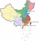
Between January 2019 and December 2019, a total of 837 MDR-TB isolates were obtained for our analysis. Based on results obtained via the MeltPro Myco assay, 22 cases (2.6%) were identified as NTM-MTB mixed infections. Significant diversity of mixed infection prevalence rates was observed across regions of China, with prevalence rates ranging from 0.0% (0/213) in Beijing to 3.4% (12/353) in Fuzhou to 3.7% (10/271) in Guangzhou (Figure 1).
 |
Figure 1 Distribution map of MDR-TB isolates included in this study. The number following the site number represents the number of isolates obtained from that site. |
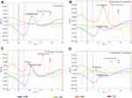
Among the 22 patients with NTM-TB mixed infections, a total of five different mycobacterial species were identified based on MeltPro Myco assay melting peaks of DNA prepared from isolates associated with these cases, as shown in Figure 2. The most prevalent species detected was Mycobacterium intracellulare (15/22, 68.2%), while prevalence rates of the other identified coinfecting mycobacterial species were as follows: Mycobacterium avium (4/22, 18.2%), Mycobacterium kansasii (1/22, 4.5%), Mycobacterium abscessus (1/22, 4.5%), and Mycobacterium malmoense (1/22, 4.5%) (Figure 3).
 |
Figure 3 Distribution of NTM species identified in mixed infection cases. |
Risk Factors Associated with Mixed Infections
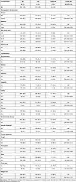
Table 1 provides an analysis of risk factors associated with incidence of MDR-TB cases co-infected with NTM in China. Based on selection of newly diagnosed MDR-TB patients as the control group, our data revealed that patients with histories of previous active pulmonary TB episodes were at higher risk of contracting mixed infections [odds ratio (OR): 3.40, 95% confidence interval (CI):1.41–8.20]. By contrast, no other demographic and clinical characteristics were significantly associated with NTM co-infection. In addition, no statistically significant difference in clinical symptoms was observed between the two groups.
 |
Table 1 Demographic and Clinical Characteristics Associated with Mixed Infection of MTB and NTM |
Discussion
The increasing incidence of NTM infections constitutes a major public health threat worldwide.16,17 As a consequence of intrinsic resistance of NTM to anti-TB drugs, patients with co-infections with MTB and NTM be misdiagnosed as MDR-TB cases. In our clinical setting, we found mixed infections of MTB and NTM in approximately 3% of previously laboratory-confirmed MDR-TB cases based on results obtained using molecular methods. Despite the fact that NTM-MTB co-infection may impact only a small proportion of the overall TB patient population, our data highlight the importance of accurate mycobacterial species identification prior to initiation of MDR TB treatment regimens. This is especially true for clinical settings in low-income countries, where a TB diagnosis is often based solely on results obtained using smear microscopy;18,19 however, this may also be true for some laboratories in the developed world, especially those that diagnose TB based on detection of MTB secreted protein MPT64. As a consequence of the above-mentioned challenges, it is essential to formulate optimal diagnostic algorithms in order to correctly identify mycobacterial species and detect NTM-MTB mixed infections. For bacterial species identification, currently used genetic sequencing methods outperform conventional biochemical testing and MPT64 antigen detection; however, current methods cannot always effectively discriminate between species of mycobacteria.20 Nevertheless, in a recent study conducted by Couto et al, a line probe assay based on hybridisation of DNA to 23S rRNA gene DNA probes was used to successfully detect mixed infections.21 Similarly, the molecular method used in the present study, which was based on genotyping of 16S rRNA gene amplicons via high-resolution melt analysis, has also been used to accurately identify co-existing species in clinical specimens.15 It is worth mentioning that accurate diagnosis of mixed infections depends on initial MTB and NTM population structures, due to different amplification efficiencies of DNA sequences of different species. Previous experiments revealed that PCR-based assays required a minimum of 20% representation of each mycobacterial species within the mixed population for successful detection of all constituent species.20 Thus, unbiased next-generation sequencing methods must be developed to facilitate identification of all potential pathogens in a single assay in a comprehensive way.22
In this study, M. intracellulare, which was detected in approximately 70% of all mixed infection cases, was the most frequently detected mycobacterial species. However, in a recent nationwide surveillance study, 52% of NTM isolates associated with pulmonary infections were M. intracellulare,23 a rate significantly lower than that observed in this study. We suggest two possible reasons for this disparity. First, the distribution of this species shows significant geographic diversity across China24 that may explain discrepancies between results. Second, another experimental study demonstrated that in vitro culture of a mixture of MTB and rapidly growing mycobacteria led to missed detection of MTB, due to the relatively lower growth rate of MTB relative to growth rates of rapidly growing mycobacteria. Nevertheless, the majority of NTM species identified in our study were slow growing mycobacterial species (21/22), further underscoring the fact that TB cases co-infected with M. intracellulare infections are becoming increasingly predominant across China. Of note, mixed infections were only observed in Guangdong and Fujian, where NTM infections are endemic, but not in Beijing, indicating that the mixed infection rate was positively correlated with NTM prevalence in only some regions of China. Thus, in view of the high potential for misdiagnosis of mixed infections as MDR-TB, more attention should be paid to investigating misdiagnoses of patients in East and South China.
Another important contribution of this work relates to information revealed through the analysis of risk factors associated with NTM-MTB co-infection cases, which revealed an association between susceptibility to mixed infections and history of previous active pulmonary TB episodes. This observation mirrored data reported in a recent systematic review that indicated that previous TB history played an important role in NTM occurrence.25 On the one hand, post-tuberculosis lung damage (PTLD) is a recognised consequence of pulmonary TB.26 As expected, in this study, 22.7% of NTM co-infection cases had histories of bronchiectasis, with preexisting lung diseases likely associated with dramatically impaired mucociliary clearance and insufficient airway drainage that facilitate NTM colonisation of individuals with histories of active pulmonary TB episodes.27 On the other hand, TB is a chronic disease that can become reactivated in previously treated TB patients with low socio-economic status and poor nutrition28,29 to explain the association between previous TB episodes and increased NTM-MTB mixed infection risk.
We also acknowledge several obvious limitations of the present study. First, despite the fact that MDR-TB isolates were obtained from three TB hospital sites, the small sample size and low prevalence of NTM-TB mixed infection among suspected MDR-TB cases may have weakened the significance of our results with regard to identification of patients at risk for mixed infections. Second, we found an association between history of previous TB episodes and increased risk of contracting mixed infections, but mechanisms underlying this association remain unclear. Thus, additional studies are needed to elucidate roles of host immunology and underlying lung conditions in susceptibility to mixed infections. Finally, antibiotic resistance profiles of MTB populations associated with mixed infections were not characterised. Despite these limitations, our findings raise concerns about accuracies of molecular diagnostic tests for use in species identification and draw attention to the possibility that NTM-MTB mixed infection cases in settings with high TB burden may be misdiagnosed as MDR-TB cases.
Conclusion
The results obtained in this work demonstrate that mixed infections with MTB and NTM occur in approximately 3% of suspected MDR-TB patients in China. M. intracellulare was the most frequently observed mycobacterial species detected in NTM-MTB mixed infections. In addition, an association was found between mixed infection risk and history of previous TB episodes that raises concerns that inaccuracies of molecular diagnostic tests used for species identification may lead to misdiagnoses of NTM-MTB co-infection cases as MDR-TB cases in high TB burden-settings.
Abbreviations
TB, tuberculosis; MDR-TB, multidrug-resistant tuberculosis; INH, isoniazid; RIF, rifampicin; WHO, World Health Organization; DST, drug susceptibility testing; MTB, Mycobacterium tuberculosis; NTM, nontuberculous mycobacteria; L-J, Löwenstein-Jensen; OR, odds ratio; CI, confidence interval.
Data Sharing Statement
The datasets used and/or analyzed during the current study are available from the corresponding author on reasonable request.
Ethics Approval and Consent to Participate
This study was conducted in accordance with the Declaration of Helsinki and approved by the ethics committee of Beijing Chest Hospital, Capital Medical University (2020KY030). The adults or the parents/legal guardians of patients under 18 years of age signed a written informed consent to agree with the anonymous use of clinical data.
Acknowledgments
We thank all the pilots that participated in this study.
Author Contributions
All authors have made a significant contribution to this study, all the way through the conception, study design, execution, acquisition of data, data analysis and interpretation to drafting, revising or critically reviewing stages of the article. The authors also gave final approval of the version to be published, agreed on the journal to which the article has been submitted, and agreed to be accountable for all aspects of the work.
Funding
The study was supported by the Beijing Hospitals Authority’ Ascent Plan (DFL20191601), the Beijing Hospitals Authority Clinical Medicine Development of Special Funding (ZYLX202122), Fuzhou Science and Technology Project (2019-SZ-59). The funders had no role in the study design, data collection and analysis, decision to publish, or preparation of the manuscript.
Disclosure
The authors declare no competing interests.
References
1. World Health Organization. Global tuberculosis report 2020. Geneva: World Health Organization; 2020. WHO/HTM/TB/2020.
2. Lange C, Chesov D, Heyckendorf J, Leung CC, Udwadia Z, Dheda K. Drug-resistant tuberculosis: an update on disease burden, diagnosis and treatment. Respirology. 2018;23(7):656–673. doi:10.1111/resp.13304
3. Nguyen TNA, Anton-Le Berre V, Banuls AL, Nguyen TVA. Molecular diagnosis of drug-resistant tuberculosis; a literature review. Front Microbiol. 2019;10:794. doi:10.3389/fmicb.2019.00794
4. Lange C, Dheda K, Chesov D, Mandalakas AM, Udwadia Z, Horsburgh CR
5. Johansen MD, Herrmann JL, Kremer L. Non-tuberculous mycobacteria and the rise of Mycobacterium abscessus. Nat Rev Microbiol. 2020;18(7):392–407. doi:10.1038/s41579-020-0331-1
6. van Ingen J, Boeree MJ, van Soolingen D, Mouton JW. Resistance mechanisms and drug susceptibility testing of nontuberculous mycobacteria. Drug Resist Updat. 2012;15(3):149–161. doi:10.1016/j.drup.2012.04.001
7. van Ingen J, Kuijper EJ. Drug susceptibility testing of nontuberculous mycobacteria. Future Microbiol. 2014;9(9):1095–1110. doi:10.2217/fmb.14.60
8. Brent AJ, Mugo D, Musyimi R, et al. Performance of the MGIT TBc identification test and meta-analysis of MPT64 assays for identification of the Mycobacterium tuberculosis complex in liquid culture. J Clin Microbiol. 2011;49(12):4343–4346. doi:10.1128/JCM.05995-11
9. Martin A, Bombeeck D, Mulders W, Fissette K, De Rijk P, Palomino JC. Evaluation of the TB Ag MPT64 Rapid test for the identification of Mycobacterium tuberculosis complex. Int J Tuberc Lung Dis. 2011;15(5):703–705. doi:10.5588/ijtld.10.0474
10. Pai HH, Chen WC, Peng CF. Isolation of non-tuberculous mycobacteria from hospital cockroaches (Periplaneta americana). J Hosp Infect. 2003;53(3):224–228. doi:10.1053/jhin.2002.1355
11. Maiga M, Siddiqui S, Diallo S, et al. Failure to recognize nontuberculous mycobacteria leads to misdiagnosis of chronic pulmonary tuberculosis. PLoS One. 2012;7(5):e36902. doi:10.1371/journal.pone.0036902
12. Thangavelu K, Krishnakumariamma K, Pallam G, et al. Prevalence and speciation of non-tuberculous mycobacteria among pulmonary and extrapulmonary tuberculosis suspects in South India. J Infect Public Health. 2021;14(3):320–323. doi:10.1016/j.jiph.2020.12.027
13. Shu W, Du J, Liu Y, et al. External quality control of phenotypic drug susceptibility testing for Mycobacterium tuberculosis in China. Eur J Clin Microbiol Infect Dis. 2020;39(5):871–875. doi:10.1007/s10096-019-03770-1
14. Liu Y, Gao M, Du J, et al. Reduced susceptibility of Mycobacterium tuberculosis to bedaquiline during antituberculosis treatment and its correlation with clinical outcomes in China. Clin Infect Dis. 2021;73:e3391–e3397.
15. Xu Y, Liang B, Du C, et al. Rapid identification of clinically relevant mycobacterium species by multicolor melting curve analysis. J Clin Microbiol. 2019;57(1). doi:10.1128/JCM.01096-18
16. Donohue MJ, Wymer L. Increasing prevalence rate of nontuberculous mycobacteria infections in five states, 2008–2013. Ann Am Thorac Soc. 2016;13(12):2143–2150. doi:10.1513/AnnalsATS.201605-353OC
17. Thomson RM. Changing epidemiology of pulmonary nontuberculous mycobacteria infections. Emerg Infect Dis. 2010;16(10):1576–1583. doi:10.3201/eid1610.091201
18. Tabarsi P, Baghaei P, Farnia P, et al. Nontuberculous mycobacteria among patients who are suspected for multidrug-resistant tuberculosis-need for earlier identification of nontuberculosis mycobacteria. Am J Med Sci. 2009;337(3):182–184. doi:10.1097/MAJ.0b013e318185d32f
19. Tortoli E, Rogasi PG, Fantoni E, Beltrami C, De Francisci A, Mariottini A. Infection due to a novel mycobacterium, mimicking multidrug-resistant Mycobacterium tuberculosis. Clin Microbiol Infect. 2010;16(8):1130–1134. doi:10.1111/j.1469-0691.2009.03063.x
20. Liang Q, Shang Y, Huo F, et al. Assessment of current diagnostic algorithm for detection of mixed infection with Mycobacterium tuberculosis and nontuberculous mycobacteria. J Infect Public Health. 2020;13(12):1967–1971. doi:10.1016/j.jiph.2020.03.017
21. Couto I, Machado D, Viveiros M, Rodrigues L, Amaral L. Identification of nontuberculous mycobacteria in clinical samples using molecular methods: a 3-year study. Clin Microbiol Infect. 2010;16(8):1161–1164. doi:10.1111/j.1469-0691.2009.03076.x
22. Naccache SN, Peggs KS, Mattes FM, et al. Diagnosis of neuroinvasive astrovirus infection in an immunocompromised adult with encephalitis by unbiased next-generation sequencing. Clin Infect Dis. 2015;60(6):919–923. doi:10.1093/cid/ciu912
23. van Ingen J, Al-Hajoj SA, Boeree M, et al. Mycobacterium riyadhense sp. nov., a non-tuberculous species identified as Mycobacterium tuberculosis complex by a commercial line-probe assay. Int J Syst Evol Microbiol. 2009;59(Pt 5):1049–1053. doi:10.1099/ijs.0.005629-0
24. Pang Y, Tan Y, Chen J, et al. Diversity of nontuberculous mycobacteria in eastern and southern China: a cross-sectional study. Eur Respir J. 2017;49(3). doi:10.1183/13993003.01429-2016
25. Sonnenberg P, Murray J, Glynn JR, Thomas RG, Godfrey-Faussett P, Shearer S. Risk factors for pulmonary disease due to culture-positive M. tuberculosis or nontuberculous mycobacteria in South African gold miners. Eur Respir J. 2000;15(2):291–296. doi:10.1034/j.1399-3003.2000.15b12.x
26. Meghji J, Lesosky M, Joekes E, et al. Patient outcomes associated with post-tuberculosis lung damage in Malawi: a prospective cohort study. Thorax. 2020;75(3):269–278. doi:10.1136/thoraxjnl-2019-213808
27. Mirsaeidi M, Machado RF, Garcia JG, Schraufnagel DE. Nontuberculous mycobacterial disease mortality in the United States, 1999–2010: a population-based comparative study. PLoS One. 2014;9(3):e91879. doi:10.1371/journal.pone.0091879
28. Liu JJ, Yao HY, Liu EY. Analysis of factors affecting the epidemiology of tuberculosis in China. Int J Tuberc Lung Dis. 2005;9(4):450–454.
29. Mishra P, Hansen EH, Sabroe S, Kafle KK. Socio-economic status and adherence to tuberculosis treatment: a case-control study in a district of Nepal. Int J Tuberc Lung Dis. 2005;9(10):1134–1139.
 © 2022 The Author(s). This work is published and licensed by Dove Medical Press Limited. The
full terms of this license are available at https://www.dovepress.com/terms.php
and incorporate the Creative Commons Attribution
- Non Commercial (unported, v3.0) License.
By accessing the work you hereby accept the Terms. Non-commercial uses of the work are permitted
without any further permission from Dove Medical Press Limited, provided the work is properly
attributed. For permission for commercial use of this work, please see paragraphs 4.2 and 5 of our Terms.

