Back to Journals » Journal of Multidisciplinary Healthcare » Volume 17
Effect of Diabetes on Wound Healing: A Bibliometrics and Visual Analysis
Authors Lang X ![]() , Li L
, Li L ![]() , Li Y, Feng X
, Li Y, Feng X
Received 1 January 2024
Accepted for publication 14 March 2024
Published 20 March 2024 Volume 2024:17 Pages 1275—1289
DOI https://doi.org/10.2147/JMDH.S457498
Checked for plagiarism Yes
Review by Single anonymous peer review
Peer reviewer comments 2
Editor who approved publication: Dr Scott Fraser
Xiaona Lang,1 Lu Li,1 Yuntao Li,2 Xin Feng1
1Pharmacy Department, Tianjin Hospital, Tianjin, People’s Republic of China; 2Integrative Chinese and Western Medicine Department, Tianjin Hospital, Tianjin, People’s Republic of China
Correspondence: Xin Feng, Pharmacy Department, Tianjin Hospital, Tianjin, People’s Republic of China, Tel +86-13652001152, Email [email protected]
Objective: The quality of life of diabetic patients is seriously affected by wound healing difficulty, which can lead to increased infection, skin deep tissue injury and continuous pain. By analyzing the research trends and hot spots in this field, the visualization analysis map is constructed.
Methods: The contents of the selected articles were sorted out and analyzed by bibliometrics. We use CiteSpace, Vosviewer and HistCite to visualize literature information, including national publication statistics, institutions, authors, journal partnerships, and citations of published articles.
Results: Among the 2942 articles, the United States and China ranked first in both article circulation and TGCS, and many countries also cooperated. The collaboration between schools and research institutions is a core part of dissertation research institution collaboration, with most authors coming from the same institution. Most of the literature studies on the mechanisms and methods of promoting diabetic wound healing. Improving cell function or making innovative attempts in local treatment are the fruits of researchers’ efforts to promote diabetic wound healing in recent years.
Conclusion: Through the metrology method, the time distribution, author institution, cooperation network, research status, research hotspot and development trend of the literature on the influence of diabetes on wound healing were intuitively displayed, which provided a reference for further research and development direction.
Keywords: diabetes, wound healing, bibliometrics, visualized analysis, Citespace
Introduction
Diabetes, a prevalent chronic metabolic disorder, poses an increasing global health threat. The World Health Organization reports that a significant number of individuals worldwide are afflicted with diabetes, a number that continues to rise.1 In addition to elevated blood glucose levels, insulin resistance, and associated complications, impaired wound healing presents a significant challenge for individuals with diabetes.2
Recent years have witnessed notable progress in strategies to improve wound healing in diabetic patients, encompassing novel pharmacological interventions, state-of-the-art wound care technologies, and innovative approaches to glycemic control and infection management. However, the field is marked by rapid evolution and a constant influx of novel methodologies and technologies, emphasizing the necessity of regularly reviewing and integrating the extensive array of emerging data.
Wound healing refers to the process by which wounds return to their normal state with the support of a series of biological processes after injury.2–4 However, diabetic wounds are skin and tissue damage caused by prolonged high blood sugar. If left untreated and unmanaged, diabetic ulcers can lead to the following consequences. 1. Infection: Due to the weak immune function of diabetic patients, ulcers are easily infected, and infection will further delay the healing rate of ulcers. Severe infections can spread to surrounding tissues and bones, threatening the health of the body. Severe cases can even lead to gangrene: When the tissue in the ulcer is severely infected, and the blood supply is insufficient, tissue necrosis may occur, forming gangrene. Gangrene severely threatens surrounding tissues and organs and requires urgent treatment, including surgical removal of dead tissue.5,6 2. Deep tissue damage: Diabetic ulcers usually occur in the foot and may cause damage to the deep tissues of the foot, such as muscles, ligaments, and bones, due to neuropathy and circulation problems. This can lead to joint stiffness, muscle dysfunction, and even fractures.7,8 3. Chronic pain: Diabetic ulcers may cause persistent pain that affects the patient’s quality of life and daily activities. Chronic pain can hurt emotional and mental health.4,9,10
Given the rapidly evolving landscape of wound healing strategies in diabetes care, there is an urgent requirement for a comprehensive analysis that comprehensively delineates the current state of research. A bibliometric analysis provides a systematic method to chart the course of these developments, discern predominant trends, and underscore areas necessitating additional exploration. Such an analysis plays a pivotal role in offering a complete perspective on the advancements achieved to date and in delineating potential pathways for future research initiatives. In recent years, there have been a number of review articles on diabetes and wounds, to summarize the new treatment modalities and developments in the rehabilitation of wounds in diabetic patients. So let us analyze it from a brand-new literature dose perspective, which is very impressive.
Citespace and Vosviewer are literature visualization and analysis software. After understanding and studying certain literature materials utilizing econometric analysis, visualization maps are made, hot spots are analyzed around the current situation of the research content and development trends are explored.11–13 HistCite is a citation analysis tool that can quickly map directions in the same field, organize the number of citations, and identify critical studies and researchers.14
This paper is based on a literature analysis spanning nearly 20 years, utilizing software to organize and visually display the literature information. It encompasses a review of relevant research on diabetes and wound healing, encompassing theoretical foundations, application effectiveness, development, and shortcomings both domestically and internationally. Additionally, it offers comprehensive data and analysis for researchers and institutions in related fields.
Materials and Methods
Data Sources
Using the core collection of Web of Science databases, with the title “Wound Healing”, synonyms include: “Wound Healings”; “Wound Healing”; “Healings, Wound”. The title is “Wound Healings”; synonyms include: “Diabetes Mellitus”; “DM”. The date range is 2003–01-01 to 2023–05-31. After combining the above two search results with “AND”, 5737 kinds of literature were selected. Through manual screening, the types of meetings, newspapers, abstracts, and books were excluded, and after reading the title and abstract of the article, the articles unrelated to the content of the subject word were deleted, and a total of 2942 articles meeting the requirements were obtained.
Analytical Tools
The HistCite software is used to visualize and analyze the data of countries/regions, institutions, and periodicals after statistics—Journal Impact Factor (IF) from Journal Citation Reports (2022). CiteSpace can be used to analyze keywords and article clustering information. Through Vosviewer, we can visualize and explore the knowledge of the publishing institution and the author. By analyzing the relevant information, you can build a co-occurrence network to get relevant research hot spots and development trends in recent years. When constructing the cluster analysis map of keywords and articles in this study, CiteSpace6.2.R2 was set as Slice=3 and g-index k=25 to ensure that the node information obtained from each time slice was as similar as possible. Set Pruning to Pathfinder and Pruning sliced networks and Pruning the merged network.
Results
Literature Statistics
As shown in Figure 1, a total of 2942 pieces of literature were selected for inclusion from 92 countries and regions, 14,082 authors, 937 journals, and 5264 keywords. By calculating the Total Global Citation Score(TGCS), we found that 127 research articles were cited more than 100 times, of which 27 were awarded more than 200 times.
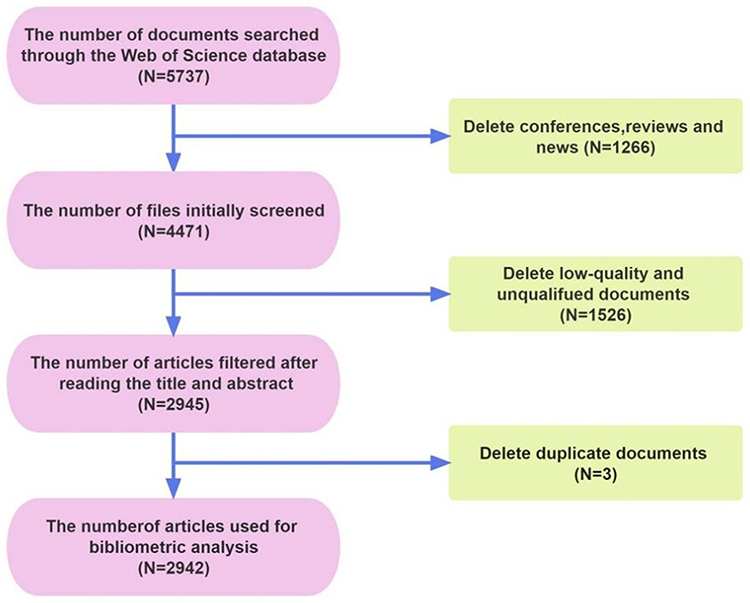 |
Figure 1 Flow chart of literature screening. |
According to the trend chart of article publication years in Figure 2A, we find that articles in 2016 and 2020 have the most significant amount of citations, which indicates that articles in these two years are of high quality and have citation value, worthy of scholars’ attention. The number of published papers has increased yearly, from 29 published in 2003 to over 400 published in 2022. In the past five months of 2023, the number of relevant articles has reached 231; the reason for the low TGCS value, we believe is because the year is too early, but the content is very innovative, and the number of citations will be significantly increased in a few years.
 |
Figure 2 (A). Yearly output and TGCS; (B). Global trends in the number of publications by country; (C). Global cooperation in publishing; (D). Global distribution of publications (by TGCS). |
Geographical Distribution of the Literature
Figure 2B shows the number of published articles by country. It can be seen that the number of pieces from China and the United States has increased year by year, and they occupy a large proportion. This also represents the increasing attention of researchers in these two countries to the relationship between diabetes and wound healing. Figure 2C shows that the United States and China have the most significant number of publications and close cooperation with many other countries. As can be seen from Figure 2D, the quality of TGCS papers published in Europe, America, and Asia in this field is high, and the content of the papers is worthy of being cited, which is of great value to the research field.
Table 1 lists the top 10 countries in terms of the number of articles in the study, of which 5 are European countries with a total of 492 pieces (16.72%), and 4 are Asian countries with 1223 articles (41.57%). The top three countries in terms of Average Citation Index(ACI) values are Sweden, the USA, and France, indicating that these three countries are more mature and have more valuable research content in this area than other countries.
 |
Table 1 The Data Ranked the Top 10 Countries by TGCS |
As shown in Table 2, most of TGCS’s top 20 institutions are in the United States, China, and Sweden. The No. 1 institution is Harvard University, which has the highest TGCS score. It was followed by the University of Pennsylvania in the United States and Shanghai Jiao Tong University in China. Kyoto Prefectural University of Medicine has an ACI of 114.14, higher than other institutions. It is the only institution in the table from Japan. Although the number of articles is only 7, the average number of citations is very high, indicating that the articles’ content value is high and worthy of study. Texas A&M University had an ACI of 105.50, and Rosalind Franklin University of Medicine and Science had an ACI of 94.08. Through this data, we found that the United States is significantly ahead of other countries in studying the impact of diabetes on wound healing.
 |
Table 2 The Top 20 Institutions (Based on Records and TGCS Respectively) |
Analysis of Journals
Table 3 lists the top 20 journals by number of publications. Among these journals, the total number OF papers published in JOURNAL OF WOUND CARE (N=80,2.72%), followed by WOUND REPAIR AND REGENERATION(N=75,2.55%). The top three TGCS are DIABETES, WOUND REPAIR AND REGENERATION, and PLOS ONE. It is worth noting that DIABETES published 56 articles, but the TGCS was very high, and the ACI value was as high as 76.84. The content of articles published in this journal is worthy of being cited by research, and it is easier for researchers to study and explore. Other journals with relatively high ACI values include DIABETES-METABOLISM RESEARCH AND REVIEWS (47.94). PLOS ONE(44.67) and JOURNAL OF INVESTIGATIVE DERMATOLOGY (39.60). The ACI value of these journals is relatively high, and the impact factor is mainly greater than 6, which means that journals have higher requirements for manuscripts, many scholars have cited their published articles, and their contents are worthy of attention.
 |
Table 3 The Top 20 Journals (Based on Recs) |
Figure 3 shows a double map overlay of journals. The left of the diagram represents citing journals, and the right side of the graph represents the relationship between the referenced journals. The orange and green lines indicate that articles published in Molecular/Biology/Immunology are mainly cited in Medicine/Medical/Clinical journals. The longer the horizontal axis of the ellipse, the more articles are published in journals. The longer the longitudinal axis of an ellipse, the more authors it represents. We found that articles in similar journals frequently cited each other, and the relationship between medicine, clinic and biology was closely linked, which provided a basis for drug exploration and discovery.
 |
Figure 3 The dual-map overlay. |
Analysis of the Cooperative Relationship Between Authors and Institutions
Figure 4A shows the collaborative relationship between institutions, and Figure 4B represents the collaborative relationship between authors. Each node in the graph is an institution; the node’s size is proportional to the number of documents issued by the institution; the connection is the cooperative relationship between different institutions, and the thickness of the link is proportional to the strength of the collaborative relationship. Most top institutions are universities or research institutes, and few hospitals. This shows that the research is still biased toward basic research. Due to the large number of authors and institutions, only central cooperative nodes in the collaborative network are selected in this paper. These are chosen according to the number of authors’ reports and the closeness of cooperation.
 |
Figure 4 (A). Academic cooperation networks between institutions; (B). Academic cooperation networks between authors; (C). Networks of keywords in the studies; (D). The top 20 strongest strength citation burst. |
Analysis of Keywords
The top five keywords in frequency were diabetes mellitus, wound healing, foot ulcer, expression, and angiogenesis. From the above words, we can see the main content of this research direction and the central nodes of other essential research directions. The map of high-frequency co-occurrence words is shown in Figure 4C, where the circular node wrapped by a purple circle indicates that the centrality of this keyword is more significant than 0.1, indicating that they are closely related to other keywords and have a relatively large proportion discussed in the paper. The nodes with red dots are the top breakout words and the trends and developments in this field. The relevant information on the top 20 high-frequency keywords is summarized in Table 4, which reflects the main keywords discussed in relevant literature in recent years. Figure 4D shows the top 20 most cited burst words; the longer the red area, the longer the word has been mentioned. These include mice, ulcers, nitric oxide, Mellitus, and complications. The most popular words in the last five years are impact, antibacterial, and hydrogel. These words are all explosive words in the research on diabetes and wound healing in recent years. We found that diabetic mice, as experimental animals, are the focus of research, and most of the primary research uses these animals. One of the complications of diabetes is diabetic foot, which is a significant problem worldwide. For this ulcer problem, the current treatment methods tend to make antibacterial hydrogels through the anti-inflammatory, bactericidal mechanism to promote wound healing. This also gives us hints for future research.
 |
Table 4 The Top 20 Keywords |
Key Article Analysis
We have summarized the top 10 TGCS articles. Table 5 is a summary of the basic information of the articles. These representative articles’ contents are precious, and the contents of the top 3 pieces are summarized. According to the content, most papers study the mechanism and methods of promoting the healing of diabetic wounds. Improving the function of cells or making innovative attempts at topical therapies are the results of researchers’ efforts to promote wound healing in diabetes in recent years. The content of these articles can be used as the research basis and foundation of other scholars.
 |
Table 5 Top 10 Articles (Based on TGCS) |
Loomans CJMet al, a novel concept of endothelial progenitor cell dysfunction in the pathogenesis of vascular complications in type 1 diabetes, published in 2004.15 It is suggested that endothelial progenitor cell dysfunction plays an essential role in vascular complications in diabetic patients. Due to the importance of endothelial cells in maintaining blood vessel function and repairing damage, restoring endothelial progenitor cell function may help prevent and treat vascular complications in patients with type 1 diabetes. This article provides new ideas about endothelial progenitor cell dysfunction in the pathogenesis of vascular complications in type 1 diabetes. Further study and understanding of the role of EPCs in diabetes may provide new therapeutic strategies for preventing and treating related vascular complications.
Wong SL et al published in 2015 on how diabetes causes neutrophil NETosis, and this process can have adverse effects on wound healing.16 In people with diabetes, the release of NETs causes a significant buildup of DNA and proteins in the wound area, creating an inflammatory environment that impedes the normal wound-healing process. These NETs components form a physical barrier that prevents new cells from migrating to the wound site and may also cause damage to surrounding healthy tissue, exacerbating the inflammatory response. Impaired wound healing in diabetic patients is a multifactorial process, and NETosis is one of the mechanisms leading to this complication. Managing blood sugar levels, controlling inflammation, and promoting proper wound care are essential strategies for improving wound healing in people with diabetes.
Armstrong DG et al. Multicenter, randomized controlled trial of adverse pressure wound treatment after partial diabetic foot amputation published in 2005.17 This paper describes a clinical trial of partial diabetic foot amputations, comparing the effect of negative pressure wound treatment with conventional dressing. The results of this multicenter, randomized controlled trial support the effectiveness and advantages of adverse pressure wound treatment in some diabetic foot amputees. Negative pressure wound treatment can promote wound healing, reduce the risk of infection, and improve quality of life and functional recovery. This has important clinical significance for clinical practice and guiding rehabilitation treatment of diabetic foot amputation patients.
Analysis of Article Structure Variation
Structural variation theory is a reorganization search in a high-dimensional common reference space. The primary purpose of structural variation analysis is to detect unprecedented inter-cluster bridges or new types of remote connections and understand why specific connections are novel and valuable capabilities. The structure variation analysis of the cited article was used here to find literature with significant impact potential. The keyword clusters with different colors in Figure 5, including Cluster #0 diabetic wound regeneration; Cluster #1 promoting wound healing; Cluster #2 diabetic ulcer; Cluster #3 no-mediated endothelial progenitor cell mobilization; Cluster #4 tissue regeneration; Cluster #5 manganese superoxide dismutase; Cluster #6 pro-apoptotic gene; Cluster #7 consensus recommendation; Cluster #8 ultrasound therapy; Cluster #9 ga-as laser. The keywords of these clusters are closely related to diabetes and wounds. By Table 6, we can find their representative articles mainly include the causes of injury nonhealing and treatment options, improving cell function, and physical ultrasound therapy are effective. The color-coded sections reflect when co-citation links appeared for the first time in those areas. Different colors represent different clusters, and each cluster has several representative articles.
 |
Table 6 Clusters Represent Articles |
 |
Figure 5 Keyword clusters of cited articles. |
Discussion
According to the literature analysis, among the 2942 literature, both the United States and China rank high in article circulation and TGCS, and many countries also cooperate. Cooperation between schools and research institutes is the central part of the research institutions for articles, and most co-authors come from the same institution in the same country. The primary focus of the literature revolves around the impact of diabetic injuries on patients, including the causes and potential solutions for non-healing wounds. In recent years, new methods of wound treatment, such as exploring the biological aspects of cell function and the physical aspects of ultrasound and laser, have emerged as prominent research areas in this field. There is a notable presence of mutual citations among journals, especially with articles cited in the early stages, indicating their significance during that time period. These articles hold great research value and are worthy of attention from researchers in the field.
After reading a lot of relevant literature, we concluded that the reasons for the problematic healing of diabetic wounds include the following: 1. High blood sugar: Chronic high blood sugar can cause damage to blood vessels, nerves, and the immune system. High blood sugar can cause blood vessels to constrict and harden, reduce blood circulation, reduce oxygen and nutrient supply to the injured site, and affect wound repair.35,36 2. Microvascular disease: Diabetic patients are prone to microvascular disease, which damages the microcirculation around the wound and causes insufficient blood supply, delaying the healing speed of the wound.37,38 3. Decreased immune function: diabetes will suppress the immune system’s function, decreasing the patient’s resistance and becoming prone to infection. Infection can aggravate the inflammatory response of the wound and interfere with the normal healing process.7,35 4. Neuropathy: Neuropathy caused by diabetes will affect the sensory nerve and autonomic nervous system, reducing pain and temperature perception, so that patients may not be able to detect the wound in time, delaying the time for treatment.2,3,39 5. Infection: Diabetic patients are prone to wound infection. Due to decreased immune function and poor blood circulation, infection will aggravate inflammation and interfere with the repair process.5,40–42 6. Other complications: Patients with diabetes are often accompanied by other complications, such as arteriosclerosis, hypertension, etc., further affecting wound healing.9,10,43 Figure 6 (by Figdraw) shows the primary classification and treatment of diabetic wounds. There are various ways to treat diabetic ulcers, and specific treatment plans should be formulated according to the condition and individual needs.44–47
 |
Figure 6 The main classification and treatment of wounds. |
At present, there are some research and developments on the new methods and directions of treating diabetic ulcer nonunion at home and abroad. The following are the more significant new approaches and tips: 1. Using growth factors: Researchers are exploring using growth factors to promote the healing of diabetic ulcers. Growth factors can promote cell proliferation and wound repair; some studies have shown that platelet-derived growth factor (PDGF) can accelerate ulcer healing.48,49 2. Stem cell therapy: Stem cell therapy is believed to promote tissue repair and regeneration potentially. Some studies are considering using stem cells to treat diabetic ulcers; for example, mesenchymal stem cells (MSCs) have been shown to promote wound healing and improve blood circulation.50–53 3. Antimicrobial techniques: Since infection is an essential factor in the difficulty of diabetic ulcers to heal, researchers are working to develop new antimicrobial techniques to prevent and treat infections. For example, nanomaterials, antibacterial surface coatings, phototherapy, and ultrasound reduce the risk of wound infection.54,55 4. 3D printing technology: 3D printing technology can produce biological materials with complex structures and individual characteristics for wound repair. Researchers are experimenting with using 3D-printed artificial skin or stents to promote the healing diabetic ulcers.56–58 5. Immunomodulatory treatment: Because of decreased immune function in diabetic patients, some new treatment methods promote the healing of ulcers through immune regulation. For example, immune inducers or biological agents are used to improve the immune response in people with diabetes.59 6. Bioactive substances and gene therapy: Researchers are also investigating the use of bioactive substances (such as DNA, RNA) and gene therapy to promote the healing of diabetic ulcer.60–62
In this study, we used bibliometrics to analyze the relevant literature in the core database of Web of Science and quantitatively and qualitatively analyzed the research contributions of different countries, institutions, journals, and authors in this field through Citespace, HistCite, VOSviewer, and other software. The advantage of this paper is to sort out the development of this discipline through the literature analysis in recent years. To identify valuable and representative articles for a thorough analysis and to elucidate the current research trends, it is essential to consider expanding the search beyond the Web of Science to include other databases. Accessing unpublished or published literature from various sources will help in achieving more comprehensive statistics and a broader perspective. While the paper focuses on analyzing essential information and key content from the literature, there is room for improvement by delving deeper into the details. In future studies, it is recommended to explore additional databases and utilize diverse software applications to enhance the ability to analyze literature content comprehensively. Moreover, researchers should continue to prioritize the treatment of diabetic wound healing, aligning with the subject requirements while staying updated on emerging trends and advancements in the field.
Data Sharing Statement
All data are presented on the proof. For further data information, please contact the correspondence author.
Author Contributions
All authors made a significant contribution to the work reported, whether that is in the conception, study design, execution, acquisition of data, analysis and interpretation, or in all these areas; took part in drafting, revising or critically reviewing the article; gave final approval of the version to be published; have agreed on the journal to which the article has been submitted; and agree to be accountable for all aspects of the work.
Funding
This research was sponsored by Tianjin Health Research Project (TJWJ2023QN053).
Disclosure
The authors declare that the research was conducted without any commercial or financial relationships construed as a potential conflict of interest.
References
1. Ogundele SO, Dada AO, Mosuro OR. Clinical profile, knowledge, and beliefs about diabetes among patients attending a Tertiary Health Centre in Lagos: a cross-sectional survey. Article. Niger J Clin Pract. 2016;19(4):508–512. doi:10.4103/1119-3077.183303
2. Riedel U, Schussler E, Hartel D, Keiler A, Nestoris S, Stege H. Wound treatment in diabetes patients and diabetic foot ulcers. Article. Hautarzt. 2020;71(11):835–842. doi:10.1007/s00105-020-04699-9
3. Okonkwo UA, DiPietro LA. Diabetes and Wound Angiogenesis. Review. Int J Mol Sci. 2017;18(7):15. doi:10.3390/ijms18071419
4. Greenhalgh DG. Wound healing and diabetes mellitus. Article. Clin Plast Surg. 2003;30(1):37. doi:10.1016/s0094-1298(02)00066-4
5. Wang W, Yan X, Lin Y, Ge HQ, Tan Q. Wnt7a promotes wound healing by regulation of angiogenesis and inflammation: issues on diabetes and obesity. Article. J Dermatol Sci. 2018;91(2):124–133. doi:10.1016/j.jdermsci.2018.02.007
6. Mohanasundaram T, Ramachandran V, Bhongiri B, et al. The promotion of antioxidant and anti-inflammatory activity by Nrf2 amplifier is a potential technique in diabetic wound healing-a review. Review. Pharm Sci. 2023;29(3):255–262. doi:10.34172/ps.2022.45
7. Male A, Kumar BN, Kumar DS, Shuaibyunus MD. Review On diabetic wound healing therapy. Article; proceedings paper. Int J Life Sci Pharma Res. 2020;3:77–80.
8. Dasari N, Jiang A, Skochdopole A, et al. Updates in diabetic wound healing, inflammation, and scarring. Article. Semin Plast Surg. 2021;35(03):153–158. doi:10.1055/s-0041-1731460
9. Baltzis D, Eleftheriadou I, Veves A. Pathogenesis and treatment of impaired wound healing in diabetes mellitus: new insights. Review. Adv Ther. 2014;31(8):817–836. doi:10.1007/s12325-014-0140-x
10. Ahmed AS. Does diabetes mellitus affect tendon healing? In: Ackermann PW, Hart DA, editors. Metabolic Influences on Risk for Tendon Disorders Advances in Experimental Medicine and Biology. Berlin: Springer-Verlag; 2016:179–184.
11. Chen C, Assoc Comp M Visualizing and exploring scientific literature with citeSpace. 2018:369–370.
12. Chen CM. CiteSpace II: detecting and visualizing emerging trends and transient patterns in scientific literature. J Am Soc Inf Sci Technol. 2006;57(3):359–377. doi:10.1002/asi.20317
13. Li B, Hu K, Lysenko V, et al. A scientometric analysis of agricultural pollution by using bibliometric software VoSViewer and Histcite (TM). Article. Environ Sci Pollut Res. 2022;29(25):37882–37893. doi:10.1007/s11356-022-18491-w
14. Wu JF, Tsai HL. An Explication of HistCite (TM): updates, modifications, and a variety of applications. Ser Rev. 2022;48(1–2):41–48. doi:10.1080/00987913.2022.2101821
15. Loomans CJM, de Koning EJP, Staal FJT, et al. Endothelial progenitor cell dysfunction - A novel concept in the pathogenesis of vascular complications of type 1 diabetes. Article. Diabetes. 2004;53(1):195–199. doi:10.2337/diabetes.53.1.195
16. Wong SL, Demers M, Martinod K, et al. Diabetes primes neutrophils to undergo NETosis, which impairs wound healing. Article. Nature Med. 2015;21(7):815. doi:10.1038/nm.3887
17. Armstrong DG, Lavery LA, Diabet Foot Study C. Negative pressure wound therapy after partial diabetic foot amputation: a multicentre, randomised controlled trial. Article. Lancet. 2005;366(9498):1704–1710. doi:10.1016/s0140-6736(05)67695-7
18. Gallagher KA, Liu ZJ, Xiao M, et al. Diabetic impairments in NO-mediated endothelial progenitor cell mobilization and homing are reversed by hyperoxia and SDF-1 alpha. Article. J Clin Invest. 2007;117(5):1249–1259. doi:10.1172/jci29710
19. Amor C, Feucht J, Leibold J, et al. Senolytic CAR T cells reverse senescence-associated pathologies. Article. Nature. 2020;583(7814):127. doi:10.1038/s41586-020-2403-9
20. Dowd SE, Wolcott RD, Sun Y, McKeehan T, Smith E, Rhoads D. Polymicrobial nature of chronic diabetic foot ulcer biofilm infections determined using bacterial tag encoded FLX Amplicon Pyrosequencing (bTEFAP). Article. PLoS One. 2008;3(10):e3326. doi:10.1371/journal.pone.0003326
21. Botusan IR, Sunkari VG, Savu O, et al. Stabilization of HIF-1 alpha is critical to improve wound healing in diabetic mice. Article. Proc Natl Acad Sci U S A. 2008;105(49):19426–19431. doi:10.1073/pnas.0805230105
22. Lerman OZ, Galiano RD, Armour M, Levine JP, Gurtner GC. Cellular dysfunction in the diabetic fibroblast - Impairment in migration, vascular endothelial growth factor production, and response to hypoxia. Article. Am J Pathol. 2003;162(1):303–312. doi:10.1016/s0002-9440(10)63821-7
23. Maruyama K, Asai J, Li M, Thorne T, Losordo DW, D’Amore PA. Decreased macrophage number and activation lead to reduced lymphatic vessel formation and contribute to impaired diabetic wound healing. Article. Am J Pathol. 2007;170(4):1178–1191. doi:10.2353/ajpath.2007.060018
24. Thangarajah H, Yao DC, Chang EI, et al. The molecular basis for impaired hypoxia-induced VEGF expression in diabetic tissues. Article. Proc Natl Acad Sci U S A. 2009;106(32):13505–13510. doi:10.1073/pnas.0906670106
25. Mohammadi MH, Molavi B, Mohammadi S, et al. Evaluation of wound healing in diabetic foot ulcer using platelet-rich plasma gel: a single-arm clinical trial. Article. Transfus Apher Sci. 2017;56(2):160–164. doi:10.1016/j.transci.2016.10.020
26. Hong S, Tian HB, Lu Y, et al. Neuroprotectin/protectin D1: endogenous biosynthesis and actions on diabetic macrophages in promoting wound healing and innervation impaired by diabetes. Article. Am J Physiol-Cell Physiol. 2014;307(11):C1058–C1067. doi:10.1152/ajpcell.00270.2014
27. Zhang SM, Ge GR, Qin Y, et al. Recent advances in responsive hydrogels for diabetic wound healing. Article. Mater Today Bio. 2023;18:25. doi:10.1016/j.mtbio.2022.100508
28. Gallagher KA, Goldstein LJ, Thom SR, Velazquez OC. Hyperbaric oxygen and bone marrow-derived endothelial progenitor cells in diabetic wound healing. Article. Vascular. 2006;14(6):328–337. doi:10.2310/6670.2006.00057
29. Badillo AT, Redden RA, Zhang LP, Doolin EJ, Liechty KW. Treatment of diabetic wounds with fetal murine mesenchymal stromal cells enhances wound closure. Article. Cell Tissue Res. 2007;329(2):301–311. doi:10.1007/s00441-007-0417-3
30. Luo JD, Wang YY, Fu WL, Wu J, Chen AF. Gene therapy of endothelial nitric oxide synthase and manganese superoxide dismutase restores delayed wound healing in type 1 diabetic mice. Article. Circulation. 2004;110(16):2484–2493. doi:10.1161/01.Cir.0000137969.87365.05
31. Alikhani ZB, Alikhani M, Boyd CM, Nagao K, Trackman PC, Graves DT. Advanced glycation end products enhance expression of pro-apoptotic genes and stimulate fibroblast apoptosis through cytoplasmic and mitochondrial pathways. Article. J Biol Chem. 2005;280(13):12087–12095. doi:10.1074/jbc.M406313200
32. Paola LD, Brocco E, Senesi A, et al. Super-oxidized solution (SOS) therapy for infected diabetic foot ulcers. Article. Wounds Compend Clin Res Pract. 2006;18(9):262–270.
33. Brem H, Sheehan P, Boulton AJM. Protocol for treatment of diabetic foot ulcers. Article; Proceedings Paper. Am J Surg. 2004;187(5A):1S–10S. doi:10.1016/s0002-9610(03)00299-x
34. Reddy GK. Comparison of the photostimulatory effects of visible He-Ne and infrared Ga-As lasers on healing impaired diabetic rat wounds. Article. Lasers Surg Med. 2003;33(5):344–351. doi:10.1002/lsm.10227
35. Azevedo FF, Moreira GV, Teixeira CJ, et al. Topical insulin modulates inflammatory and proliferative phases of burn-wound healing in diabetes-induced rats. Article. Biol Res Nurs. 2019;21(5):473–484. doi:10.1177/1099800419864443
36. Bitar MS. Glucocorticoid dynamics and impaired wound healing in diabetes mellitus. Article. Am J Pathol. 1998;152(2):547–554.
37. Ahmed AS, Antonsen EL. Immune and vascular dysfunction in diabetic wound healing. Article. J Wound Care. 2016;25(7):S35–S46. doi:10.12968/jowc.2016.25.Sup7.S35
38. Lim YC, Bhatt MP, Kwon MH, et al. Proinsulin C-peptide prevents impaired wound healing by activating angiogenesis in diabetes. Article. J Invest Dermatol. 2015;135(1):269–278. doi:10.1038/jid.2014.285
39. Swoboda L, Held J. Impaired wound healing in diabetes. Article. J Wound Care. 2022;31(10):882–885. doi:10.12968/jowc.2022.31.10.882
40. Ramachandran V, Mohanasundaram T, Tiwari R, et al. Nrf2 mediated heme oxygenase-1 activation contributes to diabetic wound healing - an overview. Review. Drug Res. 2022;72(09):487–495. doi:10.1055/a-1899-8233
41. Song HC, Xu Y, Chang WC, Zhuang JL, Wu XW. Negative pressure wound therapy promotes wound healing by suppressing macrophage inflammation in diabetic ulcers. Article. Regener Med. 2020;15(12):2341–2349. doi:10.2217/rme-2020-0050
42. Spampinato SF, Caruso GI, De Pasquale R, Sortino MA, Merlo S. The treatment of impaired wound healing in diabetes: looking among old drugs. Review. Pharmaceuticals. 2020;13(4):17. doi:10.3390/ph13040060
43. Burgess JL, Wyant WA, Abujamra BA, Kirsner RS, Jozic I. Diabetic wound-healing science. Review. Med Lith. 2021;57(10):24. doi:10.3390/medicina57101072
44. Bhadauria SS, Malviya R. Advancement in nanoformulations for the management of diabetic wound healing. Review. Endocr Metab Immune Disord Drug Targ. 2022;22(9):911–926. doi:10.2174/1871530322666220304214106
45. Da Porto A, Miranda C, Brosolo G, Zanette G, Michelli A, Da Ros R. Nutritional supplementation on wound healing in diabetic foot: what is known and what is new? Review. World J Diabetes. 2022;13(11):940–948. doi:10.4239/wjd.v13.i11.940
46. Fang WC, Lan CCE. The epidermal keratinocyte as a therapeutic target for management of diabetic wounds. Review. Int J Mol Sci. 2023;24(5):16. doi:10.3390/ijms24054290
47. Huang YW, Zhu QQ, Yang XY, et al. Wound healing can be improved by (-)-epigallocatechin gallate through targeting Notch in streptozotocin-induced diabetic mice. Article. FASEB J. 2019;33(1):953–964. doi:10.1096/fj.201800337R
48. Lassila M, Allen TJ, Cao ZM, et al. Imatinib attenuates diabetes-associated atherosclerosis. Article. Arterioscler Thromb Vasc Biol. 2004;24(5):935–942. doi:10.1161/01.ATV.0000124105.39900.db
49. Shen SH, Wang FY, Fernandez A, Hu WN. Role of platelet-derived growth factor in type II diabetes mellitus and its complications. Review. Diab Vasc Dis Res. 2020;17(7):11. doi:10.1177/1479164120942119
50. Gofron M, Mrozikiewicz-Rakowska B, Sienko D, Czupryniak L. Strategies to increase the effectiveness of wound healing therapy with mesenchymal stem cells in diabetic patients. Review. Clin Diabetol. 2021;10(2):226–233. doi:10.5603/dk.2020.0065
51. Han YF, Sun TJ, Han YQ, Xu G, Liu J, Tao R. Clinical perspectives on mesenchymal stem cells promoting wound healing in diabetes mellitus patients by inducing autophagy. Article. Eur Rev Med Pharmacol Sci. 2015;19(14):2666–2670.
52. Kose O, Pekel A. The efficacy of interfollicular epidermal stem cells versus bone marrow-derived mesenchymal stem cells in cutaneous wound healing in diabetic rats. Article. Turk Dermatol Derg. 2019;13(3):117–122. doi:10.4103/tjd.Tjd_20_19
53. Mahmoudvand G, Rouzbahani AK, Razavi ZS, Mahjoor M, Afkhami H. Mesenchymal stem cell therapy for non-healing diabetic foot ulcer infection: new insight. Review. Front Bioeng Biotechnol. 2023;11:10. doi:10.3389/fbioe.2023.1158484
54. Ngoepe MP, Battison A, Mufamadi S. Nano-enabled chronic wound healing strategies: burn and diabetic ulcer wounds. Review. J Biomed Nanotechnol. 2022;18(9):2081–2099. doi:10.1166/jbn.2022.3427
55. Pandey S, Shaif M, Ansari TM, Shamim A, Kushwaha P. Leveraging potential of nanotherapeutics in management of diabetic foot ulcer. Review. Exp Clin Endocrinol Diabet. 2022;130(10):678–686. doi:10.1055/a-1749-4909
56. Fahma F, Firmanda A, Cabral J, et al. Three-dimensional printed cellulose for wound dressing applications. Article; early access. 3D Print Addit Manuf. 2022;2022:21. doi:10.1089/3dp.2021.0327
57. Harmanci S, Dutta A, Cesur S, et al. Production of 3D printed bi-layer and tri-layer sandwich scaffolds with Polycaprolactone and Poly (vinyl alcohol)-metformin towards diabetic wound healing. Article. Polymers. 2022;14(23):19. doi:10.3390/polym14235306
58. Kesavan R, Sasikumar CS, Narayanamurthy VB, Rajagopalan A, Kim J. Management of diabetic foot ulcer with MA-ECM (Minimally Manipulated Autologous Extracellular Matrix) Using 3D bioprinting technology - an innovative approach. Article; early access. Int J Lower Extr Wound. 2021;2021:8. doi:10.1177/15347346211045625
59. Zykova SN, Balandina KA, Vorokhobina NV, Kuznetsova AV, Engstad R, Zykova TA. Macrophage stimulating agent soluble yeast beta-1,3/1,6-glucan as a topical treatment of diabetic foot and leg ulcers: a randomized, double blind, placebo-controlled Phase II study. Article. J Diabetes Investig. 2014;5(4):392–399. doi:10.1111/jdi.12165
60. Desmet CM, Preat V, Gallez B. Nanomedicines and gene therapy for the delivery of growth factors to improve perfusion and oxygenation in wound healing. Review. Adv Drug Deliv Rev. 2018;129:262–284. doi:10.1016/j.addr.2018.02.001
61. Jin W, Chen X, Kong LG, Huang CQ. Gene therapy targeting inflammatory pericytes corrects angiopathy during diabetic wound healing. Article. Front Immunol. 2022;13:9. doi:10.3389/fimmu.2022.960925
62. Luo JD, Wang YY, Zheng JS, Fu WL, Wu J, Chen AF. Gene therapy of eNOS and MnSOD, restores impaired wound healing in type 1 diabetes. Meeting Abstract. FASEB J. 2003;17(4):A686–A686.
 © 2024 The Author(s). This work is published and licensed by Dove Medical Press Limited. The
full terms of this license are available at https://www.dovepress.com/terms
and incorporate the Creative Commons Attribution
- Non Commercial (unported, 3.0) License.
By accessing the work you hereby accept the Terms. Non-commercial uses of the work are permitted
without any further permission from Dove Medical Press Limited, provided the work is properly
attributed. For permission for commercial use of this work, please see paragraphs 4.2 and 5 of our Terms.
© 2024 The Author(s). This work is published and licensed by Dove Medical Press Limited. The
full terms of this license are available at https://www.dovepress.com/terms
and incorporate the Creative Commons Attribution
- Non Commercial (unported, 3.0) License.
By accessing the work you hereby accept the Terms. Non-commercial uses of the work are permitted
without any further permission from Dove Medical Press Limited, provided the work is properly
attributed. For permission for commercial use of this work, please see paragraphs 4.2 and 5 of our Terms.
Recommended articles
Accelerated Wound Healing in Diabetic Rat by miRNA-185-5p and Its Anti-Inflammatory Activity
Wang KX, Zhao LL, Zheng LT, Meng LB, Jin L, Zhang LJ, Kong FL, Liang F
Diabetes, Metabolic Syndrome and Obesity 2023, 16:1657-1667
Published Date: 7 June 2023
MicroRNA-221-3p Targets THBS1 to Promote Wound Healing in Diabetes
Hu K, Liu X, Chang H, Zhang Y, Zhou H, Liu L, Zhang X, Jiao Z, Shen B, Zhang Q
Diabetes, Metabolic Syndrome and Obesity 2023, 16:2765-2777
Published Date: 11 September 2023
Inhibition of microRNA-139-5p Improves Fibroblasts Viability and Enhances Wound Repair in Diabetic Rats Through AP-1 (c-Fos/c-Jun)

Mo J, Zhang J, Meng X, Wang F, Tang W, Liu Y, Fu L, Liang F, Mo Z
Diabetes, Metabolic Syndrome and Obesity 2025, 18:237-248
Published Date: 30 January 2025
Diverse-Origin Exosomes Therapeutic Strategies for Diabetic Wound Healing

Wang F, Yao J, Zuo H, Jiao Y, Wu J, Meng Z
International Journal of Nanomedicine 2025, 20:7375-7402
Published Date: 12 June 2025
Exosomes in Diabetic Wound Healing: Mechanisms, Applications, and Perspectives

Yi J, Tang Q, Sun S, Xie H, Wang L, Yin X
Diabetes, Metabolic Syndrome and Obesity 2025, 18:2955-2976
Published Date: 21 August 2025