
Back to Journals » Infection and Drug Resistance » Volume 19
Eculizumab-Induced Acute Heart Failure Decompensation in a Patient with Clinically Suspected Complement-Mediated Thrombotic Microangiopathy: A Case Report
Authors Huang J, Ni T, Liu H, Yao Y, Li M, Zhao B, Zhang K, Li W, Mao E, Wang Y  , He J
Received 28 January 2026
Accepted for publication 6 June 2026
Published 21 July 2026 Volume 2026:19 599584
DOI https://doi.org/10.2147/IDR.S599584
Checked for plagiarism Yes
Review by Single anonymous peer review
Peer reviewer comments 2
Editor who approved publication: Dr Oliver Planz
Jiayang Huang,1,* Tongtian Ni,2,* Hong Liu,3 Yi Yao,2 Mengjiao Li,2 Bing Zhao,2 Ke Zhang,1 Wangsheng Li,1 Enqiang Mao,2 Yihui Wang,2 Juan He1
1Department of Pharmacy, Ruijin Hospital, Shanghai Jiaotong University School of Medicine, Shanghai, 200025, People’s Republic of China; 2Department of Emergency, The Second Hospital of Shanxi Medical University, Taiyuan, Shanxi, 030001, People’s Republic of China; 3Department of Emergency Medicine, Ruijin Hospital, Shanghai Jiaotong University School of Medicine, Shanghai, 200025, People’s Republic of China
*These authors contributed equally to this work
Correspondence: Yihui Wang; Juan He, Email [email protected]; [email protected]
Abstract: Eculizumab is a highly effective and generally safe therapy for atypical hemolytic uremic syndrome (aHUS), a rare subtype of thrombotic microangiopathy (TMA). This report presents a case of recurrent episodes of acute decompensated heart failure temporally related to its administration, a possible adverse effect that is exceedingly rare and poorly documented in the literature. A 45-year-old woman presented with altered mental status and received a clinical diagnosis of complement-mediated thrombotic microangiopathy based on the presence of thrombotic microangiopathy and marked complement activation. Within hours of the first and subsequent eculizumab infusions, she developed recurrent episodes of acute decompensated heart failure, evidenced by respiratory distress, tachycardia, soaring pro-BNP levels, and echocardiographic confirmation of left ventricular dysfunction. These episodes consistently impeded weaning from mechanical ventilation. The temporal association was reinforced when withholding eculizumab was associated with symptom resolution, while a subsequent reduced-dose challenge promptly reinduced cardiac decompensation. This case raises the possibility that eculizumab may trigger acute decompensated heart failure in susceptible patients. It alerts clinicians to monitor for this potentially life-threatening complication and underscores a significant therapeutic dilemma when first-line therapy is associated with severe adverse effects. Further investigation is needed to understand the mechanism and identify at-risk patients.
Keywords: atypical hemolytic uremic syndrome, eculizumab, adverse drug reaction, heart failure, case report
Introduction
Complement-mediated thrombotic microangiopathy (TMA), of which atypical hemolytic uremic syndrome (aHUS) is a well-recognized subtype, is an ultra-rare and life-threatening condition characterized by the triad of microangiopathic hemolytic anemia, thrombocytopenia, and renal impairment.1,2 Unlike its typical counterpart caused by Shiga toxin-producing E. coli-hemolytic uremic syndrome (STEC-HUS), aHUS is primarily driven by dysregulation of the alternative pathway (AP) of the complement system.1,3 The disease has an estimated annual incidence of 0.5 to 2 cases per million population and can manifest at any age.4 Notably, approximately 50–60% of patients experience a triggering event—such as infection, pregnancy, malignancy, or surgery—which provokes an acute episode in genetically susceptible individuals.5
In the pre-complement blockade era, aHUS was managed with supportive care, including plasma exchange, yet outcomes remained poor, reflecting the absence of effective targeted therapy.6 The introduction of eculizumab, a humanized monoclonal antibody that inhibits terminal complement activation by binding to C5, revolutionized the treatment landscape for aHUS. By preventing the formation of the membrane attack complex (C5b-9), eculizumab effectively halts the complement-mediated thrombotic process, leading to rapid hematological remission and significant improvements in renal recovery and long-term survival.7–9 Consequently, eculizumab has become the standard of care and is widely regarded as a highly effective and well-tolerated therapy for aHUS. Its safety profile is generally favorable, with the most commonly reported adverse events being an increased risk of infections, particularly by Neisseria meningitidis, necessitating mandatory vaccination and/or antibiotic prophylaxis.10
However, herein we report a highly unusual and severe adverse event temporally associated with eculizumab infusion that, to our knowledge, has been exceedingly sparsely documented in the literature. We present a case of a middle-aged woman with clinically suspected complement-mediated TMA who developed recurrent, acute episodes of heart failure decompensation following each administration of eculizumab. This case aims to alert clinicians to this potential, albeit rare, life-threatening adverse event and to stimulate further investigation into the possible mechanisms underlying complement inhibition’s impact on cardiac function.
Case Presentation
A 45-year-old woman presented with acute altered mental status, vomiting, involuntary limb movements, and urinary incontinence after 20 hours of symptom onset. Cranial computed tomography (CT) revealed lacunar infarcts in the bilateral periventricular regions but no acute intracranial hemorrhage or mass effect, which excluded acute intracranial pathology and supported TMA-related CNS involvement. Past medical history included right lung adenocarcinoma resection and rhabdomyolysis (resolved with oral medications). She denied hypertension, diabetes, or chronic cardiac disease. On admission, vital signs were as follows: temperature 37 °C, heart rate 107 bpm, respiratory rate 23 breaths/min, blood pressure 126/82 mmHg, and peripheral oxygen saturation 96%. Initial laboratory data were summarized in Table 1.
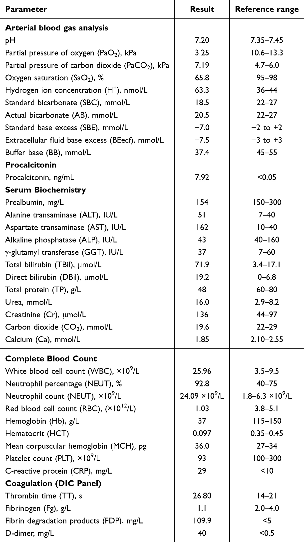 |
Table 1 Initial Laboratory Findings on Admission |
A positive direct antiglobulin test (lgG and C3d) with elevated indirect bilirubin initially suggested autoimmune hemolytic anemia, however, the subsequent development of thrombocytopenia, AKI, and laboratory evidence of hemolysis raised concern for an evolving TMA. Rhabdomyolysis (myoglobin >300 ng/mL) contributed to AKI, requiring intermittent hemodiafiltration. To support renal clearance and manage the evolving AKI, intermittent hemodiafiltration was initiated.
After starting renal replacement therapy (RRT), platelet count dropped to 21 x 109/L and fibrinogen to 1 g/L. While platelet consumption by the hemofilter was considered a possible contributor, heparin-induced thrombocytopenia (HIT) was also suspected, prompting immediate discontinuation of the heparin infusion. The suspicion was subsequently ruled out by a negative heparin-platelet factor 4 (PF4) antibody test. Given the suspicion of complement-mediated TMA, complement studies were obtained (C5b-9), and a renal biopsy was considered but contraindicated due to severe thrombocytopenia.
Peripheral blood smear examination (performed on day 8) revealed schistocytes, consistent with microangiopathic hemolytic anemia. Lactate dehydrogenase peaked at 449 IU/L, haptoglobin was <0.1 g/L, and indirect bilirubin was elevated (total bilirubin 71.9 μmol/L, direct bilirubin 19.2 μmol/L). Renal CT suggested focal infarction of both kidneys. Although the direct antiglobulin test (Coombs) was positive (IgG and C3d), indicating coexisting autoimmune hemolytic anemia, the presence of schistocytes, severe thrombocytopenia (nadir 21 × 109/L), and acute kidney injury with renal infarcts fulfilled the diagnostic criteria for complement-mediated TMA. Given the marked complement activation, exclusion of other TMA causes, and a positive response to eculizumab, a clinical diagnosis of complement-mediated thrombotic microangiopathy was made. In accordance with standard treatment guidelines, eculizumab (1.2 g) was administered promptly upon diagnosis. A favorable hematological response was evidenced by a rapid increase in the platelet count to 90 × 109/L by day 10. Given the profound immunosuppression from combined eculizumab and high-dose glucocorticoids, broad-spectrum empiric antimicrobial therapy (meropenem, vancomycin, caspofungin, plus selective digestive decontamination) was initiated. This regimen was broader than standard eculizumab-related prophylaxis (typically meningococcal vaccination or penicillin), justified by the patient’s critical illness, sepsis on admission, prolonged mechanical ventilation, and local ICU epidemiology of multidrug-resistant organisms. The regimen was de-escalated when subsequent cultures did not confirm invasive infection.
The second dose of eculizumab (1.2 g) was administered as scheduled on day 14. During the intervening period (day 8 to 14), the patient’s platelet count remained stable above 200 x 109/L, and C5b-9 steadily declined to 316.58 ng/mL. Notably, a divergent trend was observed in hs-cTnl decreased from peak 1701.8 pg/mL to 200 pg/mL. In contrast, the N-terminal pro-B-type natriuretic peptide (NT-proBNP) level demonstrated a concerning upward trend, rising to above 25,000 pg/mL, indicating significant cardiac strain or volume overload.
After the second eculizumab dose on day 14, a spontaneous breathing trial (SBT) was attempted on day 15 elicited tachycardia (heart rate 120–140 bpm) and tachypnea (respiratory rate 20–34 breaths/min), requiring reinstitution of mechanical ventilation. This failure coincided with a twofold rise in NT-proBNP, suggesting acute heart failure exacerbation. Meanwhile, infection markers improved (procalcitonin declined from 8.0 ng/mL to 3.5 ng/mL). By day 20, orthopnea was evident during morning rounds, and NT-proBNP remained markedly elevated at 26,258 pg/mL. These findings were attributed to persistent volume overload, prompting continued renal replacement therapy.
On day 22, the third scheduled dose of eculizumab (1.2 g) was administered. The following day, the patient developed acute tachycardia (130–140 bpm), and tachypnea (30–40 breaths/min) during morning rounds, with an elevated blood pressure of 148/92 mmHg. Bedside echocardiography revealed left heart enlargement, impaired left ventricular systolic function (left ventricular ejection fraction approximately 53%), mild mitral regurgitation, and a trivial pericardial effusion. Standard heart failure management was initiated, and RRT was intensified. Approximately two hours later, the heart rate decreased to 120–130 bpm and blood pressure stabilized at 140/90 mmHg.
Considering the recurrent, temporally associated episodes of acute heart failure decompensation following each eculizumab infusion, the clinically scheduled fourth dose on day 29 was withheld. Over the subsequent 30 days, the patient’s septic shock resolved, as evidenced by normalized serum lactate (<1 mmol/L), C-reactive protein (<10 mg//L), and procalcitonin (<1 ng/mL) levels. Spontaneous urine output improved to 1000–2000 mL per day independent of continuous renal replacement therapy, and serum creatinine stabilized between 300–450 μmol/L. Antihypertensive therapy was successfully de-escalated from intravenous nicardipine (60 mg via infusion pump) to oral amlodipine (5 mg twice daily) and terazosin (2 mg once daily).
The C5b-9 level was within normal range on day 24 (237.484 ng/mL) but rebounded to 349.844 ng/mL by day 40 after omission of the day 29 dose. Given the reproducible yet reversible nature of the cardiac decompensation episodes, a shared decision was made to administer a reduced dose of eculizumab (0.6 g) on day 41 to balance complement suppression against cardiovascular risk.
Approximately five hours after the infusion of the reduced-dose eculizumab (0.6 g), the patient developed acute respiratory distress characterized by chest tightness, shortness of breath, cough with clear sputum production. Vital signs deteriorated significantly: heart rate increased to 120–140 bpm, blood pressure rose to 160–170/100–110 mmHg, and peripheral oxygen saturation (SpO2) decreased markedly, fluctuating between 80% and 89%. Bibasilar moist rales were audible on pulmonary auscultation. Aggressive cardiorespiratory support including diuresis, vasodilators, and non-invasive ventilation was provided. Bedside thoracic ultrasound revealed a left-sided pleural effusion with a maximum depth of 75 mm. After 5 hours of intensive treatment, patient’s symptoms showed notable improvement with chest tightness and dyspnea alleviated, heart rate decreased to 125 bpm, blood pressure reduced to 132/80 mmHg, and SpO2 improved to 93% on oxygen support.
Following multidisciplinary consultation with nephrology and critical care services, the acute episodes of heart failure decompensation was considered to be temporally associated with eculizumab administration. Consequently, further eculizumab therapy was withheld for the remainder of the patient’s EICU stay. By day 43, thoracic ultrasound showed a significant reduction in the left pleural effusion (depth <23 mm), allowing for removal of the chest tube. Discontinuation of eculizumab coincided with a consistent downward trend in NT-proBNP levels from 35,000 pg/mL to 25,000 pg/mL, along with resolution of orthopnea and shortness of breath. However, after chest tube removal, the pleural effusion reaccumulated to a depth of 40 mm, necessitating ongoing renal replacement therapy for management of third-space fluid accumulation. On day 58, the patient was transferred to the nephrology department for long-term management. At the time of transfer, she remained alert and oriented with no recurrence of initial neurological symptoms or new deficits, her blood pressure was well controlled at 125/78 mmHg on oral antihypertensive therapy, and serum creatinine was 369 μmol/L with ongoing need for RRT due to unresolved AKI. Eculizumab had been permanently discontinued, and no further episodes of acute heart failure decompensation had occurred.
Discussion
While eculizumab has cemented its role as a life-saving therapy for aHUS with an ostensibly favorable safety profile centered on infection risk, our case highlights a possible cardiotoxic signal that has not been widely recognized. Of note, the diagnosis in this patient was clinically suspected complement-mediated thrombotic microangiopathy based on the presence of TMA, complement activation, and response to eculizumab. The temporal pattern of acute heart failure exacerbation – occurring within hours of infusion and resolving upon discontinuation – poses a formidable clinical challenge. It suggests that complement inhibition, particularly the blockade of the terminal C5 cascade, may exert unforeseen direct or indirect effects on cardiac function in susceptible individuals.
Uniqueness of the Case and Exploration of Potential Mechanisms
A review of the existing literature reveals that reports of significant cardiac adverse events directly attributable to eculizumab are exceptionally scarce. While eculizumab’s safety profile is well-documented,11,12 with a primary focus on infectious complications, our case raises the possibility of a previously underappreciated cardiac adverse effect. The uniqueness of our case is tripartite: firstly, the reproducible and life-threatening severity of the decompensation after each infusion; secondly, the notable temporal association suggested by the clear “on-off-rechallenge” phenomenon; and thirdly, the occurrence in a patient without significant pre-existing cardiac disease, which suggests eculizumab as a potential precipitant rather than solely an aggravating factor.
Given the lack of prior clinical reports, the pathophysiological mechanism underlying this phenomenon remains speculative but warrants thoughtful hypothesis generation. One hypothesis is that abrupt complement blockade might disrupt vascular tone regulation, leading to acute afterload mismatch that unmasks subclinical cardiac vulnerability. Alternatively, eculizumab could trigger a pro-inflammatory or vasoconstrictive cascade through altered anaphylatoxin (C5a) signaling.13 While these events can be challenging to differentiate from manifestations of the underlying disease (eg, malignant hypertension in complement-mediated TMA), their temporal association with drug administration suggests a potential triggering role. However, given the absence of direct mechanistic evidence (eg, endomyocardial biopsy or invasive hemodynamic monitoring), these hypotheses require further investigation. More importantly, we favor a “two-hit” model: the patient already had evidence of myocardial injury prior to eculizumab initiation (elevated hs-cTnI and NT-proBNP on admission, reflecting severe anemia, sepsis-related inflammation, and possible TMA-related microvascular ischemia).14 This pre-existing cardiac vulnerability—the “first hit”—set the stage for eculizumab to precipitate acute decompensation as the “second hit”. Therefore, our interpretation is not that eculizumab caused de novo heart failure, but rather that it may have triggered recurrent acute decompensations in a primed, susceptible myocardium. The rapid symptomatic improvement with heart failure management and the recurrence upon rechallenge argue against an irreversible cytotoxic effect and favor a functional, hemodynamic mechanism, such as the vasculopathy hypothesis.
Alternative Explanations for the Cardiac Decompensation
Although the temporal relationship is consistent with eculizumab as a potential trigger for acute heart failure decompensation, several potential alternative explanations warrant consideration. We acknowledge that multiple factors inherent to critical illness may have contributed to the cardiac decompensation in this complex clinical setting, including pre-existing myocardial injury, severe anemia, sepsis and systemic inflammation, possible TMA-related cardiac involvement, acute kidney injury, hypertension, and ICU-related hemodynamic instability. Attributing causality solely to eculizumab in such a complex scenario is not possible. Nevertheless, the reproducible temporal pattern warrants careful consideration of eculizumab as a potential trigger. First, volume overload is a common cause of acute decompensated heart failure in critically ill patients with AKI. However, in our case, heart failure episodes occurred within hours of eculizumab infusion, a time frame too short for significant positive fluid balance to develop. Moreover, the patient was receiving continuous RRT with strict ultrafiltration management, and fluid balance was closely monitored. Notably, cardiac deterioration consistently occurred within hours of eculizumab infusion, even when net negative fluid balance was achieved, making volume overload an unlikely primary cause. Second, the patient had no known history of hypertension, diabetes, coronary artery disease, or heart failure. Her admission cardiac biomarkers (hs-cTnl 1910 pg/mL, NT-proBNP 4284 pg/mL) were elevated, but these are nonspecific findings that can be explained by severe anemia (hemoglobin 39 g/L), systemic inflammation, sepsis, and TMA-related microvascular injury. An echocardiogram after the second eculizumab infusion showed left ventricular enlargement and mildly reduced ejection fraction, but no evidence of prior myocardial infarction or structural heart disease. The prompt reversibility of heart failure symptoms after stopping eculizumab, and their recurrence upon rechallenge at a reduced dose, argue against an underlying fixed cardiac pathology as the sole cause. Third, primary cardiac involvement of complement-mediated TMA via thrombotic microangiopathy is a plausible complication. Nevertheless, the temporal pattern – repeated cardiac decompensation immediately after each eculizumab dose, and resolution upon drug withholding – cannot be explained by the underlying complement-mediated TMA itself, which remained stable during this period. Collectively, while these factors may have contributed to a “primed” myocardium, none fully explains the reproducible, infusion-linked decompensation. Eculizumab appears to be a plausible trigger in the patient.
We also considered the potential role of severe hypertension, as recently highlighted by Allinovi et al in15 patients with TMA and malignant hypertension, where cardiac involvement can be significant yet reversible. In our case, blood pressure was mildly to moderately elevated (up to 160–170/100–110 mmHg) during heart failure episodes. However, hypertension alone cannot explain the reproducible, infusion-linked pattern of decompensation. Notably, when eculizumab was withheld, blood pressure was controlled with oral antihypertensives, and heart failure resolved despite persistent hypertensive susceptibility. Thus, while hypertension may have contributed to cardiac strain, it does not replace eculizumab as the acute trigger of decompensation.
In conclusion, while multiple competing factors were present, none fully explains the reproducible, infusion-linked pattern of decompensation. We interpret the temporal association as suggesting that eculizumab may have served as a potential trigger in a patient with pre-existing myocardial vulnerability, rather than the sole cause of heart failure.
Clinical Implications and Take-Home Message
This case carries several paramount implications for clinical practice. Firstly, it supports expanded vigilance among clinicians prescribing eculizumab. Beyond the well-known risk of meningococcal infections, acute heart failure may be considered a rare but possible adverse event to watch for. We recommend that baseline cardiac assessment, including echocardiography and measurement of BNP or NT-proBNP, should be considered prior to initiating eculizumab, particularly in patients with known cardiac risk factors or critical illness. Furthermore, close monitoring of vital signs, oxygen saturation, and clinical signs of pulmonary congestion during and for at least 24 hours after the infusion is crucial.
Secondly, this report highlights a potential therapeutic dilemma, challenging the conventional risk-benefit calculus for aHUS therapy. It suggests that the decision to use eculizumab may need to incorporate an assessment. In high-risk patients, strategies such as slower infusion rates, more prolonged post-infusion observation, or even prophylactic management of volume status might be prudent. Most importantly, our experience with dose reduction and subsequent recurrence suggests that simply reducing the dose may not mitigate the risk in susceptible individuals. This necessitates a highly individualized treatment approach, where the imperative to suppress complement is constantly weighted against the threat of iatrogenic heart failure.
Finally, this case reveals a critical gap in our understanding of the extra-renal effects of terminal complement blockade. It poses the fundamental question: does complement inhibition exert unforeseen, direct hemodynamic consequences on the cardiopulmonary system? This observation should stimulate further research into the physiological role of complement in maintaining vascular homeostasis and cardiac function. From a practical standpoint, it also underscores the urgent need to establish an international registry for tracking severe adverse events associated with complement inhibitors, which would help identify risk factors and incidence rates for complications like the one we describe.
However, our report still has several limitations that should be acknowledged. Firstly, given that this is a single-case report with multiple confounding factors inherent to critical illness, we cannot establish causation; rather, we present this observation to generate hypothesis and alert clinicians to a possible adverse effect that deserves further study. Secondly, the definitive pathophysiological mechanism linking eculizumab to heart failure remains speculative, as we lacked more invasive hemodynamic monitoring or endomyocardial biopsy tissue to investigate complement deposition or inflammatory changes at the cardiac level. Thirdly, the patient’s critical illness, complicated by sepsis and coagulopathy, precluded a percutaneous renal biopsy, which would have provided definitive histopathological confirmation of thrombotic microangiopathy and allowed for a more detailed comparison with known renal forms of aHUS. Fourthly, the relatively short in-hospital follow-up period limits our understanding of the long-term cardiac outcomes after discontinuation of eculizumab and the natural history of aHUS management in this complex situation. Fifthly, genetic analysis of complement factors (eg, CFH, CFI, C3, etc.) was not performed, which would have been required for a genetically confirmed diagnosis of aHUS. Therefore, our case is best characterized as a clinically suspected complement-mediated thrombotic microangiopathy with features consistent with aHUS, rather than a genetically proven aHUS. Finally, the complex ICU setting involved multiple coexisting factors—including severe anemia, sepsis, acute kidney injury, hypertension, and hemodynamic instability—which may have independently or synergistically contributed to the cardiac decompensation, further limiting our ability to attribute causality definitively to eculizumab.
Conclusion
In conclusion, this case suggests a possible association between eculizumab and acute decompensated heart failure. The rapid onset after infusion, reversibility upon discontinuation, and recurrence on rechallenge are consistent with a potential triggering role for eculizumab. This finding expands the known safety profile of eculizumab beyond infectious risks and highlights a possible cardiotoxic signal that warrants further investigation. Clinicians should closely monitor cardiac function during and after eculizumab administration, especially in critically ill patients. Further research is needed to elucidate the exact mechanism and identify at-risk individuals.
Abbreviations
aHUS, Atypical hemolytic uremic syndrome; TMA, thrombotic microangiopathy; STEC-HUS, Shiga toxin-producing E. coli-hemolytic uremic syndrome; AP, alternative pathway; C5b-9, membrane attack complex; CT, computed tomography; AIHA, autoimmune hemolytic anemia; aPCC, activated prothrombin complex concentrate; IVIG, intravenous immunoglobulin; AKI, acute kidney injury; HIT, heparin-induced thrombocytopenia; PF4, heparin-platelet factor 4; SBT, spontaneous breathing trial; TAPSE, tricuspid annular plane systolic excursion; SpO2, peripheral oxygen saturation.
Ethics Approval and Consent to Participate
The Ethics Committee of Ruijin Hospital, Shanghai Jiao Tong University has confirmed that institutional ethical approval was waived for this case report. All clinical procedures performed were in accordance with the ethical principles of the Helsinki Declaration (1975, revised 2024). Written informed consent was obtained from the patient for publication of all potentially identifiable clinical data and images included in this article.
Author Contributions
All authors made a significant contribution to the work reported, whether that is in the conception, study design, execution, acquisition of data, analysis and interpretation, or in all these areas; took part in drafting, revising or critically reviewing the article; gave final approval of the version to be published; have agreed on the journal to which the article has been submitted; and agreed to be accountable for all aspects of the work.
Disclosure
Jiayang Huang and Tongtian Ni are co-first authors for this study. The authors report no conflicts of interest in this work.
References
1. Fakhouri F, Schwotzer N, Frémeaux-Bacchi V. How I diagnose and treat atypical hemolytic uremic syndrome. Blood. 2023;141(9):984–8. doi:10.1182/blood.2022017860
2. Tsai HM. Atypical hemolytic uremic syndrome: beyond hemolysis and uremia. Am J Med. 2019;132(2):161–167. doi:10.1016/j.amjmed.2018.08.011
3. Jokiranta TS. HUS and atypical HUS. Blood. 2017;129(21):2847–2856. doi:10.1182/blood-2016-11-709865
4. Doshi K, Yusuf A, Licht C, et al. Extrarenal manifestations of atypical hemolytic uremic syndrome: a systematic review and meta-analysis. Pediatr Res. 2024;98(2):491–497. doi:10.1038/s41390-024-03771-7
5. Bogdan RG, Anderco P, Ichim C, et al. Atypical Hemolytic Uremic Syndrome: a Review of Complement Dysregulation, Genetic Susceptibility and Multiorgan Involvement. J Clin Med. 2025;14(7):2527. doi:10.3390/jcm14072527
6. Bedi GN, Sontakke T, Mapari SA, Sawant R, Reddy N. Deciphering complexity: atypical hemolytic uremic syndrome unraveled in the wake of elective hip arthroplasty. Cureus. 2024;16(9):e68690. doi:10.7759/cureus.68690
7. Campistol JM, Arias M, Ariceta G, et al. An update for atypical haemolytic uraemic syndrome: diagnosis and treatment. A consensus document. Nefrologia. 2015;35(5):421–447. doi:10.1016/j.nefro.2015.07.005
8. de Souza RM, Correa BHM, Melo PHM, et al. The treatment of atypical hemolytic uremic syndrome with eculizumab in pediatric patients: a systematic review. Pediatr Nephrol. 2023;38(1):61–75. doi:10.1007/s00467-022-05683-2
9. Ter Avest M, Bouwmeester RN, Duineveld C, et al. Proposal for individualized dosing of eculizumab in atypical haemolytic uraemic syndrome: patient friendly and cost-effective. Nephrol Dial Transplant. 2023;38(2):362–371. doi:10.1093/ndt/gfac056
10. Brocklebank V, Walsh PR, Smith-Jackson K, et al. Atypical hemolytic uremic syndrome in the era of terminal complement inhibition: an observational cohort study. Blood. 2023;142(16):1371–1386. doi:10.1182/blood.2022018833
11. Lee H, Kang E, Kang HG, et al. Consensus regarding diagnosis and management of atypical hemolytic uremic syndrome. Korean J Intern Med. 2020;35(1):25–40. doi:10.3904/kjim.2019.388
12. Avila Bernabeu AI, Cavero Escribano T, Cao Vilarino M. Atypical hemolytic uremic syndrome: new challenges in the complement blockage era. Nephron. 2020;144(11):537–549. doi:10.1159/000508920
13. Devresse A, Aydin S, Le Quintrec M, et al. Complement activation and effect of eculizumab in scleroderma renal crisis. Medicine. 2016;95(30):e4459. doi:10.1097/MD.0000000000004459
14. West EE, Woodruff T, Fremeaux-Bacchi V, Kemper C. Complement in human disease: approved and up-and-coming therapeutics. Lancet. 2024;403(10424):392–405. doi:10.1016/S0140-6736(23)01524-6
15. Allinovi M, Menale S, Querin V, et al. Potentially reversible severe cardiac involvement in thrombotic microangiopathies with malignant hypertension. J Nephrol. 2025;38(6):1655–1661. doi:10.1007/s40620-025-02334-1
 © 2026 The Author(s). This work is published and licensed by Dove Medical Press Limited. The
full terms of this license are available at https://www.dovepress.com/terms
and incorporate the Creative Commons Attribution
- Non Commercial (unported, 4.0) License.
By accessing the work you hereby accept the Terms. Non-commercial uses of the work are permitted
without any further permission from Dove Medical Press Limited, provided the work is properly
attributed. For permission for commercial use of this work, please see paragraphs 4.2 and 5 of our Terms.
© 2026 The Author(s). This work is published and licensed by Dove Medical Press Limited. The
full terms of this license are available at https://www.dovepress.com/terms
and incorporate the Creative Commons Attribution
- Non Commercial (unported, 4.0) License.
By accessing the work you hereby accept the Terms. Non-commercial uses of the work are permitted
without any further permission from Dove Medical Press Limited, provided the work is properly
attributed. For permission for commercial use of this work, please see paragraphs 4.2 and 5 of our Terms.
Recommended articles

Atypical Hemolytic-Uremic Syndrome: Genetic Basis, Clinical Manifestations, and a Multidisciplinary Approach to Management
Yerigeri K, Kadatane S, Mongan K, Boyer O, Burke LL, Sethi SK, Licht C, Raina R
Journal of Multidisciplinary Healthcare 2023, 16:2233-2249
Published Date: 4 August 2023

Misdiagnosis of Chronic Heart Failure in Patients with Type 2 Diabetes Mellitus in Primary Care: A Report of Two Cases and Literature Review


Yeshniyazov N, Posokhov IN, Medovchshikov VV, Kurmanalina G, Sartayeva A
Vascular Health and Risk Management 2024, 20:479-485
Published Date: 3 November 2024
Thrombotic Thrombocytopenic Purpura During Anti-Tuberculosis Therapy: A Case Report and Literature Review
Tang W, Xue H, He L, Zhang H, Lin S, Fang Z
Infection and Drug Resistance 2025, 18:6051-6058
Published Date: 20 November 2025

Severe Mitral Stenosis in Term Pregnancy Presenting During Labor in a Resource-Limited Setting: A Case Report from Borama, Awdal Region of Somalia

Jama AM, Osman AA, Deria HM, Mousa MM, Jama AM, Ali AO
International Medical Case Reports Journal 2026, 19:564292
Published Date: 26 February 2026