")
Back to Journals » Infection and Drug Resistance » Volume 16
Drug Susceptibility Test and Analysis of Quinolone Resistance Genes in Mycoplasma hyopneumoniae Vaccine Strains and Field Isolates from China
Received 7 February 2023
Accepted for publication 1 April 2023
Published 8 April 2023 Volume 2023:16 Pages 2075—2087
DOI https://doi.org/10.2147/IDR.S407579
Checked for plagiarism Yes
Review by Single anonymous peer review
Peer reviewer comments 2
Editor who approved publication: Professor Suresh Antony
Yapei Rui, Gang Qiu
College of Animal Science and Veterinary Medicine, Xinyang Agriculture and Forestry University, Xinyang, Henan, People’s Republic of China
Correspondence: Gang Qiu, Email [email protected]
Background: Enrofloxacin is a commonly used animal-specific drug in veterinary clinics. However, this drug has no epidemiological cutoff values (ECVs/ECOFFs) for Mycoplasma hyopneumoniae in CLSI and EUCAST. Defining the epidemiological cutoff values (ECOFFs) of enrofloxacine to Mycoplasma hyopneumoniae (M. hyo) can inform an early detection of bacterial resistance to better manage the resistance prevention and also help in establishing drug resistance breakpoints;.
Methods: We determined the susceptibility breakpoint of M. hyo to enrofloxacin by the American Clinical and Laboratory Standards Institute (CLSI) standard method based on the PCR of vaccine strains and wild strains drug resistance genes;.
Results: Eighty strains of M.hyo isolated in Tibet were moderately sensitive (S) to tetracycline, florfenicol, spiramycin, erythromycin thiocyanate, tilmicosin, tiamulin, lincomycin, clindamycin, ofloxacin, enrofloxacin, gentamicin, amikacin, with MICs below 0.5 μg/mL. For vaccine 168L, RM48, and J strains, the susceptibility to the same antibacterial drugs was lower compared to the Tibetan isolates. The resistance of J strain to erythromycin thiocyanate was confirmed. Gene point mutation was confirmed in Quinolone Resistance Determining Regions (QRDR) of HNSH strain Topoisomerase IV subunit A, this finding is compared with the sequencing results of 168L strain reference sequence (Accession number: CP003131). Arg-Lys amino acid mutation (G921A and G1179A) was confirmed for the increase of MIC value involved in M.hyo to enrofloxacine;.
Conclusion: The cut-off value of M.hyo to enrofloxacin was set as 1 μg/mLby ECOFFinder XL 2010 V2.1.
Keywords: Mycoplasma hyopneumoniae, drug resistance, Tibetan pigs, MIC, enrofloxacin
Introduction
Although imported and domestic vaccines prevented M. hyo infection in P. R. China, there is no clinical vaccine that can 100% prevent and control M. hyo infection in Tibetan pigs, possibly because of the gene variation in this species. According to reports from China and abroad, antimicrobial drugs have always played an irreplaceable role in preventing and treating swine enzootic pneumonia.1–5 High MIC values of fluoroquinolones (enrofloxacin 2.5 μg/mL; marbofloxacin 5 μg/mL) were observed against one M.hyo strain (MycSu17) by Felde, O. et al.6 Due to the long-term low-dose prophylaxis and high-dose medications,7 the resistance of Mycoplasmas (including M. hyo) to antimicrobials is becoming more widespread,8 bringing severe challenges to the prevention and control of M.hyo in P. R.China.
Epidemiological survey results have demonstrated the prevalence of Mycoplasma pneumonia of swine (MPS) in Tibetan pig herds to be 20.48% to 50%9 in the Qinghai-Tibet Plateau. MPS reduced the growth rate of Tibetan pigs, causing considerable losses to the Tibetan pig-breeding industry. However, there are relatively few basic data on drug susceptibility of M.hyo in Tibetan pigs by searching public databases.
The resistance of M.hyo to fluoroquinolones is becoming more widespread in China, with fluoroquinolones treatment failures occurring more frequently in veterinary clinics. Quinolones target DNA gyrase, an essential enzyme in bacterial replication that generates negative and positive supercoils in DNA by transiently introducing double-stranded breaks in an ATP-dependent reaction. DNA gyrase is a heterotetramer consisting of two A and two B subunits, respectively.10,11 The drug resistance mechanism includes a quinolone resistance-determining region (QRDR) target gene mutation,12–16 plasmid-mediated quinolone resistance (PMQR) gene,17 the efflux pump,18 transferable multidrug resistance element, and other not bacteria to be discovered.19 China is strengthening the monitoring of the drug resistance of animal-derived microorganisms, especially bacteria; the monitoring needs to formulate relevant drug resistance determination standards.
The International Research Programme on Comparative Mycoplasmology (IRPCM) proposed as early as 2000 the antibacterial drug susceptibility testing on animal mycoplasmas.20 However, only a few animal mycoplasmas have been tested for antimicrobial susceptibility due to various factors including the different media and the protracted cultivation process of animal mycoplasmas. A unified susceptibility breakpoint standard has not yet been established.21 At present, many countries have established systematic drug resistance detection and judgment standards. The American Clinical and Laboratory Standards Institute (CLSI) and the European Committee on Drug Susceptibility Testing (EUCAST) standards are the most widely used. Criteria for determining drug resistance are divided into three types: microbial, pharmacodynamic, and clinical breakpoints.22,23 EUCAST has replaced the term microbiological breakpoints with epidemiological cut-off values (ECOFFs) or wild-type cut-off values (COWT) to prevent confusion about the different meanings of breakpoints. The academic and clinical breakpoints are standardized as pharmacodynamic cut-off and clinical cut-off values. Wild-type strains are those that do not carry any mechanisms of acquired resistance. ECOFFs or COWT represent the minimum inhibitory concentration (MIC value) used to distinguish wild-type strains from acquired or selective drug-resistant strains. The epidemiological monitoring of bacteria can play a prompting role in the early development of drug resistance and is of great significance for optimizing clinical drug selection and slowing down the emergence of drug-resistant bacteria.
There are relatively few studies in China and abroad on determining criteria of drug resistance for animal-specific drugs. Enrofloxacin is a commonly used animal-specific drug in veterinary clinics, but there are no epidemiological cut-off values (ECVs/ECOFFs) for M.hyo in CLSI and EUCAST. Establishing ECOFFs of this drug can serve as a warning for the early development of antimicrobial resistance,24 and provide reference to initiating the formulation of drug resistance prevention and control, and establishing the final drug resistance breakpoints.25–27
It is urgently necessary to test the drug susceptibility of M.hyo and understand the occurrence and mechanism of resistance to reduce the resulting losses and improve the targeted and clinical effects of the prevention and treatment of MPS of Tibetan pigs. However, not even the reference guide of CLSI that sets standards for antimicrobial susceptibility testing provides cut-off information on Mycoplasma species of animals. This makes it difficult to evaluate results and obtain a consensus among researchers. Therefore, the current test is based on preliminary exploratory trials research methods (Reference CLSI guides), and discoloration is the primary basis for judgment.
Drugs used in Tibetan pigs have been selected for in vitro drug susceptibility testing. These drugs include macrolides, lincosamides, quinolones, tetracyclines, florfenicol, and traditional Chinese medicines.
Materials and Methods
Materials
Test Strains
All methods were performed in accordance with the relevant guidelines and regulations. Each pig was euthanized by intramuscular injection of xylazine hydrochloride (Changsha Better Biotechnology Research Institute Co., Ltd., 100 mg/mL) and Pentobarbital sodium (Shanghai Chemical Reagent Purchasing Station of Chinese pharmaceutical company, Each pig was injected 3% Pentobarbital sodium normal saline solution at a dose of 0.2 mL/kg). Then, each animal was intravenously injected with 50 mL of potassium chloride (Tianjin Zhiyuan Chemical Reagent Co., Ltd. 40%). This experiment was performed according to the AVMA (the American Veterinary Medical Association) Guidelines for the Euthanasia of Animals (2020 Edition) to minimize pain in the animals. All animal experiments have been approved by Animal Ethics and Welfare Committee (AEWC) of Xinyang Agriculture and Forestry University with the approval number of AEWC–2021011803.
Lung typical lesions were taken from the Tibetan pigs in Nyingchi, Tibet, P.R.China. Eighty M.hyo strains from Tibetan pigs, labeled TB1 (a-j), TB2 (a-j), TB3 (a-j), TB4 (a-j), TB5 (a-j), TB6 (a-j), TB7 (a-j) and TB8 (a-j) were isolated, purified, identified and expanded in the Tibetan Plateau Animal Disease Epidemiology Laboratory and Veterinary Pharmacology Laboratory. The isolation, purification, identification method of M.hyo is consistent with that of Qiu G (2019).28 Eight of the isolates were isolated in 2018 and the rest were isolated from March to May 2022. The strains were stored at −80 °C in the Laboratory of Veterinary Pharmacology, Tibet Agricultural and Animal Husbandry College, and Huazhong Agricultural University Laboratory. Other forty strains were obtained as indicated: RM48 (CVCC4049) (a-j) from China Veterinary Microbiological Culture Collection Management Center; J strain (CVCC359) (a-j) from China Veterinary Microbiological Culture Collection Management Center; and 168L strains (batch number: 20181101) (a-j) from Chuan Haojia, Qianyuanhao Biological Co., Ltd., DJ-166 strains (batch number: 20210312) (a-j) from China Animal Husbandry Industry Co., Ltd. TB1 (a-j), TB2 (a-j), TB3 (a-j), TB4 (a-j), TB5 (a-j), TB6 (a-j), TB7 (a-j), TB8 (a-j), HNSH, HNPQ, HNHC, HNNY, AHFY, and HNZZ strains are wild strains. The vaccine strains were stored at −20°C and −80°C, recently used samples are kept at −20 °C and the rest at −80 °C, within the effective period. The storage time is less than six months. The preservation conditions of wild strain and vaccine strain are consistent. The strains source and date of isolation is shown in Table 1.
 |
Table 1 The Strains Source and Date of Isolation |
Test Drugs
The current drugs used in the treatment of porcine respiratory diseases syndrome in Nyingchi, Tibet, are as follows:
tetracycline, oxytetracycline, chlortetracycline, doxycycline, florfenicol, tylosin tartrate, spiramycin, kitasamycin, erythromycin thiocyanate, tilmicosin, spectinomycin, tiamulin, lincomycin, clindamycin, ofloxacin, ciprofloxacin, enrofloxacin, ampicillin, streptomycin, gentamicin, amikacin, berberine reference material, nanosilver, mequindox, quinoquinone, cyadox, patchouli oil, tea tree oil, garlic oil, oregano oil, and cinnamic aldehyde. The above 31 drugs are commercially available as raw powders oil, drugs, or standard reference materials. Other drugs and reagents used in the tests include bacitracin and polymyxin b. All drugs, reagents, and standard reference materials are within the validity period. Detailed information was provided in Supplementary Material 1.
Methods
Preparation of Test Bacteria Suspension
One hundred and forty wild strains of M. hyo (TB1 (a-j), TB2 (a-j), TB3 (a-j), TB4 (a-j), TB5 (a-j), TB6 (a-j), TB7 (a-j), TB8 (a-j), HNPQ (a-j), HNSH (a-j), HNHC (a-j), HNNY (a-j), HNZZ (a-j), AHFY (a-j)), isolated from fourteen different pig farms (Ten strains of M.hyo from ten different pigs per farm) in China and stored in the Central Laboratory of Xinyang Agriculture and Forestry University, were inoculated in a modified KM2 liquid medium (the modified Friis medium) at a ratio of 1:10. Resuscitation was performed and then aseptically diluted to 106 CCU/mL as a bacterial test suspension. Isolation and culture method of M.hyo refer to Chinese Patent CN201710782410.X.
Farm pigs in Henan Province of P.R.China that failed enrofloxacin treatment were necropsied, the lungs were collected aseptically (Supplementary Figure 1), and M.hyo was isolated, purified, identified, and tested for drug susceptibility and drug resistance. The HNSH strain is field strain isolated from the lungs of pigs that failed enrofloxacin treatment (Figure 1); the presence of M.hyo was confirmed by throat swabs PCR testing.
 |
Figure 1 PCR Results of 16 sRNA gene from M. hyo. Notes: 1 = TB1 (field strain from Tibetan pigs), 2 = TB2 (field strain from Tibetan pigs), 3 = TB3 (field strain from Tibetan pigs), 4 = TB4 (field strain from Tibetan pigs), 5 = TB5 (field strain from Tibetan pigs), 6 = TB6 (field strain from Tibetan pigs), 7 = TB7 (field strain from Tibetan pigs), 8 = TB8 (field strain from Tibetan pigs), 9 = 168L (vaccine strain), 10 = RM48 (vaccine strain), 11 = 5722 (vaccine strain), 12 = HNPQ (field strain), 13 = HNSH (field strain), 14 = HNHC (field strain), 15 = HNNY (field strain), 16 = AHFY (field strain), 17 = HNZZ (field strain), 18=DJ-166 (vaccine strain), P = Positive Control J strain (CVCC359). |
Preparation of Drug Solution
Thirty-one drugs commonly used for the treatment of M.hyo were selected, doses were calculated according to the drug content and drug weight, and each was formulated using a modified KM2 liquid medium (the modified Friis medium) at a concentration of 1 mg/mL (2x the highest concentration of the drug) or 0.01 μL/mL (for plant essential oils).
Test Plan
Measurement Method
The specific method of MIC determination is consistent with CLSI.29 The susceptibility test was performed in a 96-well cell culture plate using a micro broth dilution method. Table 2 gives details of the sample addition method. The first column is the negative control (200 μL of KM2 medium (modified Friis medium)); the second column is the drug control (100 μL of modified KM2 medium + 100 μL of modified KM2 medium containing twice the highest concentration of drug), and the third to eleventh columns are drug gradients. In culture, each drug was tested in parallel; the 12th column shows the positive control (100 μL of modified KM2 medium + 100 μL of bacterial suspension). The detailed information is provided in Supplementary Table 1.
 |
Table 2 Adding Samples Method of Drug Sensitivity Test of M. Hyo |
Operating Procedure
0.9 mL of culture medium was placed into each tube, and 0.1 mL of the corresponding positive bacteria solution was added to each tube, and mixed thoroughly; 0.1 mL was drawn into the second tube. In this order, the solution was diluted 10−1 to 10−12 and incubated at 37 °C. Changes in the culture fluid color were used to determine the results; the highest dilution at which color changes determines the CCU. For example, When the solution was diluted to 10−1~10−6, the color changed, the solution was diluted to 10,−7 the color did not change, then the test result is CCU = 106, that is, Mh still grows when the measured bacterial liquid is diluted to 10−6.
The modified KM2 medium (200 μL) was added to the first column of the plate; 100 μL of modified KM2 medium was added to each well of columns 2–12, and 100 μL of modified KM2 base medium containing 2-times the highest concentration of the drug was added to the second and third columns. The third well was mixed thoroughly, and 100 μL was drawn into the fourth well. Then, the fourth well was mixed thoroughly, 100 μL was drawn into the fifth well, etc. to dilute wells serially to the 11th well. Then, 100 μL of the bacterial suspension was aspirated after mixing and added to columns 3–12, gently tapped the plate to mix them, making two rows of each treatment (parallel); plate was covered and cultured in a constant temperature incubator with 5% CO2 at 37 °C for 7–10 days. Then, the color changing values (CCUs) of M.hyo were determined.
When the color of the positive control wells turned yellow, and the color of the negative control wells and the drug control wells did not change, and the result of two consecutive naked-eyes observations was same, the lowest concentration of drug in the wells that did not change color was the lowest concentration of drug (MIC) that completely inhibited the growth of M. hyo. According to the result of the preliminary experiment, the observation results of the 7th day were used to judge, as shown in Figure 2.
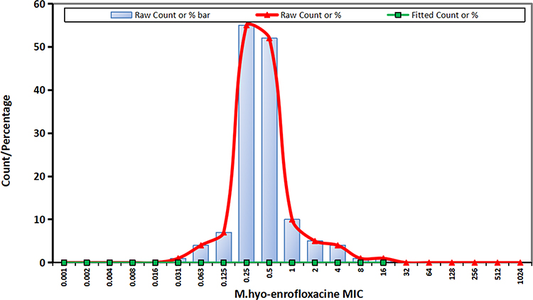 |
Figure 2 MIC distribution of M.hyo strains to enrofloxacin. |
According to the data and preliminary experimental results, the following primers were used to detect M.hyo specific genes and quinolone-related resistance genes. The primer sequence is shown in Table 3.
 |
Table 3 Primers Used to Detect M.hyo and Quinolone-Related Resistance Genes |
For amplification of the 16S rDNA, the PCR conditions included 34 cycles of initial denaturation at 95°C for 5 min, denaturation at 94°C for 1 min, annealing at 60°C for 0.5 min, and extension at 72°C for 7 min. After amplification, 5µL amplicon was electrophoreted in 1% agarose gel (Goldview Nucleic Acid Gel Stain 2µL) at 0.5×TAE at 120 V for 60 min. For amplification of the P36 gene, the PCR conditions included 34 cycles of initial denaturation at 95°C for 5 min, denaturation at 95°C for 0.5 min, annealing at 42°C for 0.5 min, and extension at 72°C for 8 min. After amplification, 5µL amplicon was electrophoreted in 1% agarose gel (Goldview Nucleic Acid Gel Stain 2µL) at 0.5×TAE at 120 V for 60 min. The PCR products were sequenced by the shotgun method. The Method of Amplification of gyrA, gyrB, parE and parC gene and follow-up experiment steps are consistent with Le Carrou J et al.30
Minimum Inhibitory Concentration Data Collection
Web of Science, China National Knowledge Infrastructure, and Wanfang databases were searched manually to obtain the minimum inhibitory concentration (MIC) data for different entities, and drug susceptibility tests and resistance assays were conducted on M.hyo strains.
MIC Data Correction
The MIC distribution data obtained in this study originated from different sources and needed to be processed with the same standard. After merging the data of the same drug, by collecting relevant literature on the quinolone-related drug resistance mechanism and epidemiological cut-off values of M. hyo, the data were revised with reference to the distribution range of wild-type M.hyo MIC; MICs of non-wild-type M.hyo strains were excluded.
ECOFFinder Fitting MIC Distribution Map and Determining Epidemiological Cut-off Values
The data were fitted using the integrated statistical software ECOFFinder XL 2010 V2.1 produced by J. Turnidge et al.
Linear regression analysis was used to fit the cumulative distribution of MIC to obtain the MIC distribution map and to determine the epidemiological cut-off value at different confidence intervals within the best fitting range.
Determination of Drug Resistance Rate and Collection of Related Literature
The epidemiological cut-off value with a confidence interval of 95% was used for the drug resistance breakpoint.
The MIC distribution of the strains was observed, and the drug resistance rate was calculated. All strains were divided into wild strains and clinical isolates based on the obtained epidemiological cut-off values and the proportion of non-wild strains to all strains calculated, giving the resistance ratio of M.hyo to the drug.
The pure culture and PCR product of M.hyo HNSH strain were sent to Shanghai Sangon Bioengineering Co., Ltd. for sequencing and used for retrieval-specific genes and quinolone-related resistance genes.
Criteria for Color Change of Drug Susceptibility Test
If M. hyo strain could grow and discolor the medium (red to yellow), the medium eventually turns yellow, indicating resistance to drug. If M. hyo strain could not grow and discolor the medium, the medium eventually appears red, indicating sensitive to enrofloxacin. Same with other drugs.
CCU=10−6, CCU is the highest dilution when the medium changes from red to yellow, then the bacterial solution concentration is calculated as CCU/mL.
The colors used to determine whether M. hyo is growing, with yellow indicating growth (resistance to drugs) and red indicating no growth (susceptibility to drugs). During the culture period, the color change was recorded every 12 hours. The color of the modified KM2 medium changed from red to yellow, indicating the growth of M. hyo.
Results
M. h field strains and vaccine strains were identified by PCR of 16 sRNA gene as shown in Figure 1.
Observation of Test results
On the 3rd day of the experiment, the MIC values of the 31 antibacterial drugs and 80 strains of M.hyo from Tibetan pigs were found not to have changed. After the 7th day of culture, the color of the 96-well culture plate used to determine the drug susceptibility test results were as follows:
Negative drug control wells did not discolor (red), the positive control wells discolored (yellow), and wells with the gradient of drug concentration from high to low gradually changed from red to yellow.
Drug Susceptibility Test Result of M. hyo Isolated from Swine
Eighty isolates from Tibet were highly sensitive to diterpenes, macrolides, tetracyclines, and quinolones (S), with MICs lower than 0.5 μg/mL; sensitivity to lincosamides was moderate, with MIC of 0.13 to 0.3 μg/mL, low sensitivity to quinocetone and ampicillin, MIC ≥ 16 μg/mL, high sensitivity to enrofloxacine, MIC50 = 0.25 μg/mL, and higher MIC for berberine than common chemical drugs MIC ≥ 32 μg/mL. The MIC of nano-silver was 0.5 ~ 1 μg/mL, considered sensitive. For the vaccine strains 168L strains, RM48 strains, and avirulent strain J strains, the susceptibility to the same antibacterial agents was lower than that of the Tibet isolates; J strains were low sensitivity to erythromycin thiocyanate more frequently. The MIC50 of HNSH strain to enrofloxacin was 16 μg/mL. To enrofloxacine, the MIC50 of HNPQ strain was 0.25μg/mL, MIC90=0.5μg/mL, MIC range was between 0.008μg/mL and 8μg/mL. The MIC50 of HNHC strain was 0.25μg/mL, MIC90=0.5μg/mL, MIC range was between 0.008μg/mL and 4μg/mL. The MIC50 of HNNY strain was 0.25μg/mL, MIC90=0.5μg/mL, MIC range was between 0.008μg/mL and 4μg/mL. The MIC50 of AHFY, strain was 0.25μg/mL, MIC90=0.5μg/mL, MIC range was between 0.008μg/mL and 4μg/mL. The MIC50 of HNZZ strain was 0.25μg/mL, MIC90=0.5μg/mL, MIC range was between 0.008μg/mL and 8μg/mL.
Drug susceptibility test resultof M.hyo isolated from swine is shown in Table 4.
 |
Table 4 MICs of Antimicrobial Agents Used Against Field Strains from Tibetan Pigs and Vaccine Strain 168L Strain, RM48 Strain and Type Strain J Strain of M. Hyo, Determined by a Serial Broth Dilution Method |
MIC distribution of M.hyo strains to enrofloxacin is shown in Figure 2.
Identification of DNA topoisomerase 4 subunit A from M.hyo HNSH strain is shown inFigure 3.
 |
Figure 3 PCR results of DNA topoisomerase 4 subunit A from M.hyo HNSH strain. 1= parC. 2= parE. 3=16sRNA gene. 4= gyrA. 5= gyrB. |
PCR results of genes (16sRNA gene, P36 gene, parC, parE, gyrA, gyrB) from M.hyo strains are shown in Figure 4.
 |
Figure 4 PCR results of genes (16sRNA gene, P36 gene, parC, parE, gyrA, gyrB) from M.hyo strains. Vaccine strains: RM48 strain, J strain, 168L strain, DJ-166 strain. Field strains: HNPQ strain, HNSH strain, AHFY strain. Conserved genes for identification of M.hyo:16sRNA gene, P36 gene. QRDR genes: gyrA, gyrB, parE and parC. Notes: A line: M = DL 2000 DAN Marker, A 1 = HNPQ strain 16sRNA gene, A 2 = J strain 16sRNA gene, A 3 = HNPQ strain P36 gene, A 4 = HNPQ strain 16sRNA gene, A 5=DJ-166 strain 16sRNA gene, A 6 = HNPQ strain gyrA gene, A 7 = AHFY strain P36 gene, A 8 = J strain parE gene, A 9 = HNSH strain parC gene, A 10=DJ-166 strain parC gene, A 11 = AHFY strain 16sRNA gene, A 12 = RM48 strain P36 gene, A 13 = RM48 strain gyrB gene, A 14 = 168L strain gyrB gene, A 15 = Negative control sample (KM2 culture medium), A 16 = 168L strain p36 gene, A 17 = J strain gyrB gene, A 18 = RM48 strain parC gene. B line: M = DL 2000 DAN Marker, B 1 = 168L strain parC gene, B 2 = RM48 strain 16sRNA gene, B 3 = AHFY strain P36 gene, B 4 = J strain P36 gene, B 5 = 168L strain P36 gene, B 6 = RM48 strain parC gene, B 7 = HNSH strain P36 gene, B 8 = AHFY strain parC gene, B 9 = HNSH strain 16sRNA gene, B 10 = 168L strain 16sRNA gene, B 11 = J strain p36 gene, B 12 = AHFY strain p36 gene, B 13 = AHFY strain gyrA gene, B 14 = 168L strain 16sRNA gene, B 15 = 168L strain 16sRNA gene, B 16 = 168L strain parC gene, B 17 = J train parC gene, B 18 = HNSH strain parC gene. |
The following gene mutations in the quinolone resistance-determining region were found by sequencing of Topoisomerase IV subunit A in the HNSH strain of M.hyo as shown in Table 5.
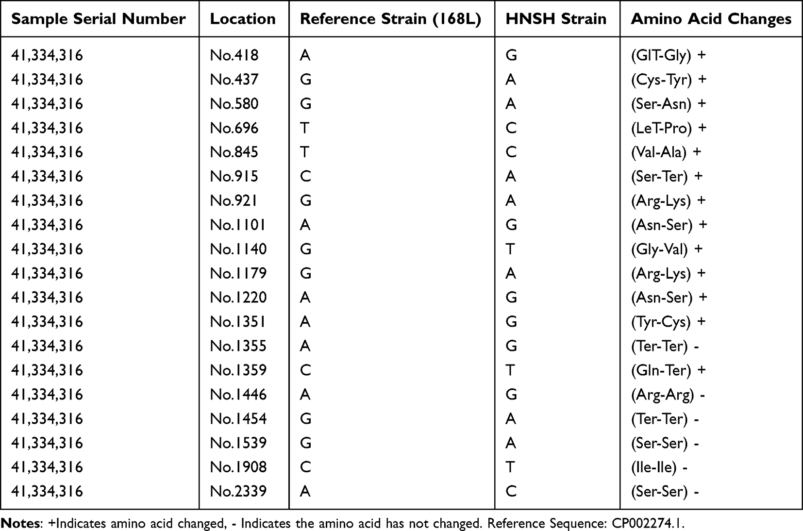 |
Table 5 Resistance-Associated Nucleotides Mutations and Amino Acid Changes in M.hyo HNSH Strain Topoisomerase IV Subunit a |
When compared the QRDRs region nucleotide sequence of HNSH strain with the reference strain CP002274.1, the results showed that the amplified sequence had multiple-mutation sites. Subsequently, we compared amino acid sequence of the gyrA, gyrB, parE and parC in HNSH strain QRDRs with the standard strain CP002274.1, specific gene polymorphisms were observed. After excluding the interference of species-specific or serotype-specific polymorphisms, we summarized the corresponding amino acid changes in QRDRs of the tested strains, as shown in Table 5. The MIC value of enrofloxacine corresponding to the above-mentioned QRDRs mutant strains was significantly higher than other strains without mutation in the QRDRs.
The MIC90 data of M. hyo strains and 31 drugs resistance rates were collected after conducting drug susceptibility tests according to CLSI standards. The cut-off values for enrofloxacin–M.hyo combinations are shown in Table 6.
 |
Table 6 Cut-off Values for Enrofloxacin–M. hyo Combinations |
Discussion
Although individual resistance strain to macrolides was found, on the whole, different from the resistance of M. hyo to Beta lactam drugs, the Tibet isolate strain showed sensitivity to enrofloxacin and macrolides in this test. HNSH field strain showed insensitivity to enrofloxacin. It suggests regional differences in drug use history and drug resistance phenotype. When compared to Mycoplasma gallisepticum,15 Mycoplasma bovis,3 Mycoplasma hominis,11 the drug resistance rate of M. hyo obtained are somewhat different. For M.hyo differences in the drug resistance rate were observed in different farms in the same year. The drug resistance rate generally shows an upward trend year by year in Central China.
Drugs with high antibacterial activity for M. hyo are mainly bisterpenes, tetracyclines, macrolides, quinolones, lincosamides, amido alcohols and some aminoglycosides. In this experiment, the 80 wild M.hyo strains (TB1 (a-j), TB2 (a-j), TB3 (a-j), TB4 (a-j), TB5 (a-j), TB6 (a-j), TB57 (a-j), TB8 (a-j)) were highly sensitive to macrolides, including tilmicosin and diterpenoid tiamulin. The results are consistent with the literature reports.31–35 The vaccine strains are moderately sensitive to lincomycin, consistent with relevant literature reports,36 possibly because lincomycin has been used as a feed additive for many years throughout the P.R.China, resulting in increased drug resistance.37 Vaccine strains are resistant to oxytetracycline attributed to long-term use of oxytetracycline in pig industry. M.hyo isolates in Tibet are more sensitive to chemotherapeutic drugs that can inhibit protein synthesis commonly used in veterinary clinics in P.R.China, while vaccine strains 168L strain, RM48 strain, and avirulent J strain are more sensitive to erythromycin and oxytetracycline. Strains 168L, RM48, and avirulent J strain are presumed to have different levels of resistance and resistance transfer due to the widespread use of oxytetracycline in different provinces and regions of the country.
Tylosin powder, tilmicosin, tiamulin, ciprofloxacin, doxycycline, and florfenicol are all highly effective with M.hyo in pigs and are recommended as the first choice for the prevention and treatment of Tibetan pig mycoplasma pneumonia. This result is consistent with the test results of Qiu H.L.38 and Zhang C.P.39 The more effective drugs, including lincomycin, spectinomycin, can be used as the second-choice drugs for the treatment of Tibetan pig mycoplasma pneumonia. The Tulathromycin is effective for eradicating M. hyo and is used abroad on pig farms in farm. However, due to the higher price of Tulathromycin and taking into account the depressed market conditions in the pig industry now, it is not recommended for large-scale eradication of M. hyo. Tylosin, tilmicosin, tiamulin, ciprofloxacin, enrofloxacin, oxytetracycline, tetracycline, doxycycline, and florfenicol are reasonable choices that can achieve adequate therapeutic effects in the treatment of Tibetan pig mycoplasma pneumonia. For industrially farmed Tibetan pigs, we can refer to the aforementioned Swiss reduction group method or partial reduction group method or unreduced population for the eradication of M. hyo, to select the appropriate program. Gene point mutation was confirmed in Quinolone Resistance Determining Regions (QRDR) of M.hyo HNSH strain Topoisomerase IV subunit A, this finding is compared with the sequencing results of Topoisomerase IV subunit A in M. hyo 168L strain reference sequence (Accession number: CP003131).40 The resistance phenotype of M.hyo to enrofloxacin is consistent with that of Enterococcus faecalis from healthy chickens and pigs in Taiwan.41
Arg-Lys amino acid mutation (G921A and G1179A) is the main reason for the increase of MIC value involved in M.hyo to enrofloxacin.30,42–48
M. Hyo-enrofloxacine cut-off value is calculated by the CLSI Official website statistical software ECOFFinder XL 2010 V2.1 (J. Turnidge et al). This method is the standard method worldwide.
Conclusions
Eighty wild M.hyo strains (TB1 (a-j), TB2 (a-j), TB3 (a-j), TB4 (a-j), TB5 (a-j), TB6 (a-j), TB7 (a-j), TB8 (a-j)) were sensitive to Tylosin, Tilmicosin, Tiamulin, Ciprofloxacin, enrofloxacin, Oxytetracycline, Tetracycline, Doxycycline, Florfenicol, and resistant to Ampicillin. Therefore, drug selection is more reasonable and efficient when considering the advantages and disadvantages of various types of drugs to prevent and treat swine mycoplasma pneumonia. Multiple genetic mutations associated with elevated MIC of M.hyo to enrofloxacin were identified. The cut-off value of M.hyo to enrofloxacin was set as 1 μg/mLby the CLSI Official website statistical software ECOFFinder XL 2010 V2.1 (J. Turnidge et al).
Data Sharing Statement
The datasets generated and/or analysed during the current study are available in the GenBank repository,(https://www.ncbi.nlm.nih.gov/nuccore/CP003131.1/), (https://www.ncbi.nlm.nih.gov/nuccore/CP002274.1/).
Institutional Review Board Statement
The animal study protocol was approved by the Animal Ethics and Welfare Committee (AEWC) of Xinyang Agriculture and Forestry University (AEWC −2021011803 and January 17, 2021).
Author Contributions
All authors made a significant contribution to the work reported, whether that is in the conception, study design, execution, acquisition of data, analysis and interpretation, or in all these areas; took part in drafting, revising or critically reviewing the article; gave final approval of the version to be published; have agreed on the journal to which the article has been submitted; and agree to be accountable for all aspects of the work.
Funding
This research was funded by The Natural Science Foundation of Henan Province OF Department of Science and Technology of Henan Province, grant number 212300410385; the Foundation of Central Laboratory of Xinyang Agriculture and Forestry University (FCL202013, Qiu Gang), the Key Scientific Research Project of Colleges and Universities of Henan Province (21A230016, Qiu Gang), and the Key Scientific Research Project of Colleges and Universities of Henan Province (21B230009, Rui Yapei). The APC was funded by The Natural Science Foundation of Henan Province, the Foundation of Central Laboratory of Xinyang Agriculture and Forestry University, and the Key Scientific Research Project of Colleges and Universities of Henan Province.
Disclosure
The authors declare no conflict of interest.
References
1. Thongkamkoon P, Narongsak W, Kobayashi H, et al. In Vitro Susceptibility of Mycoplasma hyopneumoniae Field Isolates and Occurrence of Fluoroquinolone, Macrolides and Lincomycin Resistance. J Vet Med Sci. 2013;75(8):1067–1070. doi:10.1292/jvms.12-0520
2. Beylefeld A, Wambulawaye P, Bwala DG, et al. Evidence for Multidrug Resistance in Nonpathogenic Mycoplasma Species Isolated from South African Poultry. Appl Environ Microbiol. 2018;84(21):e01660–18. doi:10.1128/AEM.01660-18
3. Hata E, Harada T, Itoh M. Relationship between Antimicrobial Susceptibility and Multilocus Sequence Type of Mycoplasma bovis Isolates and Development of a Method for Rapid Detection of Point Mutations Involved in Decreased Susceptibility to Macrolides, Lincosamides, Tetracyclines, and Spectinomycin. Appl Environ Microbiol. 2019;85(13):e00575–19. doi:10.1128/AEM.00575-19
4. World Organisation for Animal Health (OIE). Terrestrial Animal Health Code; 2021.
5. Haulisah NA, Hassan L, Bejo SK, et al. High Levels of Antibiotic Resistance in Isolates From Diseased Livestock. Front Vet Sci. 2021;8:652351. doi:10.3389/fvets.2021.652351
6. Felde O, Kreizinger Z, Sulyok KM, et al. Antibiotic susceptibility testing of Mycoplasma hyopneumoniae field isolates from Central Europe for fifteen antibiotics by microbroth dilution method. PLoS One. 2018;13(12). doi:10.1371/journal.pone.0209030.
7. Yang L, Yingbo S, Junyao J, et al. Distinct Increase in Antimicrobial Resistance Genes among Escherichia Coli During 50 Years of Antimicrobial Use in Livestock Production in China. Nature Food. 2022;3(3):197–205. doi:10.1038/s43016-022-00470-6
8. Gautier-Bouchardon AV, Ferre´ S, Le Grand D, et al. Overall Decrease in the Susceptibility of Mycoplasma bovis to Antimicrobials over the Past 30 Years in France. PLoS One. 2014;9(2):e87672. doi:10.1371/journal.pone.0087672
9. Qiu G, Rui Y, Li K, et al. Detection and phylogenetic analysis of Mycoplasma hyopneumoniae from Tibetan pigs in western China. Trop Anim Health Prod. 2017;49:1545–1551. doi:10.1007/s11250-017-1365-x
10. Haumaier F, Schneider-Fuchs A, Backert S, et al. Rapid Detection of Quinolone Resistance Mutations in gyrA of Helicobacter pylori by Real-Time PCR. Pathogens. 2022;11:59. doi:10.3390/pathogens11010059
11. Sharratt M, Sands K, Portal Edward AR, et al. Mycoplasma hominis Defining Fluoroquinolone Resistance-Mediating Mutations from Non-Resistance Polymorphisms in Topoisomerases. Antibiotics. 2021;10(11):1379. doi:10.3390/antibiotics10111379
12. Chen Z, Bai J, Zhang X, et al. Highly prevalent multidrug resistance and QRDR mutations in Salmonella isolated from chicken, pork and duck meat in Southern China, 2018-2019. Int J Food Microbiol. 2021;340:109055. doi:10.1016/j.ijfoodmicro.2021.109055
13. Herrera-Sánchez MP, Castro-Vargas RE, Fandiño-de-Rubio LC, et al. Molecular identification of fluoroquinolone resistance in Salmonella spp. isolated from broiler farms and human samples obtained from two regions in Colombia. Vet World. 2021;14(7):1767–1773. doi:10.14202/vetworld.2021.1767-1773
14. Kang JY, Lee W, Noh Gwang M, et al. Fluoroquinolone resistance of Staphylococcus epidermidis isolated from healthy conjunctiva and analysis of their mutations in quinolone-resistance determining region. Antimicrob Resist Infect Control. 2020;9(1):177. doi:10.1186/s13756-020-00841-3
15. Reinhardt AK, Bébéar CM, Kobisch M, et al. Characterization of mutations in DNA gyrase and topoisomerase IV Involved in quinolone resistance of Mycoplasma gallisepticum mutants obtained in vitro. Antimicrob Agents Chemother. 2002;46(2):590–593. doi:10.1128/AAC.46.2.590-593.2002
16. Le Carrou J, Laurentie M, Kobisch M, et al. Persistence of Mycoplasma hyopneumoniae in experimentally infected pigs after marbofloxacin treatment and detection of mutations in the parC gene. Antimicrob Agents Chemother. 2006;50(6):1959–1966. doi:10.1128/AAC.01527-05
17. Yongyan L, Xin L, Xiansheng N, et al. High Carriage Rate of the Multiple Resistant Plasmids Harboring Quinolone Resistance Genes in Enterobacter spp.Isolated from Healthy Individuals. Antibiotics. 2021;11(15):1101–1115.
18. Raherison S, Gonzalez P, Renaudin H, et al. Evidence of active efflux in resistance to ciprofloxacin and to ethidium bromide by Mycoplasma hominis. Antimicrob Agents Chemother. 2002;46:672–679. doi:10.1128/AAC.46.3.672-679.2002
19. Gong X, Ma W, Chen Q, et al. Research progress on drug resistance and drug resistance mechanism of Mycoplasma in livestock and poultry.China. Anim Husb and Vet Med. 2017;44(08):2489–2495.
20. Hannan PC. Guidelines and recommendations for antimicrobial minimum inhibitory concentration (MIC) testing against veterinary mycoplasma species. International Research Programme on Comparative Mycoplasmology. Vet Res. 2000;31(4):373–395. doi:10.1051/vetres:2000100
21. Gautier-Bouchardon AV. Antimicrobial Resistance in Mycoplasma spp. Microbiol Spectr. 2018;6(4):21. doi:10.1128/microbiolspec.ARBA-0030-2018
22. Satlin Michael J, Lewis James S, Weinstein Melvin P, et al. Clinical and Laboratory Standards Institute and European Committee on Antimicrobial Susceptibility Testing Position Statements on Polymyxin B and Colistin Clinical Breakpoints. Clin Infect Dis. 2020;71(9):e523–e529. doi:10.1093/cid/ciaa121
23. Turnidge J, Sei K, Mouton J. Polymyxin Susceptibility Testing and Breakpoint Setting. Adv Exp Med Biol. 2019;1145:117–132.
24. Espinel IA, Turnidge J, Alastruey IA, et al. Cyp51APosaconazole MIC Distributions for Aspergillus fumigatus Species Complex by Four Methods: impact of Mutations on Estimation of Epidemiological Cutoff Values. Antimicrob Agents Chemother. 2018;62(4):e01916–17.
25. Bokma J, Gille L, De Bleecker K, et al. Antimicrobial Susceptibility of Mycoplasma bovis Isolates from Veal, Dairy and Beef Herds. Antibiotics. 2020;9(12):882. doi:10.3390/antibiotics9120882
26. Bokma J, Vereecke N, Nauwynck H, et al. Genome-wide association study reveals genetic markers for antimicrobial resistance in Mycoplasma bovis. Microbiol Spectr. 2021;9:e00262–21. doi:10.1128/Spectrum.00262-21
27. Ana E-I, John T. The role of epidemiological cut-off values (ECVs/ECOFFs) in antifungal susceptibility testing and interpretation for uncommon yeasts and moulds. Rev Iberoam Micol. 2016;33(2):63–75. doi:10.1016/j.riam.2016.04.001
28. Qiu G, Rui Y, Yi B, et al. Identification and Genomic Analysis of a Pathogenic Strain of Mycoplasma hyopneumoniae(TB1) isolated from Tibetan Pigs. DNA Cell Biol. 2019;38(9):922–932. doi:10.1089/dna.2018.4560
29. CLSI. Performance Standards for Antimicrobial Susceptibility Testing.
30. Le Carrou J, Laurentie M, Kobisch M, et al. Persistence of Mycoplasma hyopneumoniae in experimentally infected pigs after marbofloxacin treatment and detection of mutations in the parC gene. Antimicrob Agents Chemother. 2006;50(6):1959–1966.
31. Felde O, Kreizinger Z, Sulyok KM, et al. Antibiotic susceptibility testing of Mycoplasma hyopneumoniae field isolates from Central Europe for fifteen antibiotics by microbroth dilution method. PLoS One. 2018;13(12):209–230.
32. Gonzaga NF, De Souza LFL, et al. Antimicrobial susceptibility and genetic profile of Mycoplasma hyopneumoniae isolates from Brazil. Braz J of Microbiol. 2020;51(1):377–384. doi:10.1007/s42770-019-00185-0
33. Huang ZL, Mao CX, Wei YZ, et al. Analysis of the mutant selection window and killing of Mycoplasma hyopneumoniae for doxycycline, tylosin, danofloxacin, tiamulin and valnemulin. PLoS One. 2020;15(6):220–350.
34. Klein U, De Jong A, Moyaert H, et al. Antimicrobial susceptibility monitoring of Mycoplasma hyopneumoniae and Mycoplasma bovis isolated in Europe. Vet Microbiol. 2017;204:188–193. doi:10.1016/j.vetmic.2017.04.012
35. Tavio MM, Poveda C, Assuncao P, et al. In vitro activity of tylvalosin against Spanish field strains of Mycoplasma hyopneumoniae. Vet Rec. 2014;175(21):538–U568. doi:10.1136/vr.102458
36. De Jong A, Youala M, Klein U, et al. Antimicrobial susceptibility monitoring of Mycoplasma hyopneumoniae isolated from seven European countries during 2015-2016. Vet Microbiol. 2021:1;253.
37. Wang Z, Xu Y, Zhu W, et al. Investigation and drug resistance analysis of prevalent strains of Haemophilus parasuis. Chinese J of Vet Med. 2019;39(10):1942–1946.
38. Huiling Q, Fu C. Observation on the therapeutic effect of tilmicosin and tylosin on Mycoplasma suis pneumonia. Anim Hus and Vet Med. 2009;41(11):69–71.
39. Zhang C, Shen Q, Hu H, et al. In vitro antibacterial drug sensitivity test of Mycoplasma hyopneumoniae. Chinese J of Vet Drugs. 2013;47:10–12.
40. Wei L, Shaobo X, Mao L, et al. Comparative genomic analyses of Mycoplasma hyopneumoniae pathogenic 168 strain and its high-passaged attenuated strain. BMC Genomics. 2013;14:80. doi:10.1186/1471-2164-14-80
41. Kuo HC, Chou CC, Chang CD, et al. Characterization of quinolone resistant enterococcus faecalis isolates from healthy chickens and pigs in Taiwan. J of Food and Drug Anal. 2009;17(6):2.
42. Lysnyansky I, Gerchman I, Mikula I, et al. Molecular Characterization of Acquired enrofloxacin Resistance in Mycoplasma synoviae Field Isolates. Antimicrob Agents Chemother. 2013;57(7):3072–3077. doi:10.1128/AAC.00203-13
43. Vicca J, Maes D, Stakenborg T, et al. Resistance mechanism against fluoroquinolones in mycoplasma hyopneumoniae field isolates. Microb Drug Resist. 2007;13(3):166–170. doi:10.1089/mdr.2007.716
44. Zhang XR, Guo MJ, Xie D, et al. Antibiotic resistance of Mycoplasma Synoviae strains isolated in China from 2016 to 2019. BMC Vet Res. 2022;18(1):021–031. doi:10.1186/s12917-021-03104-4
45. Shedko ED, Khayrullina GA, Goloveshkina EN, et al. Clinical evaluation of commercial PCR assays for antimicrobial resistance in Mycoplasma genitalium and estimation of resistance-mediated mutation prevalence in Moscow and Moscow region. Euro J of Clin Microbi & Infe Dise. 2021;40(7):1413–1418. doi:10.1007/s10096-021-04170-0
46. Garcia GA, Nouvel LX, Baranowski E, et al. Mycoplasma bovis in Spanish Cattle Herds: two Groups of Multiresistant isolates Predominate, with One Remaining Susceptible to Fluoroquinolones. Pathogens. 2020;9(7):705–745. doi:10.3390/pathogens9090705
47. Stakenborg T, Vicca J, Butaye P, et al. Characterization of In Vivo acquired resistance of Mycoplasma hyopneumoniae to macrolides and lincosamides. Microb Drug Resist. 2005;11(3):290–294. doi:10.1089/mdr.2005.11.290
48. Qiu G, Rui Y, Zhang J, et al. Macrolide-Resistance Selection in Tibetan Pigs with a High Load of Mycoplasma hyopneumoniae. Microb Drug Resist. 2018;24(7):1043–1049. doi:10.1089/mdr.2017.0254
 © 2023 The Author(s). This work is published and licensed by Dove Medical Press Limited. The full terms of this license are available at https://www.dovepress.com/terms.php and incorporate the Creative Commons Attribution - Non Commercial (unported, v3.0) License.
By accessing the work you hereby accept the Terms. Non-commercial uses of the work are permitted without any further permission from Dove Medical Press Limited, provided the work is properly attributed. For permission for commercial use of this work, please see paragraphs 4.2 and 5 of our Terms.
© 2023 The Author(s). This work is published and licensed by Dove Medical Press Limited. The full terms of this license are available at https://www.dovepress.com/terms.php and incorporate the Creative Commons Attribution - Non Commercial (unported, v3.0) License.
By accessing the work you hereby accept the Terms. Non-commercial uses of the work are permitted without any further permission from Dove Medical Press Limited, provided the work is properly attributed. For permission for commercial use of this work, please see paragraphs 4.2 and 5 of our Terms.