Back to Journals » Infection and Drug Resistance » Volume 14
Drug- and Multidrug-Resistance Pattern of Enterobacteriaceae Isolated from Droppings of Healthy Chickens on a Poultry Farm in Southwest Ethiopia
Authors Bushen A, Tekalign E, Abayneh M
Received 30 March 2021
Accepted for publication 19 May 2021
Published 2 June 2021 Volume 2021:14 Pages 2051—2058
DOI https://doi.org/10.2147/IDR.S312185
Checked for plagiarism Yes
Review by Single anonymous peer review
Peer reviewer comments 2
Editor who approved publication: Prof. Dr. Héctor Mora-Montes
Atnafu Bushen,1 Eyob Tekalign,2 Mengistu Abayneh2
1Department of Medical Laboratory Sciences, Mizan-Tepi University Teaching Hospital, Mizan-Aman, SNNPR, Ethiopia; 2Department of Medical Laboratory Sciences, Mizan-Tepi Universities, Mizan-Aman, SNNPR, Ethiopia
Correspondence: Mengistu Abayneh
Department of Medical Laboratory Sciences, Mizan-Tepi Universities, Mizan-Aman, SNNPR, Ethiopia
Tel +251-913256673
Email [email protected]
Background: In Ethiopia, the precise attribution of animals and their food products as the sources of resistant strains and the consequences of it on human health have not yet been seriously evaluated. Therefore, the aim of this study was to assess the drug- and multidrug-resistance pattern of Enterobacteriaceae isolated from chicken droppings at Jimma University poultry farm, College of Agriculture and Veterinary Medicine, southwest of Ethiopia.
Methods: A cross-sectional descriptive study was conducted from April, 2018 to June, 2018. A total of 140 fresh chicken dropping samples were collected and transported to Jimma University Medical Microbiology Laboratory for analysis. All samples were inoculated on MacConkey agar and xylose lysine deoxycholate agar. Gram stain and relevant biochemical tests were done for identification of isolates. Antimicrobial susceptibilities were tested by the Kirby–Bauer disk diffusion method.
Results: Out of 140 chicken dropping samples, 61 (43.6%) showed bacterial growth. Of these, E. coli accounts for 39.0% followed by K. pneumoniae (22.0%), P. mirabilis (19.3%), and Salmonella species (17.7%). With regard to antibiotic resistance pattern, E. coli demonstrated a high rate of resistance against ampicillin (91.7%), tetracycline (75.0%), and trimethoprim-sulfamethoxazole (70.8%). K. pneumoniae showed a high resistance rate against ampicillin, trimethoprim-sulfamethoxazole, and tetracycline, with a resistance rate ranging from 76.9% to 85.6%. P. mirabilis and Salmonella spp. also showed high resistance against ampicillin, tetracycline, and trimethoprim-sulfamethoxazole with a resistance rate ranging from 72.7% to 83.3%. All isolates relatively showed lower resistance rates with a range of 20.8% to 41.7% against third-generation cephalosporins (ceftazidime and ceftriaxone), ciprofloxacin, and gentamicin. Totally, 32 (52.5%) of the isolates showed MDR against three or more antibiotics.
Conclusion: Antibiotic resistant isolates against commonly prescribed single and multiple drugs were common. This highlights that chickens in the farm may serve as the reservoirs of antibacterial resistant bacteria that might infect humans through the food chain. Therefore, emphasis on the usage of antibiotics in chicken farms has to be considered.
Keywords: drug- and multidrug-resistance, Enterobacteriaceae, chicken droppings, Ethiopia
Background
Time-to-time emergence of antimicrobial resistant (AMR) strains of bacteria challenges the dependable treatment for many infectious diseases. The extensive and misuse of antibiotics in animals for growth promotion and disease prevention triggers high selection pressure among microbial agents which might contribute to the emergence of drug- and multidrug-resistance bacteria and put human health at risk of becoming infected with these transferred zoonotic resistant bacteria. Especially in low and middle-income countries (LMICs), due to the highest incidence of infection and inappropriate use of antibiotics in human and animals, there are significant gaps in combating the infectious diseases.1–3
Despite its benefits, inappropriate use of antibiotics leads to the emergence of antibiotic resistant bacteria and this situation has threatened the current and future advanced modern treatment efficacy.4 In veterinary practice, in addition to using antibiotics for therapy and disease prevention, antibiotics are regularly added to animal feed in sub-therapeutic doses for growth promotion. For instance, approximately 8,164,662 kg of antibiotics are used annually out of which 70% is used for non-therapeutic purposes.5 Chickens are consumed worldwide, hence poultry and poultry products for increment of human demand drive the agro-based industries of poultry to grow fast and large. The increment of demand poses a concern to the producers on the issue of care and keeping chicken health. In doing this, poultry producers employ antibiotics at therapeutic doses to prevent disease, and increase efficiency of feed utilization and growth performance.6
It was reported that Enterobacteriaceae, a large family of Gram negative bacteria, is a highly prevalent infectious agent that has increasingly shown a high rate of antimicrobial resistance.7–9 These organisms are regularly found in the intestine of animals including poultry mostly as harmless, while the pathogenic form has threatened the effective prevention and treatment interventions.9 The truth is that animal production is an important vehicle for the spread of antimicrobial resistance (AMR) to consumers.9 For instance, the emergence of AMR from poultry and poultry farms is recognized as a potential community health concern as it can be transmitted through food chains and direct contact with poultry and poultry products.3,9 The situation influenced international organizations to give emphasis over decades to recognizing its current threat on public health and veterinary medicine.10 The World Health Organization has recognized the need for an improved and coordinated global effort to contain the problem of emerging antimicrobial resistance based on global surveillance.10 The One Health paradigm has also recognized that the combined assessment of health risks across the three domains – humans, animals, and the environment – involves design and implementation of intervention strategies that preserve the usefulness of existing antibiotics for as long as possible.11
In Ethiopia, chickens are an important income source in addition to offering eggs and meat for poor smallholder households.12 The application of antimicrobial use to poultry in the country has been also practiced. Taking into account the issue, it is important to monitor the resistance of microbial agents, not only human pathogens but also pathogens of animal origin for the human. Therefore, this study aimed to assess the bacterial profile and antimicrobial resistance patterns of Enterobacteriaceae isolated from chicken droppings at Jimma University poultry farm, College of Agriculture and Veterinary Medicine, southwest of Ethiopia.
Methods
Study Area and Period
This study was conducted at Jimma University poultry farm which is found in Jimma town (administrative town of Jimma zone) from April, 2018 to June, 2018. The town is located in Jimma zone, Oromia region at a distance of 335 km away from Addis Ababa to the southwest of Ethiopia. Jimma University poultry farm was established for two basic goals, for academic and research purposes for animal science studies and for community based service through supply of chickens for the society and Jimma area farmers at affordable cost. This poultry farm was managed by two veterinary professionals and three assistants (caretakers) during the study period and holds a total of 631chickens of exotic and endogenous types.
Study Design and Study Units
A cross-sectional study was conducted and apparently healthy chickens and those aged above one week were considered eligible. A total of 140 fresh chicken dropping samples were collected from randomly selected healthy chickens of age more than one week. Based on the document of the poultry farm a total of 631 chickens were found during the study period. All chickens with age under one week and those suspected of having a symptom of illness were excluded from this study.
Sample Collection and Transport
The farm was visited once every week for consecutive eight weeks and approximately 10 g of freshly passed poultry dropping samples were aseptically collected using sterile spatulas from randomly selected apparently healthy chickens by attendants and placed into sterile universal sampling bottles. The samples were kept in an ice box containing an ice bag and immediately transported to Jimma University microbiology laboratory to be processed on the same day.
Assessment of Chicken Care Practices at the Poultry Farm
The poultry farm was managed by two veterinary professionals and three assistants (caretakers) during the study period. Chicken care practices of the workers were assessed by the use of an observational checklist and closed-ended questionnaires. During the farm visit, variables such as age of chickens, regular cleaning of chicken droppings, feeding condition (ie, whether modified feeding or antibiotic was given or added as growth promotion, whether vaccine was given for prevention of disease), living conditions/spaces, and whether diseased chickens were isolated and empirically treated were assessed.
Isolation and Identification Process
Culturing Method
About 1 g of chicken droppings were suspended in sterile universal sampling bottles containing 9 mL of 0.1% buffered peptone water (BPW) and Selenite F broth and homogenized by shaking for 5 minutes in a sterile stomacher and incubated at 37 °C for 24 hrs for enrichment purposes. One loop-full (10 μL) of the overnight broth cultures were streaked on MacConkey agar and xylose lysine deoxycholate agar (XLD) and incubated at 37 °C for 24 hrs. At the end of incubation the plates were examined for growth. First, the isolated colonies were screened by their colony morphology, lactose fermentation (pink color colonies), and Gram staining and further identifications were made with relevant biochemical tests, such as the Triple sugar iron (TSI) agar test, indole test, urease, methyl red test, gas and acid production tests, motility test, and citrate utilization test as described Cheesbrough.13,14 Colonies with red color with a black center on XLD and producing an alkaline slant with acid butt and hydrogen sulfide production on TSI, positive for lysine, negative for urea hydrolysis, negative for Indole test, and positive for citrate utilization were considered to be Salmonella spp.
Antimicrobial Sensitivity Testing
The isolated and identified bacteria were subsequently subjected to antimicrobial susceptibility testing according to the Kirby–Bauer disk diffusion technique using guidelines established by the Clinical and Laboratory Standards Institute (CLSI).15 The following 11 antibiotics were used (Oxoid Ltd, Hampshire, UK): amoxicillin-clavulanic acid (20/10 μg), ceftriaxone (30 µg), ceftazidime (30 µg), cefotaxime (30 µg), ampicillin-sulbactam (10/10 µg), cefoxitin (30 µg), gentamicin (20 µg), ciprofloxacin (10 µg), tetracycline (30 µg), trimethoprim-sulfamethoxazole (1.25/23.75 μg), and chloramphenicol (30 µg). After 24 hrs of incubation, the zone of inhibition was measured and interpreted as sensitive (S), Intermediate (I), and resistant (R) according to CLSI guidelines, 2018.15 E. coli ATCC*25922 was used as a quality control strain to monitor the quality of susceptibility testing. Multidrug resistance (MDR) is defined as resistance to three or more classes of antibiotics.16
Statistical Analysis
Data were edited, cleaned, and checked for completeness and entered into a Microsoft Excel spreadsheet and descriptive statistics, including count and percentage, were calculated using Statistical Packages for Social Sciences (SPSS) version 20. Study findings were explained in words and tables.
Results
Isolation of Bacteria from Chicken Droppings
Out of 140 chicken dropping samples, 61 (43.6%) showed bacterial growth. Only those growths with individual colonies were included, and three to five of the separate colonies were taken for identification. The predominant bacterial isolate was E. coli (n=24, 39.0%), followed by K. pneumoniae (n=14, 22.6%), P. mirabilis (n=12, 19.3%) and Salmonella spp. (n=11, 17.7%) (Figure 1).
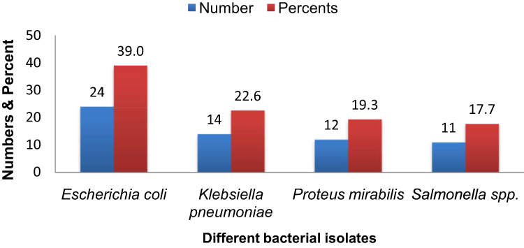 |
Figure 1 The proportions of bacterial isolates from chicken droplets. |
Feeding Practices of Chicken Attendants
Data from the interview of chicken attendants showed that none of them had any form of formal educational and short course training certificate on how to feed and on safe handling practices of chickens. There was no regular cleaning of chicken excreta which made it to be mixed with chicken feed. In addition, chickens above three months of age were supplied with antibiotics in their feed as usual practice of modified feeding. As any other feeding components, antibiotic treatment of drinking water supply for chickens was practiced. Preventive vaccine for Newcastle viral disease and other infection for all adult chickens was given on a programmed and scheduled booster dose approach. They also responded that diseased chickens might not be treated and isolated timely from the groups (Table 1).
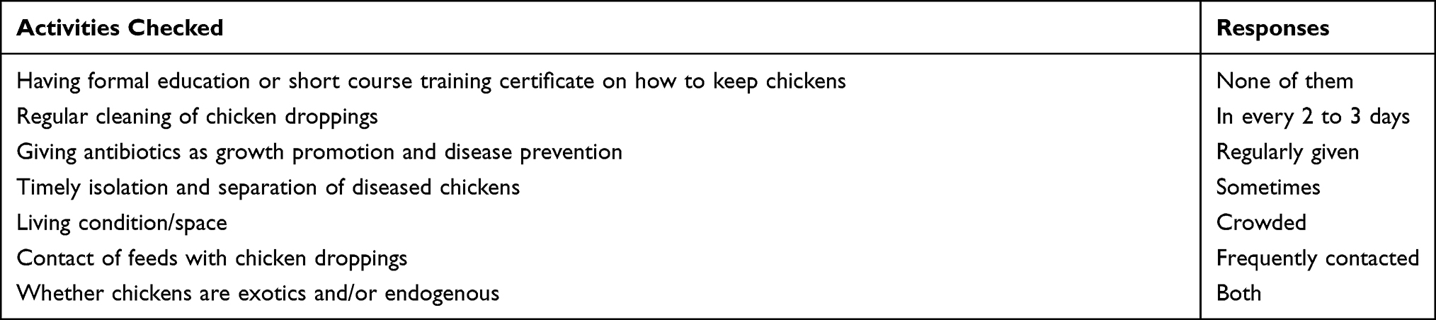 |
Table 1 Summary of Activities Done by Chicken Attendants at Jimma University Poultry Farm |
Antimicrobial Susceptibility Testing
In the current study a varying degree of susceptibility against the tested antimicrobial agents was observed. Accordingly, E. coli demonstrated a high rate of resistance against ampicillin (n=22, 91.7%), amoxicillin-clavulanic acid (n=16, 66.7%), ampicillin-sulbactam (n=16, 66.7%), tetracycline (n=18, 75.0%), trimethoprim-sulfamethoxazole (n=17, 70.8%), and chloramphenicol (n=13, 54.2%). Likewise, K. pneumoniae isolates showed a high resistance rate against ampicillin, amoxicillin-clavulanic acid, ampicillin-sulbactam, trimethoprim-sulfamethoxazole, tetracycline, cefoxitin, and chloramphenicol with resistance rates ranging from 50.0% to 85.7%. P. mirabilis and Salmonella spp. isolates also showed high resistance rate to ampicillin, amoxicillin-clavulanic acid, ampicillin-sulbactam, cefoxitin, tetracycline, trimethoprim-sulfamethoxazole, and chloramphenicol with resistance rates ranging from 45.5% to 83.3%. All isolates showed lower resistance rate with a range of 20.8% to 41.7% against third-generation cephalosporins (ceftazidime and ceftriaxone), ciprofloxacin, and gentamicin (Table 2).
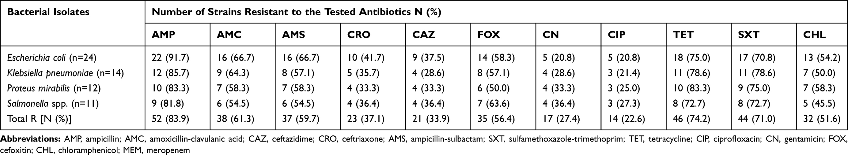 |
Table 2 Antibiotic Resistance Pattern of the Bacterial Isolates |
Multidrug-Resistance (MDR) Pattern
In this study, 13 (54.2%) E. coli isolates showed resistance against three or more antibiotics, whereas 8 (57.1%) of K. pneumoniae showed resistance against three or more antibiotics. In addition, 6 (50.0%) P. mirabilis isolates and 5 (44.4%) Salmonella spp. isolates showed resistance against three or more antibiotics. Totally, 32 (52.5%) of the isolates showed MDR phenotype (resistance to 3three or more antibiotics belonging to different families (Table 3)).
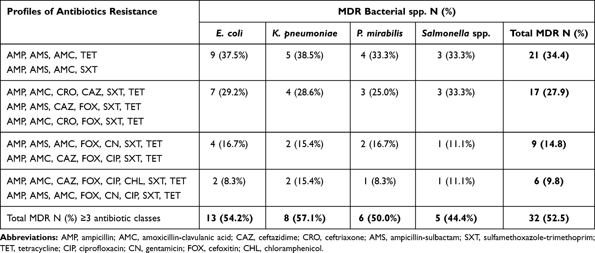 |
Table 3 Multidrug-Resistance (MDR) Pattern of the Bacterial Isolates |
Discussion
In this study, a total of 140 chicken droppings were collected from Jimma University poultry farm, southwest of Ethiopia for bacteriological analysis. Out of these, 61 (43.6%) samples showed bacterial growth. The proportion of culture positive samples in this study was lower than study findings in Nigeria17,18 and Kenya.19 However, this finding is higher than study findings in Ethiopia20 and Bangladesh.21 Differences in prevalence rates in various studies may be due to method used to isolate the bacteria, types and numbers of samples, animal management practices, and hygienic conditions. For instance, in this study, none of the chicken attendants had any form of formal educational and short course training certificate on how to feed and on safe handling practices of chickens. That is why there was no regular cleaning of chicken excreta which made it to be mixed with chicken feed that is prone for cross-contamination of chickens.
In this study, the most predominant isolate was E. coli followed by K. pneumoniae and P. mirabilis and Salmonella spp. The predominance of E. coli isolates as well as P. mirabilis and Salmonella spp. were also reported from Bangladesh21 and Kenya.19,22 The fact is that most Enterobacteriaceae, especially, E. coli, are predominantly found as normal flora gastrointestinal tracts of animals and humans.23 This study covered only one poultry farm from a focal area and thereby has generated the additional prevalence data for the bacteria in the poultry industry of this country. The outcomes of this study would be helpful in designing further extensive investigations covering more poultry farms from expanded areas of the country to find nationwide prevalence of zoonotic Enterobacteriaceae pathogen.
Emergence of MDR bacteria, especially Enterobacteriaceae, has increased in recent years. In this study, more than half (52.5%) of isolates showed MDR. The higher proportions of MDR were also reported from Bangladesh,21 Vietnam,24 and China.25 The occurrence of MDR may be linked with indiscriminate use of antimicrobial agents such as wrong indication, wrong duration, and improper route of administration, use of leftover antibiotics from a family member, and improper discontinuation of antibiotics.26–29
This study also revealed that isolates from chicken droppings showed high resistance properties toward ampicillin, augmentin, tetracycline, and trimethoprim- sulfamethoxazole followed by cefoxitin and chloramphenicol. The significant proportion of resistance against the above antibiotics was also reported from Bangladesh,21 Nigeria,18,30 Ethiopia,20 Vietnam,31 and China.25 Moreover, the current study recorded higher resistance to ampicillin-sulbactam. This demonstrates that apart from therapeutic use of antibiotics in poultry, other factors influence development of antibiotic resistance. For instance, in this study, chicken attendants responded as they were supplied antibiotics in their feed as usual practice and as they used as growth promotion and as disease prevention. In addition, chicken droppings were not cleaned regularly and it has a contact with chicken feedings. Studies from Kenya and Bangladesh showed that significant numbers of poultry feeds were contaminated with E. coli and Salmonella spp. isolates.22,32
In contrast, in this study, isolated bacterial species showed relatively lower grade of resistance against third-generation cephalosporins (ceftriaxone and ceftazidime), ciprofloxacin, and gentamicin. This finding is supported by other studies showing lower resistance rate against the above antibiotics.22,26,33 However, higher resistance rates were reported from Bangladesh21 and Nigeria.30 This might be a challenge for effectiveness of modern antimicrobial agents commonly used to treat human infections.
Limitations of This Study
The study was done from a single source of sample (one chicken farm) for the determination of prevalent bacterial isolates and antibiotic resistance patterns. Molecular techniques for more informative finding to indicate the specific antibiotic resistant genes were not conducted due to resource problems.
Conclusions
In this study, significant numbers of chicken droplets contain bacterial isolates that are resistant to single and multiple antibiotics that are commonly prescribed to humans. These findings suggested that avian farms might serve as reservoirs of antibacterial-resistant bacteria that can infect humans through the food chain. Emphasis on the usage of antibiotics in poultry farms has to be considered in order to make longer effectiveness of existing antibiotics.
Data Sharing Statement
All data were incorporated in the manuscript.
Ethical Approval and Consent to Participate
This study was done as dissertations for MSc thesis in Jimma University. That is why ethical approval of the study was obtained from the Institutional Review Board of Jimma University. Communication with the Agricultural College of Jimma University was made through formal letter before running data collection. Verbal informed consent that was approved by the Institutional Review Board of Jimma University was obtained from chicken farm attendants after fully explained the objective of the study.
Acknowledgments
We would like to thank the attendants in Jimma University chicken farm and Jimma University Medical Microbiology Laboratory staff for assisting us during microbiological investigation. We would like to thank Jimma University for their support in materials useful for investigation.
Author Contributions
All authors made a significant contribution to the work reported, whether that is in the conception, study design, execution, acquisition of data, analysis and interpretation, or in all these areas; took part in drafting, revising or critically reviewing the article; gave final approval of the version to be published; have agreed on the journal to which the article has been submitted; and agree to be accountable for all aspects of the work.
Funding
No funding was allocated for this study.
Disclosure
All authors declared that they have no conflicts of interest for this work.
References
1. World Health Organization. Antimicrobial resistance: global report on surveillance. 2014.
2. Byarugaba DK. Antimicrobial Resistance in Developing Countries. Springer; 2009:15–27.
3. Hedman H, Vasco K, Zhang L. A review of antimicrobial resistance in poultry farming within low-resource settings. Animals. 2020;10(8):1264. doi:10.3390/ani10081264
4. Boerma T, Mathers CD. The World Health Organization and global health estimates: improving collaboration and capacity. BMC Med. 2015;13(1):50. doi:10.1186/s12916-015-0286-7
5. Sridevi TD, Shankar C, Park J, Dexilin M, Rajesh RK, Thamaraiselvi R. Study on acquisition of bacterial antibiotic resistance determinants in poultry litter. PoultSci. 2009;88:1381–1387.
6. Olatoye O. Antibiotic use and resistance pattern of Salmonella species in poultry from Ibadan,Nigeria. Trop Vet. 2011;29:28–35.
7. Cortes P, Blanc V, Mora A, Dahbi G, Blanco J. Isolation and characterization of potentially pathogenic antimicrobial resistance E.coli strains from chicken and pig farm in Span. Appl Envirom Microbial. 2010;76(9):2799–2805. doi:10.1128/AEM.02421-09
8. Hubálek Z. Emerging human infectious diseases: anthroponoses, zoonoses, and sapronoses. Emerg Infect Dis. 2003;9(3):403. doi:10.3201/eid0903.020208
9. Chang Q, Wang W, Regev-Yochay G, Lipsitch M, Hanage WP. Antibiotics in agriculture and the risk to human health: how worried should we be? Evol Appl. 2015;8(3):240–247. doi:10.1111/eva.12185
10. World Health Organization. Antimicrobial Resistance: Global Report on Surveillance. World Health Organization; 2014.
11. Destoumieux-Garzon D, Mavingui P, Boetsch G, et al. The one health concept: 10 years old and a long road ahead. Front Vet Sci. 2018;5:14. doi:10.3389/fvets.2018.00014
12. Duguma R. Understanding the role of indigenous chicken during the long walk to food security in Ethiopia. Livest Res Rural Dev. 2009;21(116). Available from: http://www.lrrd.org/lrrd21/8/dugu21116.htm.
13. Cheesbrough M. District Laboratory Practice in Tropical Countries.
14. World Health Organization. (WHO): Basic Laboratory Procedures in Clinical Bacteriology.
15. CLSI. Performance Standards for Antimicrobial Susceptibility Testing.
16. Magiorakos A, Srinivasan A, Carey RB, et al. Bacteria: an international expert proposal for interim standard definitions for acquired resistance. Clin Microbiol Infect. 2011;18(3):268–281. doi:10.1111/j.1469-0691.2011.03570.x
17. Ajayi KO, Omoya FO. Antibiotic usage pattern in poultry and resistance pattern of human pathogenic bacteria isolated from poultry droppings in Akure, Nigeria. Int J Biomed Sci Eng. 2017;5(4):35–40. doi:10.11648/j.ijbse.20170504.11
18. Omoya FO, Ajayi KO. Antibiotic resistance pattern of pathogenic bacteria isolated from poultry droppings in Akure, Nigeria. Futa J Res Sci. 2016;12(2):219–227.
19. Langata LM, Maingi M, Musonye HA, Kiiru J, Nyamache AK. Antimicrobial resistance genes in Salmonella and Escherichia coli isolates from chicken droppings in Nairobi, Kenya. BMC Res Notes. 2019;12(1):22. doi:10.1186/s13104-019-40688
20. Ali DA, Tadesse B, Ebabu A. Prevalence and antibiotic resistance pattern of Salmonella isolated from caecal contents of exotic chicken in Debre Zeit and Modjo, Ethiopia. Int J Microbiol. 2020:6. Article ID 1910630. doi:10.1155/2020/1910630
21. Nahar A, Siddiquee M, Nahar S, Anwar KS, Islam S. Multidrug resistant-Proteus mirabilis isolated from chicken droppings in commercial poultry farms: bio-security concern and emerging public health threat in Bangladesh. J Biosafety Health Educ. 2014;2(02):120. doi:10.4172/2332-0893.1000120
22. Ngai DG, Nyamache AK, Ombori O. Prevalence and antimicrobial resistance profiles of Salmonella species and Escherichia coli isolates from poultry feeds in Ruiru Sub-County, Kenya. BMC Res Notes. 2021;14(1):4. doi:10.1186/s13104-021-054564
23. Carattoli A. Animal reservoirs for extended Spectrum β-lactamase producers. Clin Microbiol Infect. 2008;14:117–123. doi:10.1111/j.1469-0691.2007.01851.x
24. Vounba P, Arsenault J, Bada-Alambédji R, Fairbrother JM. Pathogenic potential and the role of clones and plasmids in beta-lactamase-producing E. coli from chicken faeces in Vietnam. BMC Vet Res. 2019;15(1):106. doi:10.1186/s12917-019-1849-1
25. Yassin AK, Gong J, Kelly P, et al. Antimicrobial resistance in clinical Escherichia coli isolates from poultry and livestock, China. PLoS One. 2017;12(9):e0185326. doi:10.1371/journal.pone.0185326
26. World Health Organization. Mapping Educational Opportunities and Resources for Health-Care Workers to Learn About Antimicrobial Resistance and Stewardship. Geneva: World Health Organization; 2017. (Human Resources for Health Observer Series No. 21). Licence: CC BY-NC-SA 3.0 IGO.
27. Muhie OA. Antibiotic use and resistance pattern in Ethiopia: systematic review and meta-analysis. Int J Microbiol. 2019;2019:8. Article ID 2489063. doi:10.1155/2019/2489063
28. Harris P, Paterson D, Rogers B. Facing the challenge of multidrug-resistant gram-negative bacilli in Australia. MJA. 2015;202(5):243–247. doi:10.5694/mja14.01257
29. Reygaert WC. An overview of the antimicrobial resistance mechanisms of bacteria. AIMS Microbiol. 2018;4(3):482–501. doi:10.3934/microbiol.2018.3.482
30. Ayandiran TO, Falgenhauer L, Schmiede J, Chakraborty T, Ayeni FA. High resistance to tetracycline and ciprofloxacin in bacteria isolated from poultry farms in Ibadan, Nigeria. J Infect Dev Ctries. 2018;12(6):462–470. doi:10.3855/jidc.9862
31. Nguyen VT, Carrique-Mas JJ, Thi Hoa N, Huynh Mai HO, Thanh Tuyen HA, Campbell JI. Prevalence and risk factors for carriage of antimicrobial-resistant Escherichia coli on household and small-scale chicken farms in the Mekong Delta of Vietnam. J Antimicrob Chemother. 2015;70(7):2144–2152. doi:10.1093/jac/dkv053
32. Islam MM, Islam MN, Sharifuzzaman FM. Isolation and identification of Escherichia coli and Salmonella from poultry litter and feed. Int J Nat Soc Sci. 2014;1:1–7.
33. Braykov NP, Eisenberg JNS, Grossman M, et al. Antibiotic resistance in animal and environmental samples associated with small-scale poultry farming in northwestern Ecuador. mSphere. 2016;1(1):e00021–15. doi:10.1128/mSphere.00021-15
 © 2021 The Author(s). This work is published and licensed by Dove Medical Press Limited. The
full terms of this license are available at https://www.dovepress.com/terms
and incorporate the Creative Commons Attribution
- Non Commercial (unported, 3.0) License.
By accessing the work you hereby accept the Terms. Non-commercial uses of the work are permitted
without any further permission from Dove Medical Press Limited, provided the work is properly
attributed. For permission for commercial use of this work, please see paragraphs 4.2 and 5 of our Terms.
© 2021 The Author(s). This work is published and licensed by Dove Medical Press Limited. The
full terms of this license are available at https://www.dovepress.com/terms
and incorporate the Creative Commons Attribution
- Non Commercial (unported, 3.0) License.
By accessing the work you hereby accept the Terms. Non-commercial uses of the work are permitted
without any further permission from Dove Medical Press Limited, provided the work is properly
attributed. For permission for commercial use of this work, please see paragraphs 4.2 and 5 of our Terms.