Back to Journals » International Journal of Chronic Obstructive Pulmonary Disease » Volume 17
Different Responses to Pulmonary Rehabilitation in COPD Patients with Different Work Efficiencies
Authors Jao LY, Hsieh PC , Wu YK , Yang MC , Wu CW, Lee C, Tzeng IS , Lan CC
Received 31 December 2021
Accepted for publication 3 April 2022
Published 26 April 2022 Volume 2022:17 Pages 931—947
DOI https://doi.org/10.2147/COPD.S356608
Checked for plagiarism Yes
Review by Single anonymous peer review
Peer reviewer comments 2
Editor who approved publication: Dr Richard Russell
Lun-Yu Jao,1,2 Po-Chun Hsieh,3 Yao-Kuang Wu,1,2 Mei-Chen Yang,1,2 Chih-Wei Wu,1,2 Chung Lee,1,2 I-Shiang Tzeng,4 Chou-Chin Lan1,2
1Division of Pulmonary Medicine, Department of Internal Medicine, Taipei Tzu Chi Hospital, Buddhist Tzu Chi Medical Foundation, New Taipei City, Taiwan; 2School of Medicine, Tzu-Chi University, Hualien, Taiwan; 3Department of Chinese Medicine, Taipei Tzu Chi Hospital, Buddhist Tzu Chi Medical Foundation; School of Post-Baccalaureate Chinese Medicine, Tzu Chi University, Hualien, Taiwan; 4Department of Research, Taipei Tzu Chi Hospital, Buddhist Tzu Chi Medical Foundation, New Taipei City, Taiwan
Correspondence: Chou-Chin Lan, Division of Pulmonary Medicine, Department of Internal Medicine, Taipei Tzu Chi Hospital, Buddhist Tzu Chi Medical Foundation, 289, Jianguo Road, Xindian District, New Taipei City, Taiwan, 23142, Tel +886-2-6628-9779 ext. 2259, Fax +886-2-6628-9009, Email [email protected]
Background: Chronic obstructive pulmonary disease (COPD) often involves the cardiopulmonary dysfunction that deteriorates health-related quality of life (HRQL) and exercise capacity. Work efficiency (WE) indicates the efficiency of overall oxygen consumption (VO2) during exercise. This study investigated whether different WEs have different effects on pulmonary rehabilitation (PR).
Methods: Forty-five patients with stable COPD were scheduled for PR. The PR programs consisted of twice-weekly sessions for three months. These patients were comprehensively evaluated by cardiopulmonary exercise testing and COPD assessment test (CAT) before and after PR. We compared these parameters between patients with a normal versus poor WE.
Results: Twenty-one patients had a normal WE and twenty-four patients had a poor WE (< 8.6 mL/min/watt). Patients with a poor WE had earlier anaerobic metabolism, a poorer oxygen pulse, lower exercise capacity, more exertional dyspnea, and a poorer HRQL than those with a normal WE. PR improved exercise capacity, HRQL, anaerobic threshold, exertional dyspnea and leg fatigue in patients with either normal or poor WE. However, significant improvement of WE, oxygen pulse, respiratory frequency (Rf) during exercise, chest tightness, activity and sleepiness by CAT were noted only in patients with a poor WE. Among the patients with a poor WE, 29% patients had WE returned to normal after PR.
Conclusion: Patients with different WE had different responses to PR. PR improved exercise capacity and HRQL regardless of a normal or poor WE. However, WE, oxygen pulse, Rf during exercise, chest tightness, activity and sleepiness were only improved in patients with a poor WE.
Keywords: chronic obstructive pulmonary disease, exercise intolerance, pulmonary rehabilitation, work efficiency
Introduction
Chronic obstructive pulmonary disease (COPD) is the third leading cause of death and it is a global health problem with increasing prevalence.1 COPD is characterized by coughing or wheezing, excess sputum production, and shortness of breath.1 A subgroup of patients of COPD have progressive disease and results in the deterioration of cardiopulmonary function.1 These patients tend to have poor exercise capacity and health-related quality of life (HRQL).
Some patients of COPD experience difficulty with physical activity and unpleasant symptoms even under optimal medical treatment. Pulmonary rehabilitation (PR) may help improve physical activity and HRQL in these patients with COPD and the Global Initiative for Chronic Obstructive Disease (GOLD) guideline recommends that PR should be an integral part of COPD treatment. PR is a cornerstone in COPD management but not all patients benefit from PR,2 and this could relate to pre-PR work efficiency (WE). We want to investigate this systematically.
Cardiopulmonary exercise testing (CPET) is used to evaluate exercise intolerance. WE, one parameter of CPET, measures an individual’s oxygen consumption (VO2) during exercise workload (WR).3 WE is a measure of overall oxygen consumption efficiency during exercise. We previously reported that COPD patients with a poor WE also had early anaerobic metabolism, lower exercise capacity, more exertional dyspnea, and poorer HRQL.4 However, whether WE affects the effect of PR in COPD patients with a poor WE remain unknown.
Since no studies to date have examined the effects of PR in patients with a poor WE, here we aimed to investigate the effects of PR in COPD patients with a normal versus poor WE. Specifically, we aimed to determine how different WE affects PR.
Materials and Methods
Study Design
Patients with COPD were recruitment from the outpatient department in Taipei Tzu-Chi Hospital. These patients underwent pre-PR assessments by CPET, questionnaire of HRQL, respiratory muscle strength. They then received a 12-week PR. After PR, they underwent post-PR assessment by CPET, questionnaire of HRQL, respiratory muscle strength again.
In this study, we aimed to investigate whether patients with different WE respond differently to PR. The normal range of WE is 8.6–10.1 mL/min/watt.3 Wasserman et al suggested that a WE <8.6 mL/min/watt indicates a poor WE.3 We therefore divided the patients into two groups: group 1 (Gr 1) were patients with a normal WE and group 2 (Gr 2) were patients with a poor WE. The primary outcome was to assess the changes in WE in patients with a normal WE versus those with a poor WE. The secondary outcomes were to assess circulatory responses, ventilatory responses, gas exchanges, exercise capacity and HRQL in patients with a normal WE versus those a with poor WE.
Patient Recruitment
Forty-five patients with stable COPD were recruited. The diagnosis and severity of COPD was defined according to GOLD guideline.5 The inclusion criteria were absence of acute exacerbations for 3 months before recruitment, ability to ambulate for completion of CPET and PR, and willingness to be included in the study. The exclusion criteria were a history of other lung diseases such as asthma, pulmonary tuberculosis, etc., orthopedic or neurological impairment that unable to perform CPET and PR, unwilling to participate in the PR programs and those had ever participated in a PR program. The ethics committee of Taipei Tzu-Chi Hospital approved the study. All patients provided informed consent.
Pulmonary Function Test
According to the standards of the American Thoracic Society (ATS), the patients used a spirometer (Medical Graphics Corporation; St Paul, MN, USA) for the pulmonary function tests.6 According to the GOLD guidelines,7 we used the percentage of forced expiratory volume in 1 second (FEV1%) to evaluate the degree of airflow obstruction.
Cardiopulmonary Exercise Test
All participants underwent the CPET using a bike ergometer (Lode Corival, Netherlands) through a progressive protocol. Their exhaled air was analyzed by breath analysis (Breeze Suite 6.1; Medical Graphics Corporation, St Paul, MN, USA) to assess their oxygen consumption (VO2), carbon dioxide output (VCO2), end tidal PCO2 (PETCO2) and tidal volume (VT). Their oxygen saturation (SpO2), respiratory frequency (Rf), electrical heart function, blood pressure (BP), and heart rate (HR) were continuously monitored during the CPET.
Peak VO2 (VO2peak) was measured at exercise capacity. WE is the relationship between VO2 and WR during exercise.8 WE is defined as the slope of VO2/WR and determined using linear regression analysis. The VO2 at the anaerobic threshold (AT) was determined by the VCO2 versus VO2 graph.9 The predicted VO2 at AT less than 40% of predicted VO2max indicates a poor AT%.3 Oxygen pulse (O2P) was defined as VO2 divided by HR (O2P = VO2/HR). An O2P at a peak exercise level lower than 80% of the predicted value is considered poor.3 Respiratory gas exchange (RER) is defined as VCO2/VO2.3
Respiratory Muscle Strength
Maximum inspiratory pressure (MIP) and maximum expiratory pressure (MEP) were measured five times with a pressure gauge (Respiratory Pressure Meter; Micro Medical Corp, England), and the highest value was recorded.4,10 For the measurement of MIP, the patients exhaled to the residual volume and then perform a rapid maximal inspiration. For the measurement of MEP, the patients inhaled to the total lung capacity and then exhaled with maximal effort.
Exertional Dyspnea and Leg Fatigue Scores
The dyspnea and leg fatigue scores were evaluated using a modified version of Borg scale at peak exercise during CPET, which is scored 0–10 points; higher scores indicate more severe dyspnea or leg fatigue.11 Dyspnea and leg scores are determined at peak exercise during the CPET.
Health-Related Quality of Life
The Chinese version of the COPD Assessment Test (CAT) was used to assess HRQL. The CAT consists of eight items (cough, phlegm, chest tightness, dyspnea, activity, confidence to leave home, sleeplessness, and energy). The score for each item ranges from 0 to 5,12 and the total score ranges from 0 (best) to 40 (worst) points.12 A total CAT score ≥10 is classified as a high-level symptom.12 The minimum clinically important difference of CAT is 2 points.12
PR Program
All patients performed a 12-week twice-weekly hospital-based PR program. In each training session, formal education, including proper use of medications, breathing exercise (purse-lip and diaphragm breathing), and self-management skills were provided. The exercise training was performed by cycle ergometer. Patients were encouraged to achieve their maximal exercise as possible. The exercise intensity was targeted to 50–100% of peak VO2 as patients’ tolerance. During the exercise training, respiratory therapists monitored the WR, SpO2, Rf, HR, BP, dyspnea, and leg fatigue.
Statistical Analysis
The parameters are shown as mean and standard deviation. A paired t-test was used to compare parameters before and after PR. An independent sample t test was used to compare the pre- and post-PR parameters between the two groups. The changes in parameters were defined as differences between pre- and post-PR and were analyzed by the independent sample t test between the two groups. We perform Normality test of variables to identify the parametric test that is used to analyze normal distribution variables. A statistically significant difference was set at p < 0.05. The statistical analyses were performed using SPSS version 24.0 (SPSS, Inc., Chicago, IL, USA).
Results
Baseline Clinical and Demographic Characteristics
Table 1 shows the demographic and clinical characteristics of all patients. Among the 45 COPD patients, 21 had a normal WE (Gr 1) and 24 had a poor WE (Gr 2). No significant differences were noted in body weight, body height, body mass index, age, gender, smoking status, COPD severity, duration of diagnosis of COPD, comorbidities of congestive heart failure, hypertension and diabetes mellitus between these two groups. Most enrolled patients were COPD group B.
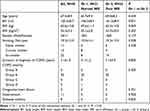 |
Table 1 Baseline Demographic Characteristics |
Effects of PR on Circulatory Parameters of Patients with a Normal versus Poor WE
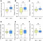
At baseline, the WE, AT, and O2P were significantly lower in Gr 2 than in Gr 1 (Table 2) but mean blood pressure (MBP) and HR did not differ significantly between Gr 1 and Gr 2. For Gr 1, at post-PR, significant improvement was seen in AT but not in WE, O2P, HR, or MBP. For Gr 2, at post-PR, significant improvement was seen in WE, AT, O2P, and MBP at rest. The improvement in circulatory parameters after PR for each group is shown in Figure 1. The improvements in WE, AT, and O2P after PR were significantly greater in Gr 2 than in Gr 1.
 |
Table 2 Effects of PR on Circulatory Parameters of Patients with a Normal and Poor WE |
Effects PR on Ventilatory Parameters of Patients with a Normal and Poor WE
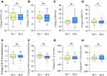
At baseline, the FVC%, FEV1%, FEV1/FVC, MIP, and MEP were significantly lower in Gr 2 than in Gr 1 (Table 3). For Gr 1, there were no significant differences in FVC, FEV1, MIP, MEP, Rf, or VT at pre- versus post-PR. For Gr 2, there were no significant differences in FVC, FEV1, MIP, MEP, and VT at pre- versus post-PR, but Rf at exercise was significantly lower at post- than pre-PR. The improvement in ventilatory parameters at post-PR for each group is shown in Figure 2. No significant intergroup difference in FVC, FEV1, MIP, MEP, Rf, or VT was noted at post-PR.
 |
Table 3 Effects of PR on Ventilatory Parameters of Patients with a Normal and Poor WE |
Effects PR on Gas Exchanges of Patients with a Normal versus Poor WE
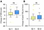
At baseline, the PETCO2, SpO2 at rest or during exercise and the RER were similar between Gr 1 and 2 (Table 4). The PETCO2 and SpO2 at rest or during exercise and the RER did not differ. The changes in PETCO2, SpO2 at rest or during exercise after PR for each group are shown in Figure 3. The changes in SpO2and PETCO2 after PR were similar between Gr 1 and Gr 2.
 |
Table 4 Effects of PR on Gas Exchanges of Patients with a Normal and Poor WE |
Effects of PR on Exercise Capacity of Patients with a Normal versus Poor WE
At baseline, the mean exercise capacity (VO2 and WR at peak exercise) was significantly greater in Gr 1 than in Gr 2 (Table 5). PR significantly improved VO2 and WR at peak exercise in patients of both groups. The changes in VO2 and WR at peak exercise after PR for both groups are shown in Figure 4. The changes in VO2 and WR at peak exercise after PR were similar between groups.
 |
Table 5 Effects of PR on Exercise Capacity of Patients with a Normal and Poor WE |
Effects of PR on HRQL, Exertional Dyspnea, and Leg Fatigue Scores of Patients with a Normal versus Poor WE
At baseline, the phlegm, breathlessness, activities, CAT total score, and modified British Medical Research Council (mMRC) score were significantly poorer in Gr 1 than in Gr 2 (Table 6). For patients in Gr 1, PR significantly improved breathlessness, CAT total score, mMRC score, dyspnea, and fatigue at peak exercise. For patients in Gr 2, PR significantly improved phlegm, chest tightness, breathlessness, activities, sleep, CAT total score, mMRC score, dyspnea, and leg fatigue during exercise.
 |
Table 6 Effects of PR on HRQL, Exertional Dyspnea and Leg Fatigue Score of Patients with a Normal and Poor WE |
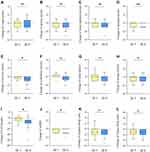
The changes in CAT, mMRC score, dyspnea, and leg fatigue during exercise after PR for each group are shown in Figure 5. The changes in breathlessness, activity, CAT total score, and mMRC score after PR were significantly greater in Gr 1 than in Gr 2.
Subgroups Analysis of Patients with Poor WE
Among the 24 patients with a poor WE, WE returned to normal in 7 patients (29%), and WE did not return to normal in 17 patients (71%). We compared the baseline data and response to PR of these two groups of patients, as shown in Table 7. For baseline data, WE, VO2 at AT and O2P were significantly higher in patients with normalized WE than those without normalized WE. However, age, gender, body mass index, exercise capacity, and CAT total score at baseline did not show significant difference between the two groups.
 |
Table 7 Subgroup Analysis of Patients with a Poor WE |
Discussion
This is the first study to assess the different effects of PR in COPD patients with a normal versus poor WE. There are some important and novel findings in this study. Compared to COPD patients with a normal WE, those with a poor WE had decreased exercise capacity, more exertional dyspnea, poor HRQL, and poor circulatory parameters. Our PR program was efficient to improve exercise capacity, exertional dyspnea, HRQL, and circulatory parameters for COPD patients with both normal or poor WE. However, greater improvements in VO2 at AT, exercise capacity, and CAT were found in patients with a poor WE than those with a normal WE. Furthermore, improvements in WE, O2P, and Rf in peak exercise, chest tightness, activity, and sleep quality were found only in patients with a poor WE, but not in patients with a normal WE.
In our study, we used high intensity (50–100%) of exercise training and these patients had improvement of exercise capacity and HRQL after such training. Patients with a poor WE further had improvement of WE and O2P. However, it is not known about the effect of low intensity (<50%) exercise training on WE. Besides, most enrolled patients were COPD group B in the current study. This was because that we enrolled stable COPD for PR and we excluded acute exacerbations within three months. Since the majority of patients were COPD group B, it is unclear whether patients of other groups would get the same results after exercise training.
The overall VO2 dynamics during exercise depend on the gas exchange by the respiratory system, oxygen delivery by the circulatory system, and oxygen extraction for exercise by the musculoskeletal system.13 Oxygen extraction further depends on the muscle mass of the limbs, muscular capillary contents, and mitochondrial function of muscle cells.8 A poor WE suggests a poor overall oxygen consumption efficiency that indicates poor oxygen delivery or impaired muscular extraction of oxygen.14
In the current study, patients with a poor WE had early AT, poorer exercise capacity, and poorer HRQL than those with a normal WE. The consequence of a poor WE is early anaerobic metabolism during exercise.14 Anaerobic glycolysis produces lactic acidosis, which further leads to excessive ventilation responses to acidosis-stimulating chemoreceptors.14,15 Hyperventilation during early exercise further results in exertional dyspnea sensation, exercise intolerance, and a poor HRQL.15 This explained that the COPD patients with a poor WE experience early anaerobic metabolism during exercise and are therefore prone to exercise intolerance and poor HRQL.
Wasserman et al previously suggested that trained and untrained patients would have a similar WE regardless of age or gender.8 However, we found that PR improved WE in COPD patients with a poor WE but not in those with a normal WE. Therefore, exercise training is not unable to change WE. It is for patients with a normal WE, exercise training will not increase their WE. But for patients with a poor WE, exercise training will still increase their WE. Patients with a normal WE indicated good oxygen delivery and extraction. Therefore, for patients with a normal WE, there is little room for improvement in WE. Patients with a poor WE indicated poor oxygen delivery or extraction. Exercise training improves their oxygen delivery or extraction.
The mechanisms of the improvement in WE after PR are multiple. One previous study suggested that PR improved the central cardiovascular response such as stroke volume during exercise in COPD.16 We here also showed that O2P, a reliable surrogate marker of stroke volume, was improved in patients with a poor WE. Nasis et al also showed that PR improved cardiac output in patients with COPD.17 The reduction in dynamic hyperinflation after PR is related to the improvement in stroke volume.16 Besides, exercise training is known to improve contractile capacity of cardiomyocyte.18
Skeletal muscle and mitochondrial dysfunction is a systemic manifestation in some patients with COPD.19 Vogiatzis et al revealed that PR increased muscle cross-sectional area and capillary contents.20 Marillier et al also found that exercise training increased muscle cell mitochondrial function.19 According to these studies, exercise training increases muscle mass, muscular capillary contents, and mitochondrial function, which are important factors of oxygen extraction. The improvement in oxygen extraction also resulted in an increase in WE. Although we did not examine mitochondrial function and muscle mass after PR, it is plausible based on these studies that PR leads to improve peripheral muscle mass and mitochondrial function.
There is one previous study about the impact of PR on severe physical inactivity in patients with COPD.2 In this study, most patients of severe physical inactivity (78%) did not change activity level after PR. The result seems to contradict our results that we showed the improvement of WE in patients with a poor WE. However, the two studies aimed at different outcomes. Thyregod et al focused on physical inactivity and we focused on WE. WE is determined by multiple factors. Physical inactivity with muscle deconditioning may be one possible reason of a poor WE, but not all patients in severe physical inactivity result in a poor WE. Most of our enrolled patients also self-reported physical inactivity, but only 24 of 45 patients had a poor WE.
Clinical Implication
Exercise intolerance, exertional dyspnea, poor HRQL are common in patients with COPD.15 An individual’s overall oxygen consumption efficiency depends on oxygen delivery, oxygen extraction, and mitochondrial function.8 WE is a good parameter that provides information about circulatory and tissue oxygen consumption function during exercise. We suggested here that COPD patients with a poor WE have more severe exertional dyspnea, lower exercise capacity, poor circulatory function, earlier anaerobic metabolism, and poorer HRQL. PR improved their exercise capacity, circulatory function, anaerobic metabolism, and HRQL.
Study Limitations
Although our research has many important findings, it still has some limitations. First, patients with a poor WE had poor circulatory function, more severe exercise intolerance, and poorer HRQL. However, the value of WE for predicting long-term outcomes such as the survival of patients with COPD is unclear. Therefore, the long-term impact of PR for improving WE is unknown. Further, PR improved WE in our study, but we could not determine whether this was due to improved circulatory, muscular, or mitochondrial function for these patients. However, it is difficult to determine muscular and mitochondrial function in patients with COPD. Therefore, WE is still an available, effective, and non-invasive parameter for monitoring an individual’s overall cardiovascular, muscular, and mitochondrial function after PR.8
Conclusions
Patients with a poor WE had poor circulatory parameters, exercise capacity and HRQL. PR improved exercise capacity, HRQL, and VO2 at AT in patients of both normal or poor WE. However, greater improvements in VO2 at AT, exercise capacity, and HRQL were found in COPD patients with a poor WE than those with a normal WE. Furthermore, improvements in WE, O2P, and Rf at peak exercise, chest tightness, activity, and sleep quality were found only in patients with a poor WE.
Data Sharing Statement
The datasets used during the current study are available from the corresponding author on reasonable request.
Ethics Approval and Informed Consent
The ethics committee of Taipei Tzu-Chi Hospital approved the study. All participants were informed about the purpose of the study, in accordance with the Declaration of Helsinki. Patient’s written consent has been obtained.
Funding
This study was supported by grants from the Taipei Tzu Chi Hospital and the Buddhist Tzu Chi Medical Foundation (TCRD-TPE-109-59 and TCRD-TPE-109-24(2/3), respectively).
Disclosure
The authors disclose no financial or other potential conflicts of interest in this work.
References
1. Hurst JR, Siddiqui MK, Singh B, Varghese P, Holmgren U, de Nigris E. A systematic literature review of the humanistic burden of COPD. Int J Chron Obstruct Pulmon Dis. 2021;16:1303–1314. doi:10.2147/copd.S296696
2. Thyregod M, Løkke A, Bodtger U. The impact of pulmonary rehabilitation on severe physical inactivity in patients with chronic obstructive pulmonary disease: a pilot study. Int J Chron Obstruct Pulmon Dis. 2018;13:3359–3365. doi:10.2147/copd.S174710
3. Herdy AH, Ritt LE, Stein R, et al. Cardiopulmonary exercise test: background, applicability and interpretation. Arq Bras Cardiol. 2016;107(5):467–481. doi:10.5935/abc.20160171
4. Yang SH, Yang MC, Wu YK, et al. Poor work efficiency is associated with poor exercise capacity and health-related quality of life in patients with chronic obstructive pulmonary disease. Int J Chron Obstruct Pulmon Dis. 2021;16:245–256. doi:10.2147/copd.S283005
5. Cheng SL, Lin CH, Chu KA, et al. Update on guidelines for the treatment of COPD in Taiwan using evidence and GRADE system-based recommendations. J Formos Med Assoc. 2021;120(10):1821–1844. doi:10.1016/j.jfma.2021.06.007
6. Culver BH, Graham BL, Coates AL, et al. Recommendations for a standardized pulmonary function report. An official American thoracic society technical statement. Am J Respir Crit Care Med. 2017;196(11):1463–1472. doi:10.1164/rccm.201710-1981ST
7. Song JH, Lee CH, Um SJ, et al. Clinical impacts of the classification by 2017 GOLD guideline comparing previous ones on outcomes of COPD in real-world cohorts. Int J Chron Obstruct Pulmon Dis. 2017;13:3473–3484. doi:10.2147/COPD.S177238
8. Wasserman K, Hansen JE, Sue DY, et al. Principles of Exercise Testing and Interpretation.
9. Wu CW, Hsieh PC, Yang MC, Tzeng IS, Wu YK, Lan CC. Impact of peak oxygen pulse on patients with chronic obstructive pulmonary disease. Int J Chron Obstruct Pulmon Dis. 2019;14:2543–2551. doi:10.2147/COPD.S224735
10. McConnell AK, Copestake AJ. Maximum static respiratory pressures in healthy elderly men and women: issues of reproducibility and interpretation. Respiration. 1999;66(3):251–258. doi:10.1159/000029386
11. Cheng ST, Wu YK, Yang MC, et al. Pulmonary rehabilitation improves heart rate variability at peak exercise, exercise capacity and health-related quality of life in chronic obstructive pulmonary disease. Heart Lung. 2014;43(3):249–255. doi:10.1016/j.hrtlng.2014.03.002
12. Wiklund I, Berry P, Lu KX. The Chinese translation of COPD assessment test (CAT) provides a valid and reliable measurement of COPD health status in Chinese COPD patients. Am J Respir Crit Care Med. 2010;181:A3575.
13. Takken T, Mylius CF, Paap D, et al. Reference values for cardiopulmonary exercise testing in healthy subjects - an updated systematic review. Expert Rev Cardiovasc Ther. 2019;17(6):413–426. doi:10.1080/14779072.2019.1627874
14. Guazzi M, Bandera F, Ozemek C, Systrom D, Arena R. Cardiopulmonary exercise testing: what is its value? J Am Coll Cardiol. 2017;70(13):1618–1636. doi:10.1016/j.jacc.2017.08.012
15. O’Donnell DE, Elbehairy AF, Faisal A, Webb KA, Neder JA, Mahler DA. Exertional dyspnoea in COPD: the clinical utility of cardiopulmonary exercise testing. Eur Respir Rev. 2016;25(141):333–347. doi:10.1183/16000617.0054-2016
16. Ramponi S, Tzani P, Aiello M, Marangio E, Clini E, Chetta A. Pulmonary rehabilitation improves cardiovascular response to exercise in COPD. Respiration. 2013;86(1):17–24. doi:10.1159/000348726
17. Nasis I, Koulouris N, Vasilopoulou M, et al. Effect of pulmonary rehabilitation on cardiac output responses during exercise in COPD. ERJ. 2012;40:P1904.
18. Kemi OJ, Haram PM, Wisløff U, Ellingsen Ø. Aerobic fitness is associated with cardiomyocyte contractile capacity and endothelial function in exercise training and detraining. Circulation. 2004;109(23):2897–2904. doi:10.1161/01.Cir.0000129308.04757.72
19. Marillier M, Bernard AC, Vergès S, Neder JA. Locomotor muscles in COPD: the rationale for rehabilitative exercise training. Front Physiol. 2019;10:1590. doi:10.3389/fphys.2019.01590
20. Vogiatzis I, Simoes DC, Stratakos G, et al. Effect of pulmonary rehabilitation on muscle remodelling in cachectic patients with COPD. Eur Respir J. 2010;36(2):301–310. doi:10.1183/09031936.00112909
 © 2022 The Author(s). This work is published and licensed by Dove Medical Press Limited. The
full terms of this license are available at https://www.dovepress.com/terms.php
and incorporate the Creative Commons Attribution
- Non Commercial (unported, v3.0) License.
By accessing the work you hereby accept the Terms. Non-commercial uses of the work are permitted
without any further permission from Dove Medical Press Limited, provided the work is properly
attributed. For permission for commercial use of this work, please see paragraphs 4.2 and 5 of our Terms.

