
Back to Journals » Diabetes, Metabolic Syndrome and Obesity » Volume 19
Dietary Antioxidant Capacity, Inflammation, and Insulin Resistance in Women with Polycystic Ovary Syndrome: An Integrated Metabolic and Nutritional Assessment
Authors Alataş H  , Arslan N , Keskin L, Ağralı C
Received 9 December 2025
Accepted for publication 15 February 2026
Published 23 May 2026 Volume 2026:19 586793
DOI https://doi.org/10.2147/DMSO.S586793
Checked for plagiarism Yes
Review by Single anonymous peer review
Peer reviewer comments 2
Editor who approved publication: Professor Melissa Olfert
Hacer Alataş,1 Nurgül Arslan,2 Lezan Keskin,3 Cansu Ağralı4
1Department of Nutrition and Dietetics, Faculty of Health Sciences, Malatya Turgut Ozal University, Malatya, Turkey; 2Department of Nutrition and Dietetics, Atatürk Faculty of Health Sciences, Dicle University, Diyarbakır, Turkey; 3Department of Internal Medicine, Faculty of Medicine, Malatya Turgut Ozal University, Malatya, Turkey; 4Department of Midwifery, Faculty of Health Sciences, Osmaniye Korkut Ata University, Osmaniye, Turkey
Correspondence : Hacer Alataş
Department of Nutrition and Dietetics, Faculty of Health Sciences, Malatya Turgut Ozal University, Malatya, Turkey
, Email [email protected]
Background: Polycystic ovary syndrome (PCOS) is characterized by metabolic dysfunction, low-grade inflammation, and oxidative stress. However, the role of dietary antioxidant capacity in shaping inflammatory and metabolic outcomes remains insufficiently explored.
Objective: To evaluate dietary antioxidant capacity and its associations with inflammation and insulin resistance among women with PCOS compared with healthy controls.
Methods: A cross-sectional study was conducted with 129 women with PCOS and 135 age-matched controls. Dietary antioxidant capacity was assessed through a composite antioxidant score. Anthropometric indices, inflammatory markers (WBC, neutrophils, NLR), and metabolic parameters (fasting glucose, insulin, HOMA-IR) were compared. Structural equation modeling (SEM) and hierarchical regression were used to test direct and indirect pathways linking diet, inflammation, and insulin resistance.
Results: Women with PCOS had significantly lower total dietary antioxidant scores than controls (11.8 vs. 14.3). Inflammatory status was higher, with NLR values elevated in the PCOS group (2.09 vs. 1.63). Insulin resistance showed the largest difference: HOMA-IR levels were nearly twice as high in women with PCOS (3.52 vs. 1.98). Structural equation modeling indicated that higher antioxidant intake reduced insulin resistance both directly (β = – 0.21) and indirectly by lowering inflammation (β = – 0.13). Regression analyses confirmed that PCOS status, higher inflammation, and lower antioxidant intake independently predicted increased HOMA-IR.
Conclusion: Reduced antioxidant intake is strongly linked with heightened inflammation and impaired insulin sensitivity in women with PCOS. Enhancing antioxidant-rich dietary patterns may offer a promising nutritional strategy to mitigate metabolic and inflammatory disturbances in PCOS.
Keywords: polycystic ovary syndrome, dietary antioxidants, inflammation, HOMA-IR
Introduction
Polycystic Ovary Syndrome (PCOS) is defined as a chronic, heterogeneous, and multifactorial endocrine disorder affecting 8–13% of women of reproductive age, with clinical manifestations spanning a wide spectrum of reproductive, metabolic, and psychosocial domains.1 The core diagnostic criteria of PCOS oligo/anovulation, hyperandrogenism, and polycystic ovary morphology are closely linked to components of metabolic syndrome, beyond the disease’s impact on reproductive health.2 In recent years, increasing evidence suggests that PCOS is not merely a hormonal disorder; it has a complex pathophysiology closely associated with insulin resistance, chronic low-grade inflammation, oxidative stress, and obesity.3
 |
Figure 1 Structural equation model showing the direct and indirect associations between dietary antioxidant capacity, inflammation, and insulin resistance in women with PCOS. Arrows indicate the direction of associations. Negative standardized beta coefficients represent inverse relationships, whereas positive coefficients indicate direct relationships. Dietary antioxidant capacity (TDAS) shows a direct negative association with inflammation (NLR) and insulin resistance (HOMA-IR), as well as an indirect effect on insulin resistance mediated through inflammation. Abbreviations: β, standardized coefficient; −, inverse association; +, direct association. |
Insulin resistance, seen in women with PCOS, occurs in 70–80% of obese women and approximately 30–40% of normal-weight women; Hyperinsulinemia, on the other hand, plays a key role in maintaining hyperandrogenism by increasing both ovarian and adrenal androgen production.4 The pathogenesis of insulin resistance is considered a metabolic process accompanied by adipocyte dysfunction, increased free fatty acids, and proinflammatory cytokine production.5 The chronic inflammation accompanying these metabolic disorders is manifested by increases in indicators such as interleukin-6 (IL-6), tumor necrosis factor-α (TNF-α), C-reactive protein (CRP), and the neutrophil-to-lymphocyte ratio (NLR).6
Insulin resistance, oxidative stress, and chronic low-grade inflammation are closely interconnected mechanisms that play a central role in the pathophysiology of polycystic ovary syndrome. Increased insulin resistance promotes hyperinsulinemia, which in turn exacerbates androgen excess and disrupts ovarian function, while simultaneously activating proinflammatory pathways.7 Oxidative stress further amplifies this process by increasing the production of reactive oxygen species, leading to endothelial dysfunction, adipocyte dysregulation, and enhanced inflammatory cytokine release. Inadequate antioxidant defenses whether due to endogenous insufficiency or low dietary antioxidant intake may accelerate this vicious cycle by failing to neutralize oxidative damage and suppress inflammatory signaling.8 Previous studies have demonstrated that reduced antioxidant capacity is associated with impaired insulin sensitivity and elevated inflammatory markers in women with PCOS, suggesting that dietary antioxidant intake may represent a modifiable determinant of metabolic dysfunction.1,9–11 If left unaddressed, these interrelated disturbances may contribute not only to short-term outcomes such as worsening insulin resistance, hyperandrogenism, and menstrual irregularities, but also to long-term complications including type 2 diabetes mellitus, cardiovascular disease, non-alcoholic fatty liver disease, and infertility.12,13 Therefore, understanding the role of dietary antioxidant capacity within this metabolic inflammatory framework is essential for developing effective nutrition-based strategies in PCOS management.14
Oxidative stress is defined as another important component of PCOS pathophysiology and represents an imbalance between reactive oxygen species and the antioxidant defense system.15 It has been reported that levels of glutathione peroxidase, superoxide dismutase, and total antioxidant capacity are decreased in women with PCOS, while lipid peroxidation products and oxidative stress biomarkers are increased.16 Increased oxidative stress exacerbates insulin resistance, leads to impaired steroidogenesis, and activates inflammatory responses, increasing the clinical severity of the syndrome.7 In women with PCOS, a Mediterranean diet, an antioxidant-rich diet, and phytochemical consumption have been reported to have improving effects on both inflammatory markers and insulin sensitivity.10
Building on the interconnected roles of insulin resistance, oxidative stress, and inflammation in the pathophysiology of PCOS, there remains a critical gap in understanding how dietary antioxidant capacity may influence these metabolic and inflammatory processes in clinical settings.17 Although previous studies have independently linked PCOS with insulin resistance, low-grade inflammation, and oxidative imbalance, evidence integrating dietary antioxidant intake with objective inflammatory and metabolic markers is limited.18 Moreover, comparative data examining antioxidant consumption patterns between women with and without PCOS are scarce. Therefore, the present study aimed to compare dietary antioxidant capacity in women with PCOS and healthy controls and to investigate its associations with insulin resistance indicators (fasting glucose, insulin, and HOMA-IR) and inflammatory markers (WBC and neutrophil-to-lymphocyte ratio). By employing hierarchical regression analysis and structural equation modeling, this study seeks to elucidate both direct and indirect pathways linking dietary antioxidant intake, inflammation, and insulin resistance, thereby providing a comprehensive metabolic–nutritional framework for understanding PCOS and supporting the development of targeted nutrition-based interventions.
Materials and Methods
Study Design
This research was designed as a cross-sectional and analytical study comparing women diagnosed with PCOS with healthy control participants, with the aim of evaluating differences in dietary antioxidant capacity, insulin resistance, inflammatory markers, and anthropometric measurements, as well as examining the interrelationships among these variables. The methodological framework was structured in accordance with international standards for observational metabolic research. The study was conducted in compliance with the ethical principles of the Declaration of Helsinki and all institutional and national regulations governing human research. Ethical approval was obtained from the Health Sciences Research Ethics Committee of Osmaniye Korkut Ata University, which reviewed and approved the study protocol during its meeting on 23 May 2025 (Decision No: 2025/5/21; Document No: E-58565088-100-236,106). All participants were informed about the study procedures in detail and provided written informed consent prior to inclusion. Confidentiality and anonymity were ensured throughout data collection, processing, and reporting, with no personal identifiers used at any stage.
Inclusion and Exclusion Criteria
Participants were eligible for inclusion if they were women aged 18 to 45 years and, for the PCOS group, had a verified PCOS diagnosis according to the Rotterdam criteria. The control group consisted of women with no endocrine or metabolic abnormalities. Exclusion criteria included pregnancy or lactation, the presence of diabetes mellitus, thyroid, adrenal, or pituitary disorders, chronic inflammatory or autoimmune diseases, and the use of hormonal therapy, insulin-sensitizing drugs, or anti-inflammatory medications in the preceding three months. Individuals with acute infection or incomplete questionnaire or laboratory data were also excluded. These criteria were applied to reduce confounding variables and ensure a metabolically homogeneous study population.
Sample Size Calculation
The required sample size was determined using the G*Power 3.1 program. A two-tailed independent samples t-test with a significance level of 0.05, power of 0.80, and a medium effect size (Cohen’s d = 0.50) suggested a minimum of 128 participants, with at least 64 individuals in each group. To enhance statistical power for multivariable regression, tertile analyses, and advanced modeling techniques, the final sample size was intentionally increased. Ultimately, the study included 129 women with PCOS and 135 women in the control group, exceeding the minimum requirement and ensuring sufficient statistical power for all planned analyses.
Data Collection Procedures
Data were collected using a structured, researcher-administered questionnaire, anthropometric measurements, and fasting blood samples. The questionnaire included sections on sociodemographic characteristics, dietary habits, lifestyle factors, PCOS-related symptoms, and consumption frequency of antioxidant-rich foods. In addition, inflammatory symptoms such as recurrent headaches, abdominal pain, acne, and frequent infections were self-reported. Blood samples were collected after 8–12 hours of fasting to evaluate biochemical indicators, including glucose, insulin, complete blood count parameters, and hormonal values.
The study was conducted between June-August 2025, at the Endocrinology and Metabolism Clinic and the Department of Obstetrics and Gynecology, Malatya Training and Research Hospital. A total of 264 women aged 18–45 years. Of these, 129 women had a confirmed diagnosis of PCOS, while 135 women constituted the healthy control group. Participants in the PCOS group were identified among individuals evaluated at the gynecology and endocrinology outpatient departments and were diagnosed by a specialist physician according to the Rotterdam 2003 criteria, which require the presence of at least two of the following: oligo- or anovulation, clinical or biochemical hyperandrogenism, and polycystic ovarian morphology on ultrasonography. Control participants were selected from women attending the same outpatient clinics for routine health assessments, with no history of PCOS, endocrine or metabolic diseases, or chronic inflammatory conditions, and all reported having regular menstrual cycles. Recruiting both groups from the same clinical environment helped minimize variability related to sociodemographic factors, environmental influences, and access to healthcare services.
All data were collected by trained dietitians and healthcare professionals using standardized procedures.
Sociodemographic Assessment
Sociodemographic data included age, marital status, education level, employment status, number of children, smoking status, alcohol use, daily water consumption, caffeine intake, and supplement use. Clinical information covered menstrual history, PCOS-related symptoms such as hirsutism, acne, and hair loss, family history of PCOS, and current treatment approaches, including dietary management, exercise, and medication use. These variables were included to identify potential confounders and to characterize the overall clinical profile of the study population.
Anthropometric Measurements
Anthropometric measurements were obtained following standardized procedures recommended by the World Health Organization.19 Body weight was measured using a calibrated digital scale, and height was assessed with a stadiometer. Body mass index was calculated by dividing weight in kilograms by height in meters squared. Waist circumference was measured at the midpoint between the lower rib and the iliac crest, while hip circumference was measured at the widest point of the gluteal region. Waist-to-hip ratio and waist-to-height ratio were subsequently calculated to assess central obesity. All measurements were taken twice, and the average value was recorded to ensure accuracy.20
Dietary Antioxidant Capacity Assessment
Dietary antioxidant capacity was evaluated using a food frequency-based scoring system included in the questionnaire. Participants reported the consumption frequency of antioxidant-rich foods over the previous week, including fruits high in vitamin C, foods rich in vitamin E, antioxidant herbal teas, red fruits, nuts, and a variety of vegetables.21 Each food category was scored on a four-point scale ranging from “never” to “three or more times per week”. The Total Dietary Antioxidant Score (TDAS) was calculated by summing the scores of all categories. Higher scores reflected greater antioxidant intake.22
Inflammation Assessment
Inflammation was assessed through both self-reported symptoms and biochemical markers. The questionnaire asked participants whether they frequently experienced headaches, abdominal pain, acne or skin lesions, oral ulcers, or recurrent infections. Biochemical inflammation indicators included neutrophil count, lymphocyte count, and white blood cell count obtained from fasting blood samples. The neutrophil-to-lymphocyte ratio (NLR) was calculated as an objective marker of systemic inflammation.23 This composite approach provided a comprehensive evaluation of inflammatory status.
Biochemical Measurements
Fasting blood samples were analyzed to measure glucose, insulin, complete blood count parameters, liver enzymes, thyroid hormones, and reproductive hormones. Insulin resistance was assessed using the Homeostatic Model Assessment of Insulin Resistance (HOMA-IR), calculated as fasting insulin multiplied by fasting glucose and divided by 405.24 Additional biochemical variables included alanine aminotransferase, aspartate aminotransferase, thyroid-stimulating hormone, free triiodothyronine, free thyroxine, follicle-stimulating hormone, luteinizing hormone, and estradiol. All laboratory analyses were performed in accredited hospital laboratories.
Physical Activity Assessment
Physical activity was evaluated using a 24-hour activity recall chart included in the questionnaire.25 Participants reported the duration of various activities such as sleeping, sitting, light household tasks, moderate physical activities, and strenuous exercise. Each activity was assigned an activity factor, and total daily energy expenditure was estimated by multiplying activity durations by their respective activity factors. This method allowed for an objective evaluation of physical activity level and its potential influence on metabolic outcomes.
Statistical Analysis
All statistical analyses were performed using SPSS (version 24),26 and statistical significance was set at p < 0.05. Prior to analysis, data were screened for missing values, outliers, and multivariate anomalies using Mahalanobis distance. Normality of continuous variables was assessed using the Shapiro–Wilk test, Q–Q plots, and skewness–kurtosis values; variables that violated normality assumptions were log-transformed as appropriate. Descriptive statistics were presented as means ± standard deviations for continuous variables and frequencies (%) for categorical variables. Group comparisons were conducted using independent samples t-tests or Welch’s t-tests when variance homogeneity was not met, while Mann–Whitney U-tests were applied for non-normally distributed variables. Categorical variables were compared using Pearson’s chi-square test. To identify independent predictors of insulin resistance, hierarchical multivariable linear regression analyses were performed using a stepwise modeling strategy. Model 1 included adiposity-related variables (body mass index and waist circumference). Model 2 incorporated clinical and inflammatory parameters (PCOS status, neutrophil-to-lymphocyte ratio, white blood cell count, and fasting insulin). Model 3 further included modifiable behavioral and dietary factors (physical activity, meal skipping, and total dietary antioxidant score). Variable selection was guided by theoretical relevance and model parsimony, and multicollinearity was assessed using variance inflation factors, with all predictors meeting acceptable thresholds (VIF < 3). To explore mechanistic pathways between dietary antioxidant capacity, inflammation, and insulin resistance, Structural Equation Modeling (SEM) was conducted using the maximum likelihood estimation method. The hypothesized model included direct effects of dietary antioxidant capacity on inflammation (NLR) and insulin resistance (HOMA-IR), as well as an indirect effect mediated by inflammation. Model fit was evaluated using χ2/df, Comparative Fit Index, Tucker–Lewis Index, Root Mean Square Error of Approximation, and Standardized Root Mean Square Residual, with conventional thresholds applied. Standardized beta coefficients were reported to facilitate interpretation.
Results
Sociodemographic, Lifestyle, and Clinical Characteristics of the Participants
Sociodemographic and clinical characteristics of participants are presented in Table 1. The mean age was 26.8 ± 5.1 years in the PCOS group and 27.3 ± 4.9 years in the control group. Married participants constituted 48.5% of the PCOS group and 52.7% of the control group. University-level education was reported by 41.4% of PCOS participants and 44.6% of controls. Smoking was reported by 27.1% of the PCOS group and 18.9% of the control group, while alcohol use was 10.0% and 12.1%, respectively. Daily water intake was 1.72 ± 0.54 L/day in the PCOS group and 1.95 ± 0.49 L/day in the control group. Meal skipping was reported by 58.6% of PCOS participants and 39.2% of controls. The number of main meals per day was 2.3 ± 0.6 in the PCOS group and 2.6 ± 0.5 in controls. Family history of PCOS was reported by 30.0% of the PCOS group and 5.4% of the control group. Menstrual irregularity was reported by 88.5% of PCOS participants and 6.7% of controls. Hirsutism rates were 62.8% in the PCOS group and 5.4% in the control group. Acne was reported by 54.2% and 12.1%, and hair loss by 41.4% and 8.1% in the PCOS and control groups, respectively.
 |
Table 1 Comprehensive Sociodemographic, Lifestyle, and Clinical Characteristics of the Participants |
Anthropometric and Body Composition Characteristics of Participants
Anthropometric measurements are shown in Table 2. Body weight was 78.4 ± 14.2 kg in the PCOS group and 66.9 ± 12.8 kg in the control group. Body mass index was 29.4 ± 4.8 kg/m2 in the PCOS group and 24.7 ± 4.2 kg/m2 in controls. Waist circumference measured 90.8 ± 12.6 cm in the PCOS group and 79.5 ± 11.4 cm in the control group. Hip circumference was 108.3 ± 11.8 cm and 102.2 ± 10.6 cm, respectively. Waist-to-hip ratio values were 0.84 ± 0.07 in the PCOS group and 0.78 ± 0.06 in controls. Waist-to-height ratio values were 0.55 ± 0.08 and 0.48 ± 0.07 in the PCOS and control groups. Additional measurements included neck circumference (34.4 ± 2.8 cm vs. 32.8 ± 2.6 cm), mid-upper arm circumference (31.2 ± 3.4 cm vs. 29.5 ± 3.1 cm), body fat percentage (37.9 ± 6.5% vs. 32.8 ± 5.9%), fat mass (30.1 ± 8.7 kg vs. 22.1 ± 7.3 kg), and fat-free mass (48.3 ± 6.4 kg vs. 44.8 ± 5.9 kg) for PCOS and control groups, respectively.
 |
Table 2 Detailed Anthropometric and Body Composition Characteristics of Participants |
Biochemical, Metabolic, and Hormonal Parameters of the Participants
Biochemical parameters are summarized in Table 3. Fasting glucose was 96.2 ± 12.1 mg/dL in the PCOS group and 89.5 ± 10.6 mg/dL in controls. Fasting insulin was 14.8 ± 6.9 µU/mL in the PCOS group and 9.2 ± 4.1 µU/mL in controls. HOMA-IR values were 3.52 ± 1.9 and 1.98 ± 1.1 in the PCOS and control groups. Triglyceride levels were 142.5 ± 46.8 mg/dL in the PCOS group and 118.3 ± 41.9 mg/dL in controls. HDL-C was 46.1 ± 10.4 mg/dL in the PCOS group and 54.8 ± 12.3 mg/dL in controls. LDL-C was measured as 128.9 ± 33.4 mg/dL in the PCOS group and 117.2 ± 29.5 mg/dL in controls. ALT values were 21.4 ± 8.7 U/L and 17.6 ± 7.2 U/L, while AST values were 19.7 ± 6.2 U/L and 18.3 ± 4.8 U/L in the PCOS and control groups, respectively. LH was 10.9 ± 5.4 mIU/mL in the PCOS group and 5.6 ± 2.8 mIU/mL in controls. FSH values were 5.1 ± 1.8 mIU/mL and 6.2 ± 2.1 mIU/mL, while estradiol was 58.7 ± 22.6 pg/mL and 63.9 ± 24.1 pg/mL for PCOS and control participants. The LH/FSH ratio was 2.24 ± 0.83 in the PCOS group and 0.91 ± 0.41 in the control group.
 |
Table 3 Biochemical, Metabolic, and Hormonal Parameters of the Participants |
Dietary Antioxidant Intake and Inflammation Markers
Dietary antioxidant scores and inflammatory markers are presented in Table 4. Vitamin C–rich fruit score was 2.05 ± 0.88 in the PCOS group and 2.41 ± 0.82 in controls. Vitamin E–rich foods scored 1.92 ± 0.71 and 2.34 ± 0.74 in the PCOS and control groups. Antioxidant herbal tea scores were 1.66 ± 0.76 and 2.08 ± 0.81. Red fruit consumption scores were 1.83 ± 0.79 and 2.29 ± 0.72. Nuts score measured 2.01 ± 0.67 in the PCOS group and 2.44 ± 0.64 in controls. Vegetable score was 2.36 ± 0.77 and 2.71 ± 0.73. Total dietary antioxidant score (TDAS) was 11.8 ± 3.1 in the PCOS group and 14.3 ± 2.9 in controls.Inflammatory biomarkers included WBC (7.86 ± 1.94 vs. 6.94 ± 1.52), neutrophils (4.32 ± 1.21 vs. 3.69 ± 1.08), lymphocytes (2.21 ± 0.69 vs. 2.36 ± 0.74), and NLR (2.09 ± 0.82 vs. 1.63 ± 0.58) in the PCOS and control groups. Reported symptoms included frequent headache (44.2% vs. 25.7%), abdominal pain (38.5% vs. 18.9%), acne or skin lesions (60.0% vs. 14.8%), and recurrent infections (25.7% vs. 10.8%).
 |
Table 4 Dietary Antioxidant Intake and Inflammation Markers |
Structural Equation Modeling (SEM)
SEM results are detailed in Table 5. The direct effect of TDAS on NLR was –0.31. The direct effect of NLR on HOMA-IR was +0.42. The direct effect of TDAS on HOMA-IR was –0.21. The indirect effect of TDAS on HOMA-IR through NLR was –0.13, resulting in a total effect of –0.34. Waist circumference showed a direct effect of +0.29 on HOMA-IR, and BMI showed a direct effect of +0.24. Model fit indices included χ2/df = 1.94, CFI = 0.964, TLI = 0.951, RMSEA = 0.052, and SRMR = 0.041.
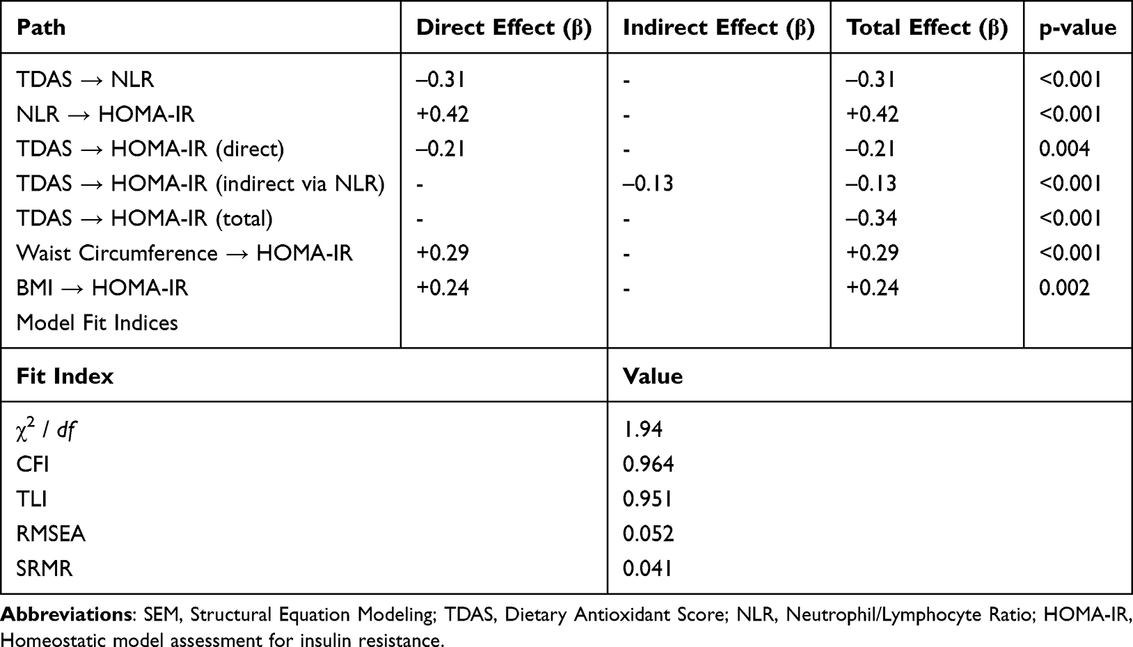 |
Table 5 Structural Equation Modeling (SEM): Direct, Indirect, and Total Effects |
Hierarchical Multivariable Regression Models
Hierarchical multivariable regression outcomes are shown in Table 6.In Model 1, BMI had a β of +0.12 (SE 0.02) and waist circumference had a β of +0.05 (SE 0.01). Model R2 was 0.32. In Model 2, PCOS status had a β of +0.67 (SE 0.20), BMI had +0.08 (SE 0.02), waist circumference had +0.03 (SE 0.01), NLR had +0.41 (SE 0.11), WBC had +0.09 (SE 0.04), and fasting insulin had +0.12 (SE 0.03). Model R2 was 0.44. In Model 3, PCOS status had a β of +0.48 (SE 0.18), BMI had +0.06 (SE 0.02), waist circumference had +0.02 (SE 0.01), NLR had +0.26 (SE 0.10), WBC had +0.07 (SE 0.03), fasting insulin had +0.10 (SE 0.03), TDAS had –0.17 (SE 0.05), physical activity had –0.06 (SE 0.03), and skipping meals had +0.22 (SE 0.10). Model R2 was 0.53.
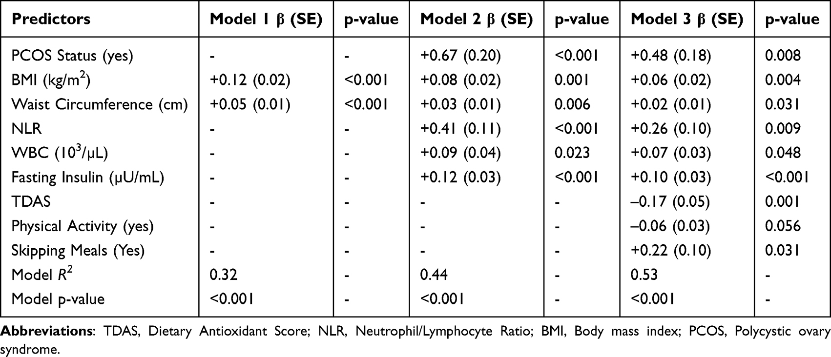 |
Table 6 Hierarchical Multivariable Regression Models Predicting HOMA-IR (Model 1–3) |
Discussion
This study presents a multidimensional profile of metabolic, inflammatory, and nutritional parameters in women with PCOS, broadly assessing the systemic aspects of the syndrome. The findings demonstrate that PCOS is not only an endocrine disorder but also a complex metabolic condition closely linked to lifestyle, inflammation, and dietary patterns.1,27
Sociodemographic and Lifestyle Characteristics
The lack of significant differences between the groups in sociodemographic variables supports the conclusion that metabolic and inflammatory differences stem from biological or lifestyle-based mechanisms.28 Furthermore, the adverse lifestyle patterns observed in the PCOS group lower water consumption, more frequent meal skipping, increased snacking tendency, and insufficient physical activity point to behavioral determinants of the syndrome. This pattern is consistent with the literature showing that eating behaviors in women with PCOS are shaped by impaired glycemic regulation, hyperandrogenism, and metabolic inflammation.11,29 PCOS habits should be reinforced with previous research explaining why lower water consumption, meal skipping, increased snacking, insufficient physical activity, and psychological variables are determinants of PCOS. Inadequate water intake, frequent meal skipping, increased snacking behavior, and low levels of physical activity observed in women with PCOS may contribute to metabolic dysregulation through multiple mechanisms. Reduced hydration status has been associated with impaired glucose metabolism and increased insulin concentration, potentially exacerbating insulin resistance. Irregular eating patterns, particularly meal skipping, may promote postprandial glycemic fluctuations, compensatory hyperinsulinemia, and increased oxidative stress, thereby worsening metabolic control. Similarly, insufficient physical activity reduces insulin sensitivity and favors a proinflammatory state, which is known to aggravate both metabolic and reproductive features of PCOS.
In addition, the higher prevalence of psychological symptoms such as fatigue, low energy, and depressed mood in the PCOS group may further interact with metabolic and inflammatory pathways. Chronic low-grade inflammation and hyperinsulinemia have been shown to influence neuroendocrine signaling and stress-related hormonal responses, potentially contributing to mood disturbances. The coexistence of adverse lifestyle behaviors, psychological burden, and elevated inflammatory markers in the present study supports the concept that PCOS is shaped by intertwined behavioral, metabolic, and inflammatory mechanisms rather than isolated endocrine dysfunction.
The higher prevalence of psychological symptoms (fatigue, low energy, and depressed mood) compared to the control group is consistent with current evidence supporting psycho-neuroendocrine interactions in PCOS.30 Chronic inflammation and hyperinsulinemia may predispose to mood disorders by affecting the modulation of the neuroendocrine axis.31,32 The co-occurrence of elevated inflammatory markers and psychological symptoms in our study points to these neuroimmune mechanisms.
Anthropometric Characteristics
The unhealthy body composition profile observed in the PCOS group, particularly the high levels of abdominal obesity indicators, points to a metabolic origin of the syndrome. Central obesity is known to be a determinant of insulin resistance, inflammation, and cardiometabolic risk,33 and our findings suggest that this risk becomes evident at an early age. The increase in neck and upper arm circumference suggests that fat accumulation is not limited to the abdominal region. This is consistent with studies suggesting that the increase in insulin and IGF-1 in PCOS may be associated with anabolic processes affecting both adipose tissue and muscle tissue.34,35 This anthropometric profile demonstrates that PCOS is not only a hormonal disorder but also a systemic metabolic disorder that reshapes body composition.
Biochemical and Hormonal Parameters
The significantly impaired glycemic indicators in the PCOS group in the study further confirm that insulin resistance is the central pathophysiological mechanism of PCOS. Recent reviews have indicated that insulin resistance is the primary process that both perpetuates hyperandrogenism and impairs ovarian function.36,37
Furthermore, the more unfavorable lipid profile in the PCOS group indicates that cardiometabolic risk is high even in the early stages of the syndrome. In particular, low HDL and elevated LDL/triglyceride levels are consistent with research demonstrating an atherogenic metabolic environment even in the young PCOS population.38,39 Elevated liver enzymes suggest that PCOS negatively impacts hepatic lipid metabolism and is associated with a higher risk of NAFLD. PCOS has been strongly demonstrated in recent years to be an independent risk factor for NAFLD.40,41 Hormonally, elevated LH and an elevated LH/FSH ratio are hallmarks of the classic PCOS phenotype. This hormonal imbalance suppresses follicular development, increasing the risk of infertility and is identified as a key element of the neuroendocrine structure of PCOS in recent studies.42
Dietary Antioxidant Capacity and Inflammation
The significantly lower dietary antioxidant capacity in the PCOS group in our study is a critical finding linked to metabolic and inflammatory disorders. A diet rich in antioxidants is known to increase insulin sensitivity, reduce oxidative stress, and exert regulatory effects on inflammation.43 Therefore, low antioxidant intake in individuals with PCOS may be one of the primary nutrition-based mechanisms that increase inflammatory burden.
The elevation of inflammatory markers (WBC, NLR) in the study supports the notion that PCOS is a syndrome characterized by chronic low-grade inflammation. The parallel course of inflammation with dermatological symptoms (acne, skin lesions) demonstrates the influence of inflammatory biomarkers on the clinical presentation. This relationship is consistent with recent findings highlighting the immunometabolic aspect of PCOS.44–46
SEM Findings and Regression Models
The direct suppressive effect of dietary antioxidant capacity on inflammation in the structural equation analysis is one of the most important nutrition-focused contributions of the study. While the effects of antioxidants on modulating inflammatory processes have long been debated, it is noteworthy that this relationship was confirmed by statistical modeling in the context of PCOS.47 The strong impact of inflammation on insulin resistance suggests that the inflammation-metabolism axis is a key determinant in PCOS. Recent literature suggests that inflammatory burden disrupts glucose homeostasis.46 This mechanistic relationship was also confirmed with a high effect size in our model. The fact that dietary antioxidant capacity reduces HOMA-IR both directly and indirectly through inflammation demonstrates the bidirectional effect of nutrition on metabolic outcomes in PCOS. This suggests that antioxidant-rich diets may be a clinically relevant target in the treatment of PCOS.48
The presence of PCOS in hierarchical regression analyses remained an independent predictor of insulin resistance, demonstrating the metabolic burden of the syndrome. This relationship persisted even when lifestyle variables and dietary antioxidant capacity were added to the model, demonstrating the biological basis of the metabolic nature of PCOS.13 The finding that dietary antioxidant capacity was negatively associated with HOMA-IR independently suggests that it is an important component of PCOS that can be modulated by diet. This finding is consistent with the concept of “nutrition-sensitive metabolic disorders”, frequently emphasized in the journal Nutrition. The positive association of skipping meals with insulin resistance is consistent with the literature demonstrating that irregular eating leads to impaired metabolic processes.49 The protective effect of physical activity supports the behavioral components of metabolic health.50
This study provides a comprehensive and multidimensional assessment of PCOS by simultaneously examining dietary antioxidant capacity, inflammatory biomarkers, metabolic indicators, and anthropometric characteristics within the same analytical framework. The use of advanced statistical approaches, including structural equation modeling and hierarchical regression, allowed for a nuanced evaluation of both direct and indirect pathways linking diet, inflammation, and insulin resistance. The inclusion of a well-defined control group and detailed phenotyping of participants enhanced the robustness and internal validity of the findings.
Conclusion
The findings suggest that PCOS is a systemic disorder shaped by the interaction between dietary patterns, inflammation, and metabolic health. In particular, the fact that low dietary antioxidant capacity exacerbates both inflammatory burden and insulin resistance highlights the clinical importance of antioxidant-rich dietary patterns in the management of PCOS. Given the focus of the Nutrition journal, these findings clearly demonstrate that PCOS should be considered a metabolic disease susceptible to nutrition-based interventions.
Data Sharing Statement
The data that support the findings of this study are available from the corresponding author upon reasonable request. Due to privacy and ethical restrictions, raw participant data cannot be shared publicly.
Ethics Statement
Ethical approval was obtained from the Health Sciences Research Ethics Committee of Osmaniye Korkut Ata University, which reviewed and approved the study protocol during its meeting on 23 May 2025 (Decision No: 2025/5/21; Document No: E-58565088-100-236106). All participants were informed about the study procedures in detail and provided written informed consent prior to inclusion.
Author Contributions
H.A.: Conceptualization; Methodology; Investigation; Data curation; Formal analysis; Writing – original draft; Writing – review & editing; Supervision.
N.A.: Conceptualization; Methodology; Investigation; Data curation; Writing – review & editing.L.K.: Investigation; Resources; Writing – review & editing.
C.A.: Investigation; Data curation; Writing – review & editing.
All authors gave final approval of the version to be published; have agreed on the journal to which the article has been submitted; and agree to be accountable for all aspects of the work.
Funding
The study was supported by Malatya Turgut Ozal University Scientific Research Projects Coordination Unit with project number 26G22.
Disclosure
The authors declare that they have no conflicts of interest regarding the publication of this paper.
References
1. Di Lorenzo M, Cacciapuoti N, Lonardo MS, et al. Pathophysiology and nutritional approaches in polycystic ovary syndrome (PCOS): a comprehensive review. Current Nutr Rep. 2023;12(3):527–14. doi:10.1007/s13668-023-00479-8
2. Calcaterra V, Verduci E, Cena H, et al. Polycystic ovary syndrome in insulin-resistant adolescents with obesity: the role of nutrition therapy and food supplements as a strategy to protect fertility. Nutrients. 2021;13(6):1848. doi:10.3390/nu13061848
3. Barrea L, Frias-Toral E, Verde L, et al. PCOS and nutritional approaches: differences between lean and obese phenotype. Metab Open. 2021;12:100123. doi:10.1016/j.metop.2021.100123
4. Zheng N, Wang C, Li Y, Fu H, Hu T. Myricetin ameliorates polycystic ovary syndrome in mice by brown adipose tissue activation. Reproduction. 2024;167(6). doi:10.1530/REP-24-0034
5. Dubey P, Reddy S, Boyd S, et al. Effect of nutritional supplementation on oxidative stress and hormonal and lipid profiles in PCOS-affected females. Nutrients. 2021;13(9):2938. doi:10.3390/nu13092938
6. Frias-Toral E, Garcia-Velasquez E, de Los Angeles Carignano M, Rodriguez-Veintimilla D, Alvarado-Aguilera I, Bautista-Litardo N. Polycystic ovary syndrome and obesity: clinical aspects and nutritional management. Minerva Endocrinol. 2021;47(2):215–241. doi:10.23736/S2724-6507.21.03349-6
7. Abraham Gnanadass S, Divakar prabhu Y, Valsala Gopalakrishnan A. Association of metabolic and inflammatory markers with polycystic ovarian syndrome (PCOS): an update. Arch Gynecol Obstetrics. 2021;303(3):631–643. doi:10.1007/s00404-020-05951-2
8. Zhang J, Li Z, Liu X-R. Dietary inflammatory index and risk of PCOS: a systematic review and meta-analysis. Nutrients. 2021;13(7):2100. doi:10.3390/nu13062100
9. Barrea L, Arnone A, Annunziata G, et al. Adherence to the Mediterranean diet, dietary patterns and body composition in women with polycystic ovary syndrome (PCOS). Nutrients. 2019;11(10):2278. doi:10.3390/nu11102278
10. Barrea L, Muscogiuri G, Pugliese G, de Alteriis G, Colao A, Savastano S. Metabolically healthy obesity (MHO) vs. metabolically unhealthy obesity (MUO) phenotypes in PCOS: association with endocrine-metabolic profile, adherence to the Mediterranean diet, and body composition. Nutrients. 2021;13(11):3925. doi:10.3390/nu13113925
11. Garad R, Shorakae S, Teede H. Assessment and management of women with polycystic ovary syndrome (PCOS). In: Advanced Practice in Endocrinology Nursing. Springer; 2019:753–769.
12. Mahabady M. The association between dietary obesity-prevention score and obesity risk in women with PCOS. Sci Rep. 2024;14(1):80238.
13. Marino JS, Iler J, Dowling AR, et al. Adipocyte dysfunction in a mouse model of polycystic ovary syndrome (PCOS): evidence of adipocyte hypertrophy and tissue-specific inflammation. PLoS One. 2012;7(10):e48643. doi:10.1371/journal.pone.0048643
14. Vassilatou E. Inflammation and insulin resistance in PCOS: the role of diet and lifestyle. J Endocrinol Invest. 2020;43(12):1721–1731.
15. Wang Q, Sun Y, Xu Q, et al. Higher dietary inflammation potential and certain dietary patterns are associated with polycystic ovary syndrome risk in China: a case-control study. Nutr Res. 2022;100:1–18. doi:10.1016/j.nutres.2021.12.006
16. Scannell N, Villani A, Moran L, Mantzioris E. The potential role of the Mediterranean diet for the treatment and management of polycystic ovary syndrome: a review of the pathophysiological mechanisms and clinical evidence.
17. Rudnicka E, Suchta K, Grymowicz M, et al. Chronic low grade inflammation in pathogenesis of PCOS. Int J Mol Sci. 2021;22(7):3789. doi:10.3390/ijms22073789
18. Shahid R, Mahnoor, Awan KA, Iqbal MJ, Munir H, Saeed I. Diet and lifestyle modifications for effective management of polycystic ovarian syndrome (PCOS). J Food Biochem. 2022;46(7):e14117. doi:10.1111/jfbc.14117
19. World Health Organization. Obesity and overweight 2022 [Available from: https://www.who.int/news-room/fact-sheets/detail/obesity-and-overweight.
20. de Onis M; WHO Multicentre Growth Reference Study Group. Reliability of anthropometric measurements in the WHO multicentre growth reference study. Acta Paediatrica. 2006;95:38–46. doi:10.1111/j.1651-2227.2006.tb02374.x
21. Bogers RP, Dagnelie PC, van den Brandt PA, et al. Using a correction factor to correct for overreporting in a food-frequency questionnaire does not improve biomarker-assessed validity of estimates for fruit and vegetable consumption. J Nutr. 2003;133(4):1213–1219. doi:10.1093/jn/133.4.1213
22. Niki E. Antioxidant capacity: which capacity and how to assess it? J Berry Res. 2011;1(4):169–176. doi:10.3233/JBR-2011-018
23. Qin B, Ma N, Tang Q, et al. Neutrophil to lymphocyte ratio (NLR) and platelet to lymphocyte ratio (PLR) were useful markers in assessment of inflammatory response and disease activity in SLE patients. Mod Rheumatol. 2016;26(3):372–376. doi:10.3109/14397595.2015.1091136
24. Lewandowski KC, Skowrońska-Jóźwiak E, Łukasiak K, et al. How much insulin resistance in polycystic ovary syndrome? Comparison of HOMA-IR and insulin resistance (Belfiore) index models. Arch Med Sci. 2019;15(3):613–618. doi:10.5114/aoms.2019.82672
25. Rosenberger ME, Fulton JE, Buman MP, et al. The 24-hour activity cycle: a new paradigm for physical activity. Med Sci Sports Exercise. 2019;51(3):454. doi:10.1249/MSS.0000000000001811
26. Meyers LS, Gamst GC, Guarino A. Performing Data Analysis Using IBM SPSS. John Wiley & Sons; 2013.
27. Mohamed AH, Albasheer O, Ghoniem MA, et al. Impact of lifestyle interventions on reproductive and psychological outcomes in women with polycystic ovary syndrome: a systematic review. Medicine. 2025;104(3):e41178. doi:10.1097/MD.0000000000041178
28. Fatima Q, Jeelani H, Abass S, Farooq M, Rashid F. Polycystic Ovary Syndrome (PCOS): clinical Features, Risk Factors, Biomarkers, Treatment, and Therapeutic Strategies. In: Toxicology and Human Health: Environmental Exposures and Biomarkers. Springer; 2023:197–229.
29. Balkrishna A, Rana M, Mishra S, et al. Exploring the therapeutic potential of phytochemicals apigenin and ellagic acid in managing polycystic ovarian syndrome and its comorbidities: a comprehensive review. Front Endocrinol. 2025;16:1633377. doi:10.3389/fendo.2025.1633377
30. Reinisch I, Ghosh A, Noé F, et al. Unveiling adipose populations linked to metabolic health in obesity. Cell Metab. 2025;37(3):640–55.e4. doi:10.1016/j.cmet.2024.11.006
31. Sharma S, Mahajan N. Polycystic ovarian syndrome and menopause in forty plus women. J Mid-Life Health. 2021;12(1):3–7. doi:10.4103/jmh.jmh_8_21
32. Khan A, Sundus H, Zaidi S, Khan H, Khan SA. Comparison of sleep quality, fatigue and physical performance among working and non-working females with polycystic ovary syndrome: a cross-sectional study. Sleep Vigilance. 2025;2025:1–9.
33. Singh S, Arya V, Mishra RK, Rajput SK. Clinical efficacy of yoga practices in the management of polycystic ovarian syndrome: insights from a single-arm pilot trial. Int Res J Ayurveda Yoga. 2025;8:1–10. doi:10.48165/IRJAY.2025.80901
34. Ibáñez L, Oberfield SE, Witchel S, et al. An international consortium update: pathophysiology, diagnosis, and treatment of polycystic ovarian syndrome in adolescence. Hormone Res Paediatrics. 2017;88(6):371–395. doi:10.1159/000479371
35. Park S-J, Kim YN, Oh BK, Kang J. Risk factors for metabolic syndrome in the premetabolic state assessed using hierarchical clustering study in a health screening group. Sci Rep. 2024;14(1):31169. doi:10.1038/s41598-024-82513-5
36. Fijałek P, Karczmarz J, Paprocka A, et al. Inositol–multidimensional support for metabolic and mental health. Med Sci. 2025;29:e23ms3530. doi:10.54905/disssi.v29i156.e23ms3530
37. Fernandez RC, Moore VM, Van Ryswyk EM, et al. Sleep disturbances in women with polycystic ovary syndrome: prevalence, pathophysiology, impact and management strategies. Nat Sci Sleep. 2018;Volume 10:45–64. doi:10.2147/NSS.S127475
38. Guillot CC, Martínez RM, Alfonso YMM. Relative impact of probiotics on improving insulin resistance in women with PCOS: an evaluation with neutrosophic plithogenic statistics. Neutrosophic Sets Syst. 2025;89:266–274.
39. Kar S. Changing Profile of the PCOS Women: a 15 Year Retrospective Cohort Study. J Human Reprod Sci. 2025;18(4):225–232.
40. Waldrop SW, Buenaventura M, Reyes KJC, Stanford FC. Disparities in the diagnosis and management of polycystic ovarian syndrome in adolescents. Endocrinol Metab Clin. 2025;54(2):233–250. doi:10.1016/j.ecl.2025.02.016
41. Eslam M, Fan J-G, Yu M-L, et al. The Asian Pacific association for the study of the liver clinical practice guidelines for the diagnosis and management of metabolic dysfunction-associated fatty liver disease. Hepatol Internat. 2025;19:1–41. doi:10.1007/s12072-024-10773-4
42. Di Michele S, Fulghesu AM, Pittui E, et al. Ultrasound assessment in polycystic ovary syndrome diagnosis: from origins to future perspectives—a comprehensive review. Biomedicines. 2025;13(2):453. doi:10.3390/biomedicines13020453
43. Dumesic DA, Oberfield SE, Stener-Victorin E, Marshall JC, Laven JS, Legro RS. Scientific statement on the diagnostic criteria, epidemiology, pathophysiology, and molecular genetics of polycystic ovary syndrome. Endocrine Rev. 2015;36(5):487–525. doi:10.1210/er.2015-1018
44. Ntikoudi A, Spyrou A, Evangelou E, Dokoutsidou E, Mastorakos G. The effect of menopausal status, insulin resistance and body mass index on the prevalence of non-alcoholic fatty liver disease. In: Healthcare. MDPI; 2024.
45. Liao M, Li X, Zhang H, et al. Effects and plasma proteomic analysis of GLP-1RA versus CPA/EE, in combination with metformin, on overweight PCOS women: a randomized controlled trial. Endocrine. 2024;83(1):227–241. doi:10.1007/s12020-023-03487-4
46. Xia J, Ding L, Liu G. Metabolic syndrome and dermatological diseases: association and treatment. Nutr Metab. 2025;22(1):36. doi:10.1186/s12986-025-00924-1
47. Reivan Ortiz GG, Maraver-Capdevila L, Granero R. Predicting metabolic and cardiovascular healthy from nutritional patterns and psychological state among overweight and obese young adults: a neural network approach. Nutrients. 2025;17(16):2651. doi:10.3390/nu17162651
48. Roya R, Mohammad Akbar A, Wajeeda T, et al. An Indian evidence-based study of prevalence, phenotypic features, lifestyle modifications of polycystic ovarian syndrome patients. J Gynecol Women’s Health. 2021;21:556069.
49. Ashraf A, Singh R, Mir S. Clinical profiling of polycystic ovary syndrome patients in Kashmir population. Matrix Sci Pharma. 2022;6(1):23–33. doi:10.4103/mtsp.mtsp_4_22
50. Khan GH. The Diagnostic and Prognostic Role of Proteomics and Metabolomics in Polycystic Ovary Syndrome. University of Nottingham; 2018.
 © 2026 The Author(s). This work is published and licensed by Dove Medical Press Limited. The
full terms of this license are available at https://www.dovepress.com/terms
and incorporate the Creative Commons Attribution
- Non Commercial (unported, 4.0) License.
By accessing the work you hereby accept the Terms. Non-commercial uses of the work are permitted
without any further permission from Dove Medical Press Limited, provided the work is properly
attributed. For permission for commercial use of this work, please see paragraphs 4.2 and 5 of our Terms.
© 2026 The Author(s). This work is published and licensed by Dove Medical Press Limited. The
full terms of this license are available at https://www.dovepress.com/terms
and incorporate the Creative Commons Attribution
- Non Commercial (unported, 4.0) License.
By accessing the work you hereby accept the Terms. Non-commercial uses of the work are permitted
without any further permission from Dove Medical Press Limited, provided the work is properly
attributed. For permission for commercial use of this work, please see paragraphs 4.2 and 5 of our Terms.
Recommended articles

Semaglutide Alleviates Ovary Inflammation via the AMPK/SIRT1/NF‑κB Signaling Pathway in Polycystic Ovary Syndrome Mice
Liu M, Guo S, Li X, Tian Y, Yu Y, Tang L, Sun Q, Zhang T, Fan M, Zhang L, Xu Y, An J, Gao X, Han L, Zhang L
Drug Design, Development and Therapy 2024, 18:3925-3938
Published Date: 4 September 2024
Integrative Mendelian Randomization and Whole-Blood Transcriptomic Analysis Implicate a Myeloid–Inflammation Axis in Polycystic Ovary Syndrome
Fu Y, Li W, Ma L
International Journal of Women's Health 2026, 18:576568
Published Date: 14 January 2026