Back to Journals » Journal of Inflammation Research » Volume 14
Diagnostic and Prognostic Value of Immune/Inflammation Biomarkers for Venous Thromboembolism: Is It Reliable for Clinical Practice?
Authors Xue J, Ma D, Jiang J , Liu Y
Received 1 July 2021
Accepted for publication 19 September 2021
Published 2 October 2021 Volume 2021:14 Pages 5059—5077
DOI https://doi.org/10.2147/JIR.S327014
Checked for plagiarism Yes
Review by Single anonymous peer review
Peer reviewer comments 2
Editor who approved publication: Dr Monika Sharma
Junshuai Xue,1,* Delin Ma,2,* Jianjun Jiang,1 Yang Liu1
1Department of General Surgery, Vascular Surgery, Shandong University Qilu Hospital, Jinan City, Shandong Province, People’s Republic of China; 2Department of General Surgery, Shandong University Qilu Hospital, Jinan City, Shandong Province, People’s Republic of China
*These authors contributed equally to this work
Correspondence: Yang Liu
Department of General Surgery, Vascular Surgery, Shandong University Qilu Hospital, No. 107, Road Wen Hua Xi, Jinan City, Shandong Province, 250012, People’s Republic of China
Tel +86 18560088317
Email [email protected]
Abstract: Venous thromboembolism (VTE), which includes deep vein thrombosis (DVT) and pulmonary embolism (PE), has been an important cause of sudden in-hospital death. Studies have shown that the immune/inflammatory response plays an important role in the pathogenesis of vascular disease, with representative markers in the blood including the neutrophil/lymphocyte ratio (NLR), platelet/lymphocyte ratio (PLR), monocyte/lymphocyte ratio (MLR), systemic immune/inflammatory index (SII), etc. However, there is a variety of immune/inflammatory indicators. Moreover, most previous studies have been single-center investigations involving one or two indicators, with varying nature of cases, number of cases and study objectives, thereby making it difficult to reach consensus conclusions with good clinical guidelines. This article reviews the clinical value of immunoinflammatory indicators for VTE based on previous studies, including the diagnostic and prognostic capabilities. In conclusion, NLR provides promising predictive capability for the onset and prognosis of VTE and deserves extensive application in clinical practice. PLR also has certain diagnostic and prognostic value, but further studies are warranted to identify its reliability and stability. Monocytes, eosinophils and platelet-related indicators show some clinical association with VTE, although the predictive capabilities are mediocre. SII is of promising potential value for VTE and deserves further investigations. This review will provide new clues and valuable clinical guidance for the diagnosis and therapy of VTE.
Keywords: venous thromboembolism, immune/inflammation biomarker, diagnosis, prognosis
Venous thromboembolism (VTE), which includes deep vein thrombosis (DVT) and pulmonary embolism (PE), has been an important cause of sudden in-hospital death.1 Until now, a number of risk factors have been identified to be associated with VTE, nevertheless, risk factors alone cannot promptly and effectively predict VTE occurrence and prognosis of mortality and therefore blood biomarkers are often required. The sensitivity of D-dimer for the diagnosis of VTE is sufficiently high for clinical practice. In contrast, the specificity of D-dimer is indeed suboptimal in clinical practice, as many pathophysiological processes may stimulate an increase in D-dimer. Recent studies have attempted to identify new biomarkers that balance sensitivity and specificity and act as a complement to D-dimers. Studies have shown that the immune/inflammatory response plays an important role in the pathogenesis of vascular disease, with representative markers in the blood including the neutrophil/lymphocyte ratio (NLR), platelet/lymphocyte ratio (PLR), monocyte/lymphocyte ratio (MLR), systemic immune/inflammatory index (SII), etc.2–6 These indicators are calculated from readily available biomarkers in the blood panel and are routinely measured. Over the past decades, the clinical value of these indicators of immune/inflammatory response for VTE has gradually become one of the hot topics of research, as reflected not only by the increasing number of studies published by researchers from different countries over the years, as well as their impact. (Figure 1) These studies have identified and confirmed the clinical value of these inexpensive and readily available indicators in the diagnosis and prognosis of mortality in VTE, helping to improve the specificity of VTE detection and suggesting high-risk factors for poor prognosis. However, there is a variety of immune/inflammatory indicators. Moreover, most previous studies have been single-center investigations involving one or two indicators, with varying nature of cases, number of cases and study objectives, thereby making it difficult to reach consensus conclusions with good clinical guidelines. Therefore, there is still a need for multicenter prospective studies to further verify the clinical value of different immune/inflammatory indicators. This article reviews the clinical value of immune/inflammatory indicators for VTE based on previous studies, including the diagnostic and prognostic capabilities, that will provide new clues and valuable clinical guidance for the diagnosis and therapy of VTE.
We systematically searched PubMed, Embase, and Web of Science for relevant literature up to June 2021. The search terms used to search literature were as followed: (“neutrophil/lymphocyte ratio” OR “neutrophil to lymphocyte ratio” OR “NLR”) OR (“platelet/lymphocyte ratio” OR “platelet to lymphocyte ratio” OR “PLR”) OR (“monocyte/lymphocyte ratio” OR “MLR” OR “lymphocyte/monocyte ratio” OR “LMR”) OR (“systemic immune/inflammatory index” OR “SII”) OR (eosinophils) OR (platelets) AND (“pulmonary embolism” OR “PE”) OR (“deep venous thrombosis” OR “DVT”) OR (“venous thromboembolism” OR “VTE”). In addition, related articles and the references in the relevant studies were manually screened for additional eligible studies. The studies were included if they met the following criteria: 1) patients in the studies were diagnosed PE or DVT or VTE; 2) studies reported the specificity/sensitivity/cutoff value of immune/inflammatory indicators; 3) studies assessed associations between immune/inflammatory indicators and diagnosis/prognosis. The studies were excluded if they met the following criteria: 1) studies were not available of sufficient data; 2) studies were abstracts, letters, editorials, expert opinion, case reports; 3) studies were non-clinical or non-human research.
NLR and VTE
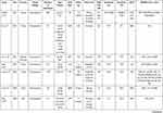
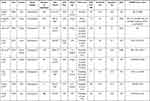
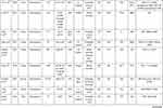
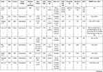
There are a number of studies on the clinical value of NLR in VTE. (Table 1) Logistic regression analysis and COX regression analysis have shown that NLR has good diagnostic value and prognostic capability for VTE. The receiver operating characteristic curve (ROC) analysis results are displayed in Figure 2A.
 |
 |
 |
 |
Table 1 Studies of NLR for VTE |
NLR provides some clinical diagnostic value for acute pulmonary embolism (APE). Ates et al identified NLR as an independent predictor of massive APE in 639 patients with an odds ratio (OR) of 2.22.7 ROC analysis determined that NLR had good clinical diagnostic value for massive APE (AUC=0.893, p<0.001). Ateş et al also demonstrated a comparison of 34 patients with pulmonary embolism (PE) and 38 patients with community-acquired pneumonia (CAP) and found that patients with PE had lower levels of NLR on both the day of admission and the third day of admission (p=0.002, p=0.004).8 The NLR/D-dimer ratio provided better differential diagnostic power between PE and CAP than traditional indicators such as D-dimer, PCT and CRP, with a sensitivity of 97.4%, a negative predictive value of 96.7% and a positive predictive value of 91.7%, and an AUC of 0.921. Köse et al also observed in 103 patients with PE that NLR was significantly associated with the onset of PE (OR=1.52, p<0.001).9 Furthermore, NLR was strongly associated with the prognosis of mortality in low-medium risk PE and low risk PE (hazard ratio [HR]=1.17, p=0.033), while NLR was not associated with the prognosis of mortality in medium-high risk PE and high risk PE (HR=0.99, p=0.859). Celik et al identified significantly higher NLR levels in patients with acute PE than in those with non-acute PE (6.2±2.9 vs 5.4±3.0, p=0.03) and a high positive correlation between NLR and PLR (r=0.488, p<0.001).10 However, regression analysis showed that NLR did not predict the onset of acute PE (OR=0.887, p=0.068).
NLR is strongly correlated with in-hospital mortality, 30-day mortality and overall mortality in patients with PE. Akgüllü et al observed in 206 patients with APE that NLR was associated with early mortality (OR=1.079, 95% CI:1.005–1.160).11 ROC analysis showed that NLR had favorable prognostic value with a sensitivity of 76.67%, a specificity of 81.82% and an AUC of 0.825. In addition, NLR showed a higher sensitivity (90%), specificity (79.55%) and AUC (0.906) when combined with sPESI. In 142 patients with APE, Soylu et al have found that the group with a high NLR (≥4.4) was more likely to have massive PE (66.2% vs 36.6%, P<0.001) than the group with a low NLR (<4.4), and its in-hospital mortality (21.1% vs 1.4%, P<0.001) was also higher.12 Multivariate regression analysis identified NLR as an independent predictor of in-hospital mortality, and ROC analysis showed that patients with an NLR value over 5.7 had a 10.8-fold higher mortality rate than those with an NLR value below 5.7. Bi et al found that NLR was significantly higher in the patients who died than in the survival group (P<0.05) in 72 patients with PE. The AUC for NLR to predict mortality was 0.861, with a sensitivity of 0.779 and specificity of 0.890.13 And a combination of NLR with BNP, TnI and D-dimer showed a higher predictive value with an AUC of 0.92, sensitivity of 0.889 and specificity of 0.904.
Kasapoğlu et al retrospectively analysed 550 patients with APE, where NLR levels were significantly higher in patients who died within 30 days (p=0.003).14 Short-term mortality was also notably higher in patients with NLR >7.3 (p<0.05). Although Cox regression analysis found that NLR was not an independent predictor of death for overall patients (HR=1.27, p=0.669). A subgroup analysis in patients without comorbidities showed that NLR was an independent predictor of mortality (HR=3.3, p=0.016). Kayrak et al performed multivariate Cox regression analysis on 359 patients with APE and found that NLR was an independent predictor of 30-day mortality (HR=1.03, 95% CI:1.01–1.06, p=0.008) with a cut-off value of 9.2 and sensitivity, specificity, negative predictive value and positive predictive value of 68.6%, 80.5%, 93.9% and 36.5%, respectively.15 Ma et al also found in 248 patients with APE that a 1 unit increase of NLR was associated with a 13% increased risk of death at 30 days (OR=1.13, 95% CI:1.04–1.23, p=0.003).16 NLR predicted 30-day mortality with a sensitivity of 80%, specificity of 66.7% and AUC of 0.79 (95% CI: 0.703–0.880).
Liu et al found that 24 patients (23.8%) died within 30 days in 101 patients with postoperative acute pulmonary embolism (PAPE).17 Multivariate analysis showed that NLR was an independent predictor of 30-day mortality in patients with PAPE, with a 1-unit increase in NLR related to a 17.1% increase in the probability of death (OR=1.171, 95% CI:1.073–1.277, p=0.000). ROC analysis showed that NLR had a predictive sensitivity of 62.5%, specificity of 90.9% and AUC of 0.823. Jia et al detected significantly higher levels of NLR in 154 APE patients with right heart insufficiency and regression analysis showed that NLR was associated with right heart insufficiency (OR=1.075, p=0.024).18 Besides, NLR was associated with 30-day mortality (P<0.001) with a predictive sensitivity of 83.3%, specificity of 75.6% and AUC of 0.803 (95% CI: 0.730–0.875).
NLR has also been verified to predict the mortality when risk levels are stratified for PE patients. Telo et al revealed that NLR was significantly higher in PE patients with high-risk for mortality than in those with low-risk for mortality based on the sPESI score (p<0.01).19 NLR had a predictive sensitivity of 66%, a specificity of 53% and an AUC of 0.675 (95% CI: 0.556–0.794). All patients were divided into two groups according to the NLR cut-off value of 3.56, showing statistically significant increase in-hospital mortality, 3rd month mortality and overall 3-month mortality in the groups above 3.56 (χ2=4.771, p<0.05; χ2=4.383, p<0.05; χ2=9.101, p<0.01). Phan et al found there was no difference in NLR levels among 191 APE patients with low-risk, sub-risk and high-risk for mortality (p=0.58).20 However, NLR was significantly higher in patients who died than in those who survived (8.10 [4.28–13.7] vs 3.91 [2.46–6.71]; p<0.01). Furthermore, elevated NLR was associated with all-cause mortality with a predictive sensitivity of 75.0%, specificity of 66.9% and AUC of 0.692 (95% CI: 0.568–0.816).
Duman et al found that an NLR above 6.1 increased mortality at 30 days by 13.8-fold in a cohort of 828 PE patients.21 NLR over 3.25 was an independent risk factor for 6-month mortality (HR=2.77, 95% CI: 1.48–5.18, p=0.001) and NLR over 3.14 was an independent risk factor for 1-year mortality (HR=2.20, 95% CI: 1.32–3.66, p=0.003). Karataş et al followed 203 patients with APE for a mean of 20 months and 34 patients (17%) died during the follow-up period.22 Patients who died within 30 days had significantly higher NLR levels than those who survived (9.9 vs 4.5, p=0.01). NLR levels were also markedly elevated during long-term follow-up (8.4 vs 4.1, p=0.01). Cox regression analysis also demonstrated elevated NLR was an independent risk factor for overall mortality (HR = 1.13, 95% CI: 1.04–1.23, p=0.01).
However, a few studies have shown that there is no significant correlation between NLR and death in patients with PE. For example, Ghaffari et al found no significant difference in NLR in 492 APE patients with and without major cardiopulmonary adverse events (p=0.206) and that NLR was not associated with patient mortality.23 Ertem et al followed 294 patients with APE, of whom 64 (21.8%) died and 230 (78.2%) survived.24 Although NLR was higher in patients who died (P<0.001), there was no significant correlation between NLR and death (HR: 0.990, P=0.240).
Meaningful findings regarding NLR have also been obtained when some investigators have analysed PE and DVT together as VTE. Farah et al discovered significantly elevated NLR level in 272 patients with acute VTE (p<0.001).25 Multivariate logistic regression models identified NLR for early detection of potential acute VTE (OR=1.2, 95% CI: 1.01–1.4, p=0.041), with a predictive sensitivity of 69%, specificity of 57% and AUC of 0.67 (95CI: 0.60–0.75). However, Artoni et al observed no increment in the risk of VTE in patients with high NLR among 486 patients (OR=0.69, 95% CI: 0.34–1.39).26 Fuentes et al identified 13 patients (11.6%) with VTE during follow-up of 112 gastric cancer patients, but NLR was not associated with the onset of VTE (HR=0.8, p=0.8), with a sensitivity of 46.2% and specificity of 47.5%.27
NLR can also predict the occurrence of VTE after osteopathic surgery. Yao et al found that 120 patients who suffered postoperative DVT (16.4%) had a significantly higher preoperative NLR (p=0.030) and postoperative NLR (p=0.015) than those who did not suffer from DVT among 733 patients after total joint arthroplasty.28 Multiple logistic regression analysis showed that both preoperative NLR (OR=1.11, P<0.035) and postoperative NLR (OR=1.20, P<0.001) were independently associated with the occurrence of DVT, with an AUC of 0.533 (95% CI: 0.473–0.592) and an AUC of 0.613 (95% CI: 0.564–0.662), respectively. Seo et al observed that VTE occurred in 102 cases (38.6%) at 1 week postoperatively in 264 patients after total knee arthroplasty.29 Preoperative NLR≥1.90 was the only independent predictor of postoperative VTE (OR=1.95, 95% CI: 1.16–3.31, p=0.013). ROC analysis revealed a predictive sensitivity of 57.8%, specificity of 55.6% and AUC of 0.589. Barker et al demonstrated higher levels of NLR in patients with total knee arthroplasty than in patients with unicondylar knee arthroplasty (p=0.02).30 NLR could predict VTE during hospitalization in patients after total knee arthroplasty (OR=1.38; 95% CI:1.05–1.80, p=0.02). However, Peng et al found that NLR was not an independent predictor of VTE after hip fracture among 52 patients over 60 years old, although the NLR was higher in them than in those without VTE (p=0.078).31
NLR was also correlated with death in patients with VTE. Grimnes et al found although NLR was not associated with first occurrence or recurrence of VTE, NLR was associated with increased mortality in 25,107 patients (HR=2.13, 95% CI:1.26–3.58, p=0.02).32 Grilz et al identified 128 (16.4%) patients who experienced VTE during follow-up of 1469 cancer patients, and NLR was statistically associated with the occurrence of VTE after multivariate adjustment for age, gender and cancer stage (HR=1.2, 95% CI:1.0–1.4, p=0.049).33 In addition, NLR was associated with 24-month mortality with a HR of 1.5 (95% CI: 1.0–1.4, p<0.001). Ming et al also observed that NLR was an independent risk factor for death in 115 patients with acute DVT (OR=1.889, 95% CI: 1.086–3.286, p=0.024).34 However, Ferroni et al revealed from 55 cancer patients receiving chemotherapy that symptomatic VTE patients had higher post-chemotherapy NLRs than pre-chemotherapy NLRs (p=0.015).35 NLR > 3 was associated with the incidence of symptomatic VTE, with an HR of 2.5 (95% CI: 1.0–6.4, p=0.06). And patients with NLR > 3 had a worse survival rate at one year (Log rank test: 2.32, p = 0.020). Although ROC analysis showed a sensitivity of 59%, specificity of 57% and AUC of 0.55, NLR was not an independent risk factor for the occurrence of VTE (p>0.05). Go et al found that among 114 lung cancer patients with VTE, those with high NLR responded poorly to anticoagulation therapy (p=0.004).36 Moreover, NLR was associated with prognosis of mortality in lung cancer patients with VTE, but NLR did not predict mortality (p>0.05).
NLR may have an association with endothelial cell function and pulmonary hypertension. In a study of 106 patients with chronic thromboembolic pulmonary hypertension, Yanartas et al found that patients with higher NLR on admission experienced significantly higher mortality and NLR was an independent predictor of death (OR=2.767, p=0.002).37 ROC analysis showed an optimal cut-off value of 2.54 for NLR, with a sensitivity of 86%, specificity of 40%, and AUC of 0.825. Correlation analysis also showed a clear correlation between NLR and preoperative pulmonary vascular resistance (r=0.214, p=0.027). Kurtipek et al observed significantly higher NLR values in the 71 patients with APE than in healthy controls (4.2±4.90 vs 1.89±1.46, p=0.0001), suggesting possible endothelial cell dysfunction.38
NLR can also indicate the location of venous thrombosis in the lower extremities. Kuplay et al found that NLR correlated with thrombus location in 933 patients with DVT.39 Mean NLR was higher in patients with proximal DVT than distal DVT (4.40±4.28 vs 3.54±3.55; p<0.05) and NLR was increased with the number of venous segments involved (p<0.001). Its predictive sensitivity was 65.4% and specificity 55.0%, with an AUC of 0.6 (95% CI: 0.56–0.64). Bakirci et al identified significant differences in NLR between patients with PE, proximal DVT and distal DVT in 77 patients (p<0.001).40 An NLR>1.84 predicted VTE with a sensitivity of 88.2%, specificity of 67.6% and AUC of 0.849 (95% CI. 0.765–0.913, p<0.001).
NLR may be related to iliac vein stent occlusion. Jahangiri et al observed higher NLR level in patients with early stent occlusion (p=0.026) in 50 patients with iliac vein stent placement.41 However, COX analysis showed that NLR was not an independent predictor of stent occlusion and only had borderline significance (p=0.050, HR=12.19, 95CI:1–147.85).
Interesting results can also be obtained when an analysis is performed on neutrophil counts alone. Kushnir et al have found that an elevated neutrophil count is associated with an increasing risk of venous thrombosis in 43,538 outpatients and that neutrophilia may be a marker of vulnerability to VTE.42 A neutrophil count≥7.8x10⁹/L was not significantly associated with the development of VTE. Patients with a neutrophil count≥9.0x10⁹/L had a twofold increased risk of VTE (OR=2.0, 95% CI:1.3–3.1, p=0.003). Patients with a neutrophil count≥10.0x10⁹/L had a higher risk of VTE (OR=2.3, 95% CI:1.2–4.8, p=0.019).
In patients with corona virus disease 2019 (COVID-19), elevated NLR may increase the risk of VTE development. Mareev et al observed a 2.5-fold increase in NLR levels when high-dose glucocorticoid (GCS) pulse therapy was administered to patients with novel coronavirus pneumonia (p=0.006), and the increase was significantly correlated with D-dimer (r=0.49, p=0.04).43 The authors suggested that therapy with high doses of GCS exerted a rapid anti-inflammatory effect, but also elevated NLR and D-dimer levels, increasing the risk of VTE.
NLR also has a predictive value for the prognosis of mortality in patients with portal vein/hepatic vein tumour thrombosis (PVTT/HVTT). Li et al found that preoperative NLR was a prognostic predictor after hepatic resection in 81 patients with PVTT/HVTT.44 The median overall survival (OS) and disease-free survival (DFS) were significantly shorter in the high NLR (>2.9) group than in the low NLR group (OS: 6.2 months vs 15.7 months, p=0.007; DFS: 2.2 months vs 3.7 months, p=0.039). NLR>2.9 was considered an independent predictor of prognosis of mortality (p=0.034, HR=1.866, 95% CI:1.048–3.322). There was also a significant positive correlation between NLR and Child-Pugh score (r=0.276, p=0.015) and the maximum diameter of the tumour (r=0.435, p<0.001).
PLR and VTE
PLR has certain diagnostic value for the onset of APE and is associated with in-hospital mortality, 30-day mortality and overall mortality (Table 2). ROC analysis results for PLR are displayed in Figure 2B. Ates et al identified PLR (OR=1.59, p<0.001) as an independent predictor of massive APE in 639 patients.7 ROC analysis provided good differential diagnostic value of PLR for massive APE with an AUC of 0.877 (p<0.001). However, Celik et al found that PLR was not an independent predictor of APE in 112 patients (OR=0.998, p=0.340).10
 |
 |
 |
Table 2 Studies of PLR for VTE |
Ghaffari et al did not find a difference in PLR between patients with and without major cardiopulmonary adverse events in 492 patients with APE (p=0.338), but PLR was associated with in-hospital mortality (p<0.01) with a sensitivity of 61%, specificity of 63.2% and AUC of 0.61 (95% CI:0.50, 0.72).23 Karataş et al observed significantly higher levels of PLR in 203 patients with APE who died within 30 days (n=14, p=0.01), as well as in long-term follow-up (n=20, p=0.01).15 Elevated PLR levels were independently associated with total mortality with a HR of 1.002 (95% CI: 1.001–1.004, p=0.01).
PLR could also predict prognosis of mortality in subgroups of patients with different risks of PE. Köse et al found that PLR was associated with prognosis of mortality in patients with moderate-high risk PE (HR=1.01, p=0.046) while not with prognosis of mortality in patients with low risk and moderate-low risk PE (HR=1.0, p=0.545) among 103 patients.9 Phan et al identified no difference in PLR between low-risk, sub-risk and high-risk for mortality in 191 PE patients (p=0.02).20 However, patients who died had significantly higher PLR levels (263 vs 148; p<0.01) and elevated PLR was associated with all-cause mortality with a sensitivity of 53.6%, specificity of 82.8% and AUC of 0.693 (95% CI:0.580–0.805, p<0.01). Telo et al observed a statistically significant increase in PLR in patients with high-risk for mortality compared to those with low-risk for mortality according to the sPESI score (p<0.01).19 And ROC analysis showed that PLR was associated with total mortality at 3rd month (OR=6.325, p<0.01) with a sensitivity of 74%, specificity of 64% and an AUC of 0.717 (95% CI: 0.539–0.896). Among 646 patients with APE, Kundi et al found higher levels of PLR in patients with high sPESI scores (p<0.001).45 PLR was independently associated with in-hospital mortality in patients with APE (p<0.001) with a sensitivity of 77.1%, a specificity of 76.3% and an AUC of 0.860 (95% CI:0.50, 0.72).
However, several more studies have also found that PLR could not predict death in PE patients, either at 30 days or in the long term. Kasapoğlu et al discovered a significantly higher level of PLR in APE patients who died within 30 days (p=0.022) and a significantly higher short-term mortality in patients with PLR>170.14 However, Cox regression analysis showed that PLR was not an independent risk factor for APE mortality (HR=1.4, p=0.503). Ma et al observed a higher PLR (p<0.01) in 248 APE patients who died, which was associated with short-term mortality (OR:1.002, 95% CI:0.997–1.008, p=0.003).16 But PLR was not an independent predictor of overall mortality. Jia et al also found significantly higher levels of PLR in APE patients with right heart insufficiency (p=0.041).18 While there was no significant difference in PLR levels between dead and surviving patients (p=0.246), and regression analysis showed that PLR was not associated with 30-day mortality (p>0.05). Duman et al also identified there was no significant difference in PLR levels between dead and surviving subgroups in 828 PE patients (p=0.13) and that PLR was not an independent risk factor for death (p>0.05).21 Ertem et al found that PLR was also not significantly associated with death in 294 patients with APE (p=0.241).24 Liu et al also revealed that PLR was not an independent predictor of 30-day mortality in patients with PAPE (p=0.193).17
PLR is a predictor of the occurrence of VTE in cancer patients. Grilz et al identified a statistically significant association between PLR and the occurrence of VTE in 1469 cancer patients, with a HR of 1.5 (95% CI:1.4–1.7, p<0.001).33 Yamagata et al identified a strong correlation between preoperative PLR and the incidence of postoperative VTE in 101 patients with oral cancer (OR=13.375, 95% CI:2.950–63.044, p=0.001).46 The predictive sensitivity of PLR was 75.0%, specificity was 74.2%, AUC was 0.772 (p=0.002). Yang et al observed that significantly elevated PLR level was an independent predictor of VTE in cancer patients (OR=2.757, 95% CI: 1.655–3.862, p=0.025).47 Ferroni et al showed a higher PLR in symptomatic VTE patients after chemotherapy than before chemotherapy (p=0.040) and PLR was an independent risk factor for the onset of VTE (HR=2.4, p=0.027).35 ROC analysis showed a sensitivity of 30% and specificity of 80% for PLR, with an AUC of 0.54.
Some studies have also found that PLR is not significantly associated with the occurrence of VTE. Fuentes et al revealed an association between PLR and mortality in 112 patients with gastric cancer (p<0.01), but PLR was not significantly different in patients with and without VTE (p=0.751).48 Artoni et al detected that patients with high PLR did not have an increased risk of VTE in 486 patients (OR=0.89, 95% CI:0.46–1.76).26 Farah et al observed a significantly higher PLR in 272 patients with acute VTE (p=0.014), but PLR was not an independent predictor (p>0.05).25 Ming et al discovered a significantly higher PLR in 105 patients with acute DVT than in controls (p=0.005), while PLR was also not an independent risk factor (p>0.05).34
PLR also has some predictive value for the onset of VTE in patients after surgical procedures. Yao et al identified a higher preoperative PLR than postoperative PLR in 733 patients after total joint replacement (p=0.002).28 Multiple logistic regression analysis showed that postoperative PLR was independently associated with the occurrence of DVT (OR=0.99, 95% CI: 0.98–0.99, p<0.001). ROC analysis showed an AUC of 0.513 (95% CI: 0.453–0.573) for preoperative PLR and an AUC of 0.561 (95% CI: 0.510–0.611) for postoperative PLR. Tham et al found seven patients (22.9%) with VTE among a total of 306 patients with head and neck cancer who underwent surgery.49 PLR was strongly associated with the occurrence of VTE with an OR of 95.95, sensitivity of 97.66%, specificity of 71.43% and AUC of 0.905 (95% CI, 0.82–0.98). Nevertheless, among patients with hip fracture, Peng et al found that PLR levels were higher in patients with VTE than in those without VTE, but PLR was not an independent predictor of VTE after hip fracture (p=0.051).31
Furthermore, Kurtipek et al found significantly higher PLR values in 71 patients with APE compared to healthy controls (140.64±126.68 vs 112.45±57.62, p=0.003), suggesting that PLR may be associated with pulmonary artery endothelial cell dysfunction.38 Kuplay et al discovered that PLR was related to thrombus location, with a higher mean PLR in proximal DVT than distal DVT (1.77x10⁷±1.3x10⁷ vs 1.49x10⁷±1.08x10⁷; p=0.03), but PLR was not increased with the number of venous segments involved (p=0.097).39
There are relatively few meta-analyses of the clinical value of NLR and PLR in VTE disease. Wang et al conducted a meta-analysis of seven studies and found that NLR and PLR were both significantly associated with mortality in patients with PE.50 NLR correlated significantly with overall (short-term and long-term) mortality (OR 10.13, 95% CI 6.57–15.64, p<0.001) and short-term (in-hospital and 30-day) mortality (OR 8.43, 95% CI 5.23–13.61, p<0.001) in patients with PE. PLR was significantly associated with overall mortality (OR 6.32, 95% CI 4.52–8.84, p<0.001), short-term mortality (OR 6.69, 95% CI 2.86–15.66, p<0.001) and long-term mortality (OR 6.11, 95% CI 3.90–9.55, p<0.001).
Monocyte-Related Indicators and VTE
Lymphocyte/monocyte ratio (LMR), monocyte/lymphocyte ratio (MLR) and monocyte count have been applied in studies concerning the diagnosis and prognosis of mortality in VTE. Köse et al found that LMR was associated with prognosis of mortality in low risk and medium-low risk PE (HR=1.58, p=0.046) and not with prognosis of mortality in medium-high and high-risk PE (HR=0.95, p=0.751).9 Ertem et al discovered that LMR was higher among deaths in PE patients (p<0.001).24 LMR was an independent risk factor for death (HR=0.211, p=0.001). ROC analysis showed a cutoff value of 1.96 for LMR, a sensitivity of 77%, a specificity of 79% and an AUC of 0.851 (95% CI: 0.802–0.900, p<0.001). However, Liu et al identified that MLR was not an independent predictor of 30-day mortality in patients with PAPE (p>0.05).17 Peng et al also identified that MLR was not an independent predictor of the occurrence of VTE after hip fracture, although MLR was higher than in patients without VTE (p=0.062).31
The monocyte count also has some predictive value for VTE. Rojnuckarin et al found that monocyte count was significantly higher in 28 patients of solid tumours with VTE compared to 280 patients without VTE (p=0.013).51 Monocyte count over 0.5 × 10⁹/L was significantly associated with VTE and was an independent predictor of VTE (OR=5.0, 95% CI:1.62–15.5, p=0.005). Among 70 VTE patients, Wypasek et al found that VTE was associated with increased non-classical and intermediate monocytes, which were higher in VTE patients than in controls (16.8 ±9.3 vs 10.4±4.0 cells/μL and 64.1±25.2 vs 44.1± 19.2 cells/μL, p<0.001).52 Maldonado-Peña et al identified a significantly higher monocyte count in 46 patients with DVT, which was strongly associated with the development of DVT (OR=9.35, 95% CI: 3.2–27.3).53 ROC analysis showed an AUC of 0.742, a sensitivity of 67.3%, a specificity of 80%, a positive predictive value (PPV) of 79.49% and a negative predictive value (NPV) of 63.9%. Nonetheless, Basavaraj et al identified 429 (17.1%) VTEs in 25,127 patients with a mean follow-up time of 12.5 years.54 In the first year, the risk of VTE was increased 2.5-fold in patients in the upper tertile of monocyte count (≥0.7x10⁹/L) compared to those in the lower tertile (≤0.4x10⁹/L) (HR:2.51; 95% CI:0.69,9.12, p=0.07). In the third year, the risk of VTE was increased 1.5-fold (HR:1.58; 95% CI:0.84,2.99, p=0.07) in patients in the upper tertile of monocyte counts (≥0.7x10⁹ /L) compared to those in the lower tertile (≤0.4x10⁹/L). Throughout the study period, the risk estimates decreased progressively and there was no association between monocyte count and VTE at the end of follow-up.
The monocyte count has shown limited clinical value in cancer patients with VTE. Go et al found that VTE occurred in 134 (7.9%) of 1707 patients with lung cancer and that monocyte count was associated with mortality (HR:1.994, 95% CI 1.137–3.498, p=0.016).55 However, Lopez-Salazar et al identified 16 of 119 (13.4%) patients with ovarian cancer who experienced VTE as having a significantly higher monocyte count (p=0.042), whereas monocyte count was not an independent predictor of VTE and mortality.56
The other two reports proposed possible mechanisms for the involvement of monocytes in VTE. Chirinos et al showed significantly higher levels of endothelial microparticle (EMP)-monocyte conjugates in 25 patients with VTE (3.3 fluorescence intensity units [FLIU], IQR 2.96–3.91) than in controls as well (2.5 FLIU; IQR 2.13–2.83; p=0.002).57 Shih et al revealed significantly higher levels of circulating platelet-monocyte aggregates (PMAs) expression (p<0.001) in 32 elderly patients experiencing postoperative VTE, which correlated with CRP levels (r2=0.536, p=0.004).58
SII and VTE
SII has been confirmed to be associated with cancer, but recent studies have shown its remarkable clinical value for both the diagnosis and prognosis of mortality in VTE.59–61 Gok et al identified elevated SII levels in 442 patients with APE and a progressive increase from non-massive to massive APE (p<0.001).62 It was also significantly higher in patients who died in-hospital (p<0.001). Multivariate analysis showed that SII was an independent predictor of massive APE (OR:1.005, 95% CI:1.002–1.007, p<0.001), with an optimal cut-off value of 1161, sensitivity of 91%, specificity of 90% and AUC of 0.957 (95% CI:0.935–0.979). Peng et al discovered an increased SII in patients with VTE compared to those without VTE.31 And SII was an independent predictor of VTE after hip fracture in elderly patients with an OR of 1.004 (95% CI: 1.001–1.008, p=0.001). ROC analysis revealed a cut-off value of 847.78, a sensitivity of 53.8%, a specificity of 92.3%, and an AUC of 0.795 (95% CI: 0.710–0.880, p<0.001).
Eosinophils and VTE
The eosinophil count also has a predictive capability for VTE and may be associated with in-hospital mortality. Among 63 patients with eosinophilia, Liu et al discovered that PE occurred in 31 (50.9%) patients with a peak absolute eosinophil count>6.3x10⁹/L (OR:5.55, 95% CI:1.292–23.875, p=0.021), with a sensitivity of 83.9%, a specificity of 75.0% and an AUC of 0.684.63 Persistence of eosinophilia over 13.9 months was an independent risk factor for PE with an OR of 4.51 (95% CI: 1.123–18.09, p=0.034). Réau et al identified VTE in 29 (53.7%) of 54 patients with eosinophilia, and persistent eosinophilia was closely associated with a shorter time to VT recurrence (HR 7.48, CI95%:1.94–29.47, p=0.015).64 The eosinophil count was not significantly elevated in patients with solid tumours combined with VTE (p=0.138), whereas it was significantly correlated with in-hospital deaths (p=0.027), as reported by Rojnuckarin et al.51 Nevertheless, the opposite conclusion was reached by two studies on eosinophil count. Wypasek et al identified that there was no significant increase in eosinophil count among 70 patients with VTE compared to controls (p=0.62).52 Maldonado-Peña et al also identified there was no apparent rise of eosinophil count in 46 patients with DVT (p=0.092).53
Platelet-Related Indicators with VTE
Among 639 patients, Ates et al detected WMR (white blood cell to mean platelet volume ratio) (OR:1.22, AUC:0.762, p<0.001); MPR (mean platelet volume to platelet ratio) (OR:0.33, AUC:0.656, p<0.001); RPR (red distribution width/platelet ratio) (OR:0.68, AUC:0.719, p<0.001) were all of certain diagnostic value for massive APE.7 Ghaffari et al found that mean platelet volume (MPV) was predictive of in-hospital mortality in 492 APE patients, with a cut-off value of 9.85fl, an AUC of 0.67, predictive sensitivity of 81% and specificity of 49.6%.23 In-hospital mortality was also highly correlated with platelet distribution width (PDW) with a cut-off value of 13.6%, and AUC of 0.66, predictive sensitivity of 66.7% and specificity of 65.2%. However, Duman et al observed that among 828 PE patients, the Platelet/MPV ratio was not significantly different between the deaths and survivors (p=0.73) as well as not being an independent risk factor for death.21
Farah et al identified MPV to be significantly elevated in 272 patients with acute VTE (p=0.008).25 A multivariate logistic regression model found that MPV could predict acute VTE (OR:1.5, 95% CI:1.07–2.12, p=0.02). ROC analysis showed a cut-off value of 8.6, an AUC of 0.61, a sensitivity of 52% and a specificity of 67%. Chirinos et al found increased activation of platelets (p-selectin 35.2 vs 5.0 fluorescence intensity units; p<0.0001) and leukocytes (CD11b 13.9 vs 7.7 U; p=0.004) in 25 patients with VTE.57 Platelet and leukocyte polymer (PLC) (61.7% vs 39.6%; p=0.01) was also significantly increased. CD11b expression in leukocytes was strongly correlated with PLC (r=0.74; p<0.0001), suggesting that PLC formation regulates leukocyte activation and participates in the link between thrombosis and inflammation. Ming et al, however, also detected a significantly higher MPV/PDW/MPVLR in 105 patients with acute DVT than in controls (p=0.044/0.017/0.001), but neither was an independent risk factor.34 From the 119 patients with ovarian cancer, Lopez-Salazar et al discovered that 16 (13.4%) patients experienced VTE, 37.8% with a baseline platelet value over 450 × 109/L and 63.8% with a value over 350 × 109/L, although there was no significant correlation between thrombocytosis and the incidence of VTE and mortality.56
Few meta-analyses have been conducted on the clinical value of platelet-related indicators in VTE. A meta-analysis by Kovács et al showed that MPV was elevated in patients with DVT than in controls (p<0.001), while there was no significant increase in patients with PE.65 Subgroup analysis showed that PLT was significantly lower in patients with PE than in controls (p<0.05). Summary ROC (SROC) analysis showed a diagnostic OR (DOR) of 4.76 (95% CI: 2.3–9.85), diagnostic accuracy of 0.66 and AUC of 0.745 (95% CI: 0.672–0.834) for MPV.
In conclusion, current studies have confirmed the diagnostic and prognostic value of immune/inflammatory biomarkers regarding VTE. NLR appears to provide promising predictive capability for VTE onset and prognosis of mortality, which deserves to be validated in more clinical practice. PLR also has certain diagnostic and prognostic value, but further research is needed to determine its reliability and stability. Monocytes, eosinophils and platelet-related indicators show some clinical association with VTE, although the predictive capabilities are mediocre. SII is of promising potential value for VTE and deserves more investigations.
Abbreviations
APE, acute pulmonary embolism; AUC, area under the curve; CI, confidence interval; COVID-19, corona virus disease 2019; DVT, deep venous thrombosis; DOR, diagnostic OR; DFS, disease-free survival; GCS, glucocorticoid; HR, hazard ratio; LMR, lymphocyte/monocyte ratio; MLR, monocyte/lymphocyte ratio; MPR, mean platelet volume to platelet ratio; MPV, mean platelet volume; NA, not available; NLR, neutrophil/lymphocyte ratio; OR, odds ratio; OS, overall survival; PE, pulmonary embolism; PAPE, postoperative acute pulmonary embolism; PLR, platelet/lymphocyte ratio; PLC, platelet and leukocyte polymer; PVTT/HVTT, portal vein/hepatic vein tumour thrombosis; ROC, receiver operating characteristic curve; SII, systemic immune/inflammatory index; SROC, Summary ROC; VTE, venous thromboembolism; WMR, white blood cell to mean platelet volume ratio.
Disclosure
The authors have declared that no competing interest exists. This report is supported by National Natural Science Foundation of China (Grant No.82000451).
References
1. Wendelboe AM, Raskob GE. Global burden of thrombosis: epidemiologic aspects. Circ Res. 2016;118(9):1340–1347. doi:10.1161/CIRCRESAHA.115.306841
2. Bhat TM, Afari ME, Garcia LA. Neutrophil lymphocyte ratio in peripheral vascular disease: a review. Expert Rev Cardiovasc Ther. 2016;14(7):871–875. doi:10.1586/14779072.2016.1165091
3. Mozos I, Malainer C, Horbanczuk J, et al. Inflammatory markers for arterial stiffness in cardiovascular diseases. Front Immunol. 2017;8:1058. doi:10.3389/fimmu.2017.01058
4. Teperman J, Carruthers D, Guo Y, et al. Relationship between neutrophil-lymphocyte ratio and severity of lower extremity peripheral artery disease. Int J Cardiol. 2017;228:201–204. doi:10.1016/j.ijcard.2016.11.097
5. Kremers B, Wubbeke L, Mees B, et al. Plasma biomarkers to predict cardiovascular outcome in patients with peripheral artery disease: a systematic review and meta-analysis. Arterioscler Thromb Vasc Biol. 2020;40(9):2018–2032. doi:10.1161/ATVBAHA.120.314774
6. Neupane R, Jin X, Sasaki T, et al. Immune Disorder in Atherosclerotic Cardiovascular Disease- Clinical Implications of Using Circulating T-Cell Subsets as Biomarkers. Circ J. 2019;83(7):1431–1438. doi:10.1253/circj.CJ-19-0114
7. Ates H, Ates I, Kundi H, Yilmaz FM. Diagnostic validity of hematologic parameters in evaluation of massive pulmonary embolism. J Clin Lab Anal. 2017;31(5):e22072. doi:10.1002/jcla.22072
8. Ates H, Ates I, Bozkurt B, et al. What is the most reliable marker in the differential diagnosis of pulmonary embolism and community-acquired pneumonia? Blood Coagul Fibrinolysis. 2016;27(3):252–258. doi:10.1097/MBC.0000000000000391
9. Kose N, Yildirim T, Akin F, Yildirim SE, Altun I. Prognostic role of NLR, PLR, and LMR in patients with pulmonary embolism. Bosn J Basic Med Sci. 2020;20(2):248–253.
10. Celik A, Ozcan IT, Gundes A, et al. Usefulness of admission hematologic parameters as diagnostic tools in acute pulmonary embolism. Kaohsiung J Med Sci. 2015;31(3):145–149. doi:10.1016/j.kjms.2014.12.004
11. Akgullu C, Omurlu IK, Eryilmaz U, et al. Predictors of early death in patients with acute pulmonary embolism. Am J Emerg Med. 2015;33(2):214–221. doi:10.1016/j.ajem.2014.11.022
12. Soylu K, Gedikli O, Eksi A, et al. Neutrophil-to-lymphocyte ratio for the assessment of hospital mortality in patients with acute pulmonary embolism. Arch Med Sci. 2016;12(1):95–100. doi:10.5114/aoms.2016.57585
13. Bi W, Liang S, He Z, et al. The Prognostic Value of the Serum Levels of Brain Natriuretic Peptide, Troponin I, and D-Dimer, in Addition to the Neutrophil-to-Lymphocyte Ratio, for the Disease Evaluation of Patients with Acute Pulmonary Embolism. Int J Gen Med. 2021;14:303–308. doi:10.2147/IJGM.S288975
14. Kasapoglu US, Olgun YS, Arikan H, et al. Comparison of neutrophil to lymphocyte ratio with other prognostic markers affecting 30 day mortality in acute pulmonary embolism. Tuberk Toraks. 2019;67(3):179–189. doi:10.5578/tt.68519
15. Kayrak MB, Erdogan HI, Solak Y, et al. Prognostic value of neutrophil to lymphocyte ratio in patients with acute pulmonary embolism: a restrospective study. Heart Lung Circ. 2014;23(1):56–62. doi:10.1016/j.hlc.2013.06.004
16. Ma Y, Mao Y, He X, et al. The values of neutrophil to lymphocyte ratio and platelet to lymphocyte ratio in predicting 30 day mortality in patients with acute pulmonary embolism. BMC Cardiovasc Disord. 2016;16:123. doi:10.1186/s12872-016-0304-5
17. Liu C, Zhan HL, Huang ZH, et al. Prognostic role of the preoperative neutrophil-to-lymphocyte ratio and albumin for 30-day mortality in patients with postoperative acute pulmonary embolism. BMC Pulm Med. 2020;20(1):180. doi:10.1186/s12890-020-01216-5
18. Jia D, Liu F, Zhang Q, et al. Rapid on-site evaluation of routine biochemical parameters to predict right ventricular dysfunction in and the prognosis of patients with acute pulmonary embolism upon admission to the emergency room. J Clin Lab Anal. 2018;32(4):e22362. doi:10.1002/jcla.22362
19. Telo S, Kuluozturk M, Deveci F, Kirkil G. The relationship between platelet-to-lymphocyte ratio and pulmonary embolism severity in acute pulmonary embolism. Int Angiol. 2019;38(1):4–9. doi:10.23736/S0392-9590.18.04028-2
20. Phan T, Brailovsky Y, Fareed J, et al. Neutrophil-to-lymphocyte and platelet-to-lymphocyte ratios predict all-cause mortality in acute pulmonary embolism. Clin Appl Thromb Hemost. 2020;26:1076029619900549. doi:10.1177/1076029619900549
21. Duman D, Sonkaya E, Yildirim E, et al. Association of inflammatory markers with mortality in patients hospitalized with non-massive pulmonary embolism. Turk Thorac J. 2021;22(1):24–30. doi:10.5152/TurkThoracJ.2021.190076
22. MB Karataş, İpek G, Onuk T, et al. Assessment of prognostic value of neutrophil to lymphocyte ratio and platelet to lymphocyte ratio in patients with pulmonary embolism. Acta Cardiol Sin. 2016;3(32):313–320.
23. Ghaffari S, Parvizian N, Pourafkari L, et al. Prognostic value of platelet indices in patients with acute pulmonary thromboembolism. J Cardiovasc Thorac Res. 2020;12(1):56–62. doi:10.34172/jcvtr.2020.09
24. Ertem AG, Yayla C, Acar B, et al. Relation between lymphocyte to monocyte ratio and short-term mortality in patients with acute pulmonary embolism. Clin Respir J. 2018;12(2):580–586. doi:10.1111/crj.12565
25. Farah R, Nseir W, Kagansky D, Khamisy-Farah R. The role of neutrophil-lymphocyte ratio, and mean platelet volume in detecting patients with acute venous thromboembolism. J Clin Lab Anal. 2020;34(1):e23010. doi:10.1002/jcla.23010
26. Artoni A, Abbattista M, Bucciarelli P, et al. Platelet to lymphocyte ratio and neutrophil to lymphocyte ratio as risk factors for venous thrombosis. Clin Appl Thromb Hemost. 2018;24(5):808–814. doi:10.1177/1076029617733039
27. Fuentes HE, Paz LH, Wang Y, et al. Performance of current thromboembolism risk assessment tools in patients with gastric cancer and validity after first treatment. Clin Appl Thromb Hemost. 2018;24(5):790–796. doi:10.1177/1076029617726599
28. Yao C, Zhang Z, Yao Y, et al. Predictive value of neutrophil to lymphocyte ratio and platelet to lymphocyte ratio for acute deep vein thrombosis after total joint arthroplasty: a retrospective study. J Orthop Surg Res. 2018;13(1):40. doi:10.1186/s13018-018-0745-x
29. Seo WW, Park MS, Kim SE, et al. Neutrophil-Lymphocyte Ratio as a Predictor of Venous Thromboembolism after Total Knee Replacement. J Knee Surg. 2021;34(2):171–177. doi:10.1055/s-0039-1694043
30. Barker T, Rogers VE, Henriksen VT, et al. Is there a link between the neutrophil-to-lymphocyte ratio and venous thromboembolic events after knee arthroplasty? A pilot study. J Orthop Traumatol. 2016;17(2):163–168. doi:10.1007/s10195-015-0378-3
31. Peng J, Wang H, Zhang L, Lin Z. Construction and efficiency analysis of prediction model for venous thromboembolism risk in the elderly after hip fracture. Zhong Nan Da Xue Xue Bao Yi Xue Ban. 2021;46(2):142–148.
32. Grimnes G, Horvei LD, Tichelaar V, Braekkan SK, Hansen JB. Neutrophil to lymphocyte ratio and future risk of venous thromboembolism and mortality: the Tromso Study. Haematologica. 2016;101(10):e401–e404. doi:10.3324/haematol.2016.145151
33. Grilz E, Posch F, Konigsbrugge O, et al. Association of platelet-to-lymphocyte ratio and neutrophil-to-lymphocyte ratio with the risk of thromboembolism and mortality in patients with cancer. Thromb Haemost. 2018;118(11):1875–1884. doi:10.1055/s-0038-1673401
34. Ming L, Jiang Z, Ma J, et al. Platelet-to-lymphocyte ratio, neutrophil-to-lymphocyte ratio, and platelet indices in patients with acute deep vein thrombosis. Vasa. 2018;47(2):143–147. doi:10.1024/0301-1526/a000683
35. Ferroni P, Riondino S, Formica V, et al. Venous thromboembolism risk prediction in ambulatory cancer patients: clinical significance of neutrophil/lymphocyte ratio and platelet/lymphocyte ratio. Int J Cancer. 2015;136(5):1234–1240. doi:10.1002/ijc.29076
36. Go SI, Lee A, Lee US, et al. Clinical significance of the neutrophil-lymphocyte ratio in venous thromboembolism patients with lung cancer. Lung Cancer. 2014;84(1):79–85. doi:10.1016/j.lungcan.2014.01.014
37. Yanartas M, Kalkan ME, Arslan A, et al. Neutrophil/Lymphocyte Ratio Can Predict Postoperative Mortality in Patients with Chronic Thromboembolic Pulmonary Hypertension. Ann Thorac Cardiovasc Surg. 2015;21(3):229–235. doi:10.5761/atcs.oa.14-00190
38. Kurtipek E, Buyukterzi Z, Buyukterzi M, Alpaydin MS, Erdem SS. Endothelial dysfunction in patients with pulmonary thromboembolism: neutrophil to lymphocyte ratio and platelet to lymphocyte ratio. Clin Respir J. 2017;11(1):78–82. doi:10.1111/crj.12308
39. Kuplay H, Erdogan SB, Bastopcu M, et al. The neutrophil-lymphocyte ratio and the platelet-lymphocyte ratio correlate with thrombus burden in deep venous thrombosis. J Vasc Surg Venous Lymphat Disord. 2020;8(3):360–364. doi:10.1016/j.jvsv.2019.05.007
40. Bakirci EM, Topcu S, Kalkan K, et al. The role of the nonspecific inflammatory markers in determining the anatomic extent of venous thromboembolism. Clin Appl Thromb Hemost. 2015;21(2):181–185. doi:10.1177/1076029613494469
41. Jahangiri Y, Endo M, Al-Hakim R, Kaufman JA, Farsad K. Early venous stent failure predicted by platelet count and neutrophil/lymphocyte ratio. Circ J. 2019;83(2):320–326. doi:10.1253/circj.CJ-18-0592
42. Kushnir M, Cohen HW, Billett HH. Persistent neutrophilia is a marker for an increased risk of venous thrombosis. J Thromb Thrombolysis. 2016;42(4):545–551. doi:10.1007/s11239-016-1398-4
43. Mareev VY, Orlova YA, Pavlikova EP, et al. [Steroid pulse -therapy in patients With coronAvirus Pneumonia (COVID-19), sYstemic inFlammation And Risk of vEnous thRombosis and thromboembolism (WAYFARER Study)]. Kardiologiia. 2020;60(6):15–29. doi:10.18087/cardio.2020.6.n1226. Esperanto.
44. Li SH, Wang QX, Yang ZY, et al. Prognostic value of the neutrophil-to-lymphocyte ratio for hepatocellular carcinoma patients with portal/hepatic vein tumor thrombosis. World J Gastroenterol. 2017;23(17):3122–3132. doi:10.3748/wjg.v23.i17.3122
45. Kundi H, Balun A, Cicekcioglu H, et al. The relation between platelet-to-lymphocyte ratio and Pulmonary Embolism Severity Index in acute pulmonary embolism. Heart Lung. 2015;44(4):340–343. doi:10.1016/j.hrtlng.2015.04.007
46. Yamagata K, Fukuzawa S, Uchida F, et al. Is Preoperative Plate-Lymphocyte Ratio a Predictor of Deep Vein Thrombosis in Patients With Oral Cancer During Surgery? J Oral Maxillofac Surg. 2021;79(4):914–924. doi:10.1016/j.joms.2020.10.024
47. Yang W, Liu Y. Platelet-lymphocyte ratio is a predictor of venous thromboembolism in cancer patients. Thromb Res. 2015;136(2):212–215. doi:10.1016/j.thromres.2014.11.025
48. Fuentes HE, Oramas DM, Paz LH, et al. Venous thromboembolism is an independent predictor of mortality among patients with gastric cancer. J Gastrointest Cancer. 2018;49(4):415–421. doi:10.1007/s12029-017-9981-2
49. Tham T, Rahman L, Persaud C, Olson C, Costantino P. Venous thromboembolism risk in head and neck cancer: significance of the preoperative platelet-to-lymphocyte ratio. Otolaryngol Head Neck Surg. 2018;159(1):85–91. doi:10.1177/0194599818756851
50. Wang Q, Ma J, Jiang Z, Ming L. Prognostic value of neutrophil-to-lymphocyte ratio and platelet-to-lymphocyte ratio in acute pulmonary embolism: a systematic review and meta-analysis. Int Angiol. 2018;37(1):4–11. doi:10.23736/S0392-9590.17.03848-2
51. Rojnuckarin P, Uaprasert N, Sriuranpong V. Monocyte count associated with subsequent symptomatic venous thromboembolism (VTE) in hospitalized patients with solid tumors. Thromb Res. 2012;130(6):e279–82. doi:10.1016/j.thromres.2012.09.015
52. Wypasek E, Padjas A, Szymanska M, et al. Non-classical and intermediate monocytes in patients following venous thromboembolism: links with inflammation. Adv Clin Exp Med. 2019;28(1):51–58. doi:10.17219/acem/76262
53. Maldonado-Pena J, Rivera K, Ortega C, et al. Can monocytosis act as an independent variable for predicting deep vein thrombosis? Int J Cardiol. 2016;219:282–284. doi:10.1016/j.ijcard.2016.06.020
54. Basavaraj MG, Braekkan SK, Brodin E, Osterud B, Hansen JB. Monocyte count and procoagulant functions are associated with risk of venous thromboembolism: the Tromso study. J Thromb Haemost. 2011;9(8):1673–1676. doi:10.1111/j.1538-7836.2011.04411.x
55. Go SI, Kim RB, Song HN, et al. Prognostic significance of the absolute monocyte counts in lung cancer patients with venous thromboembolism. Tumour Biol. 2015;36(10):7631–7639. doi:10.1007/s13277-015-3475-2
56. Lopez-Salazar J, Ramirez-Tirado LA, Gomez-Contreras N, et al. Cancer-associated prothrombotic pathways: leucocytosis, but not thrombocytosis, correlates with venous thromboembolism in women with ovarian cancer. Intern Med J. 2020;50(3):366–370. doi:10.1111/imj.14762
57. Chirinos JA, Heresi GA, Velasquez H, et al. Elevation of endothelial microparticles, platelets, and leukocyte activation in patients with venous thromboembolism. J Am Coll Cardiol. 2005;45(9):1467–1471. doi:10.1016/j.jacc.2004.12.075
58. Shih L, Kaplan D, Kraiss LW, et al. Platelet-Monocyte Aggregates and C-Reactive Protein are Associated with VTE in Older Surgical Patients. Sci Rep. 2016;6:27478. doi:10.1038/srep27478
59. Cannon NA, Meyer J, Iyengar P, et al. Neutrophil-lymphocyte and platelet-lymphocyte ratios as prognostic factors after stereotactic radiation therapy for early-stage non-small-cell lung cancer. J Thorac Oncol. 2015;10(2):280–285. doi:10.1097/JTO.0000000000000399
60. Dolan RD, McSorley ST, Park JH, et al. The prognostic value of systemic inflammation in patients undergoing surgery for colon cancer: comparison of composite ratios and cumulative scores. Br J Cancer. 2018;119(1):40–51. doi:10.1038/s41416-018-0095-9
61. Wang BL, Tian L, Gao XH, et al. Dynamic change of the systemic immune inflammation index predicts the prognosis of patients with hepatocellular carcinoma after curative resection. Clin Chem Lab Med. 2016;54(12):1963–1969. doi:10.1515/cclm-2015-1191
62. Gok M, Kurtul A. A novel marker for predicting severity of acute pulmonary embolism: systemic immune-inflammation index. Scand Cardiovasc J. 2021;55(2):91–96. doi:10.1080/14017431.2020.1846774
63. Liu Y, Meng X, Feng J, Zhou X, Zhu H. Hypereosinophilia with concurrent venous thromboembolism: clinical features, potential risk factors, and short-term outcomes in a Chinese cohort. Sci Rep. 2020;10(1):8359. doi:10.1038/s41598-020-65128-4
64. Reau V, Vallee A, Terrier B, et al. Venous thrombosis and predictors of relapse in eosinophil-related diseases. Sci Rep. 2021;11(1):6388. doi:10.1038/s41598-021-85852-9
65. Kovacs S, Csiki Z, Zsori KS, Bereczky Z, Shemirani AH. Characteristics of platelet count and size and diagnostic accuracy of mean platelet volume in patients with venous thromboembolism. A systematic review and meta-analysis. Platelets. 2019;30(2):139–147. doi:10.1080/09537104.2017.1414175
 © 2021 The Author(s). This work is published and licensed by Dove Medical Press Limited. The full terms of this license are available at https://www.dovepress.com/terms.php and incorporate the Creative Commons Attribution - Non Commercial (unported, v3.0) License.
By accessing the work you hereby accept the Terms. Non-commercial uses of the work are permitted without any further permission from Dove Medical Press Limited, provided the work is properly attributed. For permission for commercial use of this work, please see paragraphs 4.2 and 5 of our Terms.


