
Back to Journals » International Journal of Nanomedicine » Volume 21
Development and Evaluation of Liposomal Nanobiotics for Combating Antibiotic Resistance Among Enterobacteriaceae
Authors Devi T B  , K V DF, Kini S, Deekshit VK
Received 15 April 2026
Accepted for publication 5 June 2026
Published 16 June 2026 Volume 2026:21 617238
DOI https://doi.org/10.2147/IJN.S617238
Checked for plagiarism Yes
Review by Single anonymous peer review
Peer reviewer comments 2
Editor who approved publication: Dr Kamakhya Prakash Misra
Barani Devi T,1 Dhiya Fathima K V,1 Sudarshan Kini,2 Vijaya Kumar Deekshit1,3
1Department of Infectious Diseases and Microbial Genomics, Nitte University Centre for Science Education and Research (NUCSER), Nitte (Deemed to be University), Paneer Campus, Mangalore, Karnataka, 575018, India; 2Department of Bio and Nanotechnology, Nitte University Centre for Science Education and Research (NUCSER), Nitte (Deemed to be University), Paneer Campus, Mangaluru, Karnataka, 575018, India; 3Department of Microbiology, KS Hegde Medical Academy (KSHEMA), Nitte (Deemed to be University), Mangaluru, Karnataka, 575018, India
Correspondence: Vijaya Kumar Deekshit, Department of Infectious Diseases and Microbial Genomics, Nitte University Centre for Science Education and Research (NUCSER), Nitte (Deemed to be University), Paneer Campus, Mangaluru, 575018, India, Email [email protected]
Background: The increasing prevalence of antimicrobial-resistant bacteria, such as Escherichia coli, Klebsiella pneumoniae, and non-typhoidal Salmonella, poses a significant healthcare problem, leading to increased mortality. Liposomal nanocarriers have already shown their potential in overcoming resistance and improving antibiotic delivery.
Methods: In this study, liposome-based nanocarriers encapsulating conventional antibiotics were developed, characterised and evaluated for their antibacterial efficacy and interactions with bacteria. The liposomal nanobiotics were prepared using the thin-film hydration method and loaded with antibiotics.
Results: The prepared nanobiotics of tetracycline, chloramphenicol and nalidixic acid showed average particle size of 120– 190 nm and entrapment ranging between 30% and 85% for the antibiotics used. Furthermore, the formulations exhibited minimal cytotoxic effects on HEK293 cells, indicating favourable biocompatibility. Compared to the free antibiotics the nanobiotics demonstrated significantly enhanced antibacterial activity against MDR isolates. The time kill curves showed significant reduction in viable bacterial count, while in vitro release profile showed sustained release of the encapsulated liposomes around 85% within 24 hours. The TEM analysis and PI/calceinAM assay demonstrated effective liposme-bacterial interaction and enhanced intracellular delivery of the encapsulated antibiotics. Further, the analysis of the efflux-pump associated genes showed impact of nanobiotic formulations on efflux pump gene expressions and suggests a role in mitigating the resistance mechanism. Also, antibiotics encapsulated in liposomes significantly reduced the bacterial load across all conditions in the food-spiking experiment with enhanced antibiotic delivery.
Conclusion: The liposomal formulation of tetracycline, chloramphenicol and nalidixic acid showed better physicochemical properties, sustained release profile and increased antibacterial activity against MDR bacterial isolates compared to free drug. Overall findings suggest that encapsulation enhances the therapeutic potential of the conventional antibiotics and can be a promising strategy to overcome the multi-drug resistance in bacteria. The diagram illustrates antimicrobial resistance mechanisms: efflux pump, impermeable, enzymatic inactivation, target alteration, gene mutation and plasmid mediated. It presents nanobiotic delivery as an alternative approach, focusing on liposomal formulation and characterization. The process involves dissolving lipid and cholesterol in solvent, forming a dry lipid layer, hydration and creating liposomes using the thin film hydration method. Efficacy validation includes agar well diffusion, MIC determination, time kill curves, transmission electron microscopy, permeability assay, in vitro release study, cytotoxicity study and spiking assay on food matrices like sprouts, chicken and shrimps. It also covers bacterial enumeration and qPCR-DNA copy number variation.Diagram of antimicrobial resistance and nanobiotic delivery with liposomal formulation and efficacy validation.
Keywords: Enterobacteriaceae, multi-drug resistance, nanobiotics, liposomes, antibacterial activity, nanocarrier delivery
Introduction
Antimicrobial resistance (AMR) compromises the effectiveness of disease treatment and is recognised as a significant global health threat. Approximately 1.27 million of the estimated 13.7 million infection-related deaths observed in 2019 were directly attributable to antimicrobial resistance (AMR), which was associated with nearly 4.95 million fatalities globally.1 Pathogens such as Escherichia coli, Staphylococcus aureus, Klebsiella pneumoniae, Streptococcus pneumoniae, Acinetobacter baumannii, and Pseudomonas aeruginosa were responsible for 73% of AMR-related deaths in 2019. These bacteria are included in the priority list of pathogens by the WHO. They also overlap with the ESKAPEE group, a collection of notorious multidrug-resistant organisms responsible for nosocomial infections.1 Infections caused by these pathogens are becoming harder to manage and often necessitate the use of last-resort antibiotics, which are associated with severe adverse effects. Among them, extra-intestinal pathogenic Escherichia coli and Klebsiella pneumoniae pose the greatest concern.2 Salmonella species are also members of this bacterial family, and are major foodborne pathogens affecting humans and animals, causing an estimated 90 million cases of gastroenteritis and over 155,000 deaths globally each year.3 They are prevalent in food animals such as poultry, pigs, and cattle. They can pass through the entire food chain from animal feed to industrial food-service establishments.4 Non-typhoidal Salmonella (NTS) causes an estimated 150 million infections and 60,000 deaths annually, posing a major global health threat. The infection leads to significant short- and long-term complications, and growing antibiotic resistance among NTS strains further heightens concern.5 Antibiotics are also widely used in livestock for multiple purposes, including treating active infections, preventing disease, controlling infections within groups, promoting growth, and enhancing feed efficiency. Such extensive and varied use contribute to the emergence and spread of antimicrobial resistance.6 The wide use of antibiotics for growth promotion in livestock, poultry, and aquaculture has prompted regulatory efforts to limit their use. However, such low-level, extensive applications have exerted selective pressure on farm bacteria, fostering the emergence of antibiotic-resistant bacteria that threaten both animal and human health.7 In addition, the presence of antibiotic residues in the environment exposes bacteria to sub-MIC levels, promoting the gradual development of resistance. With few new antibiotic classes being developed, multidrug-resistant (MDR) bacteria now pose a serious challenge to both human and veterinary healthcare systems.8 Tetracycline, chloramphenicol, and nalidixic acid are broad-spectrum antibiotics widely used in both clinical and veterinary settings; however, their extensive use has led to increased bacterial resistance.
The global antimicrobial crisis has worsened due to delays in new antibiotic development and the rise in bacterial resistance to multiple drugs. In addition to developing novel antibiotics, alternative approaches are essential. Nanotechnology offers promising options, particularly lipid-based delivery systems, which enhance targeting while reducing toxicity and improving antibiotic efficacy. Current antibiotics can be made more effective against resistant bacteria by encapsulating them in liposomes. The nanocarrier-based repurposing of existing antibiotics is referred to as “nanobiotics”. This provides a simple, less expensive and more efficient alternative to new antibiotics.9–11
Liposomes are phospholipid vesicles with an aqueous core that can self-assemble. They are promising antibiotic carriers due to their low toxicity and their ability to encapsulate both hydrophilic and hydrophobic antibiotics. Notably, several liposomal formulations have already been approved by regulatory agencies, indicating that they can be utilised in healthcare applications.6 Encapsulating conventional antibiotics within liposomes has emerged as a promising strategy to enhance their efficacy against bacteria that have become resistant to antibiotics. Although many pathogens have developed resistance to widely used antibiotics through different mechanisms, liposomal delivery can bypass these barriers by enhancing drug uptake, protecting antibiotics from degradation, and promoting targeted release at the bacterial surface. This approach not only enhances the antibacterial performance of existing antibiotics but also delays the development of resistance, thereby reducing the burden of discovering new antibiotics. Thus, nanocarrier-based delivery systems offer an effective means of repurposing older antibiotics for combating multidrug-resistant infections. However, limited information is available on the efficacy of liposomal formulations of conventional antibiotics against clinically relevant resistant isolates. Therefore, the present study aimed to formulate liposome-encapsulated antibiotics, characterise their physicochemical properties, evaluate their in vitro drug release profiles, determine the biocompatibility and their antibacterial performance against E. coli, K. pneumoniae and Salmonella spp. This work presents a comprehensive evaluation of liposomal nanobiotics as a potential strategy to enhance therapeutic outcomes against drug-resistant bacterial infections.
Methodology
Chemicals
Lipoid S100 obtained as a gift sample from Lipoid (Lipoid GmbH), Cholesterol (HiMedia), Chloroform, antibiotics – tetracycline, chloramphenicol and nalidixic acid, phosphate-buffered saline, calcein AM and Propidium Iodide, CTAB/NaCl.
Bacterial Strains
In this study, Escherichia coli, Klebsiella pneumoniae, and Non-Typhoidal Salmonella (NTS) strains were used as model Gram-negative organisms due to their clinical importance, foodborne pathogenicity, and well-documented mechanisms of multidrug resistance. This study includes isolates of E. coli- J269, J186, J180, J111, J274, J103 and J113; Klebsiella pneumoniae- KP22, KP03, KP33, KP30, KP39, KP42 and KP49 isolated from clinical samples (urine, pus, and sputum) and the Non-typhoidal Salmonella serovars Salmonella Weltevreden-SW9 and S. Newport-SN33, SN34 and SN35 isolated from different seafood, preserved at the institutional repository. These isolates were characterised and screened for their antibiotic susceptibility pattern and were considered to be multidrug-resistant.
Preparation of Liposomes
Liposomes were prepared using the thin-film hydration method with a simple modification of the Bangham method.12 Briefly, a lipid film was prepared dissolving phosphatidylcholine and cholesterol (2:1 molar ratio) in 10 mL of chloroform. The organic solvent was removed through rotary flash evaporator under reduced pressure at 40°C maintaining the rotation at 60rpm till thin lipid film is formed. The resulting lipid film was further desiccated for complete removal of the organic solvent. It was then hydrated using pre-warmed phosphate buffer (10 mL) under continuous stirring resulting in multilamellar vesicles formation. Based on solubility, antibiotics were added either during lipid film formation or during hydration (chloramphenicol (10 mg), tetracycline (5 mg), nalidixic acid (20 mg)). It was then subjected to sonication (30–40% amplitude) to obtain uniform sized particles. The prepared liposomes are stored at 4°C for further analysis.
Characterisation of Liposomes
Particle size, polydispersity index and zeta potential of the liposomal formulation were determined by the dynamic light scattering technique using a zeta analyser (Horiba Scientific, SZ-100V2). Transmission Electron Microscopy (TEM) was performed to analyse the morphology and distribution of the liposomes. A drop of the sample was placed on a carbon-coated copper grid, air-dried, and imaged at an accelerating voltage of 80–200 kV. Particle size and distribution were determined from the acquired TEM micrographs (Hitachi HD-2000).
Determination of Percentage Encapsulation Efficiency (%EE)
Liposomal nanobiotic formulations were centrifuged at 4°C, and the resulting pellet was lysed with ethanol, followed by bath sonication. The lysate was again centrifuged under the same conditions, and the supernatant was collected for analysis. Drug content was quantified by measuring absorbance at the respective antibiotic peak wavelengths (TE: 280 nm; CHL: 270 nm; NA: 254 nm) and interpolating from the standard calibration curve (R2 > 0.99). The encapsulation efficacy was calculated using the formula

Efficacy Validation of Liposomal Nanobiotic Formulation
Agar Well Diffusion Method
The antimicrobial efficacy of the nanoformulations against E. coli, Klebsiella pneumoniae and NTS was assessed using the well diffusion assay. A 0.5 McFarland standardised culture was evenly swabbed onto Mueller–Hinton agar plates, and wells were punched into the medium. Different volumes of the nanoformulations, along with plain antibiotic and empty liposome controls, were added to the wells. Plates were incubated at 37°C for 16–18 h, and zones of inhibition were measured to evaluate antibacterial activity.
MIC Determination Microbroth Dilution Method
MIC was determined by broth dilution following CLSI guidelines. Cultures adjusted to 0.5 McFarland were inoculated into Mueller–Hinton broth containing serial concentrations of nanobiotics and incubated at 37°C for 18 h. The lowest concentration without visible growth was recorded as MIC, with nanoparticle-only tubes as controls. MBC was assessed by plating samples from the MIC tube and the next higher concentration to check for bacterial survival.13
Time Kill Assay
Killing curve assays were performed to determine the MIC values of the nanobiotics compared with plain antibiotics. Briefly, overnight cultures of E. coli and K. pneumoniae in a final inoculum of 5 × 10 ^5 CFU/mL were incubated with either free- or liposome-encapsulated antibiotics at 0.25, 0.5 times the MIC, and the MIC. Control tubes contained no antibiotics. The tubes were then incubated at 37°C for 0, 2, 4, 6, 8, 10, 12 and 24 h. At the end of each time period, serial dilutions were prepared, and the CFU on triplicate Muller-Hinton agar plates were determined.14
In vitro Release Study
In vitro drug release was assessed using the dialysis bag diffusion method. A known volume of nanosuspension containing a defined amount of antibiotic was sealed in a pre-activated dialysis membrane (12–14 kDa) and immersed in 100 mL of PBS (pH 7.4) at 37°C under gentle stirring. At fixed intervals, 1 mL of medium was withdrawn and replaced with fresh buffer. Samples were filtered and analysed spectrophotometrically to quantify the released antibiotic.15 Release profiles were recorded in triplicate and expressed as cumulative drug release over time. The cumulative drug release percentage was calculated using the formula

Analysis of Liposome-Bacterium Interactions
Microscopic Method
Transmission electron microscopy (TEM) was employed to visualize liposome–bacteria interactions. Overnight bacterial cultures (~1.5 × 10⁸ CFU/mL) were incubated with antibiotic-free liposomes and antibiotic-loaded liposomes for 1 h at 37°C under gentle agitation. Following incubation, samples were placed onto formvar-coated copper grids and examined using a Hitachi HD-2000 TEM to assess liposome association with the bacterial surface.14
Determination of Bacterial Membrane Permeability of the Nanobiotics
A lipid film was prepared by mixing lipoid S100 (PC) and cholesterol (in a 2:1 molar ratio) in a round-bottom flask, followed by solvent evaporation under nitrogen and subsequent desiccation. Propidium iodide (PI) (4 μM concentration) was added to the buffer to hydrate the dried lipid film, and the suspension was intermittently vortexed for 1 h to ensure complete lipid dispersion. Unencapsulated dye was removed by size-exclusion chromatography. It was further used to study the interaction of liposomes with bacteria. The PI loaded liposomes were incubated with bacteria for 15 minutes and then stained with 2 μM calcein AM, followed by incubation in the dark for 10–15 minutes. The sample was then centrifuged, and the pellet was resuspended in buffer and mounted onto a clean glass slide for observation under fluorescence microscopy (Olympus BX53).
Cytotoxicity Study
The biocompatibility of the nanobiotics was assessed using the colourimetric MTT assay after 24 h treatment with free antibiotic and nanobiotics. The HEK293 cell line (procured from the National Centre for Cell Science, Pune and maintained under standard cell culture conditions) was cultured using Dulbecco’s Modified Eagle’s Medium (DMEM, Gibco) containing 10% fetal bovine serum (FBS; Gibco) and incubated at 37°C at 5% CO2. The cells were seeded on to a 96-well plate and allowed to adhere. The adhered cells were then treated with antibiotics and the nanobiotics for 24 hours. Following treatment, the cells were washed with PBS, and 100 µL of MTT solution (5 mg/mLin PBS) was added to each well. The cells were then incubated for 4 h at 37°C in a 5% CO2 atmosphere. The medium was removed, and the formazone precipitates were dissolved in dimethyl sulfoxide (DMSO; Sigma-Aldrich). Optical absorbance at 570 nm was measured using a UV–Visible spectrophotometer to quantify cell viability.
Efflux Pump Gene Expression Study
The efflux pump gene expression levels in E. coli were determined under different conditions, including in the presence of antibiotics, empty liposomes, and antibiotic-encapsulated liposomes. The total RNA was extracted from 1.5 mL of culture (OD600= 0.6) using the RNAiso Plus Kit (TaKaRa, Shiga, Japan). RNA was reverse transcribed using Prime Script RT Reagent Kit (Takara, Japan). Quantitative PCR was performed using gene-specific primers and SYBR Green Master Mix according to the manufacturer’s instructions. Gene expression was calculated using the 2−ΔΔCT method, and all samples were normalised using gapA for E. coli as an internal control. The data obtained from the real-time PCR were statistically analysed by a sample t-test with a 5% significance level.16
Validation of Nanobiotics Efficacy: Bacterial Spiking Assay
Selection of Food Matrices
The food matrices, such as seafood (shrimp), poultry (chicken), and vegetables (sprouts), were selected to determine the effect of nanobiotics. The surface of the food matrices was sterilised by ethanol (75% ethanol) and UV sterilisation to eliminate existing microorganisms.
Bacterial Spiking on Food Matrices
The NTS inoculum of 0.6 OD was taken for different treatment conditions, such as NTS spiked onto the matrices (T2), NTS along with MIC concentration of antibiotic (T3), and NTS along with specific nanobiotic (T4), which were incubated at 37°C for 4 hours, along with sterilised food matrices as a control (T1).
Enumeration of Bacteria from the Spiked Food Matrices
After 4 hours of incubation, bacterial cells from the food surface were collected using a sterile swab from different conditions and suspended in 1 mL of 1x PBS. These cells were then used for further studies, such as Total Plate Count and DNA extraction. The total plate count was performed by serially diluting samples collected from each condition, then plating the dilutions onto an LBA plate to obtain countable colony-forming units (CFU/mL). The plates were incubated overnight at 37°C. The results were calculated as log10CFU. The CFU was calculated from the formula given below/CFU/mL = (No. of colonies × dilution factor)/volume plated (in mL).
DNA Extraction
DNA was extracted from the different spiked conditions (T1, T2, T3 & T4) using the CTAB/NaCl method. Bacterial cells were collected and centrifuged; the pellet was then lysed in TE buffer containing SDS, proteinase K, and RNase at 37°C for 1 h. CTAB-NaCl was added for further lysis, followed by extraction with chloroform–isoamyl alcohol and phenol–chloroform–isoamyl alcohol. DNA was precipitated using isopropanol, washed with 70% ethanol, dried, and resuspended in TE buffer for subsequent use.
PCR Assay
PCR was performed in a 30 μL reaction containing 10× buffer, 200 μM of each dNTP, 10 pmol of each forward and reverse primer, 1 U Taq DNA polymerase, and the colony obtained from the enumeration. The invA primer (Table S1) was used to identify Salmonella at the genus level. Amplification was performed on a programmable thermocycler, and PCR products were analyzed by agarose gel electrophoresis.
Absolute DNA Quantification by Real-Time qPCR
Quantitative real-time PCR (qPCR) was used to assess DNA copy number variation after treatment. Reactions contained SYBR Green master mix, invA primers (100 nM), 10–100 ng of DNA template, and were made up to the required volume using nuclease-free water. A standard curve from known DNA concentrations was used to calculate absolute copy numbers from Ct values (R2 ≥ 0.99).
Statistical Analysis
Statistical analysis was performed using GraphPad Prism version 10. One-way ANOVA was applied to compare differences among groups, followed by a post-hoc Tukey’s test to determine pairwise significance. A p-value <0.05 was considered statistically significant.
Results
Characterization of Liposomal Nanobiotic
The antibiotic-loaded liposomes (nanobiotics) containing 5 mg tetracycline, 10 mg chloramphenicol, and 20 mg nalidixic acid were successfully formulated and exhibited desirable physicochemical characteristics. The average and standard deviation of size, polydispersity index (PDI) and zeta potential of liposomal formulations were measured using the SZ-100 software (Horriba SZ-100V2) (Table S2). Dynamic light scattering analysis revealed that the liposomes had an average particle size of approximately 124.2 ± 3.22 nm for unloaded liposomes, 132.7 ± 3.043 nm for TE nanobiotics, 153.4 ± 2.94 nm for CHL-loaded nanobiotics, and 186.1 ± 2.53 nm for NA nanobiotics, all of which are suitable for efficient cellular interaction and antimicrobial delivery. The PDI was in the 0.3–0.4 range, indicating a narrow size distribution and good colloidal stability. The zeta potential was measured at −31 mV for plain liposomes and approximately −50 mV for the nanobiotics, indicating surface charge to maintain dispersion stability and prevent particle aggregation. The morphological characteristics of unloaded liposomes and nanobiotics were assessed, revealing well-formed liposomes that are moderately uniform and spherical. Transmission electron microscopy (TEM) further confirmed the formation of uniformly spherical vesicles with smooth surfaces. The liposomes appeared well-defined and discrete, with no signs of structural deformation or aggregation, confirming their suitability for subsequent biological assays (Figure 1i and ii).
 |
Figure 1 Transmission electron microscopy (TEM) images showing [i and ii] morphology and size of the nanobiotic; [iii and iv] interaction of nanobiotic with the bacterial cell membrane indicated using arrows. The nano formulation exhibited a uniform spherical structure with smooth surface morphology. TEM micrographs also reveal membrane disruption and structural alterations in treated bacterial cells, indicating effective interaction and possible membrane permeabilization by the formulation. |
Percentage Encapsulation Efficiency (%EE)
The encapsulation efficiency (EE%) of the formulated nanobiotics was determined to assess the drug-loading capacity of the nanosystem. The liposomes showed EE% of 30 ± 2.64% for tetracycline, 81.6 ± 3.78% for chloramphenicol, and 23 ± 2.64% for nalidixic acid, indicating effective incorporation of the antibiotic within the lipid bilayer. The encapsulation efficiency reflects strong drug–lipid interactions and confirms the effectiveness of the formulation for sustained release and enhanced antimicrobial delivery.
Efficacy Validation of Nanobiotic Formulation
Agar Well Diffusion Method
The antibacterial efficacy of the antibiotic-loaded liposomes was evaluated using the agar well diffusion assay against clinical strains of E. coli, K. pneumoniae, and foodborne NTS strains. Clear and measurable zones of inhibition were observed around wells containing the liposomal formulation, indicating strong antimicrobial activity. The nanobiotic produced a larger inhibition zone compared with the plain antibiotic at equivalent concentrations, demonstrating enhanced potency and improved diffusion through the agar matrix (Figure S1) (Table 1). In contrast, unloaded liposomes did not exhibit any detectable antibacterial activity, indicating that the observed inhibitory effect was attributable to the encapsulated antibiotic rather than the liposomal carrier itself.
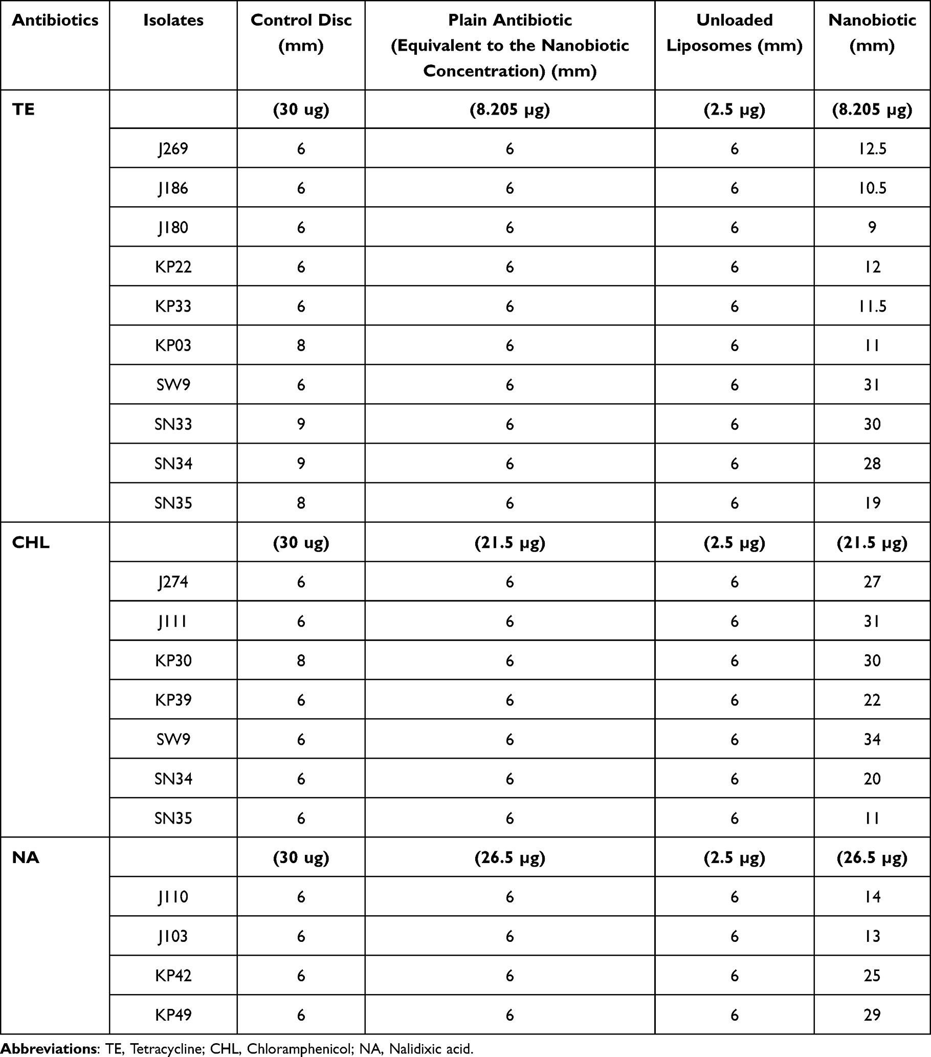 |
Table 1 Agar Well Diffusion Assay of Nanobiotics |
MIC Determination
The liposome-encapsulated antibiotic exhibited markedly lower MIC values for individual isolates than the plain antibiotic. The data for the bacterial strains in the presence of different nanobiotics, compared to their original MIC, are given in Table 2. From the results, it was clear that many tetracycline-resistant isolates (KP33, KP03, SW9, SN33, SN34, and SN35) showed susceptibility to the TE nanobiotic, with MIC reductions from 256 μg/mL to 16 μg/mL. While few of the resistant isolates (J269, J186, J180, and KP22) did not become susceptible, they showed a marked reduction in MIC from 125 μg/mL for plain tetracycline to 41.025 μg/mL for TE nanobiotic. Similarly, in the case of CHL nanobiotic, the resistant isolates became sensitive with MIC reduction for the isolates J274 (MIC 156 μg/mL to 13.4 μg/mL), J111 (MIC > 256 μg/mL to 26.87 μg/mL), KP30 (MIC 78 μg/mL to 6.7 μg/mL), KP39 (MIC156 μg/mL to 13.4 μg/mL), SW9 (MIC 62.5 μg/mL to 6.9 μg/mL), SN34 and SN35 (MIC > 256 μg/mL to 4 μg/mL). Furthermore, the E. coli isolates J110 and J103 (MIC > 256 μg/mL) exhibited a significant reduction in the MIC (64.23 μg/mL) when treated with the NA nanobiotic. Also, Klebsiella pneumoniae isolates KP42 (MIC > 256 μg/mL) and KP49 (MIC > 256 μg/mL) showed a marked reduction in MIC to 32 μg/mL and 16 μg/mL, respectively. This reduction in MIC demonstrates an enhanced antibacterial effect resulting from improved drug delivery and sustained release provided by the liposomal system. In contrast, unloaded liposomes showed no inhibitory activity, confirming that the observed antimicrobial effect was attributable to the encapsulated antibiotic. Further, the MBC determination showed the enhanced antibacterial activity of the nanobiotic formulations at the concentration equivalent to that of the MIC compared to the corresponding plain antibiotics. The nanobiotic formulations exhibited lower viable bacterial counts and improved antibacterial efficiency, indicating the potential role of liposomal encapsulation (Table S3). The obtained MBC values correlate with the MIC associated concentrations used in time kill assay, thereby supporting the observed antibacterial activity.
 |
Table 2 MIC Determination of Nanobiotics |
Time Kill Assay
To confirm the MIC data and evaluate the ability of nanobiotics to eliminate MDR Enterobacteriaceae, killing curve assays were performed on various MDR E. coli and K. pneumoniae strains. Liposomal antibiotic formulations exhibited significantly greater antibacterial activity (P ≤ 0.05) against multidrug-resistant (MDR) Enterobacteriaceae compared with their corresponding free antibiotics. For instance, the TE nanobiotic at the MIC completely inhibited the growth of the isolates J269 and J186 (Figure 2a and c) at 4 hours, whereas for the isolate J180 (Figure 2b), it was around 1 hour. Although the plain antibiotic at the MIC level inhibited the growth in the initial phase, it failed to eliminate the bacterial growth. Similarly, the TE nanobiotic eradicated the bacterial cells within 4 hours and 2 hours for the K. pneumoniae isolates KP22, KP33, and KP03, respectively (Figure 2d–f).
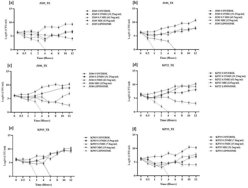 |
Figure 2 Time–kill curves showing the antibacterial activity of TE nanobiotic against MDR E. coli and K. pneumoniae under different conditions: Bacteria were exposed to 0.25, 0.5 times of MIC and MIC and TE nanobiotic along with the untreated control. The broken lines represent the TE nanobiotic. Bacterial viability was determined at different time intervals (0–24 h) by plating serial dilutions and counting colony-forming units (log10 CFU/mL). Each point represents the mean ± SD of three independent experiments. [a–c]-E. coli; [d–f]-K. pneumoniae. |
The CHL nanobiotic significantly reduced the bacteria at the 10th and 6th hours for the J111 and J274 isolates, respectively (Figure 3.1a and b). In contrast, the MIC of the antibiotic inhibited bacterial growth at 12 hours. In the case of K. pneumoniae KP39 and KP30 isolates, the CHL nanobiotic showed effective killing activity within 10 hours. In contrast, the MIC concentration of chloramphenicol initially decreased CFU counts and later allowed normal growth after 4 to 6 hours (Figure 3.1c and d). All plain antibiotic conditions showed a decrease in CFU until 4 hours, followed by an increase. Whereas, in the presence of NA nanobiotics, the bacteria were significantly reduced within 4 hours, indicating that encapsulation enhances the antibacterial activity of the antibiotic without its elimination from the system (Figure 3.2a–d). Collectively, the MICs shown in Table 2 and the killing curves described above confirm the higher potency of the antibiotic-encapsulated liposome compared with free antibiotics against resistant strains of clinically significant E. coli and K. pneumoniae.
 |
Figure 3 Time–kill curves showing the antibacterial activity of [3.1] CHL nanobiotic and 32.2] NA nanobiotic against MDR E. coli and K. pneumoniae under different conditions: Bacteria were exposed to 0.25, 0.5 times of MIC and MIC and CHL & NA nanobiotic along with the untreated control. The broken lines represent the CHL and NA nanobiotic. Bacterial viability was determined at different time intervals (0–24 h) by plating serial dilutions and counting colony-forming units (log10 CFU/mL). Each point represents the mean ± SD of three independent experiments. [a and b]-E. coli; [d and e]-K. pneumoniae. |
In vitro Release Study
The plain tetracycline antibiotics exhibited a rapid release pattern, with the majority of the drug diffused within the first 1–2 hours, indicating an immediate burst release followed by a plateau phase, reaching a maximum cumulative release of 95% by 8 hours. In contrast, the liposome-encapsulated tetracycline exhibited a sustained and controlled release profile, characterised by a gradual increase in drug diffusion over time (Figure 4a). Only 20% of the drug was released in the initial phase, followed by a gradual and continuous release over the study period, ultimately reaching around 85% at 24 hours for CHL (Figure 4b) and NA nanobiotics (Figure 4c), whereas the plain antibiotic of same concentration diffuses rapidly reaching more than 90% within 10 hours. The in vitro drug release study was performed using two independent experiments to evaluate the reproducibility of the liposomal formulations. The release profiles obtained from the independent experiments exhibited closely overlapping patterns with minimal variation, indicating good formulation stability and reproducibility. The sustained release pattern observed in our study clearly demonstrates the ability of the liposome to retain the drug and release it in a controlled manner. The difference in the release pattern between the plain antibiotic and the nanobiotic shows that encapsulation has efficiently reduced premature drug leakage and prolonged its availability.
 |
Figure 4 In vitro release profile of (a) TE, (b) CHL & (c) NA nanoformulation from two independent experiments compared with plain drug, determined using the dialysis method. The release study was carried out in [phosphate-buffered saline, pH 7.4] at 37 ± 0.5°C under constant stirring. Samples were collected at specific time intervals up to 24h and analyzed spectrophotometrically at 280nm-TE; 270nm-CHL & 254nm-NA. Data are represented as mean ± standard deviation (SD). |
Analysis of Liposome-Bacterium Interactions
Microscopic Method
Transmission electron microscopy results gave us a clear view of how the antibiotic-loaded liposomes interacted with bacterial cells. The bacterial cells, upon treatment with liposomes, exhibited membrane deformation, and liposome binding to the surface was clearly visible. Few liposomes appeared to have merged or been partially trapped in the membrane. It was also observed that the bacterial outer membrane was becoming thinner, the surfaces appeared uneven, and there was leakage in the cytoplasm. These TEM observations demonstrate that the liposome-encapsulated antibiotic exerts a direct, membrane-targeted effect, facilitating drug entry and contributing to enhanced antibacterial activity compared to the plain antibiotic (Figure 1iii and iv).
Determination of Bacterial Membrane Permeability of Nanobiotics
PI-encapsulated liposomes were successfully formulated and were characterized using DLS (Horriba SZ-100V2). The average size was 117.2 ± 0.64 nm, PDI was 0.222 ± 0.03, and the zeta potential was −3.2 ± 0.43. Fluorescence microscopy (Figure 5) showed the bacterial membrane permeability using PI encapsulated liposomes. Here, the control groups, Figure 5i Live bacterial cells treated only with PI, exhibited minimal red fluorescence, indicating the limited or no penetration of free PI across intact bacterial membrane. Figure 5ii shows the bacterial cells stained with calcein AM and the Figure 5iii is the merged image showing the live (green) and dead cells (red). In contrast, the treatment group bacterial cells treated with PI-encapsulated liposomes showed enhanced intracellular fluorescence, suggesting uptake of PI through liposomal formulation (Figure 5iv). Further, the bacterial cells were stained with caleinAM showed strong green fluorescence which confirms that cells are metabolically active (Figure 5v). Figure 5vi is the merged image showing the co-localization of red and green fluorescence. This observation suggests that liposomal encapsulation has helped penetration of PI into the bacterial cells, through interaction with the bacterial membrane. The presence of green fluorescence indicates that some of the cells retained its metabolic activity despite the uptake of PI. This also implies partial membrane penetration rather than complete membrane disruption. Thus, the fluorescence permeability assay served as complementary evidence supporting the TEM observations by demonstrating intracellular uptake of PI following liposomal interaction with bacterial cells. The combined TEM and fluorescence microscopy findings collectively suggest membrane-associated interaction and permeability effects induced by the nanobiotic formulations.
 |
Figure 5 Fluorescence microscopic analysis of liposome–bacteria interaction and membrane permeability. Control groups: [i] Bacterial cells treated with propidium iodide (PI) alone; [ii] Bacterial cells stained with calcein AM showing green fluorescence; [iii] Merged image of PI- and calcein AM-stained bacterial cells. Treatment groups: [iv] Bacterial cells treated with propidium iodide (PI)–encapsulated liposomes exhibiting red fluorescence due to intracellular uptake of PI following liposomal interaction; [v] PI-encapsulated liposome-treated bacterial cells subsequently stained with calcein AM showing green fluorescence in metabolically active cells; [vi] Merged image demonstrating the co-localization of calcein AM (green) and PI (red), confirming effective liposomal interaction and controlled release of encapsulated contents into bacterial cells. Scale bar = 10 µm. |
Cytotoxicity Study
The biocompatibility and the toxicity of the antibiotic-loaded liposomes were evaluated using HEK-293 human embryonic kidney cells. Cells were treated with two different concentrations such as 5% and 10% of encapsulated liposomes, plain antibiotic and empty liposomes for (a) tetracycline, (b) chloramphenicol, and (c) nalidixic acid (Figure 6). The result obtained from our study showed that plain liposomes had improved biocompatibility with nearly 100% cell viability and minimal cytotoxicity. Cell viability remained above 90% and the liposomal formulation showed very little cytotoxicity at all tested concentrations. In contrast, treatment with plain antibiotics resulted in a comparatively lower cell viability (Figure 6). Overall, liposomal encapsulation improved the cytocompatibility of all tested antibiotics without compromising cell viability.
 |
Figure 6 Cytotoxicity assessment of nanobiotics/plain antibiotic on HEK293 cell lines using the MTT assay. Cells were exposed to the indicated concentrations of tetracycline (TE), chloramphenicol (CHL), and nalidixic acid (NA) nanobiotics and their respective free antibiotics for 24 hand cell viability was expressed as a percentage relative to untreated control cells. Data represent mean ± SD from three independent experiments, indicating the biocompatibility and safety of the developed nanoformulation. (a) TE nanobiotic and free TE; (b) CHL nanobiotic and free CHL; and (c) NA nanobiotic and free NA; Statistical analysis was performed using Student’s t-test. *p < 0.05, **p < 0.01, ***p < 0.001, ****p < 0.0001 were considered statistically significant compared with the control group. |
Efflux Pump Gene Expression Analysis
Relative gene expression analysis of the efflux pump genes acrA, acrB, tolC, acrD, acrE, and acrF showed that the expression levels of the isolates treated with unloaded liposomes and those exposed to free antibiotics (tetracycline, chloramphenicol, and nalidixic acid) were similar (Figure 7). In the presence of antibiotic-encapsulated liposomes there was a significant decrease in efflux pump gene expression. This implies that antibiotics encapsulated in liposomes may inhibit efflux-mediated resistance mechanisms. Improved intracellular drug transport and less stimulation of efflux systems may be responsible for the lower expression.
 |
Figure 7 Relative gene expression of efflux pump genes on exposure to Ab- antibiotics, E- Empty liposomes and nanobiotics; (a–c) TE-Tetracycline encapsulated liposomes; (d and e) CHL- Chloramphenicol encapsulated liposomes; (f and g) NA- Nalidixic acid encapsulated liposomes against E. coli isolates. Statistical analysis was performed using Student’s t-test. *p < 0.05, **p < 0.01, and ***p < 0.001 were considered statistically significant compared with the control group. |
Validation of Nanobiotics
The formulated nanobiotics were validated using a food spiking assay on various food matrices, including shrimp, chicken, and sprouts, against the food-borne pathogenic Non-Typhoidal Salmonella strains.
Enumeration of Bacteria from the Spiked Food Matrices
Bacterial cells from the spiked matrices under different conditions were analyzed for Total Plate Count (TPC) and Colony Forming Units (CFU). The CFU in T1 condition for all the isolates in food matrices (shrimps, chicken and sprouts) were observed to have no colonies as they were sterilized. When compared to the other condition, T2 had higher CFU as it was spiked only with the NTS strains. In the case of T3 and T4, the levels varied depending on the nanobiotic, isolate, and food matrix of choice. Statistical significance between groups was determined using one-way ANOVA followed by Tukey’s post hoc test (p < 0.05). The NTS isolates SW9, SN33, SN34, and SN35 showed significantly lower CFU/mL in the presence of TE nanobiotic (Figure S2) than in the free antibiotic and untreated conditions. A similar result was observed for the isolates SW9, SN34 and SN35 under treatment with CHL nanobiotic (Figure S3) on the shrimp matrix. The reduction in the CFU/mL was highly significant in SW9 and SN34 in comparison with the plain antibiotic and the untreated control. It was also observed that there was a reduction in the bacterial CFU in T4 condition when compared with T2 and T3 conditions when TE nanobiotic was used for treating chicken matrix, and this was found to be statistically significant. Similarly, CHL nanobiotic was found to be more effective than plain antibiotic in reducing bacterial count in a spiked chicken matrix. The TE nanobiotic and CHL nanobiotic on spiked sprout samples, though found to be effective, did not show much reduction when compared to other food matrices. The reduction in CFU in the presence of nanobiotics demonstrates the efficacy of the nanoformulation compared to the plain antibiotic.
Real-Time qPCR for Absolute DNA Quantification
The bacterial load in spiked food samples was further quantified using real-time PCR to determine DNA copy number variation (CNV) under different treatment conditions. Species-specific primers targeting invA gene were used to ensure accurate detection of NTS bacteria from the spiked food matrices. In untreated control samples (T1 and T2), the bacterial DNA copy number remained high, reflecting the expected proliferation of spiked organisms. Treatment with the plain antibiotic (T3) resulted in a moderate reduction in DNA copy number, whereas samples treated with the liposome-encapsulated antibiotic (T4) showed a marked decrease, indicating antimicrobial efficacy of nanobiotics. A significant DNA CNV was observed in Salmonella spiked shrimp matrices treated with TE nanobiotics when compared with plain antibiotics (Figure S4). Further, it was clear that the amount of DNA in the presence of liposomes encapsulated with the chloramphenicol antibiotic was significantly reduced compared to the T2 and T3 conditions in shrimp matrix. However, the isolates (SW9, SN33, SN34, and SN35) showed variations in the presence of the tetracycline antibiotic and tetracycline-loaded nanobiotic in the chicken matrix. No particular trend was observed. In case of CHL nanobiotic, the isolates (SW9, SN34 and SN35) showed extreme reduction in DNA copy number in chicken matrix, which was determined to be significant, confirming that the encapsulation is effective in controlling these food-borne pathogens (Figure S5). In the case of sprouts, the results were similar to those obtained from shrimp and chicken. There was a reduction in the DNA copy number in (T4), compared with the T2 and T3 conditions for both TE and CHL nanobiotics. Quantitative analysis of DNA copy numbers revealed statistically significant differences among treatment groups (p < 0.05). These results corroborate the CFU-based findings from the food-spiking assay and confirm that the nanoformulation effectively reduces bacterial load at the genetic level, demonstrating enhanced delivery and functional antimicrobial activity in complex food matrices.
Discussion
The increasing prevalence of antimicrobial resistance has necessitated the development of innovative drug delivery strategies capable of improving the efficacy of existing antibiotics.17 In the present study, liposome-based nanobiotics demonstrated enhanced antibacterial activity and modulation of efflux pump-associated resistance mechanisms. The increasing multidrug resistance in clinical isolates of E. coli and K. pneumoniae, as well as foodborne pathogens such as non-typhoidal Salmonella, is rapidly reducing the effectiveness of conventional antibiotics. A 2019 study on cattle-derived Salmonella reported that over half of the isolates were resistant to seven different antibiotic classes, with extremely high resistance rates to tetracycline (97%), sulfisoxazole (96%), streptomycin (95%), and ampicillin/ceftriaxone (85%).18 In comparison, our study showed that NTS isolates required very high MIC’s 39–500 µg/mL for tetracycline, 62.5–500 µg/mL for chloramphenicol, and 250–2500 µg/mL for nalidixic acid indicating strong resistance. Similar findings have been reported on widespread resistance in NTS to antibiotic overuse in livestock and plasmid-mediated resistance.19 Another multi-year surveillance study (2015–2019) also confirmed high resistance in foodborne Salmonella, particularly to tetracycline (53.9%), ciprofloxacin (47.2%), ampicillin (44.4%), and nalidixic acid (42.7%).20
A wide range of Gram-negative and Gram-positive bacterial strains that are resistant to conventional, plain antibiotics have been shown to regain susceptibility when they are delivered in liposomal form. This enhanced response represents a key advantage of liposomes over many other nanocarrier platforms. Although this improvement is often linked to the ability of liposomes to shield antibiotics from enzymatic inactivation, growing evidence suggests that the liposomal delivery system itself plays a crucial role in boosting antimicrobial activity.21–23
Our study highlights that liposome-encapsulated tetracycline, chloramphenicol, and nalidixic acid can significantly enhance antimicrobial activity against resistant strains by improving drug administration and overcoming efflux pumps. Liposomes made of phospholipid bilayer enable close interaction with bacterial membrane through adsorption, fusion and lipid exchange thereby facilitating increased membrane permeability and intracellular accumulation of antibiotics. The lipid bilayer of liposomes may destabilize the bacterial membrane architecture, resulting in transient pore formation or membrane perturbation that enhances intracellular uptake. Encapsulation can protect the antibiotic from enzymatic degradation and enable localized release leading to higher concentration inside the cells. This in turn overwhelms the bacterial defense systems. Additionally, it can improve the penetration of encapsulated antibiotic across the outer membrane barrier in resistant pathogen and biofilms.11,24–26 Over the last fifty years, numerous drug delivery systems, including liposomes, micelles, and dendrimers, have been investigated for the encapsulation of therapeutic agents and other biomolecules.24 In our study, liposomes were prepared using a thin-film hydration method, a simple yet reliable method.15 The primary advantage of this method is its high reproducibility, even with small amounts of material, while the low encapsulation efficiency remains a major drawback. It has been widely used by researchers in several studies and is especially well suited for adding lipophilic compounds in small pharmaceutical quantities.27
The older antibiotics, such as tetracycline, nalidixic acid, and chloramphenicol, were chosen due to their well-established association with multidrug resistance mechanisms in Gram-negative bacterial pathogens though they are not clinically used at present. These antibiotics are commonly affected by efflux pump-mediated resistance, reduced intracellular accumulation, and adaptive bacterial survival mechanisms, making them suitable candidates for evaluating nanobiotic-based drug delivery approaches. Furthermore, tetracycline, nalidixic acid, and chloramphenicol represent distinct antimicrobial classes with different cellular targets and resistance profiles, thereby allowing broader assessment of the effectiveness of the prepared liposomal formulations. The use of these antibiotics also enabled evaluation of whether liposomal encapsulation could enhance antibacterial efficacy, improve sustained drug release, and potentially overcome resistance-associated limitations observed with conventional antibiotic therapy.7,28–31 Also, the nanobiotic formulations demonstrated improved antibacterial efficacy, which may be associated with enhanced intracellular antibiotic delivery and potential reduction of efflux-mediated drug expulsion in resistant bacterial isolates. These findings suggest that liposomal encapsulation could serve as a promising strategy to improve antibiotic effectiveness against efflux-associated antimicrobial resistance.
In this study, the prepared nanobiotics had particle sizes ranged from 120 to 180 nm, and their encapsulation efficiencies were 12–33% for TE, 15–86% for CHL, and 5–25% for NA. These results are consistent with other studies that found liposomes prepared by the thin-film technique to have an average particle size of 107–152 nm and encapsulation efficiencies of 3.7–7.2% for meropenem-loaded liposomes.32 As reported in previous studies, the entrapment efficiency of polymyxin B in extruded DPPC: Chol (2:1) liposomes was relatively low at 3.7 ± 0.5%, with a loading capacity of 5.6 μmol polymyxin B per μmol DPPC.33 Similarly, another study showed liposomal tylosin exhibited approximately 50% encapsulation with an average diameter of 185.23 ± 3.23 nm, a size well-suited for delivering antibacterial agents.34 A recent study employed a sustainable microfluidic technique to encapsulate amoxicillin into liposomes, producing particles with an average size of ~130 nm and a low polydispersity index of 0.2, indicating a highly uniform formulation with an encapsulation efficiency of 77%.34 Our results are consistent with earlier observations that liposomes between 100 and 200 nm in size are suitable for achieving extended systemic circulation. The variation in encapsulation efficiency among tetracycline, chloramphenicol, and nalidixic acid is likely due to differences in their physicochemical properties, including solubility, lipophilicity, and drug–lipid interactions. These factors influence drug partitioning within the liposomal system and ultimately affect drug retention and entrapment efficiency. Similar observations have been reported in previous studies, where encapsulation efficiency was found to be strongly dependent on drug properties and phospholipid interactions.35
After confirming that the liposomes were within the intended size range and demonstrated effective drug loading, we used the agar well diffusion method and MIC determination to investigate whether these formulations improved antibacterial activity. In the present study, the agar well diffusion assay showed that the nanoformulations were effective, as evidenced by measurable zones of inhibition against the test organisms on comparison to the controls used (Figure S1). Additionally, the MICs of nanobiotics against multidrug-resistant E. coli, K. pneumoniae, and NTS isolates were significantly lower than those of the corresponding plain antibiotics. The majority of the drug-resistant isolates in our study became susceptible upon exposure to antibiotic nanoformulations, with MICs reduced from 256 µg/mL to 16 µg/mL. Furthermore, the time-kill assay provides a comprehensive evaluation of antibacterial activity by monitoring bacterial viability over time and is particularly useful for assessing whether nanobiotic formulations enhance the rate, magnitude, and duration of bacterial killing compared with their free antibiotic counterparts. The results of the time-kill assay clearly demonstrate that, after loading the drug into the nanocarrier, its bactericidal effect increases significantly. The nanobiotics used in our study inhibited bacterial growth within 2 to 6 hours of exposure, compared with plain antibiotics. A similar result was reported in a study against Pseudomonas aeruginosa, wherein the nonmucoid strain PA 48912–2, which showed high resistance to amikacin (MIC 256 µg/mL) and tobramycin (MIC 64 µg/mL), restored susceptibility significantly in liposomal formulation by reducing the MIC to 8 µg/mL for both antibiotics. A previous study demonstrated that liposomal amikacin reduced the MIC against a resistant P. aeruginosa strain from 256 to 8 µg/mL and achieved complete bacterial eradication in time-kill assays, whereas the free antibiotic remained ineffective. These findings support the enhanced antibacterial efficacy of liposomal antibiotic formulations. 22Similarly, exposure of A. baumannii isolates to ciprofloxacin- or levofloxacin-loaded liposomes at MIC levels resulted in rapid growth reduction within 2–3 hours (P < 0.05), followed by complete bacterial killing within 5–6 hours. In contrast, plain levofloxacin at the same concentration showed only ~10% inhibition after 24 hours, while plain ciprofloxacin exhibited no measurable antibacterial effect. Plain nanoparticles displayed no bactericidal activity, as no reduction in viable cell count was observed even after 24 hours.36 Similarly, the liposomal antibiotic of amoxicillin showed strong activity against Proteus mirabilis and Staphylococcus aureus, achieving MIC values as low as 3 µg/mL.34
In our study, the release profiles of tetracycline, chloramphenicol, and nalidixic acid from liposomes and their free forms were assessed using dialysis. Free antibiotics displayed fast diffusion, with most of the antibiotics released within 1–2 hours, reaching over 90% by 8–10 hours. Liposome-encapsulated formulations, on the other hand, demonstrated controlled and sustained release, with only about 20% released initially within 30 minutes and a steady diffusion over 24 hours, reaching approximately 85%. This shows that the drug is efficiently retained by the lipid bilayer whereas weakly associated drugs are released more rapidly through diffusion into the external medium. Overall, encapsulation prolongs therapeutic activity and enhances drug availability, possibly at a lower dose. This release behavior suggests that the nanobiotics does not maintain prolonged sub-inhibitory exposure over extended periods, but rather provides an initial therapeutic burst followed by sustained drug availability sufficient to achieve antibacterial efficacy. The outcome is comparable to the tylosin release profile from liposomes, which showed an initial burst phase followed by a slower, sustained release regardless of the medium pH. Within the first six hours, about 40% of the drug was released. This early burst release is beneficial because it ensures a high local drug concentration at the infection site and allows for rapid onset of action.15 Although sustained drug release form the liposomal formulations may enhance antibacterial activity, prolonged exposure to sub-inhibitory concentration could potentially contribute to bacterial tolerance or adaptive resistance development. Therefore, optimization of release kinetics and maintenance of effective therapeutic concentrations are important considerations for future investigations.
In Gram-negative bacteria, particularly members of the Resistance-Nodulation-Division (RND) family, efflux systems play a critical role in maintaining sub-therapeutic intracellular drug concentrations and promoting multidrug resistance. To the best of our knowledge, this study is among the first to evaluate efflux pump gene expression in response to liposome-based nanobiotic treatment, thereby providing preliminary molecular evidence supporting the role of nanocarrier systems in overcoming efflux-mediated antimicrobial resistance.
The efflux pump associated gene expression analysis suggests a potential interference of the liposomal formulation with bacterial efflux-mediated resistance mechanisms. The reduced expression of efflux-associated genes such as acrA, acrB, acrD, acrE, acrF and tolC was observed in the present study indicates that the nanobiotic formulation may modulate bacterial stress responses linked to efflux activity, thereby improving antibacterial susceptibility. Similar studies have suggested that nanobiotics can interfere with efflux mechanisms either by direct inhibition of transporter activity, disruption of membrane energetics required for active efflux or enhancement of intracellular drug concentration beyond the extrusion capacity of bacterial pumps.11,37–39
Earlier studies have shown that aminoglycosides can be efficiently delivered to resistant bacterial strains, such as P. aeruginosa and Burkholderia cenocepacia, using conventional DPPC or DSPC/cholesterol liposomes.21,22 Similarly, it has been demonstrated that negatively charged vesicles with low phase transition temperatures increase the intracellular release of tobramycin in resistant bacteria.40 Our study revealed significant interaction of liposomes with bacterial membrane, as observed in TEM analysis. It also revealed the extensive attachment of liposomal vesicles to the bacterial surface, demonstrating close interaction between the nanobiotic formulation and the bacterial membrane (Figure 1iii and iv). These results demonstrate how a liposomal carrier can enhance antibiotic administration and combat bacterial resistance by facilitating antibiotic delivery intracellularly and promoting membrane fusion. Further, the interaction of liposomes with the bacterial membrane was investigated using a calcein-AM/PI assay. The enhanced intracellular fluorescence observed in bacterial cells treated with PI-encapsulated liposomes suggests effective liposome–bacteria interaction and improved delivery of the encapsulated antibiotic (Figure 5). Combined with the TEM observations, these findings indicate that liposomes may increase membrane permeability and facilitate intracellular uptake, thereby potentially improving antibiotic accumulation and reducing the impact of resistance mechanisms such as decreased permeability and active efflux. Based on the findings, liposomes may interact with the bacterial membrane, leading to membrane deformation through surface binding or possible membrane penetration. This is one of the important features of the lipid bilayer that is related to its fluidity. Calcein-AM is a fluorescent dye that can self-quench at high concentrations but fluoresces only after release from liposomes. The rate of calcein-AM release is largely influenced by bilayer fluidity, with more fluid membranes showing faster release, which is likely similar to the data obtained in the present study.27
In the biocompatibility study, the antibiotic-encapsulated liposomes also demonstrated significantly higher cell viability (90%) than the respective plain antibiotics, indicating that encapsulation effectively mitigated the cytotoxic effects of the antibiotics. This is similar to the results of a study demonstrating that, at lower concentrations, both free and liposome-encapsulated tylosin maintained over 70% cell viability in NIH3T3 and NHDF cells. However, at the highest dose (1024 µg/mL), viability dropped below 70% for free tylosin, while liposomal tylosin preserved cell viability above 70% (P < 0.0001).15
As a first-of-its-kind study, the developed nanoformulations of Tetracycline (TE) and chloramphenicol (CHL) were validated in Salmonella-spiked food matrices. Interestingly, even at the lower concentration, liposome-encapsulated antibiotics excelled the plain antibiotic in different food matrices spiked with drug-resistant S. Weltevreden and S. Newport isolates. The increased effectiveness of the liposomal formulations was further validated by total plate count analysis and DNA copy number quantification. All the nanobiotics developed in the study have significantly reduced both the bacterial load and the DNA copy number. These results demonstrate that liposomal encapsulation is an effective alternative approach for combating drug-resistant pathogens in food. However, several challenges and regulatory issues must be considered for the use of liposomal antibiotics in industrial food applications. Further, the intricacy of their production is a significant drawback of liposomal antibiotic delivery. Maintaining batch-to-batch consistency is challenging, especially for drug release kinetics, surface charge, and particle size distribution. Furthermore, achieving effective antibiotic encapsulation remains challenging. Therefore, additional optimization is needed for antibiotic-loaded liposomes to achieve a dependable and clinically useful therapeutic value, even though liposomal formulations have proven successful in cancer therapy.25
Conclusion
The present study highlights the potential of liposomal delivery as a promising approach for restoring antibiotic efficacy against multidrug-resistant pathogens. Antibiotic-encapsulated nanobiotics showed increased activity against multidrug-resistant E. coli, K. pneumoniae, and Salmonella spp. against their respective antibiotics. The formulated nanobiotics demonstrated suitable physicochemical characteristics, including stable particle size distribution <200 nm, encapsulation efficiency ranging from 12% to 85% for the all three antibiotics, and sustained drug release behaviour. The nanobiotics were biocompatible and non-cytotoxic. Antibacterial assessment analysis revealed enhanced inhibitory activity of the nanobiotics compared to the corresponding free antibiotics. The nanobiotics exhibited improved bacterial killing efficiency in MIC, MBC, and time-kill assays. The liposomal tetracycline and nalidixic acid showed notable antibacterial performance against resistant isolates, while chloramphenicol-loaded liposomes demonstrated a favorable antibacterial activity. TEM analysis further indicated active interaction of nanobiotic with bacterial membrane. Also, the enhanced intracellular fluorescence observed in PI-encapsulated liposome-treated bacterial cells suggests improved membrane interaction and intracellular delivery mediated by the nanobiotic. To the best of our knowledge, this study is among the first to investigate the expression of bacterial efflux pump-associated genes in response to liposome-based nanobiotic treatment, providing novel insight into the potential of nanocarrier systems to modulate efflux-mediated antimicrobial resistance. The reduced expression of efflux pump-associated genes in the presence of the nanobiotic formulation suggests that liposomal encapsulation may interfere with bacterial efflux-mediated resistance mechanisms. Further studies involving protein-level expression and functional efflux assays are required to elucidate whether the nanobiotic formulation directly inhibits, bypasses, or modulates bacterial efflux pump activity. The results of the food-spiking assay showed that the nanobiotics significantly reduced bacterial load on food surfaces compared with plain antibiotics. Therefore, the increased bactericidal activity of antibiotics within liposomes (nanobiotics) at lower concentrations enables successful approaches to controlling multidrug-resistant bacteria. Overall, the findings suggest that liposomal encapsulation (nanobiotics) may improve the antibacterial efficacy and therapeutic potential of conventional antibiotics and could serve as a promising strategy for repurposing the older drugs and addressing antimicrobial-resistant bacterial pathogens. Further, in vivo and mechanistic investigations are recommended to validate the clinical applicability of these formulations.
Highlights
- This study offers a practical and innovative solution to the growing global threat of antimicrobial resistance (AMR).
- This work demonstrates that liposome-encapsulated formulations of tetracycline, chloramphenicol and nalidixic acid can significantly enhance antimicrobial efficacy against multidrug-resistant strains.
- This approach supports antimicrobial stewardship by minimising unnecessary antibiotic exposure and offering a more targeted treatment.
- Nanocarrier-based delivery systems offer an effective means of repurposing older antibiotics for combating multidrug-resistant infections.
Abbreviations
AMR, Antimicrobial resistance; MDR, Multi drug resistance; NTS, Non-Typhoidal Salmonella; TE, Tetracycline; CHL, Chloramphenicol; NA, Nalidixic acid; MIC, Minimum inhibitory concentration; DLS, Dynamic Light Scattering; PDI, Polydispersity Index; CFU, colony forming units; TEM, Transmission electron microscopy; CNV, Copy number variation.
Data Sharing Statement
All the data have been included in the manuscript.
Acknowledgments
The authors thank Nitte University Centre for Science Education and Research, Nitte (Deemed to be University), for providing the necessary research facilities and research fellowships.
Author Contributions
BDT performed study design, execution of the experiments, acquisition of data, analysis and interpretation and drafting the manuscript. DFKV performed execution, acquisition of data. SK supervised the research, critically reviewed the article. DVK conceptualised, supervised the research, critically reviewed the article and gave final approval of the version to be published.
Funding
This study was supported by the ICMR-funded project (AMR/DHR/GIA/3/ECD-II/2020).
Disclosure
The authors declare that they have no known competing financial interests or personal relationships that could have influenced the work reported in this paper.
References
1. Ho CS, Wong CTH, Aung TT, et al. Antimicrobial resistance: a concise update. Lancet Microbe. 2025;6(1):1. doi:10.1016/j.lanmic.2024.07.010
2. Cabrera A, Mason E, Mullins LP, Sadarangani M. Antimicrobial resistance and vaccines in Enterobacteriaceae including extraintestinal pathogenic Escherichia coli and Klebsiella pneumoniae. Npj Antimicrob Resist. 2025;3:1–21. doi:10.1038/s44259-025-00100-8
3. World Health Organization. Critically Important Antimicrobials for Human Medicine.
4. Bhutta ZA. Typhoid fever: current concepts. Infect Dis Clin Pract. 2006;14:266–272. doi:10.1097/01.idc.0000222625.11629.f4
5. Sima CM, Buzilă ER, Trofin F, et al. Emerging strategies against non-typhoidal Salmonella: from pathogenesis to treatment. Curr Issues Mol Biol. 2024;46:7447–7472. doi:10.3390/cimb46070442
6. Gonzalez Gomez A, Hosseinidoust Z. Liposomes for antibiotic encapsulation and delivery. ACS Infect Dis. 2020;6:896–908. doi:10.1021/acsinfecdis.9b00357
7. Granados-Chinchilla F, Rodríguez C. Tetracyclines in food and feedingstuffs: from regulation to analytical methods, bacterial resistance, and environmental and health implications. J Anal Methods Chem. 2017;2017:1315497. doi:10.1155/2017/1315497
8. World Health Organization. WHO Global Principles for the Containment of Antimicrobial Resistance in Animals Intended for Food. Geneva, Switzerland: World Health Organization; 2000.
9. Jeong YI, Na HS, Seo DH, et al. Ciprofloxacin-encapsulated poly (DL-lactide-co-glycolide) nanoparticles and its antibacterial activity. Int J Pharm. 2008;352:317–323. doi:10.1016/j.ijpharm.2007.11.001
10. Chamundeeswari M, Jeslin J, Verma ML. Nanocarriers for drug delivery applications. Environ Chem Lett. 2019;17:849–865. doi:10.1007/s10311-018-00841-1
11. Ferreira M, Ogren M, Dias JN, et al. Liposomes as antibiotic delivery systems: a promising nanotechnological strategy against antimicrobial resistance. Molecules. 2021;26:2047. doi:10.3390/molecules26072047
12. Bangham AD, De Gier J, Greville GD. Osmotic properties and water permeability of phospholipid liquid crystals. Chem Phys Lipids. 1967;1:225–246. doi:10.1016/0009-3084(67)90030-8
13. Shaaban MI, Shaker MA, Mady FM. Imipenem/cilastatin encapsulated polymeric nanoparticles for destroying carbapenem-resistant bacterial isolates. J Nanobiotechnol. 2017;15(1):29. doi:10.1016/j.jgar.2025.01.010
14. Omri A, Ravaoarinoro M. Comparison of the bactericidal action of amikacin, netilmicin and tobramycin in free and liposomal formulation against Pseudomonas aeruginosa. Chemotherapy. 1996;42:170–176.
15. Saddiqi ME, Kadir AA, Abdullah FF, Zakaria MZ, Banke IS. Preparation, characterization and in vitro cytotoxicity evaluation of free and liposome-encapsulated tylosin. OpenNano. 2022;8:100108.
16. Devi BT, Aditya V, Kini S, Kumar BK, Deekshit VK. Synergistic effects of antibiotics and efflux pump inhibitors on multidrug-resistant Escherichia coli and Klebsiella pneumoniae. J Appl Microbiol. 2025;136(7):lxaf169. doi:10.1093/jambio/lxaf169
17. Hochvaldová L, Večeřová R, Kolář M, et al. Antibacterial nanomaterials: upcoming hope to overcome antibiotic resistance crisis. Nanotechnol Rev. 2022;11:1115–1142. doi:10.1515/ntrev-2022-0059
18. Srednik ME, Lantz K, Hicks JA, Morningstar-Shaw BR, Mackie TA, Schlater LK. Antimicrobial resistance and genomic characterization of Salmonella Dublin isolates in cattle from the United States. PLoS One. 2021;16:e0249617. doi:10.1371/journal.pone.0249617
19. McDermott PF. Antimicrobial Resistance in Nontyphoidal Salmonellae. In: Antimicrobial Resistance in Bacteria of Animal Origin. 2005;293–314. doi:10.1016/S0005-2736(99)00217-5
20. Peruzy MF, Capuano F, Proroga YTR, Cristiano D, Carullo MR, Murru N. Antimicrobial susceptibility testing for Salmonella serovars isolated from food samples: five-year monitoring (2015–2019). Antibiotics. 2020;9:365. doi:10.3390/antibiotics9070365
21. Mugabe C, Halwani M, Azghani AO, Lafrenie RM, Omri A. Mechanism of enhanced activity of liposome-entrapped aminoglycosides against resistant strains of Pseudomonas aeruginosa. Antimicrob Agents Chemother. 2006;50:2016–2022. doi:10.1128/AAC.01547-05
22. Halwani M, Mugabe C, Azghani AO, Lafrenie RM, Kumar A, Omri A. Bactericidal efficacy of liposomal aminoglycosides against Burkholderia cenocepacia. J Antimicrob Chemother. 2007;60:760–769. doi:10.1093/jac/dkm289
23. Wang DY, Van der Mei HC, Ren Y, Busscher HJ, Shi L. Lipid-based antimicrobial delivery systems for the treatment of bacterial infections. Front Chem. 2020;7:872. doi:10.3389/fchem.2019.00872
24. Bekele T, Alamnie G. Treatment of antibiotic-resistant bacteria by nanoparticles: current approaches and prospects. Ann Adv Chem. 2022;6:1–9. doi:10.29328/journal.aac.1001025
25. Ghosh R, De M. Liposome-based antibacterial delivery: an emergent approach to combat bacterial infections. ACS Omega. 2023;8(39):35442–35451.
26. Faraag AH, Shafaa MW, Elkholy NS, Abdel-Hafez LJ. Stress impact of liposomes loaded with ciprofloxacin on the expression level of MepA and NorB efflux pumps of methicillin-resistant Staphylococcus aureus. Int Microbiol. 2022;25(3):427–446. doi:10.1007/s10123-021-00219-4
27. Šturm L, Poklar Ulrih N. Basic methods for preparation of liposomes and studying their interactions with different compounds, with emphasis on polyphenols. Int J Mol Sci. 2021;22:6547. doi:10.3390/ijms22126547
28. Kounatidis D, Dalamaga M, Grivakou E, et al. Third-generation tetracyclines: current knowledge and therapeutic potential. Biomolecules. 2024;14(7):783. doi:10.3390/biom14070783
29. Oong GC, Tadi P. Chloramphenicol. [Updated 2023 Jul 3]. In: StatPearls [Internet]. Treasure Island (FL): StatPearls Publishing; 2025 Jan-.Available from: https://www.ncbi.nlm.nih.gov/books/NBK555966/.
30. Pandey A, Aggarwal N, Adholeya A, Kochar M. Resurrection of nalidixic acid: evaluation of water-based nanoformulations as potential nanomedicine. Nanoscale Res Lett. 2018;13(1):298. doi:10.1186/s11671-018-2718-8
31. Lashaki SB, Moulavi P, Ashrafi F, Sharifi A, Asadi S. Imipenem/Cilastatin encapsulation in UIO-66-NH2 carrier as a new strategy for combating imipenem-resistant Pseudomonas aeruginosa isolates. J Global Antimicrob Resist. 2025;42:15–27. doi:10.1016/j.jgar.2025.01.010
32. Drulis-Kawa Z, Gubernator J, Dorotkiewicz-Jach A, Doroszkiewicz W, Kozubek A. In vitro antimicrobial activity of liposomal meropenem against Pseudomonas aeruginosa strains. Int J Pharm. 2006;315:59–66. doi:10.1016/j.ijpharm.2006.02.017
33. Omri A, Suntres ZE, Shek PN. Enhanced activity of liposomal polymyxin B against Pseudomonas aeruginosa in a rat model of lung infection. Biochem Pharmacol. 2002;64:1407–1413. doi:10.1016/S0006-2952(02)01346-1
34. Weaver E, Macartney RA, Irwin R, et al. Liposomal encapsulation of amoxicillin via microfluidics with investigation of PEGylated therapeutics. Int J Pharm. 2024;650:123710. doi:10.1016/j.ijpharm.2023.123710
35. Akbarzadeh A, Rezaei-Sadabady R, Davaran S, et al. Liposome: classification, preparation, and applications. Nanoscale Res Lett. 2013;8(1):102.
36. Aboelenin AM, El Mowafy M, Saleh NM, Shaaban MI, Barwa R. Ciprofloxacin- and levofloxacin-loaded nanoparticles suppressed fluoroquinolone resistance and biofilm formation in Acinetobacter baumannii. Sci Rep. 2024;14:3125. doi:10.1038/s41598-024-53441-1
37. Puvanendran D, Cece Q, Picard M. Reconstitution of the activity of RND efflux pumps: a “bottom-up” approach. Res Microbiol. 2018;169(7–8):442–449. doi:10.1016/j.resmic.2017.11.004
38. Devi Thillai B, Kini S, Deekshit VK. Nanobiotics to tackle efflux-mediated resistance in bacterial pathogens. Crit Rev Microbiol. 2025. doi:10.1080/1040841X.2025.2597220
39. Kavanaugh LG, Dey D, Shafer WM, Conn GL. Structural and functional diversity of Resistance-Nodulation-Division (RND) efflux pump transporters with implications for antimicrobial resistance. Microbiol Mol Biol Rev. 2024;88(3):e00089–23. doi:10.1128/mmbr.00089-23
40. Sachetelli S, Khalil H, Chen T, Beaulac C, Sénéchal S, Lagacé J. Demonstration of a fusion mechanism between a fluid bactericidal liposomal formulation and bacterial cells. Biochim Biophys Acta Biomembr. 2000;1463:254–266. doi:10.1016/S0005-2736(99)00217
41. Rahn K, De Grandis SA, Clarke RC, et al. Amplification of an invA gene sequence of Salmonella typhimurium by polymerase chain reaction as a specific method of detection of Salmonella. Mol Cell Probes. 1992;6(4):271–279. doi:10.1016/0890-8508(92)90002-F
 © 2026 The Author(s). This work is published and licensed by Dove Medical Press Limited. The
full terms of this license are available at https://www.dovepress.com/terms
and incorporate the Creative Commons Attribution
- Non Commercial (unported, 4.0) License.
By accessing the work you hereby accept the Terms. Non-commercial uses of the work are permitted
without any further permission from Dove Medical Press Limited, provided the work is properly
attributed. For permission for commercial use of this work, please see paragraphs 4.2 and 5 of our Terms.
© 2026 The Author(s). This work is published and licensed by Dove Medical Press Limited. The
full terms of this license are available at https://www.dovepress.com/terms
and incorporate the Creative Commons Attribution
- Non Commercial (unported, 4.0) License.
By accessing the work you hereby accept the Terms. Non-commercial uses of the work are permitted
without any further permission from Dove Medical Press Limited, provided the work is properly
attributed. For permission for commercial use of this work, please see paragraphs 4.2 and 5 of our Terms.