Back to Journals » Infection and Drug Resistance » Volume 16
Detection of Mycobacterium avium-intracellulare Complex (MAC) by Bronchial Lavage and the Relationship with Titers of Anti-Glycopeptidolipid-Core IgA Antibodies to MAC in Patients with Pulmonary MAC Disease
Authors Shimada D , Sagawa M, Seki M
Received 4 December 2022
Accepted for publication 10 February 2023
Published 17 February 2023 Volume 2023:16 Pages 977—984
DOI https://doi.org/10.2147/IDR.S400200
Checked for plagiarism Yes
Review by Single anonymous peer review
Peer reviewer comments 3
Editor who approved publication: Prof. Dr. Héctor Mora-Montes
Daishi Shimada,1 Motoyasu Sagawa,2 Masafumi Seki1,3
1Division of Infectious Diseases, Tohoku Medical and Pharmaceutical University Hospital, Sendai City, Japan; 2Division of Endoscopy, Tohoku Medical and Pharmaceutical University Hospital, Sendai City, Japan; 3Division of Infectious Diseases and Infection Control, Saitama Medical University International Medical Center, Hidaka City, Japan
Correspondence: Masafumi Seki, Division of Infectious Diseases and Infection Control, Saitama Medical University International Medical Center, Yamane 1397-1, Hidaka City, Saitama, Japan, Tel +81-42-984-4392, Fax +81-42-984-0280, Email [email protected]
Background: Higher rates of diagnosis of pulmonary Mycobacterium avium-intracellulare complex (MAC) disease by bronchoscopy (BS) in patients who could not diagnose by sputum cultures have been suggested, but the detailed utility of BS, especially in combination with anti-glycopeptidolipid-core IgA antibodies (anti-MAC Ab), is still unclear.
Methods: A total of 111 patients at our hospital with suspected MAC who underwent BS because they were sputum-negative from April 2018 to March 2022 were analyzed prospectively. These patients were also divided into two groups, anti-MAC Ab-positive and anti-MAC Ab-negative, and compared.
Results: A total of 111 patients underwent BS, though 95 (38.0%) of 250 enrolled patients were sputum smear/culture-positive. The age of the 111 patients was 69.14 (31.0– 89.0) years, and 90 (81.0%) were female; 69 (62.2%) of 111 patients were either smear-positive (n = 42, 37.8%) or culture-positive (n = 27, 24.3%) by BS. Of the total 111 patients, 69 (62.2%) were anti-MAC Ab-positive and 57 (82.6%) of 69 patients were also positive by BS. In contrast, only 12 (28.6%) of the 42 anti-MAC Ab-negative patients were positive by BS. The sensitivity and specificity of anti-MAC Ab for positive by BS were 82.6% and 71.4%, respectively, and the area under the curve (AUC) on receiver-operating characteristic (ROC) curve analysis was 0.807.
Conclusion: BS and anti-MAC Ab showed similar usefulness to confirm the diagnosis in patients who could not be diagnosed by sputum examination, but pulmonary MAC disease was strongly suspected based on chest radiography/CT findings. These two examinations were correlated, and their combination appeared to provide more accurate diagnosis and earlier therapy.
Keywords: anti-MAC antibody, bronchiectasis, bronchoscopy, nontuberculous mycobacteria, sputum
Introduction
Mycobacterium avium complex (MAC) is the most commonly isolated non-tuberculous mycobacterium (NTM) in Japan and Eastern Asia, and it has become a major issue as the cause of chronic lung disease leading to hemoptysis, cough, and dyspnea in middle-aged women.1
Although the patients show the characteristic chest radiograph (X-ray) and computed tomography (CT) findings, such as fibro-cavitary (FC) radiographic changes or bronchiectasis with nodular and reticulonodular radiographic changes, termed the nodular bronchiectatic (NB) type, these chest imaging presentations are sometimes similar to those of pulmonary tuberculosis and lung cancers; therefore, a definitive diagnosis by culture is important for determining the appropriate therapeutic agents, such as azithromycin (AZM)/clarithromycin (CAM), rifampicin (RIP), and ethambutol (EB).2,3
However, the detection of MAC from sputum smear/culture might unfortunately fail because the number of mycobacteria is few in the lungs, and some patients, especially elderly patients, cannot produce sputum. Therefore, bronchoscopy (BS) and/or surgery is performed to isolate the mycobacterium from bronchial lavage fluid and dissected tissue sections. Gene analysis is also required to detect MAC in tissue sections that have already been embedded in paraffin.4 In addition, serum biomarkers, such as anti-glycopeptidolipid (GPL)-IgA antibodies (anti-MAC Abs), support the diagnosis of MAC and are used worldwide.5 The GPL core antigen is an important epitope of the cell wall of MAC,6 and, currently, an anti-MAC Abs assay kit for diagnosing MAC is commercially available; a systematic review and meta-analysis examined the precise diagnostic accuracy of the anti-MAC Abs for MAC pulmonary disease, with estimates of sensitivity and specificity of 0.696 and 0.906, respectively, with a cutoff value of 0.7 U/mL.7
In the present study, patients whose chest X-ray/CT findings strongly suggested pulmonary MAC diseases, but no mycobacteria could be detected by sputum examinations, were prospectively analyzed. These patients underwent BS, and the bronchial lavage fluid was collected. Anti-MAC Ab levels in the sera were also measured, and the detection rates of MAC from the bronchial lavage fluid were compared between the serum anti-MAC Ab-positive and anti-MAC Ab-negative groups.
Methods
Patients and Bronchoscopy Procedures
Patients who visited Tohoku Medical and Pharmaceutical University Hospital from April 2015 to March 2022 were analyzed. Outpatients who fulfilled the following three criteria were enrolled: (1) age ≥20 years; (2) symptoms suggestive of pulmonary MAC diseases; and (3) findings suggestive of MAC on chest X-ray or CT. Bronchial lavage was performed in patients suspected of having pulmonary MAC disease based on chest X-ray or CT findings, but in whom a diagnosis could not be obtained because of negative cultures from at least 3 consecutive sputum samples or the absence of expectorated sputum even though sputum induction was conducted by inhalation of nebulized sterile saline solution followed by coughing and expectoration of airway secretions.
The BS procedures were as follows: the segmental or subsegmental bronchus with abnormal lesions on chest CT was wedged after brushing twice, 20 mL of sterile normal saline was injected, and the lavage fluid was aspirated via BS, as previously reported.8 Collected samples were cultured by MIGT (Mycobacterium growth indicator tube, Becton-Dickinson, Franklin Lakes, NJ, USA) and Ogawa culture media (Kyokuto Pharmaceutical Industries CO. LTD, Tokyo, Japan) to isolate the mycobacteria, in addition to Ziehl-Neelsen staining to detect mycobacteria. Definition and differentiation of the MAC were performed by polymerase chain reaction (PCR) methods and/or matrix-assisted laser desorption/ionization time-of-flight mass spectrometry (MALDI-TOF MS) (Bruker Daltonics, Billerica, MA, USA).9,10
Ethics
This study was approved by the Committee for Clinical Scientific Research of Tohoku Medical and Pharmaceutical University Hospital on July 8 and October 9, 2015 (No. ID2015-2-008 and ID2015-2-025), respectively, as trials for mycobacteria (https://www.hosp.tohoku-mpu.ac.jp/department/infection.html). All the patients whose specimens were used and who participated this study provided written, informed consent to have their case details and any accompanying images published. The Declaration of Helsinki was also adhered in this study.
Measurement of Anti-MAC Ab Levels
Serum samples were collected from the patients, and anti-MAC Ab levels were measured using a serological test based on an enzyme immunoassay (Kyokuto Pharmaceutical Industries CO. LTD, Tokyo, Japan). Briefly, microtiter plates were coated with 0.5 μg of GPLs and GPL core of M. avium serotype 4/well. Serum samples were diluted 40-fold with phosphate-buffered saline containing 1% bovine serum albumin. Diluted serum samples were added, followed by incubation for 1 h at 37 °C. Plates were washed, then peroxidase-conjugated F(ab′)2 of goat antibody against human immunoglobulin G (IgG), IgA, or IgM was added, and plates were incubated for 2 h at 37 °C. Unbound labeled antibody was removed by washing, and the substrate, o-phenylenediamine dihydrochloride, was added. Following color development, the optical densities of the wells on the plates were read for absorbance at 492 nm in a reader (model 550, Bio-Rad Laboratories, Tokyo, Japan).11 The cut-off point was defined as 0.7 U/mL.12 The sera from the patients were aliquoted into individual 1.0-mL doses in tubes, stored at −80 °C until use, and thawed at room temperature just before the assay.
Statistical Analysis
The Mann–Whitney U-test was used to compare continuous variables between two groups. Sensitivity and specificity were calculated as follows: Sensitivity = (True Positives)/(True Positives +False Negatives), and Specificity = (True Negatives)/(True Negatives + False Positives), respectively. A receiver-operating characteristic (ROC) curve and the area under the ROC curve (AUC) were assessed as appropriate. A p-value of less than 0.05 denoted a significant difference. All analyses were carried out using Stat View software (Abacus Concepts, Cary, NC, USA) and SPSS statistics ver. 25.
Results
Patients’ Characteristics
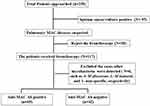
Overall, 250 adult patients with suspected pulmonary MAC were approached; 95 (38.0%) of the 250 patients were sputum smear/culture-positive, and 38 patients rejected the BS procedures and requested observational follow-up at the outpatient clinic (Figure 1). A total of 117 patients with suspected pulmonary MAC disease with no organisms isolated from sputum underwent BS, and from 6 of them, other mycobacteria, such as 4 Mycobacterium abscessus, 1 Mycobacterium kansasii, and 1 non-specific mycobacteria were isolated by BS, but these 6 cases were excluded from the analysis because non-MAC mycobacteria, especially Mycobacterium abscessus, are known to also express the GPL, and potential false-positive results of the anti-MAC Abs have been reported.13
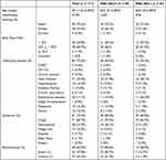
Finally, a total of 111 patients with suspected pulmonary MAC disease with no organisms isolated from sputum underwent BS and were analyzed. The median age of the 111 patients was 69.14 (31.0–89.0) years, and 92 (82.8%) of the 111 patients were female (Table 1). The age distribution and male/female ratio are shown in Figure 2. The number of patients increased with age and peaked in the seventies, with females predominant in all age groups. Most of the patients were non-smokers and thin with a body mass index <25 kg/m2 (Table 1). Chronic pulmonary diseases, such as COPD and old tuberculosis, were present in only 2 (2.6%) and 3 (3.4%) patients, respectively, as underlying diseases, although 41 (35.0%) of the 111 patients had heart disease. However, most patients showed some respiratory symptoms, such as cough (71/111, 60.7%) and hemosputum (28/111, 23.9%).
 |
Table 1 Characteristics of Patients with Suspected Pulmonary Mycobacterium avium/intracellulare Complex (MAC) Who Underwent Bronchoscopy (n = 111) |
 |
Figure 2 Age distribution and the male/female ratio of the patients with suspected pulmonary Mycobacterium avium complex disease who underwent bronchoscopy. White bars: female, and black bars: male. |
Isolated Mycobacteria and Anti-MAC Ab Levels
MAC was detected by BS from 69 (62.2%) of 111 patients who could not be definitively diagnosed as having MAC by sputum examination. Of the 69 patients who could be diagnosed by BS, 42 (37.8%) patients became smear-positive, and the other 27 (24.3%) were confirmed by culture of the bronchial lavage by BS.
The serum anti-MAC Ab levels were as follows: 69 (62.2%) of 111 patients were serum anti-MAC Ab-positive (Table 1), and 57 (82.6%) of these 69 patients were also bronchial lavage fluid positive (Table 1). In contrast, 42 (37.8%) of 111 patients were anti-MAC Ab-negative, with isolation of MAC from only 12 (28.9%) of these 42 patients (Table 1). The detection rates of MAC were significantly higher in serum anti-MAC Ab-positive patients than in serum anti-MAC Ab-negative patients (p < 0.01).
In addition, 31 (44.9%) of the 69 BS-positive patients had an anti-MAC Ab level greater than 10 U/m and 18 (26.1%) of the 69 BS-positive patients showed negative anti-MAC Ab (Figure 3A). In the present study, the sensitivity and specificity of anti-MAC Ab for BS-positive cases were 82.6% and 71.4%, respectively, and false-positive and false-negative results were seen in 17.4% and 28.6%, respectively (Table 1). The AUC in the ROC curve analysis was 0.807 (Figure 3B).
Discussion
Identification of mycobacteria and their subtypes is essential for the diagnosis and treatment of mycobacterial infections, though their isolation from sputum cultures is sometimes difficult.1,14 It has been reported that high rates of sputum-positive were seen in a retrospective, observational study,1,5 but we unfortunately sometimes could not detect MAC by sputum smear/culture, and elderly patients frequently cannot produce sputum. Overall, sputum smear/cultures were positive for 95 (38.0%) of 250 patients in the present study, although 34 (81.0%) of 42 patients were reported sputum smear/culture positive in a previously reported, retrospective, single-center-study in Japan.15 This discrepancy might be because patients who could not produce sputum or had negative cultures were not distinguished, and sputum induction was also not performed uniformly in our study. The results of sputum smear/cultures might be unstable and dependent on the skill levels of the physicians and medical staff handling.
In contrast, BS could be performed in patients who could not produce sputum, and sample collection was absolutely stable under anesthesia. The effectiveness of bronchial lavage or biopsy by BS for the diagnosis of pulmonary MAC disease has been reported in some previous studies,16,17 and in the American Thoracic Society guidelines for the diagnosis of NTM infection, the diagnosis of pulmonary NTM disease required the patient to meet clinical and microbiologic criteria, including either (1) positive culture results from at least two separate expectorated sputum samples or (2) positive culture results from one bronchial wash or lavage.1 These data suggest that BS is as effective as sputum culture for the diagnosis of pulmonary MAC diseases.1,5,8 Furthermore, the anti-MAC Ab test may be a valuable laboratory test, with high sensitivity (84.3%) and specificity (100%),5 but there are few clinical data elucidating the potential effects of patients’ characteristics and other clinical data.
In the present study, the effectiveness of BS for the diagnosis of pulmonary MAC disease was examined, especially when it was combined with diagnosis by a serum biomarker, such as anti-MAC Abs. The 111 patients who could not be diagnosed by sputum and underwent BS were evaluated. They were middle-aged to elderly patients, primarily women, and they showed slight cough, hemosputum, and weight loss. These characteristics were similar to previously reported representative patients.1,8,18 In the distribution of isolated NTM, most were MAC, and these data also matched with the many previous reports.1,18–20 These data suggest that the present patients had the common characteristics of MAC patients.
In the analysis of these patients, there was a relatively high rate of MAC isolation by BS, especially in the anti-MAC Ab-positive patients. Overall, 69 (62.2%) of 111 patients were anti-MAC Ab-positive, and, similarly, 69 (62.2%) of 111 patients were definitively diagnosed as having MAC by BS. The rate of MAC detection by BS was slightly lower, compared with 84.6% in the previous study,15 but the 62.2% detected by BS was significantly higher than the 38.0% sputum smear/culture-positive rate in the present study. These data suggest the usefulness of BS, especially when diagnosing patients strongly suspected of having MAC by chest X-ray/CT, but who could not be diagnosed by sputum examination.
Furthermore, of the 69 anti-MAC Ab-positive patients, 57 (82.6%) were also BS-positive, although 12 of 42 (28.9%) anti-MAC Ab-negative patients were BS-positive in the present study. In addition, there were no significant differences in the prevalence of each chest X-ray/CT finding, including nodules, air-space disease, bronchiectasis, and cavities, between the BS-positive and sputum-positive patients (data not shown), as previously reported.8 Therefore, it might be much better to perform not only sputum culture but also BS when MAC is strongly suspected based on chest X-ray/CT findings and anti-MAC Ab-positive by serum examinations, although BS needs sedation and caution regarding respiratory suppression, especially in elderly persons with chronic pulmonary diseases.
The MAC-Ab test is available as a diagnostic examination for pulmonary MAC diseases in clinical settings in Japan, though this test might not be frequently used in the USA.11 The GPL-core is a common structure of the GPL antigen of cell walls of MAC, but not of Mycobacterium tuberculosis (MTB) complex and Mycobacterium kansasii,21 though Mycobacterium abscessus is known to also express the GPL, and potential false-positive results of the anti-MAC Abs have been reported.13 In addition, high sensitivity and specificity of anti-MAC Ab have been reported, but relatively low sensitivity of 58.6% and a high false-negative rate were also observed in patients with malignant disease in some hospitals.5 They found that only 26 of 296 anti-MAC Ab-positive patients were retrospectively diagnosed with definitive pulmonary MAC diseases according to the guidelines, and this definite MAC group showed no significant differences in strains, treatment history, or number of segments involved compared with anti-MAC Ab-negative patients. These data suggest that anti-MAC Ab tests are sometimes unstable and are the reason that they are unfortunately not included as definitive diagnostic criteria in several NTM guidelines.1,5 It has been reported that radiological severity scores were associated with anti-MAC Ab titers.22 During the median 364-day follow-up, 114 patients (49.8%) required treatment. From the USA, it was also reported that sensitivity and specificity for MAC were 48% and 89% (cutoff point 0.7 U/mL), respectively, and the authors concluded that anti-MAC Ab was relatively specific for MAC pulmonary diseases and decreased with treatment.23 However, a meta-analysis suggested that the positive and negative likelihood ratios of the anti-MAC Ab test were 7.4 and 0.34, respectively, the diagnostic odds ratio was 24.8, and the area under the hierarchical summary ROC curves was 0.873 with a cutoff value of 0.7 U/mL.7 These data were similar to the present study results and suggested the test’s usefulness, but the demanding clinical diagnostic criteria may be a cause of a false-positive index test.
The results of the present study showed high sensitivity, but relatively low specificity of the anti-MAC Ab for definitive MAC diagnosis in BS-positive cases. The AUC on the ROC curve analysis suggested high accuracy. Furthermore, 31 (44.9%) of the 69 BS-positive patients had an anti-MAC Ab level greater than 10 U/m, but 18 (26.1%) of the 69 BS-positive patients had a negative anti-MAC Ab. Relatively high false-positive and false-negative results were also found in the present study. These data also suggested the usefulness of the anti-MAC Ab test for diagnosis of MAC pulmonary diseases, though it is not absolutely definitive. Therefore, we should perform the BS combined with anti-MAC Ab tests in addition to chest X-ray/CT.
The present study has limitations. This was a single-center study with a small number of definite MAC patients and a short observation period of MAC patients. In addition, patients who could not produce sputum or had negative cultures could not be clearly distinguished. Most of the pathogens diagnosed as MAC by BS were M. avium, not M. intracellulare in the present study. These limitations may be associated with the differences of the results compared to previous studies.
In conclusion, a total of 111 patients with suspected pulmonary MAC disease based on chest X-ray/CT findings, but whose sputum examinations did not detect MAC, were examined. Overall, 62.2% of the patients were positive on bronchial lavage fluid and could be definitively diagnosed as having MAC, although 38.0% of patients were definitively diagnosed as having MAC by sputum examination before BS. Similarly, 62.2% of the patients who could not be diagnosed by sputum examination showed positive anti-MAC Ab results. Furthermore, 82.6% of BS-positive patients were also anti-MAC Ab-positive. BS, in combination with measurement of anti-MAC Ab levels, might be useful for accurate diagnosis and appropriate treatment of pulmonary MAC disease after standard chest imaging and sputum examinations to prevent progression of MAC diseases. BS may be an invasive procedure for elderly patients because they need sedation and caution for respiratory suppression, but we should perform it for patients who are anti-MAC Ab-positive, but cannot be diagnosed by sputum examination and pulmonary MAC is strongly suspected based on chest X-ray/CT findings.
Author Contributions
All authors made a significant contribution to the work reported, whether that is in the conception, study design, execution, acquisition of data, analysis and interpretation, or in all these areas; took part in drafting, revising or critically reviewing the article; gave final approval of the version to be published; have agreed on the journal to which the article has been submitted; and agree to be accountable for all aspects of the work.
Funding
This research received no specific grant from any funding agency in the public, commercial, or not-for-profit sectors.
Disclosure
The authors have no competing interests to disclose for this work.
References
1. Daley CL, Iaccarino JM, Lange C, et al. Treatment of nontuberculous mycobacterial pulmonary disease: an official ATS/ERS/ESCMID/IDSA clinical practice guideline: executive summary. Clin Infect Dis. 2020;71:e1–e36. doi:10.1093/cid/ciaa241.
2. Griffith DE. Treatment of Mycobacterium avium Complex (MAC). Semin Respir Crit Care Med. 2018;39:351–361. doi:10.1055/s-0038-1660472
3. Wallace RJ, Brown-Elliott BA, McNulty S, et al. Macrolide/Azalide therapy for nodular/bronchiectatic mycobacterium avium complex lung disease. Chest. 2014;146:276–282. doi:10.1378/chest.13-2538
4. Koyama K, Ohshiman N, Kawashima M, et al. Characteristics of pulmonary Mycobacterium avium complex disease diagnosed later in follow-up after negative mycobacterial study including bronchoscopy. Respir Med. 2015;109:1347–1353. doi:10.1016/j.rmed.2015.08.016
5. Numata T, Araya J, Yoshii Y, et al. Clinical efficacy of anti-glycopeptidolipid-core IgA test for diagnosing Mycobacterium avium complex infection in lung. Respirology. 2015;20:1277–1281. doi:10.1111/resp.12640
6. Kitada S, Maekura R, Toyoshima N, et al. Serodiagnosis of pulmonary disease due to Mycobacterium avium complex with an enzyme immunoassay that uses a mixture of glycopeptidolipid antigens. Clin Infect Dis. 2002;35:1328–1335. doi:10.1086/344277
7. Shibata Y, Horita N, Yamamoto M, et al. Diagnostic test accuracy of anti-glycopeptidolipid-core IgA antibodies for Mycobacterium avium complex pulmonary disease: systematic review and meta-analysis. Sci Rep. 2016;6:29325. doi:10.1038/srep29325
8. Maekawa K, Naka M, Shuto S, Harada Y, Ikegami Y. The characteristics of patients with pulmonary Mycobacterium avium-intracellulare complex disease diagnosed by bronchial lavage culture compared to those diagnosed by sputum culture. J Infect Chemother. 2017;23:604–608. doi:10.1016/j.jiac.2017.05.008
9. Hariu M, Watanabe Y, Oikawa N, Seki M. Usefulness of matrix-assisted laser desorption ionization time-of-flight mass spectrometry to identify pathogens, including polymicrobial samples, directly from blood culture broths. Infect Drug Resist. 2017;10:115–120. doi:10.2147/IDR.S132931
10. Hariu M, Watanabe Y, Oikawa N, Manaka T, Seki M. Evaluation of blood culture broths with lysis buffer to directly identify specific pathogens by matrix-assisted laser desorption/ionization time-of-flight mass spectrometry methods. Infect Drug Resist. 2018;11:1573–1579. doi:10.2147/IDR.S169197
11. Kitada S, Maekura R, Toyoshima N, et al. Use of glycopeptidolipid core antigen for serodiagnosis of mycobacterium avium complex pulmonary disease in immunocompetent patients. Clin Diagn Lab Immunol. 2005;12:44–51. doi:10.1128/CDLI.12.1.44-51.2005
12. Kitada S, Kobayashi K, Ichiyama S, et al. Serodiagnosis of Mycobacterium avium-complex pulmonary disease using an enzyme immunoassay kit. Am J Respir Crit Care Med. 2008;177:793–797. doi:10.1164/rccm.200705-771OC
13. Kobayashi T, Tsuyuguchi K, Yoshida S, et al. Serum immunoglobulin a antibodies to glycopeptidolipid core antigen for Mycobacteroides abscessus complex lung disease. Int J Mycobacteriol. 2020;9:76–82. doi:10.4103/ijmy.ijmy_14_20
14. Adachi Y, Tsuyuguchi K, Kobayashi T, et al. Effective treatment for clarithromycin-resistant Mycobacterium avium complex lung disease. J Infect Chemother. 2020;26:676–680. doi:10.1016/j.jiac.2020.02.008
15. Ikedo Y. The significance of bronchoscopy for the diagnosis of Mycobacterium avium complex (MAC) pulmonary disease. Kurume Med J. 2001;48:15–19. doi:10.2739/kurumemedj.48.15
16. Tanaka E, Amitani R, Niimi A, Suzuki K, Murayama T, Kuze F. Yield of computed tomography and bronchoscopy for the diagnosis of Mycobacterium avium complex pulmonary disease. Am J Respir Crit Care Med. 1997;155:2041–2046. doi:10.1164/ajrccm.155.6.9196113
17. Sugihara E, Hirota N, Niizeki T, et al. Usefulness of bronchial lavage for the diagnosis of pulmonary disease caused by Mycobacterium avium-intracellulare complex (MAC) infection. J Infect Chemother. 2003;9:328–332. doi:10.1007/s10156-003-0267-1
18. Watanabe Y, Sugimoto H, Konno S, et al. Sinobronchial syndrome patients with suspected non-tuberculous mycobacterium infection exacerbated by exophiala dermatitidis infection. Infect Drug Resist. 2022;15:1135–1141. doi:10.2147/IDR.S359646
19. Shin SH, Juhn B, Kim SY, et al. Nontuberculous mycobacterial lung diseases caused by mixed infection with mycobacterium avium complex and mycobacterium abscessus complex. Antimicrob Agents Chemother. 2018;62. doi:10.1128/AAC.01105-18.
20. Seki M, Kamioka Y, Takano K, et al. Mycobacterium abscessus associated peritonitis with CAPD successfully treated using a linezolid and tedizolid containing regimen suggested immunomodulatory effects. Am J Case Rep. 2020;21. doi:10.12659/AJCR.924642
21. Brennan PJ, Nikaido H. The envelope of mycobacteria. Annu Rev Biochem. 1995;64:29–63. doi:10.1146/annurev.bi.64.070195.000333
22. Matsuda S, Asakura T, Morimoto K, et al. Clinical significance of anti-glycopeptidolipid-core IgA antibodies in patients newly diagnosed with Mycobacterium avium complex lung disease. Respir Med. 2020;171:106086. doi:10.1016/j.rmed.2020.106086
23. Hernandez AG, Brunton A, Ato M, et al. Use of anti-glycopeptidolipid-core antibodies serology for diagnosis and monitoring of mycobacterium avium complex pulmonary disease in the United States. Open Forum Infect Dis. 2022;9:ofac528. doi:10.1093/ofid/ofac528
 © 2023 The Author(s). This work is published and licensed by Dove Medical Press Limited. The full terms of this license are available at https://www.dovepress.com/terms.php and incorporate the Creative Commons Attribution - Non Commercial (unported, v3.0) License.
By accessing the work you hereby accept the Terms. Non-commercial uses of the work are permitted without any further permission from Dove Medical Press Limited, provided the work is properly attributed. For permission for commercial use of this work, please see paragraphs 4.2 and 5 of our Terms.


