
Back to Journals » Infection and Drug Resistance » Volume 19
Detection Blind Spots in Microbial Culture, tNGS, and mNGS: Anaerobic Bacterial Infections in the Lung—A Retrospective Analysis of Two Cases
Authors Zhuang J  , Yu Z, Jin C , Qiu H , Wu Y, Feng Q, Zheng S, Wang J
Received 7 April 2026
Accepted for publication 2 July 2026
Published 9 July 2026 Volume 2026:19 611567
DOI https://doi.org/10.2147/IDR.S611567
Checked for plagiarism Yes
Review by Single anonymous peer review
Peer reviewer comments 2
Editor who approved publication: Dr Hazrat Bilal
Jingying Zhuang, Ziyi Yu, Chengji Jin, Hangqi Qiu, Yaoqiao Wu, Qiong Feng, Shaomao Zheng, Jing Wang
Department of Respiratory Medicine, Second Affiliated Hospital of Hainan Medical University, Haikou, People’s Republic of China
Correspondence: Shaomao Zheng; Jing Wang, Department of Respiratory Medicine, Second Affiliated Hospital of Hainan Medical University, Haikou, People’s Republic of China, Tel +86-17396432319; +86-18889859906, Email [email protected]; [email protected]
Abstract: Aspiration pneumonia is often associated with specific obligate anaerobic bacteria, particularly oral commensals, as causative agents; however, these infections are frequently misdiagnosed in clinical settings. This retrospective analysis of two patients presenting with fever and cough demonstrates that, in the setting of inconclusive routine microbiological testing and tNGS results, along with ineffective empirical antimicrobial therapy, comprehensive mNGS analysis of BALF microbiota—combined with the presence of high-risk oral factors (such as dental caries and severe periodontitis)—facilitated the diagnosis of anaerobic pneumonia. In both cases, tNGS was unable to detect anaerobic pathogens due to the limited scope of anaerobic bacterial targets in commercial panels. In contrast, comprehensive mNGS, when correctly interpreted in conjunction with clinical context, can detect anaerobic sequences. The key difference lies in that mNGS offers a broader detection capability, but it requires careful correlation with clinical circumstances to distinguish between true pathogens and colonizing bacteria. Specifically, Case 1 revealed the presence of Bacteroides timidum and Fusobacterium nucleatum. Case 2 identified Prevotella oralis, Streptococcus australis, and Actinomyces caries. The administration of targeted anti-anaerobic therapy (metronidazole, ornidazole) subsequently resulted in significant improvement in clinical symptoms and radiographic findings. These cases underscore the diagnostic value of integrating metagenomic next-generation sequencing (mNGS) with clinical risk factor assessment when conventional diagnostics produce negative results.
Keywords: anaerobic bacteria, aspiration pneumonia, metagenomic next-generation sequencing, targeted sequencing, bronchoalveolar lavage fluid, diagnostic blind spots
Background
Aspiration pneumonia, caused by selective obligate anaerobes like oral commensal bacteria, occurs when oral or upper digestive tract contents are inhaled.1 Poor oral hygiene increases anaerobic bacteria in the mouth, raising the risk of this condition,2 which constitutes 5% to 15% of community-acquired pneumonia cases and has a higher mortality rate.3 Key pathogens include melanin-producing bacteria, Fusobacteria, Bacteroides fragilis, and Streptococcus viridans, which are normally harmless but can become harmful when inhaled, especially in those with poor oral hygiene, swallowing difficulties, or altered consciousness. Common routine microbiological diagnostic techniques for determining the etiology of pulmonary include sputum culture, bronchoalveolar lavage fluid culture, serological testing, antigen/antibody testing, and PCR, each with limitations. To enhance pathogen detection in lung infections, metagenomic (mNGS) and targeted next-generation sequencing (tNGS) are increasingly used in clinical settings. However, these technologies often fail to accurately identify anaerobic bacteria due to limited coverage in tNGS panels and misclassification in mNGS reports. This report discusses two severe pneumonia cases caused by anaerobic bacteria, highlighting the importance of combining mNGS data with clinical risk assessments, especially when conventional methods yield negative results, and stressing the need for clinical context in interpreting molecular diagnostics.
Case Presentation
Patient 1 (P1)
A 60-year-old man was admitted to a local hospital on December 20, 2024, with a week-long cough, sputum, and fever. He was treated with piperacillin-tazobactam and moxifloxacin for five days, which briefly improved his symptoms. However, by December 26, 2024, his condition worsened, featuring a high fever (39.0°C), persistent cough, and exertional dyspnea, prompting transfer to the Respiratory Intensive Care Unit (RICU) at the Second Affiliated Hospital of Hainan Medical University. He had a history of pulmonary tuberculosis. On admission, his vitals were temperature 39.0°C, pulse 90 bpm, respiratory rate 23 bpm, blood pressure 110/67 mmHg, oxygen saturation 93%. Wet rales were heard in the left lung. A chest CT showed exudative lesions in both lungs, with consolidation and small cavitation in the left lower lobe (Figure 1a). Lab tests showed a significant inflammatory response (WBC 23.85×10⁹/L, CRP 254.17 mg/L, PCT 29.00 ng/mL). Sputum culture identified yeast-like fungi and BALF culture found Candida albicans (+) and carbapenem-resistant Acinetobacter baumannii (++). BALF tNGS identified Candida albicans, Herpes simplex virus type 1. Initial empirical treatment included meropenem, moxifloxacin, and doxycycline, but fever persisted. Repeat BALF mNGS confirmed Candida albicans and Human Herpesvirus 1. We noted substantial anaerobic bacterial sequences in the background flora of mNGS (such as, Staphylococcus epidermidis, Bacteroides fragilis, Faecal streptococcus sodium) (Table 1). Oral examination revealed extensive dental calculus and residual roots. Considering the patient’s clinical presentation, oral condition, laboratory inflammatory markers, radiological findings, multiple culture results, tNGS, mNGS results, and response to treatment, we suspected a pulmonary anaerobic bacterial infection complicated by fungal infection. Antibiotic therapy was changed to piperacillin-tazobactam (4.5 g q8h, IV) combined with metronidazole (0.4 g q8h, IVGGT) for anaerobic infections and caspofungin (50 mg qd, IVGGT) for fungal infections. After 3 days of treatment, the patient’s temperature normalized and inflammatory markers showed improvement (Figure 1c–e), allowing transfer to a general ward. Subsequent consolidation therapy with ornidazole (0.5 g q12h, IV) and fluconazole (0.4 g qd, IV) for 16 days led to clinical improvement and discharge. Follow-up CT on January 26, 2025, demonstrated marked lesion resolution (Figure 1b). Primary pathogens (anaerobic): Bacteroides timidum (mNGS relative abundance 12.3%) and Fusobacterium nucleatum (mNGS relative abundance 8.7%) were identified as the primary causative pathogens based on: (1) high relative abundance in mNGS; (2) compatible clinical presentation (fever, purulent sputum, cavitation on CT); (3) presence of high-risk oral factors (dental calculus, residual roots); (4) clinical response to metronidazole. Secondary pathogens: Candida albicans (sputum and BALF culture positive, tNGS positive) was considered a secondary fungal infection requiring antifungal therapy. Carbapenem-resistant Acinetobacter baumannii (BALF culture ++) was considered a nosocomial superinfection managed with meropenem. Colonizing flora: Staphylococcus epidermidis (mNGS background) was considered skin contaminant/ colonizer and not treated.
 |
Table 1 mNGS and tNGS Read Distribution and Relative Abundance Analysis |
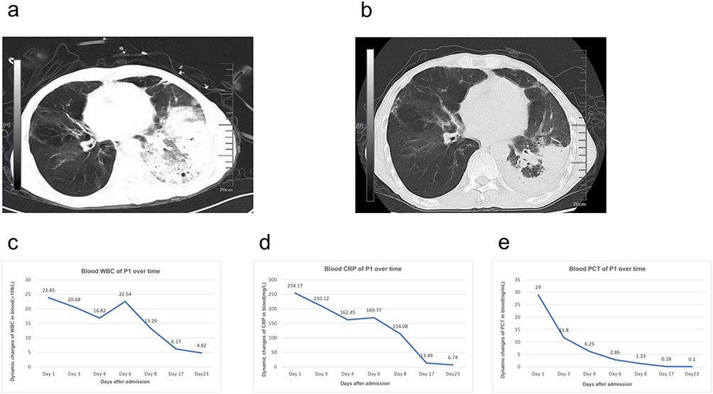 |
Figure 1 Clinical course and diagnostic findings of Patient 1. (a) Chest CT image (day 1 of treatment); (b) Chest CT image (after 1 month of treatment); (c) WBC (white blood cell) dynamic changes. (d) CRP (C-reactive protein) dynamic changes; (e) PCT (procalcitonin) dynamic changes; CRP: mg/L; PCT: ng/mL; WBC: 109/L. |
Patient 2 (P2)
A 24-year-old male fire-breathing acrobat, was admitted on June 28, 2025, with a 7-day history of recurrent fever. Previous penicillin treatment was ineffective. He had a high fever (up to 40°C), cough, and purulent, and occasional bloody sputum. On admission, his vital signs were stable, but wet rales were heard in both lungs. A chest CT showed scattered lung, inflammation, consolidation, and a cavitation in the left lower lobe (Figure 2a). Inflammatory markers were elevated (CRP 83.00 mg/L, WBC 14.99×10⁹/L). Routine microbiological tests (including BALF culture and tuberculosis-related tests) and tNGS (reporting only human parainfluenza virus) were negative. Admitted with suspected pulmonary infection, the patient received empirical treatment with piperacillin-tazobactam (4.5 g q12h IV) and moxifloxacin tablets (0.4 g qd PO), which proved ineffective. We observed poor oral hygiene with multiple carious teeth (Figure 2c), and there were a large number of anaerobic bacteria in mNGS, such as Australian streptococcus, infant streptococcus, oral Prevotella, and dental actinomyces (Table 1). Considering the patient’s clinical presentation, occupational history, oral condition, laboratory inflammatory markers, radiological findings, culture results, and tNGS response to treatment, we suspected a pulmonary anaerobic infection. We discontinued moxifloxacin and switched to piperacillin-tazobactam (4.5 g q8h, IVGGT) combined with metronidazole (1 g q8h, IVGGT) for anaerobic coverage. BALF mNGS was resubmitted with a focus on anaerobic organisms. mNGS detected multiple anaerobic species. IVGGT) combined with metronidazole (1 g, q8h, IVGGT) for anaerobic coverage. After 3 days of extra metronidazole, the patient’s temperature and symptoms improved, and inflammatory markers decreased (Figure 2d–f). The patient was discharged after 9 days with instructions to continue metronidazole (400mg, q8h, p.o.) post-discharge. A follow-up CT scan approximately one month after treatment showed lesion absorption and cavity closure (Figure 2b). Primary pathogens (anaerobic): Prevotella oralis (mNGS relative abundance 15.2%) was identified as the most likely primary pathogen based on highest relative abundance, clinical correlation, and treatment response. Streptococcus australis (mNGS relative abundance 6.8%) and Actinomyces caries (mNGS relative abundance 4.3%) were considered contributing anaerobic pathogens. Viral detections: Human parainfluenza virus (tNGS) and Human Herpesvirus 1 (mNGS) were considered incidental findings or viral reactivation not requiring specific antiviral therapy in the absence of clinical evidence of active viral disease. Treatment protocol: Initial empirical therapy (piperacillin-tazobactam + moxifloxacin) was ineffective. Revised to piperacillin-tazobactam (4.5 g q8h IV) + metronidazole (1 g q8h IV) targeting anaerobic pathogens, with clinical improvement within 3 days.
 |
Figure 2 Clinical course and diagnostic findings of Patient 2. (a) Chest CT image (day 1 of treatment); (b) Chest CT image (after 1 month of treatment); (c) The oral condition of P2 (d) White blood cell (WBC) count over time. (e) CRP (C-reactive protein) dynamic changes; f. Blood neutrophil count (ANC) dynamics; CRP: mg/L; WBC: 109/L; ANC: 109/L. |
Materials and Methods
BALF was obtained via sterile fiberoptic bronchoscopy by skilled pulmonologists in a dedicated suite. Samples were promptly sent to the microbiology lab in sterile containers and processed within 2 hours. Quality control involved: (1) Gram staining and cell count for sample adequacy; (2) ensuring squamous epithelial cells were <1% to confirm lower respiratory origin;4 (3) maintaining aseptic technique to reduce oral contamination; (4) collecting blood cultures to distinguish true infection from contamination. These consistent measures ensured reliable results.
Metagenomic sequencing was conducted on the Illumina NextSeq 550 platform5. DNA from BALF samples was extracted with the QIAamp DNA Mini Kit. Libraries were prepared using the Nextera DNA Flex protocol with dual-index adapters, producing 150 bp paired-end reads with at least 20 million reads per sample6. Bioinformatics involved: (1) quality control and adapter trimming with FastQC and Trimmomatic; (2) removal of human sequences by mapping to GRCh38 using Bowtie;5 (3) microbial identification by aligning to the NCBI RefSeq database with Kraken2/Bracken;6,7 (4) pathogen reporting based on thresholds (>5% for bacteria), coverage depth, and clinical correlation. Targeted next-generation sequencing on the Ion Torrent S5 platform with a respiratory pathogen panel covering 185 targets was conducted, excluding anaerobic bacteria, which explains the failure to detect them.
Susceptibility testing for aerobic bacteria used the VITEK 2 system, while anaerobic bacteria were tested via broth microdilution per CLSI guidelines. Patient 1 showed susceptibility to piperacillin-tazobactam and metronidazole; Patient 2 to metronidazole and piperacillin-tazobactam. Antibiotic choices were guided by susceptibility results, clinical guidelines, and local resistance patterns.
We implemented a diagnostic process with six steps: 1) mNGS identifies anaerobic sequences above 5% relative abundance; 2) align findings with symptoms like fever, cough, and aspiration risk; 3) confirm with radiological signs of anaerobic pneumonia; 4) check for high-risk oral factors such as dental issues; 5) rule out other diagnoses through thorough testing; 6) assess response to anti-anaerobic treatment.
Discussion
Anaerobic aspiration pneumonia frequently manifests in individuals with high-risk factors for aspiration. These include ischemic stroke, cerebral hemorrhage, neurodegenerative disorders, dementia, advanced malignancies, the administration of sedatives or antipsychotics, alcohol consumption, gastroesophageal reflux, oropharyngeal dysphagia, periodontal disease, and inadequate oral hygiene.8 Symptoms include cough, and purulent or foul-smelling sputum. Severe cases may lead to shortness of breath, respiratory distress, and tachycardia. Lab tests usually show high WBC counts and CRP levels, while imaging reveals pulmonary infiltrates, possibly with cavitation or lung abscesses.
Diagnosis faces multiple challenges. Standard pathogen detection methods include microbial culture, serology, antigen, antibody assays, and PCR-based nucleic acid testing.9 Anaerobic bacterial culture is time-consuming and requires special conditions, being relevant only for BALF, and not sputum, which is not easily available at all medical facilities. It also needs a separate physician order and equipped labs, making it rare in routine practice.5 Antigen/antibody detection has limited sensitivity, and PCR testing, though sensitive and specific requires knowing the pathogen type in advance.
To improve the detection and diagnosis of pathogens causing lung infections, an increasing number of hospitals have adopted mNGS and tNGS testing in clinical settings in recent years. As a culture-independent detection method, mNGS offers the capability to impartially detect nucleic acid sequences of all microorganisms in samples,10 enabling the identification of rare or specialized pathogens that are difficult to detect using traditional methods.11 However, this technology has certain limitations: these include data quality and contamination issues, significant interference from human host sequences,12 the need to balance database size with clinical demands, relatively high costs,13 and difficulties in distinguishing colonization, contamination, and infection.14 Bioinformatics workflows struggle to automatically identify and prioritize the primary pathogen(s),15 often misclassifying anaerobic sequences as background or colonizing bacteria, or even excluding them from reports. Although tNGS offers higher sensitivity and resolution, its detection is limited to predefined panels targeting specific pathogen genes.16 Most testing companies exclude anaerobic bacterial gene sequences from pulmonary infection panels due to concerns about oral contamination and colonizing bacteria, missing anaerobic genetic information. Both mNGS and tNGS face contamination issues from orally colonized bacteria in respiratory samples, leading to false negative or false positive results.4 As demonstrated in the two cases presented, initial tNGS/mNGS analyses failed to identify anaerobic bacteria, and confirmation required integrating high-risk clinical factors with microbial community data analysis.
mNGS findings require careful differentiation between colonization, contamination, and true infection. We applied the following criteria: (i) relative abundance threshold (>5% of total bacterial reads for anaerobic sequences); (ii) clinical correlation (fever, purulent sputum, radiological consolidation or cavitation, elevated inflammatory markers); (iii) risk factors (poor oral hygiene, dental caries, periodontal disease); (iv) culture correlation (anaerobic culture positivity when available); (v) treatment response (clinical improvement following targeted anti-anaerobic therapy). Contamination was minimized through strict bronchoscopy protocols and sample quality assessment. Colonization was suspected when anaerobic sequences were detected without compatible clinical features or risk factors. True infection was diagnosed when multiple criteria were met, as in both cases.
Our findings align with studies on mNGS performance in pulmonary infections. Chen et al6 demonstrated that mNGS detected pathogens in a substantial proportion of culture-negative lower respiratory tract infections, with higher sensitivity than conventional methods. Langelier et al17 established an integrated host-microbe model for LRTI diagnosis in critically ill adults, highlighting the challenge of distinguishing pathogenic microbes from commensal flora—a limitation similarly encountered in our cases. Duan et al further validated the diagnostic value of mNGS across infectious diseases, emphasizing its utility when conventional diagnostics fail.
Predominant anaerobic pathogens in aspiration pneumonia include Prevotella species, Fusobacterium nucleatum, Streptococcus anginosus group, and Bacteroides species. Bartlett identified these organisms as established pathogens in anaerobic lung infections, with Prevotella and Fusobacterium among the most frequently isolated.18 In Case 1, detection of Bacteroides timidum and Fusobacterium nucleatum aligns with these pathogenic anaerobes. In Case 2, Prevotella oralis and Streptococcus australis correspond to oral commensal flora typically implicated in aspiration-related infections.
The correlation between identified pathogens and clinical pneumonia can be established through: (i) clinical presentation consistent with anaerobic infection (fever, purulent sputum, aspiration risk factors); (ii) radiological findings of consolidation with cavitation or abscess formation; (iii) elevated inflammatory markers; (iv) presence of high-risk oral factors; (v) clinical response to targeted anti-anaerobic therapy. In both cases, this multi-modal correlation supported the diagnosis of anaerobic pneumonia despite negative conventional cultures.
Anaerobic bacteria are generally treatable with nitroimidazoles, beta-lactam/beta-lactamase inhibitors, and carbapenems.19 Clinically, nitroimidazoles and β-lactams are often chosen for these infections. Nitroimidazoles like metronidazole, tinidazole, and ornidazole specifically target against anaerobic bacteria20 and exhibit relatively stable resistance patterns.21 β-lactams are effective against most anaerobes with low toxicity, though resistance to some is increasing22, which may affect treatment outcomes. Recent research questions the need for routine anaerobic coverage in aspiration pneumonia. Many recommend excluding anaerobic coverage in initial treatment for patients with good oral hygiene and no lung abscess or empyema.23,24 If treatment fails, reassess oral status, radiographic findings, and molecular testing results to adjust the regimen promptly.25
Clinicians may consider anaerobic infection in pneumonia patients with aspiration risk, poor oral hygiene, and negative conventional pathogen tests. mNGS can detect anaerobic sequences, but it cannot distinguish between colonization and infection alone. Potential strategies include: (1) Collecting BALF for anaerobic culture when clinically feasible; (2) Carefully analyzing mNGS reports for anaerobic sequences and their relative abundance; (3) Communicating with laboratories to request anaerobic bacterial analysis when indicated; (4) Selective empirical anti-anaerobic therapy guided by clinical risk factors and m findings, with reassessment based on culture results and clinical response. Increasing clinical awareness and diagnostic vigilance is crucial to overcoming detection challenges and preventing treatment delays. However, these recommendations are based on observational data from two cases and require validation in prospective studies before generalization to broader clinical practice.
Conclusion
Careful interpretation of mNGS profiles, combined with BALF anaerobic cultures and selective empirical anti-anaerobic therapy guided by high-risk oral factors, can facilitate timely diagnosis and improve outcomes in patients with suspected anaerobic pneumonia. However, these findings are based on only two cases and require validation in larger prospective studies.
Case 1: The initial treatment with meropenem, moxifloxacin, and doxycycline was ineffective, so it was changed to piperacillin-tazobactam and metronidazole for anaerobic bacteria, with caspofungin added for fungal infection. Consolidation therapy with ornidazole and fluconazole for 16 days led to clinical recovery. Case 2: The initial treatment with piperacillin-tazobactam and moxifloxacin was ineffective, so the regimen was adjusted to piperacillin-tazobactam and metronidazole, both administered intravenously every 8 hours. After discharge, metronidazole was continued orally. Clinical improvement was observed with the targeted anti-anaerobic therapy.
This study has several design limitations: a small sample size (n=2) limits generalizability; potential contamination and variability in BALF collection may have affected mNGS results; mNGS cannot distinguish between viable and non-viable organisms, risking detection of non-pathogenic nucleic acid; the retrospective design lacked standardized data collection; differing mNGS detection thresholds and bioinformatics pipelines hinder cross-study comparability; findings are specific to certain clinical contexts and may not apply to other populations; and no formal statistical analysis was conducted as the report covers two retrospective cases. Caution is advised in interpreting these results until validated in diverse settings; all analyses were descriptive.
Abbreviations
BALF, Bronchoalveolar lavage fluid; CAP, community-acquired pneumonia; PCR, polymerase chain reaction; mNGS, Metagenomic next-generation sequencing; tNGS, Targeted next-generation sequencing; CRP, C-reactive protein; PCT, procalcitonin; WBC, white blood cell; ANC, neutrophil count.
Data Sharing Statement
Data on the case clinical information and images are available for review from the corresponding author upon request.
Ethics and Consent Statements
This was retrospective case report study conducted in the Department of Infectious Diseases at a tertiary hospital, and has been approved by the Ethics Committee of the Second Affiliated Hospital of Hainan Medical University (No. LW202607). Both patients have consented to the publication of the details of their cases.
Acknowledgments
This study was partially supported by Department of respiratory medicine, the Second Affiliated Hospital of Hainan Medical University. We thank the staff at the Second Affiliated Hospital of Hainan Medical University for their dedication to diagnosis and treatment.
Author Contributions
All authors made a significant contribution to the work reported, whether that is in the conception, study design, execution, acquisition of data, analysis and interpretation, or in all these areas; took part in drafting, revising or critically reviewing the article; gave final approval of the version to be published; have agreed on the journal to which the article has been submitted; and agree to be accountable for all aspects of the work.
Disclosure
The authors report no conflicts of interest in this work.
References
1. Mandell LA, Niederman MS. Aspiration pneumonia. N Engl J Med. 2019;380(7):651–8. doi:10.1056/NEJMc1903636
2. Terpenning MS, Taylor GW, Lopatin DE, Kerr CK, Dominguez BL, Loesche WJ. Aspiration pneumonia: dental and oral risk factors in an older veteran population. J Am Geriatr Soc. 2001;49(5):557–563. doi:10.1046/j.1532-5415.2001.49114.x
3. Marik PE. Aspiration pneumonitis and aspiration pneumonia. N Engl J Med. 2001;344(9):665–671. doi:10.1056/NEJM200103013440908
4. Chen H, Yin Y, Gao H, et al. Clinical utility of in-house metagenomic next-generation sequencing for the diagnosis of lower respiratory tract infections and analysis of the host immune response. Clin Infect Dis. 2020;71(Suppl 4):S416–S426. doi:10.1093/cid/ciaa1516
5. Duan H, Li X, Mei A, et al. The diagnostic value of metagenomic next-generation sequencing in infectious diseases. BMC Infect Dis. 2021;21(1):62. doi:10.1186/s12879-020-05746-5
6. Langelier C, Kalantar KL, Moazed F, et al. Integrating host response and unbiased microbe detection for lower respiratory tract infection diagnosis in critically ill adults. Proc Natl Acad Sci U S A. 2018;115(52):E12353–E12362. doi:10.1073/pnas.1809700115
7. Vallianou NG, Skourtis A, Kounatidis D, et al. The role of the respiratory microbiome in the pathogenesis of aspiration pneumonia: implications for diagnosis and potential therapeutic choices. Antibiotics. 2023;12(1):xx. doi:10.3390/antibiotics12010140
8. Zhang N, Wang L, Deng X, et al. Recent advances in the detection of respiratory virus infection in humans. J Med Virol. 2020;92(4):408–417. doi:10.1002/jmv.25674
9. Nagy E, Boyanova L, Justesen US. How to isolate, identify and determine antimicrobial susceptibility of anaerobic bacteria in routine laboratories. Clin Microbiol Infect. 2018;24(11):1139–1149. doi:10.1016/j.cmi.2018.02.008
10. Yin Y, Zhu P, Guo Y, et al. Enhancing lower respiratory tract infection diagnosis: implementation and clinical assessment of multiplex PCR-based and hybrid capture-based targeted next-generation sequencing. EBioMedicine. 2024;107:105307. doi:10.1016/j.ebiom.2024.105307
11. Zheng Y, Qiu X, Wang T, et al. The diagnostic value of metagenomic next-generation sequencing in lower respiratory tract infection. Front Cell Infect Microbiol. 2021;11:694756. doi:10.3389/fcimb.2021.694756
12. Lin Q, Yao Y, Li X, et al. The application of nanopore targeted sequencing for pathogen diagnosis in bronchoalveolar lavage fluid of patients with pneumonia: a prospective multicenter study. Infect Dis. 2024;56(2):128–137. doi:10.1080/23744235.2023.2276785
13. Liu H, Zhang Y, Yang J, et al. Application of mNGS in the etiological analysis of lower respiratory tract infections and the prediction of drug resistance. Microbiol Spectr. 2022;10(1):e0250221. doi:10.1128/spectrum.02502-21
14. Zhong J, Liu Y, Luo N, et al. Metagenomic next-generation sequencing for rapid detection of pulmonary infection in patients with acquired immunodeficiency syndrome. Ann Clin Microbiol Antimicrob. 2023;22(1):57. doi:10.1186/s12941-023-00608-9
15. Sun W, Zheng L, Kang L, et al. Comparative analysis of metagenomic and targeted next-generation sequencing for pathogens diagnosis in bronchoalveolar lavage fluid specimens. Front Cell Infect Microbiol. 2024;14:1451440. doi:10.3389/fcimb.2024.1451440
16. Huang J, Jiang E, Yang D, et al. Metagenomic next-generation sequencing versus traditional pathogen detection in the diagnosis of peripheral pulmonary infectious lesions. Infect Drug Resist. 2020;13:567–576. doi:10.2147/IDR.S235182
17. Bartlett JG. Anaerobic bacterial infection of the lung. Anaerobe. 2012;18(2):235–239. doi:10.1016/j.anaerobe.2011.12.004
18. Snydman DR, Jacobus NV, McDermott LA, et al. Update on resistance of Bacteroides fragilis group and related species with special attention to carbapenems 2006–2009. Anaerobe. 2011;17(4):147–151. doi:10.1016/j.anaerobe.2011.05.014
19. Bai AD, Srivastava S, Digby GC, et al. Anaerobic antibiotic coverage in aspiration pneumonia and the associated benefits and harms: a retrospective cohort study. Chest. 2024;166(1):39–48. doi:10.1016/j.chest.2024.02.025
20. Wybo I, Van den Bossche D, Soetens O, et al. Fourth Belgian multicentre survey of antibiotic susceptibility of anaerobic bacteria. J Antimicrob Chemother. 2014;69(1):155–161. doi:10.1093/jac/dkt344
21. Forbes JD, Kus JV, Patel SN. Antimicrobial susceptibility profiles of invasive isolates of anaerobic bacteria from a large Canadian reference laboratory: 2012–2019. Anaerobe. 2021;70:102386. doi:10.1016/j.anaerobe.2021.102386
22. Kitsios GD, Nguyen VD, Sayed K, et al. The upper and lower respiratory tract microbiome in severe aspiration pneumonia. iScience. 2023;26(6):106832. doi:10.1016/j.isci.2023.106832
23. Rodriguez AE, Restrepo MI. New perspectives in aspiration community-acquired pneumonia. Expert Rev Clin Pharmacol. 2019;12(10):991–1002. doi:10.1080/17512433.2019.1663730
24. Yoshimatsu Y, Tobino K, Ko Y, et al. Careful history taking detects initially unknown underlying causes of aspiration pneumonia. Geriatr Gerontol Int. 2020;20(8):785–790. doi:10.1111/ggi.13978
25. Yoshimatsu Y, Tobino K, Ortega O, et al. Development and implementation of an aspiration pneumonia cause investigation algorithm. Clin Respir J. 2023;17(1):20–28. doi:10.1111/crj.13557
 © 2026 The Author(s). This work is published and licensed by Dove Medical Press Limited. The
full terms of this license are available at https://www.dovepress.com/terms
and incorporate the Creative Commons Attribution
- Non Commercial (unported, 4.0) License.
By accessing the work you hereby accept the Terms. Non-commercial uses of the work are permitted
without any further permission from Dove Medical Press Limited, provided the work is properly
attributed. For permission for commercial use of this work, please see paragraphs 4.2 and 5 of our Terms.
© 2026 The Author(s). This work is published and licensed by Dove Medical Press Limited. The
full terms of this license are available at https://www.dovepress.com/terms
and incorporate the Creative Commons Attribution
- Non Commercial (unported, 4.0) License.
By accessing the work you hereby accept the Terms. Non-commercial uses of the work are permitted
without any further permission from Dove Medical Press Limited, provided the work is properly
attributed. For permission for commercial use of this work, please see paragraphs 4.2 and 5 of our Terms.
Recommended articles

A Paired Comparison of Plasma and Bronchoalveolar Lavage Fluid for Metagenomic Next-Generation Sequencing in Critically Ill Patients with Suspected Severe Pneumonia
Sun T, Liu Y, Cai Y, Zhai T, Zhou Y, Yang B, Wu X, Zhan Q
Infection and Drug Resistance 2022, 15:4369-4379
Published Date: 9 August 2022
Diagnostic Value of Metagenomic Next-Generation Sequencing for Multi-Pathogenic Pneumonia in HIV-Infected Patients
Xie Y, Dai B, Zhou X, Liu H, Wu W, Yu F, Zhu B
Infection and Drug Resistance 2023, 16:607-618
Published Date: 27 January 2023
Diagnosis of Non-Tuberculous Mycobacterial Pulmonary Disease by Metagenomic Next-Generation Sequencing on Bronchoalveolar Lavage Fluid
Zhang X, Chen H, Lin Y, Yang M, Zhao H, Hu J, Han D
Infection and Drug Resistance 2023, 16:4137-4145
Published Date: 26 June 2023
Comprehensive Analysis of Pathogen Diversity and Diagnostic Biomarkers in Patients with Suspected Pulmonary Tuberculosis Through Metagenomic Next-Generation Sequencing
Li Y, Li C, Fang Y, Zhang L, Ying X, Ren R, Zang Y, Ying D, Zhu S, Liu J, Cao X
Infection and Drug Resistance 2025, 18:2215-2227
Published Date: 2 May 2025