Back to Journals » Clinical, Cosmetic and Investigational Dermatology » Volume 16
Dermatosis Neglecta Based on Exanthematous Drug Eruption Following Head Trauma: A Case Report and Literature Review
Authors Shi L , Zhao J, Zeng L , Wang L, Zhang G
Received 28 April 2023
Accepted for publication 29 July 2023
Published 7 August 2023 Volume 2023:16 Pages 2083—2088
DOI https://doi.org/10.2147/CCID.S419092
Checked for plagiarism Yes
Review by Single anonymous peer review
Peer reviewer comments 5
Editor who approved publication: Dr Jeffrey Weinberg
Liping Shi,1,2 Jiaqing Zhao,1,2 Linxi Zeng,1,2 Lihong Wang,3 Guoqiang Zhang1,2
1Department of Dermatology, The First Hospital of Hebei Medical University, Shijiazhuang, People’s Republic of China; 2Candidate Branch of National Clinical Research Center for Skin Diseases, Shijiazhuang, People’s Republic of China; 3Department of Dermatology, Luancheng People’s Hospital, Shijiazhuang, People’s Republic of China
Correspondence: Guoqiang Zhang, The First Hospital of Hebei Medical University, 89 Donggang Road, Yuhua District, Shijiazhuang City, Hebei Province, People’s Republic of China, Tel +8618633888122, Email [email protected]
Abstract: Dermatosis neglecta (DN) is a rare psychogenic dermatosis. To our knowledge, this is the first reported case of DN based on exanthematous drug eruption. We report a 68-year-old Chinese male patient who presented with thick, yellowish-brown crusting on his face and scalp and scaly skin for 6 days. Dermoscopy revealed diffusely distributed yellow-green serescrust-like plaques with different sizes and uneven thickness on a red background, and some demonstrated dot or globular hemorrhages. We considered DN and exanthematous drug eruptions based on the combination of the clinical medication and the history before the rash.
Keywords: cutaneous dirt-adherent, malassezia infection, drug allergy, autoantibody
Introduction
Dermatosis neglecta (DN) is a rare mental dermatosis characterized by localized, persistent, and dirty substances adhering to the skin, which is more common for young Asian women. DN occurs on the skin of the whole body, and especially on the head and face, hands, ankles, armpits, mammary areola, and around the mammary areola. Here, we report the development of DN based on exanthematous drug eruption following head trauma in an elderly male patient.
Case Presentation
A 68-year-old male patient presented to our department for a 2-month history of erythema and pruritus all over his body and a 1-week history of yellowish-brown crusting on his face and scalp and scaly skin. He developed erythema and pruritus all over his body 2 months ago, following a 20-day duration of vancomycin infusion due to a brain abscess. The patient was diagnosed with exanthematous drug eruption after consultation with the Department of Dermatology and Pharmacy, and was treated with intravenous methylprednisolone, oral ebastine and topical desonide cream, and the rash area was slightly reduced and the itching was relieved, but the lesions on his face and neck remained unaffected. Yellowish-brown crusts and scales appeared on his face and scalp 1 week ago. The crusts and scales gradually increased and thickened, extending to the whole face, without special treatment.
Medical history revealed hypertension for >30 years, with a maximum of 200/130 mmHg. He was hit in the jaw with a blunt object 34 years ago, resulting in a mandibular fracture and oral trauma. He underwent a thoracotomy for pleurisy >10 years ago. He underwent cerebral contusion and hematoma removal 2 months ago due to multiple cerebral contusions and lacerations, subdural hematoma, skull fracture, and traumatic subarachnoid hemorrhage.
Dermatological examination revealed densely distributed yellowish to brown serous crusts on the scalp, bilateral zygomatic, eyebrow, temporal, and cheek, which tightly adhered and were difficult to peel off. Further, mild swelling of both eyelids and diffuse erythema on the neck, with unclear boundaries and slightly higher skin temperature, were seen (Figure 1A–E).
Laboratory tests revealed red blood cell counts of 3.08 × 10¹²/L (↓) and hemoglobin of 99 g/L (↓). Biochemistry + immunoglobulin test + blood homocysteine test + complement test revealed γ-glutamyl transpeptidase of 91 U/L (↑), total protein of 61.5 g/L (↓), and high-sensitivity C-reactive protein of 12.41 mg/L (↑). Antinuclear antibody screening + antineutrophil cytoplasmic antibody full item+ENA15 revealed anti-Ro-52 antibody of >400.00RU/mL, anti-SSA antibody of >400.00RU/mL, and anti-SSB antibody of 66.95RU/mL, as well as a positive antinuclear antibody (ANA) with a titer of 1:320 and a granular type of ANA. Urine routine, stool routine, serum oxcarbazepine concentration determination, infectious diseases examination, five male tumor tests, blood coagulation, fungal dextran, bacterial endotoxin, Aspergillus, procalcitonin, and electrocardiogram were normal. Compared with 2 months ago, chest computed tomography (CT) revealed no significant changes, showing patchy high-density shadows in the middle lobe of the right lung, the lingual segment of the left upper lobe of the lung, and the lower lobe of both lungs, and foliar high-density shadows in the lower lobe of the right lung. Fibrous and calcified lesions were seen in the upper lobe of the right lung. Left and right coronary artery calcification, aortic sclerosis, a small amount of pericardial effusion, and multiple small bilateral axillary lymph nodes, partially full, were seen. Brain CT plain scan + enhanced revealed left frontal lobe postoperative changes, bilateral frontal lobe contusion and laceration, and left temporal lobe and insular lobe, as well as small nodular abnormal enhancement near the anterior horn of the left frontal lateral ventricle. The right frontal bone and the right parietal bone were fractured. Pus bacterial culture revealed Staphylococcus aureus growth. Mycological microscopic examination of skin lesions was negative. Dermoscopy revealed diffusely distributed yellow-green serescrust-like plaques with different sizes and uneven thickness on a red background, and some demonstrated dot or globular hemorrhages (Figure 2).
The patient was diagnosed with 1) exanthematous drug eruption, 2) DN, 3) postoperative traumatic subarachnoid hemorrhage, 4) postoperative subdural hematoma, 5) skull fractures, 6) hypertension grade 3 (very high risk), 7) postoperative pleurisy, 8) pericardial effusion, 9) aortic sclerosis, and 10) chronic lung inflammation.
Ebastine and mometasone furoate were used to treat drug-induced dermatitis. Levofloxacin was used against infection following the bacterial culture results. The scaly scab on the head and face was removed with benzalkonium chloride solution, followed by a spray of recombinant human acid fibroblast growth factor for external use, and finally, a topical application of compound polymyxin B ointment. The patient’s rash completely resolved after 10 days of treatment (Figure 1F–J). We performed followed-up for 4 months without recurrence, and the patient was satisfied with the efficacy.
Discussion
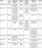
DN is a rare psychogenic skin disease first described in 1995. DN was previously known as Cutaneous dirt-adherent disease (COAD). COAD is diagnosed mainly based on the typical clinical presentation, which is characterized by thick brown verrucous crusts that adhered to the skin surface with well-defined borders and are difficult to peel off.1 The disease etiology is unknown. The pathogenesis of it may be related to mental factors, endocrine disorders, trauma, long-term non-scrubbing, and Malassezia infection.2 However, COAD is a vague diagnosis, which can be accurately diagnosed as DN, terra firma–forme dermatosis, confluent and reticulated papillomatosis, or other skin diseases with detailed clinical data.3 Four cases were accurately diagnosed as DN based on previous COAD reports. DN can complicate with other diseases, such as Sjogren’s syndrome, pemphigus, Darier’s disease, pityriasis rubra pilaris, alopecia areata, and schizophrenia and so on1,4–10 (Table 1).
 |
Table 1 Clinical Features of Some Recently Reported Dermatosis Neglecta Complicated with Other Diseases |
Our patient was bedridden for several days postoperatively due to trauma and then developed a rash with itching all over the body due to vancomycin allergy. The mental health symptom checklist (SCL-90) revealed symptoms of depression and anxiety, thus we speculate that the combination of a variety of diseases caused his mental problems. Additionally, psychiatric factors may cause endocrine disturbances that promote sebum secretion. Moreover, head and face sebum secretion is vigorous, and long-term inadequate washing causes abnormal bacteria and fungi growth. The patient refused to wash the diseased skin, thereby forming a vicious cycle, because the dirt substances adhering to the head and face cannot be washed off easily with water. The unifying stepwise approach to dirt-like lesion diagnosis,3 combined with the patient’s history of inadequate washing and the removal of scaly scab with benzalaceum chloride solution, made the DN diagnosis easily. The diagnosis in this patient requires differentiation from acanthosis nigricans, Darier’s disease, Terra Firme-Forme Dermatosis, and confluent and reticulated papillomatosis.11
The occurrence of DN after trauma has been previously reported, as was the case in our patient. The patient’s jaw and oral cavity were damaged when he was young, but DN did not occur at that time, which may be related to the faster skin metabolism and stronger repair ability of young people. The patient suffered a second head and face injury and developed DN 34 years later. Alternatively, it may be related to the superposition of two traumas causing impaired metabolism, repair, and barrier function. Additionally, the patient developed DN based on drug eruption. We speculated that the damage to the skin barrier function and bacterial colonization disorder caused by drug eruption, with the above reasons together, contributed to DN occurrence.
The fungal examination was negative in this case, and the pus bacterial culture revealed the growth of Staphylococcus aureus. The patient showed clinical improvement with oral and topical antibacterial medication. Therefore, bacterial infection may play a role in DN pathogenesis.
Drug allergy is a specific and repeatable immune response to a particular drug. The most common type of drug allergy is cutaneous manifestation, drug eruption. Antibiotics and nonsteroidal anti-inflammatory drugs are the most frequent causes of drug allergy.12 The types of drug allergy caused by vancomycin include exanthem drug eruption, systemic symptoms syndrome, linear IgA bullous dermatosis (LABD), and Stevens–Johnson syndrome/toxic epidermal necrolysis (SJS/TEN),13 and LABD is the most common vancomycin hypersensitivity reaction.13 Our patient’s rash was consistent with an exanthematous drug eruption with a Naranjo score of 6,14 indicating that vancomycin was most likely the causative agent of the patient’s drug eruption. Patients with autoimmune diseases have a high incidence of drug allergy, such as Sjogren’s syndrome, systemic lupus erythematosus, and adult-onset Still’s disease,11 additionally DN could be accompanied by Sjogren’s syndrome,4 several autoantibodies were positive in this patient, and connective tissue disease could not be excluded after consultation with the rheumatology department, because the patient had no symptoms, such as the dry mouth and eyes, oral ulcers, joint pain, and photaesthesia, thus he could not be further diagnosed. The relationship between a patient’s autoantibody and drug eruptions, DN, or both remains uncertain. This study indicated that the patient regularly reviewed the relevant indicators and followed up with the rheumatology department.
Conclusion
To our knowledge, this is the first report of DN occurring after drug eruption. Positive autoantibodies are also relatively rare in patients with DN. DN can be accompanied by a variety of diseases, and a variety of factors cause its occurrence. Clinicians should pay attention to patients who are bedridden, postoperative, and with physical and mental disorders to prevent DN.
Abbreviations
DN, dermatosis neglecta; ANA, antinuclear antibody; CT, chest computed tomography; LABD, linear IgA bullous dermatosis; SJS/TEN, Stevens–Johnson syndrome/toxic epidermal necrolysis.
Consent Statement
Written informed consent was obtained from the patient for publication of this manuscript and any accompanying images. Institutional approval was not required to publish the case details.
Author Contributions
All authors made a significant contribution to the work reported, whether that is in the conception, study design, execution, acquisition of data, analysis and interpretation, or in all these areas; took part in drafting, revising or critically reviewing the article; gave final approval of the version to be published; have agreed on the journal to which the article has been submitted; and agree to be accountable for all aspects of the work.
Funding
This work is supported by the Graduate Education and Teaching Reform Project of Hebei Medical University (2022-20) and Medical science research project of Hebei Province Health Commission (20190470).
Disclosure
All authors have no conflicts of interest in this work.
References
1. Jun L, Liu JW, Sun QN. Cutaneous dirt-adherent disease on a base of pemphigus erythematosus. Int J Dermatol. 2014;53(4):e269. doi:10.1111/ijd.12236
2. Tajima M, Amaya M, Sugita T, Nishikawa A, Tsuboi R. Malassezia の菌相を解析したアカツキ病の3例 [Molecular analysis of Malassezia species isolated from three cases of Akatsuki disease (pomade crust)]. Nihon Ishinkin Gakkai Zasshi. 2005;46(3):193–196. Japanese. doi:10.3314/jjmm.46.193
3. Tan C. Dirt-adherent dermatosis: not worth an additional name. Arch Dermatol. 2010;146(6):679–680. doi:10.1001/archdermatol.2010.106
4. Jiang T, He C. Cutaneous dirt-adherent disease with Sjögren’s syndrome. Dermatol Ther. 2022;35(4):e15316. doi:10.1111/dth.15316
5. Shan SJ, Xu TH, Liu J, et al. Cutaneous dirt-adherent disease with single apparent transverse leukonychia on the fingernails. Arch Dermatol. 2009;145(9):1070–1071. doi:10.1001/archdermatol.2009.208
6. Zhu Q, Guo SJ, Wang B, Xu LL, Zhang GQ. Cutaneous dirt-adherent disease complicated with Darier’s disease, schizophrenia, and cutis verticis gyrata: a case report. Front Med. 2022;9:939107. doi:10.3389/fmed.2022.939107
7. Chen X, Zhang J, Zhou C. Dermatosis neglecta of the scalp complicated with Alopecia Areata. Int J Trichology. 2020;12(3):138–139. doi:10.4103/ijt.ijt_46_19
8. Singh P, Kar SK, Kumari R, Gupta SK. Dermatosis neglecta in schizophrenia: a rare case report. Indian J Psychol Med. 2015;37(1):93–95. doi:10.4103/0253-7176.150851
9. Saha A, Seth J, Sharma A, Biswas D. Dermatitis neglecta - a dirty dermatosis: report of three cases. Indian J Dermatol. 2015;60(2):185–187. doi:10.4103/0019-5154.152525
10. Wattanawinitchai K, Suchonwanit P. Case report: dermatosis neglecta mimicking pemphigus foliaceus in association with obsessive-compulsive disorder. Front Med. 2023;10:1076397. doi:10.3389/fmed.2023.1076397
11. Tan C, Zhu WY. Nonmelanotic pigmentary disorders with abnormal deposits. In: Atlas of Pigmentary Skin Disorders. Springer; 2023:503–517.
12. Watanabe Y, Yamaguchi Y. Drug allergy and autoimmune diseases. Allergol Int. 2022;71(2):179–184. doi:10.1016/j.alit.2022.02.001
13. Minhas JS, Wickner PG, Long AA, Banerji A, Blumenthal KG. Immune-mediated reactions to vancomycin: a systematic case review and analysis. Ann Allergy Asthma Immunol. 2016;116(6):544–553. doi:10.1016/j.anai.2016.03.030
14. Naranjo CA, Busto U, Sellers EM, et al. A method for estimating the probability of adverse drug reactions. Clin Pharmacol Ther. 1981;30(2):239–245. doi:10.1038/clpt.1981.154
 © 2023 The Author(s). This work is published and licensed by Dove Medical Press Limited. The full terms of this license are available at https://www.dovepress.com/terms.php and incorporate the Creative Commons Attribution - Non Commercial (unported, v3.0) License.
By accessing the work you hereby accept the Terms. Non-commercial uses of the work are permitted without any further permission from Dove Medical Press Limited, provided the work is properly attributed. For permission for commercial use of this work, please see paragraphs 4.2 and 5 of our Terms.


