Back to Journals » International Journal of Nanomedicine » Volume 20
Current Applications and Future Challenges of Mesenchymal Stem Cell-Extracellular Vesicles in Tissue Engineering: A Bibliometric Analysis
Authors Chen S ![]() , Di J, Zhang Z, Guo Z, Tian Z, Qin Y
, Di J, Zhang Z, Guo Z, Tian Z, Qin Y ![]() , Long Y, Xu J
, Long Y, Xu J ![]() , Xiang C
, Xiang C ![]() , Cao F
, Cao F
Received 25 June 2025
Accepted for publication 6 December 2025
Published 11 December 2025 Volume 2025:20 Pages 14885—14910
DOI https://doi.org/10.2147/IJN.S549684
Checked for plagiarism Yes
Review by Single anonymous peer review
Peer reviewer comments 2
Editor who approved publication: Dr RDK Misra
Shuai Chen,1,2,* Jingkai Di,1,2,* Zhibo Zhang,1 Zijian Guo,1,2 Zui Tian,1,2 Yingda Qin,1,2 Yingi Long,1 Jiake Xu,3– 5 Chuan Xiang,1 Fuyang Cao1,2
1Department of Orthopedics, The Second Hospital of Shanxi Medical University, Taiyuan, Shanxi, People’s Republic of China; 2Shanxi Key Laboratory of Bone and Soft Tissue Injury Repair, Department of Orthopedics, Second Hospital of Shanxi Medical University, Taiyuan, Shanxi, People’s Republic of China; 3Center for AI-Driven Medical Research, Chinese Academy of Sciences, Shenzhen, Guangdong, People’s Republic of China; 4Faculty of Pharmaceutical Sciences, Shenzhen University of Advanced Technology, Shenzhen, Guangdong, People’s Republic of China; 5School of Biomedical Sciences, The University of Western Australia, Perth, WA, Australia
*These authors contributed equally to this work
Correspondence: Chuan Xiang, Email [email protected] Fuyang Cao, Email [email protected]
Background: Extracellular vesicles derived from mesenchymal stem cells (MSCs-EVs) are nano-sized vesicles and have become key mediators in tissue engineering and emerging therapeutic agents in regenerative medicine. This study systematically assessed the global research trend of MSCs-EVs for tissue engineering through bibliometric analysis of literature from 2014 to 2024.
Methods: A comprehensive search of Web of Science retrieved 752 eligible articles. We visualized and further analyzed collaboration, co-citation, co-authorship and co-occurrence through VOSviewer and Citespace, focusing on their application and future development trends.
Results: Annual publications increased continuously, with China and the United States accounting for 48.9% and 19.1% of research output, respectively, accompanied by intensive transnational cooperation. China leads in number of publications, number of years, total citations, H index and collaboration. The evolution of research trends confirms that the current field of application has expanded from cellular repair to drug delivery systems and biomaterial integration. Keyword cooccurrence reveals three clusters of research: artificial editing of exosomes (membranes), drug delivery (drugs, nucleic acids), and regeneration mechanisms (bone morphogenesis, angiogenesis regulation). The International Journal of Molecular Science became the most influential journal, and Shanghai Jiaotong University was a leader in institutional productivity.
Conclusion: This study is the first comprehensive quantitative analysis of tissue-engineered EVs and details trends and advances in tissue-engineered EVs research within the field of regenerative medicine. It portrays recent frontiers and hotspots, providing valuable insights for researchers in this particular area of research.
Keywords: extracellular vesicles, exosomes, mesenchymal stem cells, tissue engineering, regenerative medicine, bibliometrics, visualization
Introduction
Tissue Engineering (TE) is an interdisciplinary field that combines principles of biology, medicine, and engineering to design biological substitutes to maintain, restore, or improve tissue function and is an important component of regenerative medicine.1–3 Seed cells, as one of the three elements of tissue engineering, provide the necessary cell source for constructing tissues and organs.4 In recent years, mesenchymal stem cells (MSC) have shown great potential as hot spot cells in regenerative medicine research in the fields of tissue repair, immune regulation and disease therapy.5 For example, umbilical cord MSCs have been shown to improve the progression of cartilage degeneration in osteoarthritis through mitochondrial transfer.6 However, clinical data show that MSCs survive less than 10% after transplantation and their paracrine function is susceptible to inhibition by oxidative stress in the recipient microenvironment.7 Therefore, there is a need to find more suitable tools on this basis. Extracellular vesicles (EVs) of MSCs origin are considered to be a good alternative to MSCs. Because they naturally carry mother cell bioactive molecules, such as miRNAs, cytokines) and nanoscale lipid membrane structures, they exhibit superior organ targeting and immune evasion capabilities, and have become emerging vectors for precision therapy.8 EVs derived from stem cells are heterogeneous and can be divided into exosomes, microvesicles and apoptotic bodies. Their contents depend on the cellular and environmental context of the parent cell. Exosomes, in particular, are being studied for biomarker and therapeutic purposes.9
A growing body of research confirms the role of MSC-EVs in regulating tissue regeneration. Due to its cell-free therapy and low immunogenicity, it shows great advantages in anti-inflammatory and immunomodulatory,10 reversing mitochondrial damage,11 osteoarthritis,12 corneal therapy13 and other fields. Despite the many advantages of natural MSC-EVs, their clinical application remains constrained by bottlenecks such as the limited efficiency of passive targeting and the inability to regulate drug loading.14 In recent years, the employment of engineering modification techniques has been explored as a means to enhance the therapeutic benefits of natural EVs. However, despite the rapid advancements in this field, a systematic evaluation and organization of existing knowledge remains deficient.
Although MSC-EVs show great potential for tissue engineering applications, the field of research is experiencing explosive growth, resulting in a large, fragmented and complex scientific literature.15,16 This rapid development makes it difficult for researchers to fully grasp the overall pattern, evolution path and research frontier of the field through traditional methods. Therefore, it is necessary to make a systematic and objective quantitative analysis of this field by using bibliometrics.17–19
This study aims to visualize the knowledge structure of the field, identify core authors, institutions and collaborative networks, analyze keyword evolution trends, and highlight current research hotspots and future directions.20 This will not only provide an efficient “domain map” for new researchers, but also provide data-driven decision support for senior scholars and scientific policy makers to jointly push the field in a deeper and more efficient direction.20
Therefore, to address the aforementioned challenges and bridge the existing research gaps, this study aims to conduct a systematic quantitative analysis of the literature on the application of MSC-EVs in tissue engineering from the Web of Science Core Collection using bibliometric methods. The specific objectives of this study are as follows: (1) to quantify and visualize the overall research trends in this field, including annual publication volume and collaborative networks among core countries/institutions and authors, in order to untangle the global distribution of research capabilities; (2) to map the foundational knowledge and intellectual structure of the field through co-citation analysis and keyword analysis, thereby identifying key publications and research themes; (3) to track the dynamic evolution of research hotspots through burst-term detection and timeline analysis, and to attempt to predict future research frontiers. Unlike existing traditional reviews, this study provides, for the first time, macro-level data-driven insights into this field, aiming to delineate its knowledge structure, trace developmental trajectories, and identify emerging frontiers, thereby offering objective decision-making support for researchers and promoting the structured development of this interdisciplinary field.
Materials and Methods
Search Strategy
Literature searches were carried out via Web of Science (https://www.webofscience.com/wos/woscc/basic-search) to obtain sufficient and efficient coverage. A comprehensive search was conducted of articles published between January 1, 2014 and December 31, 2024, inclusive. The specific search formula ((TS= (tissue engineering)) AND TS=((Extracellular vesicles) OR (Exosomes))) AND TS= (mesenchymal stem cells) was applied to database. We narrowed the search results by limiting the document type to articles and reviews. After merging the results and removing duplicates using reference management software (Zotero), the language of inclusion in the article was restricted to English. Finally, the combined dataset was subjected to manual screening, where articles unrelated to the research content were removed manually.
Data Analysis
To compare research contributions more effectively, we have combined England, Scotland, Wales, and Northern Ireland into the “United Kingdom”, to make statistical analysis more in line with international practices. The Sankey Diagram to show the relationship between institutions, keywords, and countries, with the width of the connecting line indicating the traffic size.
The VOSviewer software (version 1.6.19) imported bibliographic data, including citations, references, abstracts, keywords, and other information, and it was used to generate visual collaborative, co-cited, co-occurring network views. In our study, collaborative analyses of countries and institutions were conducted, along with co-citation analyses of citations, authors, and journals. In the comprehensive analysis of country/regional and institutional collaboration, a global collaborative network was carefully reviewed, focusing on publications with at least 2 documents per country and academic institutions a minimum of 3 documents and a maximum of 20 organizations per document through VOS viewer. The co-author analysis focuses on authors who have published three or more articles, focusing on collaborative networks within the research community. For the country-based co-author analysis, countries with more than three papers were screened for follow-up studies. In The institutional co-authorship analysis, institutions with more than 3 documents were selected for subsequent analysis and visualization. Co-citation analysis was used by citation frequency ≥20 as threshold to screen high-impact literature as analysis unit, which is used to unravel complicated academic relationship network and determine groundbreaking or high-quality literature in the field. Citespace is a widely utilized bibliometric analysis software. In this study, version 6.3.1 of the software was employed to generate keyword cluster analysis plots. In this study, the keywords and references were analyzed to identify the strongest citation bursts over time. The evolution of knowledge in key areas within the discipline was demonstrated through timeline plots.
The R language (version 4.4.1), RStudio, and the R package “bibliometrix” (version 4.3.0) (https://www.bibliometrix.org) were used to parse the research in the field from multiple dimensions. A comprehensive co-citation analysis of journals using the Bibliometrix R package and adhering to the criteria of excluding journals with less than 20 citations yielded important insights. We designed three field plots, and annual production charts to show the different levels of posting in this area.
Strategic Daram is a visualization tool for revealing the distribution of topics in a research field, identifying research hotspots and emerging trends in the field, and helping researchers to quickly locate the key research directions in the field by quantifying the Densihy and Centrality of the topics and dividing them into different quadrants. The first quadrant (upper right): characterized by high centrality + high density; indicating core topics (mature hotspots in the field), the second quadrant (upper left): low centrality + high density; indicating siloed topics (internally tight but externally weakly connected), and the third quadrant (lower left): low centrality + low density; Marginal receding themes (low research attention), fourth quadrant (lower right): high centrality + low density; potential themes (emerging cross-cutting areas or cutting-edge directions).
In addition, Trending Topics Maps, thematic maps, and Lotka’s Law were used to analyze potential hotspots and research depth in the field. Author productivity was quantified by fitting the observed publication counts to Lotka’s inverse-square power law, a classical bibliometric model widely employed to describe the frequency distribution of scholarly output within a given research domain. The quantitative analysis of the publication was conducted using Microsoft Office Excel 2021.
Results
Overview of Global Literature
In the area of global scholarly output, a comprehensive review of the literature from 2014 to 2024 yielded 758 publications that met the established search parameters. Subsequent improvements to the corpus included the exclusion of online publications (1), meeting abstracts (1), editorial material (1), and along the removal of three non-English studies. This meticulous process culminated in the identification of 752 relevant studies, as shown in Figure 1. Compared with the existing literature, the unique contribution of this analysis lies in its objectivity and systematicness. It fills the gap of lack of large-scale data-driven analysis in this field, and provides cooperation network, topic evolution and frontier prediction information that previous qualitative reviews could not provide. Analyzing the time course from 2014 to 2024, the global publication volume shows a consistent upward trajectory. The number of annual publications increases significantly, from 3 in 2014 to 176 in 2024 (Table 1). This upward trend in publication volume has intensified over the past five years (2020–2024) in particular, as shown in Figure 2A. The number of publications is expected to reach 684 globally in 2050, as shown in Figure 2B.
 |
Table 1 The Annual Number of Publications on Exosome Tissue Engineering from 2014 to 2024 |
 |
Figure 1 Flowchart of the screening of the retrieved publications for this bibliometric analysis. Purple blocks represent the remaining sections after each cull, while green blocks represent the culled literature. Numbers in parentheses are the corresponding number of publications. A total of 758 relevant publications were collected from 2014 to 2024 After excluding 3 publications including online publication, meeting abstract and editorial material, a total of 755 publications were retrieved (including 348 articles and 407 reviews). Excluding 3 not in English language, 725 publications were finally identified. |
 |
Figure 2 Global trends and the contributions of countries/regions to the research field of extracellular vesicles for tissue engineering from 2014 to 2024. (A) The yearly count of publications related to extracellular vesicles for tissue engineering from 2014 to 2024. (B) A linear regression plot based on curve fitting of the global publication volume from 2014 to 2024, predicting that there will be a total of 684 articles published by the middle of 2050. (C) A world map illustrating the distribution of extracellular vesicles for tissue engineering from 2014 to 2024. The total (D) and yearly (E) number of publications in the top 10 most productive countries from 2014 to 2024 are analyzed. |
In addition, the analysis of national and regional contributions shows a wide variety of international participation, with 60 countries/regions contributing to the literature in this area, as shown in Figure 2C. Of these, the top 10 countries/regions are significantly dominant in terms of publication output. China led the way with a notable contribution of 368 papers, accounting for 48.9% of the total publications, followed by the United States (144, 19.1%), Iran (59, 7.8%), India (44, 5.8%), Italy (42, 5.6%), South Korea (34, 4.5%), the United Kingdom (26, 3.4%), and Germany (26, 3.4%), as shown in detail in Figure 2D, E and Table 2 (Detailed information can be found in Supplementary Table 1).
 |
Table 2 The Number of Citations in the Top Ten Countries by Volume of Publication (Detailed Information Can Be Found in Supplementary Table 1) |
Quality of Publications in Different Countries/Areas
In the context of the total citation frequency analysis, publications from China showed the highest total number of citations with a total of 11,872 citations. The United States ranked second with 5801 citations, Iran with 1794, Italy with 1711, and Australia with 1218, as shown in Figure 3A and Table 2. Furthermore, when examining the average citation frequency, publications from Australia were the leaders with an average citation frequency of 52.96. Italy follows with the second highest average citation frequency of 40.74. The United States (40.28), Germany (36.04) and France (32.65) also excel in this metric, as shown in Figure 3B. Furthermore, in terms of the H-index, a key indicator of research impact, publications from China lead with the highest H-index of 58. The H-index of the United States is 44, Italy 22, Iran 21, and India 18, emphasizing the significant research impact of these countries in this field, as shown in Figure 3C and Table 2.
 |
Figure 3 A summary of the citation frequency of related publications worldwide to assess the quality of the publications. (A) The total citation frequency of each country in the top 10. (B) The average number of citations of published articles from different countries. (C) The enumeration and statistics of the highest H-index of the countries. |
Analysis of Country/Regional and Institutional Collaboration
The review (shown in Figure 4A) involved publications from 44 countries. China and the United States emerged as a dominant force, demonstrating high output areas and strong international cooperation, as evidenced by their apparent interaction with other countries. In terms of total link strength, the top five countries were the United States (109,107), China (321,59), India (39,38), Italy (30,35) and Iran (48,33). In general, cooperation between countries globally was close.
 |
Figure 4 Mapping of countries/regions and institutions associated with extracellular vesicles for tissue engineering spanning from 2014 to 2024. (A) Examination of country/regional collaboration utilizing Vosviewer. (B) Analysis of institutional collaboration using Vosviewer. Nodes represent countries/regions and institutions, varying in size based on the number of publications attributed to each. Connecting lines denote cooperation, with line thickness indicating collaboration strength; thicker lines denote closer cooperation. (C) Keywords plus 3-field graphical annotation for the analysis of extracellular vesicles for tissue engineering: 3-field plot of the countries/regions and institutions analysis: (middle field: keywords; left field: institutions; right field: countries/regions). |
Further analysis, delineated in Figure 4B, was directed towards academic institutions. This survey covered 269 institutions, and the institutions showing the maximum total link strength were Shanghai Jiaotong University (50 times), University of Tehran Medical College (35 times), Sichuan University (27 times), and Zhejiang University (27 times).
Figure 4C shows that China and the United States dominate this research field, with mesenchymal stem, extracellular vesicles, exosomes, in-vitro, stromal cells, delivery, differentiation, repair, bone-marrow, system-cells, angiogenesis and other keywords occupy the core research in this field, sun yat sen university, zhejiang university, shanghai jiao tong universit, sichuan university, china medicaluniversity occupied the top five positions in this analysis.
Analysis of Co-Authorship by Authors, Countries and Institutions
The co-author analysis involved 214 authors, and as shown in Figure 5A, the best co-authors were identified based on their total link strength: Zhang, Y (41 times), Zhang, X (33 times), Liu, YS (30 times), Ravindran, S (28 times) and Huang, CC (27 times) (Table 3 and Detailed information can be found in Supplementary Table 2). This analysis emphasizes the importance of collaborative efforts in advancing research.
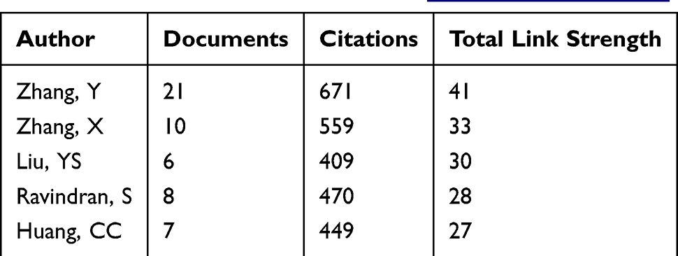 |
Table 3 Author Collaboration Analysis Conducted (Top 5) (Detailed Information Can Be Found in Supplementary Table 2) |
 |
Figure 5 Network visualization of author collaboration analysis concerning extracellular vesicles for tissue engineering spanning from 2014 to 2024. (A) Author collaboration analysis conducted using Vosviewer. (B) Visualization diagram illustrating authorship-country analysis based on Vosviewer. (C) Network visualization diagram illustrating authorship- institutions analysis based on Vosviewer. Author collaborations are depicted by nodes, with the size of each node scaling in accordance with the number of collaborations. Collaboration relationships are represented by connecting lines between nodes. (D) Most local cited authors survey. (E) Author Productivity through Lotka’s Law. |
For the country-based co-authorship analysis, 40 countries were selected for review. The results of this analysis are encapsulated in Figure 5B. The United States emerged as the leader in co-authorship intensity, with 128 times the total link intensity. China followed with 73 times the total link strength. Other notable countries include Italy (44 times), the India (39 times), Iran (38 times), and Germany (36 times), all of which exhibit significant collaborative links in the research community (Table 4 and Detailed information can be found in Supplementary Table 3).
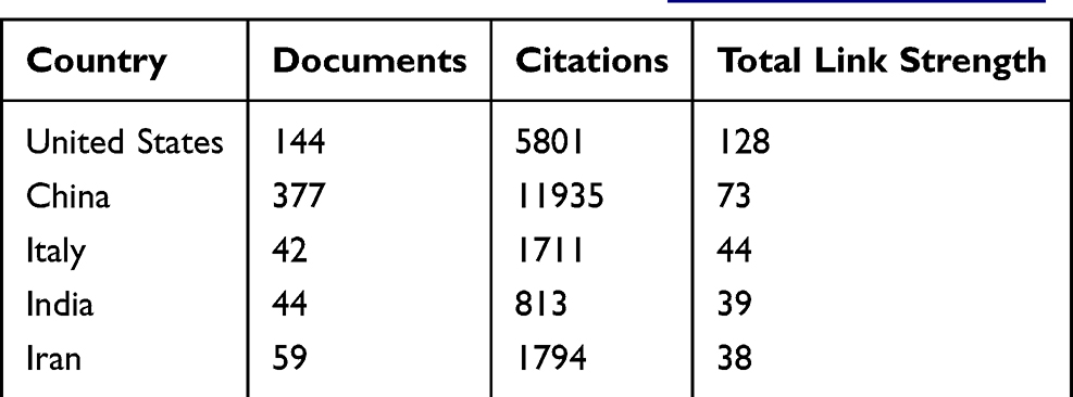 |
Table 4 Analysis of Author-Country Collaboration (Top 5) (Detailed Information Can Be Found in Supplementary Table 3) |
The institutional co-authorship analysis extends to 164 institutions. The findings shown in Figure 5C indicate that Shanghai Jiao Tong University has the highest total link strength at 51 times. Sichuan University follows with 33 times. Other institutions in the top five include Tehran Medical University (28 times), Shahid Beheshti University (22 times), and Sun Yat-sen University (21 times) (Table 5 and Detailed information can be found in Supplementary Table 4). Conducted at both the national and institutional levels, this comprehensive co-authored analysis underscores the interrelationships and collaborative efforts in the field, highlighting the strength of key players and their scientific networks.
 |
Table 5 Analysis of Author-Organization Collaboration (Top 5) (Detailed Information Can Be Found in Supplementary Table 4) |
Most Local Cited Authors survey, revealing their core contributions to the field of exosome tissue engineering. Figure 5D illustrates these findings, including the top 10 authors, with the most cited authors being Ravindran S (Citations= 66), Cooper LF (65), Huang CC (65), Lu Y (54), and Shirazi S (54).
As shown in Figure 5E, in the field of exosomal tissue engineering, the number of authors who have published one document is 3047, accounting for 84.9%, and the number of authors who have published more than or equal to 5 or more related documents is 39, accounting for 1.1% (10 for 5, 15 for 6, 7 for 7, 4 for 8, 2 for 9, and 1 for 19). The above data show that the distribution of research productivity in the field of exosome tissue engineering is more in line with the pattern of scientific research development “core-marginal” structure, ie, a few high-yield authors (core) contribute a large number of papers, and the majority of authors (marginal) publish only a small number of papers.
Analysis of Research Fields and Journals
A complex bimapping overlay depicting the journals associated with exosome tissue engineering has been carefully constructed and is presented in Figure 6A. The bimap overlay is a “panoramic map” that reveals the “Physics, Materials, Chemistry” at the heart of the field. In addition, “Ophthalmology” and its related subfields (such as “Ophthalmic” and “Ophthalmologica”) have appeared many times, suggesting the importance of ophthalmology in health research. Interdisciplinary disciplines such as “Molecular Biology, Genetics, Immunology” intersect closely with other disciplines, suggesting the development of this research field in the direction of interdisciplinary intersection and integration.
 |
Figure 6 Articles published in various journals on extracellular vesicles for tissue engineering from 2014 to 2024. (A) Dual-map overlay depicting journals relevant to extracellular vesicles for tissue engineering from 2014 to 2024. (B) Bibliographic analysis of journals utilizing VOSviewer. (C) Comprehensive co-citation analysis of journals. |
Figure 6B illustrates the scholarly publishing area, the journals that contributed to this study. International Journal of Molecular Sciences leads the cohort with a commendable number of 38 publications, followed closely by Frontiers in Bioengineering and Biotechnology with 37 publications, Stem cell Research and Therapy with 21, Journal of Nanobiotechnology, 20 publications, and Frontiers in Cell and Developmental Biology contributed 19 articles (Table 6 and Detailed information can be found in Supplementary Table 5).
 |
Table 6 Analysis of Published Journals (Top 5) (Detailed Information Can Be Found in Supplementary Table 5) |
As shown in Figure 6C, the co-citation analysis demonstrates the top ten cited journals. The frontier of the series is dominated by five journals; Stem Cell Research and Therapy (2292 citations), Biomaterials (2194 citations), International Journal of Molecular Sciences (1493 citations), Journal of Extracellular Vesicles (1299 citations) and Scientific Reports (1282 citations), with each journal demonstrating substantial citation impact.
Analysis of Co-Citation
In order to elucidate the profile of seminal literature in the field, a complex co-citation analysis was conducted using VOSviewer, as shown in Figure 7A and B. The top five articles from 174 publications at the apex of this assessment highlight their strong total link strength, including Théry C’s 2018 publication in Journal of Extracellular Vesicles21 (123 citations, total link strength=1889), Kalluri R’s 2020 work in Science22 (123 times, total link strength=1590), Dominici M’s 2006 publication in Cytotherapy23 (81 times, total link strength=540), Alvarez-Erviti l 2011 2011 in Nature Biotechnology24 (76 times, total link strength=1591), and Van Niel G 2018 in Nature Reviews Molecular Cell Biology25 (same 76 times, total link strength=1137). The information of the top 6–10 articles is shown in Figure 7B.26–30
 |
Figure 7 Mapping of references in studies on extracellular vesicles for tissue engineering from 2014 to 2024. (A) Network visualization of reference analysis using VOSviewer. (B) Most local cited references. (C) Identification of the top 10 references exhibiting the most pronounced citation bursts in publications. |
Complementing this, the concept of “citation explosion” is a key indicator that highlights publications that have generated considerable interest over a given period of time, which we carefully analyzed using CiteSpace. This analysis ultimately identified the strongest citation bursts, led by Zhang S’s research,30 with a burst strength of 13.51 between 2017 and 2021, followed by Zhang JY,29 cementing an understanding of the trends and foci in the research trajectory of the field,8,31–37 as illustrated in Figure 7C.
Keyword Co-Occurrence Analysis
Figure 8A shows the keyword co-occurrence network generated by VOSviewer. The network reveals key themes such as “regenerative medicine”, “biomaterials”, “wound healing” and “bone regeneration”. These themes indicate that current research is focused on the use of tissue-engineered EVs for tissue repair, drug delivery, binding to materials and wound healing.
 |
Figure 8 Bibliometric analysis of active topics, keywords, and orientation in the future. (A) Co-occurrence analysis of keywords. (B) Identification of the top 10 keywords exhibiting the most pronounced citation bursts in publications. (C) The timeline map shows the evolution of research trends and keywords for different topics in the field between 2014 and 2024. |
Figure 8B highlights the top 10 keywords with the strongest citation bursts, indicating specific topics that have gained significant attention in recent years. Citation bursts for keywords like “myocardial infarction”, “periodontal ligament”, “surface”. The explosion of citations for keywords like “myocardial infarction”, “periodontal ligament”, “surface functionalization”, and “autophagy” suggests that tissue-engineered exosomes are well suited for use in different disease contexts as well as in different cytosolic and intracytoplasmic functional regions. The presentation of the timeline illustrates the shift of the field from the original phenotypic exploration of disease treatment to the exploration of specific mechanisms, for example, in recent years, studies have mostly explored whether it is chemical and molecular functional modification of the exosome surface to optimize its effects and achieve certain specific functions38,39 or mitochondria-mediated autophagy in target cells.40,41
The analysis extends to examine the shift in keywords over the decade from 2014 to 2024, as shown in the visualization in Figure 8C. This timeline reveals significant changes in keyword frequency over time, implying a shift in research focus. The change in keywords such as #0 wound healing; #1 osteoarthritis suggests that exosome tissue engineering has a wide range of applications in the wound healing and osteoarthritic fields, and suggests that researchers in other disease specialties who want to develop tissue-engineered exosome therapeutic solutions for diseases in their field can refer to them. The keyword changes of #2 tissue regeneration and #4 drug delivery suggest that tissue-engineered EVs are hot in the field of regenerative medicine and drug delivery, which is more in line with the current research hotspots. Changes in the frequency of the keywords of #3 tissue engineering; #6 bone tissue engineering; #8 cardiac tissue engineering reflect the wide application of exosomes in the fields of bone tissue engineering and cardiac tissue engineering in the past decade, in which researchers have invested great efforts.
Hot Spots and Trends in Research
As the use of EVs continues to expand, understanding the key research trends and hotspots in in vivo studies is critical to guide future research. In Figure 9A, publication counts and relative research interest indicate a significant upward trend in the number of studies on EVs, particularly over the past 5 years. This rise reflects the growing recognition of EVs as promising candidates for drug delivery and regenerative therapies. The corresponding increase in RRIs further emphasizes the interest of the scientific community in RRIs.
 |
Figure 9 Analysis of in vivo studies on MSC-derived exosome delivery trends and research hotspots. (A) Publication count and relative research interest over time. (B) The Thematic Map displaying the distribution and evolution of topics related to the study of Extracellular Vesicles in Tissue Engineering, sorted by degree of development and relevance. Horizontal axis: centrality, vertical axis: density. (C) The Trend Topics Chart shows the trend in the frequency of different terms used in tissue engineering extracellular vesicle research from 2016 to 2024, reflecting the evolution of research hotspots over time. |
Figure 9B shows the research hotspots and cutting-edge topics in the field of exosome tissue engineering. The combination with the field related to biomimetic nanoparticles is currently a mature hotspot in the field, and numerous researchers are involved in it. autologous chondrocyte implantation has also demonstrated more mature research results, but the communication between disciplines is not yet close, suggesting that we can continue to expand the cooperation between related disciplines to promote the development of this field. Alzheimers-disease and parkinsons-disease have also been widely used in the field of exosomal tissue engineering. At the same time, we acquired that biomaterials research is still a cutting-edge direction, and the fields of tissue regeneration and exosome delivery will develop into hot spots in the future.
The Trending Topics Map demonstrates the dynamic changes in research topics by integrating the time dimension with topic correlations. As shown in Figure 9C, “fibrosis”, “growth-factors”, “blood-brain-barrier”, “nanoparticles”, “hydrogel”, and “cells” are emerging technology trends in current research. Keywords such as “autologous chondrocyte implantation”, “promote tumor-growth”, “horizontal transfer”, etc. are emerging technologies. Keywords such as “autologous chondrocyte implantation”, “promote tumor-growth”, and “horizontal transfer” are recognized as declining areas, prompting researchers to seek breakthroughs and avoid over-reliance.
Discussion
Trend of Global Publications
In today’s era of information explosion, if you want to accurately grasp the industry’s development context and keep up with cutting-edge research results, you undoubtedly face more and more difficulties and challenges. In view of this, in order to efficiently and clearly present the global research in the field of “exosome research based on tissue engineering technology”, especially between 2014 and 2024, we decided to use the objective method of bibliometric analysis. The advantage of this method is that it can sort out and clarify the research frameworks in specific fields, and also make predictions about the future direction to a certain extent. It provides a clear “domain map” for researchers who are bogged down in a large amount of detailed literature. Indicating innovation paths: identifying gaps and weaknesses points the way for future experimental design, grant applications, and interdisciplinary collaboration.
Quality and Status of Global Publications
This study uses professional tools such as CiteSpace, VOSviewer and Bibliometrix R software packages to conduct in-depth bibliometric analysis and visualization of literature in this field. It is expected to provide valuable reference information and a new perspective for in-depth exploration of the development history of this field, accurately capture research trends, and keen insight into research hotspots. Through the analysis of 754 original documents and reviews, it was found that the number of documents involving EVs and tissue engineering technology increased year by year. Our findings indicate that the annual number of publications in the field of exosome tissue engineering has shown a continuous upward trend between 2014 and 2024, as shown in Figure 2, with a particularly significant growth rate in the last five years, while the relative research interest (RRI) value has grown in parallel, suggesting that the academic attention and resource investment in this field has continued to expand. Globally, 44 countries and 269 academic institutions are involved in this research process, with China (48.9%) and the United States (19.1%) dominating the field, contributing a combined 68% of publications, and reinforcing its research impact through high-frequency international collaborations (eg, 380 total link strengths between China and the United States).
In order to clarify the academic impact and quality of research contributions in the field of EVs-derived tissue engineering in different countries, the focus was on in-depth analysis of the total citations, Average citations and H index, and the results are shown in Figure 3. China is leading among the countries with the highest total citation times of 11,872 citations and the H index is the highest 58. The United States ranked behind by total citations and second in the H index. It shows China and the United States’ core position in this field and its outstanding scientific research image. Interestingly, Australia and Italy performed well in average citation rates, surpassing the United States and China, indicating that Australia and Italy played a key role in this specialized field. This phenomenon of high publications and high total citations in China is closely related to China’s continuous reform and innovation in scientific development.42 However, China has shown a great disadvantage in the average number of citations, which may be because academic articles written in Chinese are affected by language barriers and communication path restrictions during the dissemination process to the international academic community, which makes it difficult for international scholars to obtain and cite these research results, which in turn affects the number of citations. But this does not mean that the research results of Chinese articles are lacking in depth and breadth, but there are some areas that need to be optimized in the information dissemination process. On the other hand, China’s academic evaluation system tends to pay too much attention to quantity rather than quality, which has led some scholars to ignore the improvement of research quality in order to pursue quantity. Some scientific research institutions and academic institutions have relatively broad requirements for the quantity of articles published in terms of evaluation standards, which may reduce the strictness of quality control to a certain extent.43
Collaboration and Co-Authorship Analysis
By analyzing the cooperation situation by countries/regions and institutions, we can clearly grasp the global research distribution and pattern in specific fields. Collaborations often revolve around current research hotspots and frontiers, and by pursuing these collaborations we can clearly track research hotspots and frontiers. In addition, the parties to the cooperation often have higher research levels and strong scientific research capabilities, and their research results are more innovative and influential. In this study, as shown in Figure 4, we found that more than 44 countries worldwide participated in research in this field, among which China and the United States performed prominently in terms of research output and international cooperation. In addition, the relationship between institutions, keywords and countries is shown through Sangji map. It is found that China and the United States occupy a dominant position in the research field, with key words such as mesenchymal stem cells as the core research content, and some universities in China rank high in the analysis. Academic research is not an isolated individual behavior, but is constantly promoted on the basis of mutual communication and cooperation among researchers. By analyzing the co-authorship relationship between authors, various characteristics of academic cooperation can be excavated. As shown in Figure 5: Co-authority Analysis indicates that the author Zhang Y, China and the United States, Shanghai Jiaotong University have outstanding performance in cooperation and have high influence. The Most Local Cited Authors survey reveals that Ravindran S has the highest individual contribution in the field. In academic research, Lotka’s Law provides us with a classic perspective on understanding and quantifying author productivity. In the field of exosome tissue engineering, its research productivity distribution shows an obvious “core-edge” structure. This distribution pattern clearly reveals that a small number of prolific authors are the core force in this field and contribute a lot of research results. They often have significant advantages in terms of research depth, resource acquisition and academic influence, and can continuously produce high-quality academic papers. Most authors belong to marginal groups and publish only a few papers, which may be related to their limited research time, relatively scarce research resources or scattered research interests. The existence of this “core-periphery” structure reflects, on the one hand, the elite effect of academic research, that is, a few top researchers lead the development direction of the discipline; on the other hand, it also reflects the competition and imbalance of resource allocation in the academic field, and at the same time provides the direction and goal for the new researchers entering the field, that is, how to move from the marginal group to the core researchers.
Research Frontiers and Hotspots
The New Direction from the Perspective of Bibliometrics
Keyword and strategy mapping analysis reveals that current research focuses on EVs for applications such as wound healing, bone/heart tissue engineering, and drug delivery, while emerging themes such as “biomimetic nanoparticles”, “hydrogels”, and “blood-brain barrier”, mark the beginning of a new era in EVs, “and emerging themes” such as “biomimetic nanoparticles”, “hydrogels”, and “blood-brain barrier” mark cutting-edge directions. Our keyword analysis revealed that the vast majority of studies focused on “efficacy validation” and “mechanism of action”, whereas research on “large-scale production” and “pharmacokinetics” remained relatively limited. This indicates that the field is currently transitioning from “phenomenological observation” and “mechanistic investigation” toward “translational application”, although the latter remains a significant bottleneck. Notably, MSCs have laid the theoretical foundation for the field through exosome-mediated paracrine mechanism (Baglio et al, 2012) and their transcellular communication ability (Chang et al, 2018), and it is expected that future research will continue to deepen around exosome functional modifications (such as surface engineering) and mechanism explorations (such as mitochondria-mediated autophagy) to promote regenerative medicine and precision therapeutic innovation breakthroughs. The above information reveals that tissue engineering technology combined with EVs has become a key research direction in regenerative medicine in the future.
Recent Achievements and Critical Analysis of MSC-EVs
As nanoscale biological vesicles, extracellular vesicles (exosomes), especially those derived from mesenchymal stem cells, have been extensively studied in the diagnosis, prevention and treatment of many diseases.44,45 To date, exosomes and extracellular vesicles have been shown to resemble the biological activities of MSCs, including anti-inflammatory, anti-apoptotic, and immunomodulatory activities, and are therefore considered an ideal cell-free therapeutic option.46 Recent studies have further elucidated the functional roles and underlying mechanisms of EVs. For instance, extensive research has demonstrated that EVs are well-known for their direct involvement in regenerative and biological responses of target cells, as well as signal transduction. This is primarily attributed to their crucial role in intercellular communication through the delivery of bioactive molecules, including microRNAs (miRNAs), proteins, and lipids.47,48 Upon transfer of certain nucleic acids from EVs to recipient cells, gene expression is altered, leading to the regulation of cellular proliferation, differentiation, and transcription.49,50 Furthermore, the beneficial effects of EVs in the regeneration and repair of the musculoskeletal system are largely ascribed to their ability to induce osteogenic differentiation, promote angiogenesis, and modulate immune responses.34,50
However, upon reviewing the existing body of research, we observe that the parental cells are specific, and the overall sensitivity of downstream signaling pathways varies under different experimental parameters. Moreover, current research trends have predominantly focused on evaluating only one or a few predetermined pathways or signaling factors. Consequently, it remains challenging to delineate the detailed mechanisms through which EVs induce tissue regeneration based on available evidence. Obstacles arising from batch-to-batch heterogeneity persist, yet this represents a critical direction for future investigation. Current vesicle isolation protocols may lead to the enrichment of multiple subtypes rather than a purified homogeneous population. Exceptionally, although the majority of studies support that exosomes promote angiogenesis,51,52 particularly under hypoxic conditions,53 research by Roberta et al54 demonstrated inhibition of endothelial cell migration and neovascularization within an inflammatory microenvironment. This discrepancy reinforces the concept of mesenchymal stem cells acting as environmental “sensors”. These findings underscore the necessity for future studies to place high emphasis on the coupling of temporal and spatial factors.
Evolution of EVs Isolation Technologies
EVs’ isolation has undergone significant advancements, transitioning from an early “extensive” stage to the current “precise” stage.55 Initially, ultracentrifugation (UC) was predominantly used. Although capable of processing large sample volumes, UC often introduces contaminating proteins, and the high centrifugal forces can compromise EV integrity, leading to considerable variability and poor reproducibility in downstream analyses.56 To overcome these limitations, methods such as size exclusion chromatography (SEC)57 and polymer-based precipitation (eg, polyethylene glycol, PEG)58 were introduced during a transitional period. While these approaches improved purity, they often came at the cost of reduced yield and were time-consuming. Presently, the technology is leaping toward “precision, speed, and high purity”. Novel physical techniques, such as the EXODUS system, which utilizes negative pressure oscillation and dual-frequency lateral waves, enable ultra-rapid and high-yield isolation.59 Microfluidic chips allow for the manipulation of minute fluid volumes, facilitating high-throughput and automated EV capture.60,61 These devices can be integrated with downstream analytical modules, offering potential for clinical diagnostics and real-time monitoring. Furthermore, immunoaffinity and nanomaterial-based technologies leverage antibodies or specific nanomaterials (eg, magnetic nanoparticles)62 that bind to EV surface markers, enabling the “targeted fishing” of specific EV subpopulations. This greatly enhances the specificity and purity of isolation.63 These technological advances now allow researchers to obtain well-defined EV populations of single subpopulations. Consequently, different EV subpopulations can be studied with precision, much like pharmaceuticals, to elucidate their specific roles in tissue regeneration, which paves the way for designing more precise and effective “cell-free therapies”.
Revolution in EV Characterization Technologies
EV characterization has evolved from an initial stage of “visualization” to the current stage of “comprehension”, achieving a leap from physical property analysis to molecular-level profiling. Early efforts primarily focused on physical characteristics; techniques such as dynamic light scattering (DLS) and nanoparticle tracking analysis (NTA) provided information on EV “size” and “concentration”.64,65 In an intermediate phase, transmission electron microscopy (TEM) and atomic force microscopy (AFM) allowed visualization of EV “morphology” and “structure”.66 Today, the technology is advancing toward “multidimensional, single-vesicle level” molecular characterization. High-throughput omics technologies—including proteomics,67,68 transcriptomics,69 and lipidomics69—can simultaneously resolve the composition of hundreds to thousands of molecules within a single EV, untangling their complex “cargo inventory”. Single-vesicle analysis technologies, such as single-vesicle RNA sequencing and ultra-sensitive protein detection platforms,69 enable the study of heterogeneity at the level of individual EVs, moving beyond the “average” characteristics of population-based analyses.70,71 This shift is analogous to progressing from observing the “forest” to examining each individual “tree”, allowing the discovery of critical information previously obscured by bulk data. These advances empower scientists to establish precise “composition-function” correlations. This enables researchers to, much like drug design, “customize” EV cargo through engineering approaches, thereby manufacturing tissue engineering products with more stable efficacy and clearly defined mechanisms.
Breakthroughs in EV Delivery Mechanisms
The understanding of EV delivery mechanisms has shifted from an early “bystander” phase to the current “intelligent courier” phase, marking a transition from natural uptake to engineered targeted delivery. Initially, scientists recognized that EVs could be naturally taken up by cells, but little was known about how they cross biological barriers (eg, the blood-brain barrier, BBB) or home to specific tissues. Current research focuses on understanding and harnessing the innate “homing” and “barrier-crossing” capabilities of EVs, while also employing engineering strategies to endow them with “targeted delivery” intelligence.72 Studies indicate that EVs derived from different cell types exhibit inherent tissue tropism. For instance, EVs from tumor cells preferentially return to tumor tissue.73 Through genetic engineering or chemical modification, “navigation devices” (eg, specific antibodies, peptides) can be installed on the EV surface, enabling precise targeting of diseased tissues or specific cells. Moreover, methods such as electroporation, incubation, or genetic engineering allow efficient “packaging” of therapeutic cargo—including drugs (eg, chemotherapeutic agents), nucleic acids (eg, siRNA, CRISPR-Cas9), and even gene-editing tools—into EVs, transforming them into highly effective drug delivery vehicles. We will make the statement in the subsequent chapters.
Recent Achievements and Critical Analysis of MSC-EVs with Biomaterials
In recent years, research results on tissue engineering of EVs have been accumulating and EVs edited by tissue engineering techniques have been found to exert therapeutic effects on a wide range of disease research areas, including orthopedic diseases and bone tissue engineering fields, wound healing, oral diseases, and cardiovascular diseases.74–76 The introduction of engineered strategies for biomaterials has provided novel approaches to address challenges encountered in disease diagnosis and treatment. Scientists have turned their attention to bioactive scaffolds, utilizing them as carriers for EVs to achieve stable and sustained release. Based on this rationale, EVs can be incorporated into biomaterials through various mechanisms, directly or indirectly facilitating signal transduction in diverse regenerative processes.77 Tissue engineering technologies can provide scaffolds for EVs, for example, Li et al combined EVs derived from human adipose-derived stem cells (hASCs) with poly(lactic-co-glycolic acid) (PLGA) scaffolds to construct a new type of cell-free tissue-engineered bone and showed excellent potential for bone defect repair.27
In addition, biomaterials themselves possess robust functionalities, providing both physical and chemical support for tissue regeneration.78,79 To date, studies on biomaterial-mediated EVs therapy have emerged rapidly, demonstrating significant regenerative potential in tissue engineering.80 In fact, the selection of biomaterial carriers is also based on specific clinical application requirements. For bone tissue, scaffolds should exhibit favorable mechanical strength, while for soft tissue injuries such as wound healing, hydrogels are currently the mainstream choice.81,82 Indeed, some researchers have already noted subsequent challenges associated with combining biomaterials and EVs.83 For instance, EVs often cannot be uniformly loaded throughout bioactive scaffolds, particularly in the innermost regions.84 Moreover, few studies have verified the integrity of EV membranes after delivery, which is essential to attribute the observed functional outcomes specifically to EVs, thereby strengthening and supporting their findings. Pore parameters of bioactive scaffolds—such as optimal pore size and pore size distribution—often influence both mechanical properties and diffusion processes; however, no consensus has yet been reached on these characteristics. Furthermore, the long-term fate of biomaterials must also be considered.85
Recent Achievements and Critical Analysis of MSC-EVs with Engineering Technologies
Furthermore, tissue engineering techniques are employed to address the limitations of natural EVs—such as insufficient therapeutic efficacy, poor targeting capability, and inadequate local concentration—through genetic modification of parental cells,86 external environmental stimulation,87 and optimization of EV culture conditions. For instance, in the field of osteoporosis, Cui et al prepared an engineered exosome that not only realized the anti-osteoporosis function of traditional exosome, but also combined siRNA and targeted peptide parts, realizing the multiple therapeutic potentials of killing three birds with one stone.88 Xu et al used the exosome tissue engineering technology to solve the problem of the chondrogenic differentiation of the small molecule Kartogenin (KGN) in the synovial membrane of the synovial membrane MSCs, which could not play its physiological role. KGN formed a precipitate and could not play its physiological role, which led to the homogeneous dispersion of KGN in the cytoplasm, increased its effective concentration and strongly promoted the chondrogenesis of SF-MSC in vitro and in vivo, showing the advantages of tissue engineering in the field of exosome treatment of osteoarthritis.89 At the same time, tissue engineering technology also achieves the effect of localized intensive treatment: Song et al precisely utilized the principle of tissue engineering to achieve slow release and high local concentration of EVs at the wound to promote wound healing.90 Despite the aforementioned advantages of these technologies, the off-target effects of gene editing, the long-term consequences of genetic modifications, and the subsequent increase in costs due to technical complexity remain issues that require consideration.91 It should also be noted that precise control of external stimuli and inter-individual variability in stimulation efficacy often impede the maturation of this technology.92
Future Development Trend
The Future of MSC-EVs
Rapid advances in tissue engineering technology have not only promoted the high efficiency of the extraction and characterization process of EVs, but also opened up completely new horizons for their functional studies. Due to the special properties of EVs, such as low toxicity, low immunogenicity, low biodegradability, the ability to encapsulate endogenous bioactive molecules and to cross part of the biological barriers, EVs have demonstrated a great potential for drug delivery in many diseases.93 As previously mentioned, significant challenges remain in the future development of engineered EVs. Beyond improving purity and specificity, increasing EV yield per unit time appears to be a more pressing challenge in the near future. The development of large-scale ultracentrifugation systems and the design of more user-friendly commercial EV extraction kits represent promising directions for scalable production of therapeutic EVs. Furthermore, there is an urgent need to further explore manufacturing processes compliant with Good Manufacturing Practice (GMP) to achieve stable and scalable production of homogeneous EVs.
The Future of MSC-EVs Combined with Biological Materials
Given their excellent biocompatibility and physicochemical properties, biomaterials such as hydrogels have been recognized as the most suitable carriers for EVs.94 In the field of tissue engineering, hydrogels can protect EVs from the in vivo environment, especially some harsh environments related to trauma and inflammation, and can be released slowly to reach the desired location.95 With the increase of cooperation in the medical-industrial crossroads, some magnetic materials and metal-organic frameworks have gradually attracted the attention of exosome researchers. Studies have shown that magnetic nanoparticles (eg, Fe3O4, γ-Fe2O3) have stronger osteogenic and angiogenic abilities compared with EVs secreted by bone marrow mesenchymal stem cells pretreated with a static magnetic field.96 In addition, Kang et al successfully combined Mg2+ with human adipose-derived stem cell-derived exosomes (hADSCs-Exos) and found that this Mg2+-based metal-organic framework promoted both osteogenesis and angiogenesis.97 Multifunctional, smart or conductive hydrogels, nanoparticles, and the combination of research with engineering and mechanics will be a major hotspot for future research. The combination of these technologies not only enhances the potential for the application of EVs in basic research, but also provides new directions for clinical treatment.
The Future of Engineered-Modified MSC-EVs
Thanks to the unique editable nature and superior membrane penetrability of EVs, we have skillfully modified the membrane surface of EVs and their internal contents through innovative nanotechnologies, such as electroporation, sonication, extrusion, freeze-thaw cycling, and transfection, so as to enable them to acquire the active targeting ability for specific tissues or specific cell types,98,99 and thus they have been regarded as a highly promising tool for drug delivery.100 For example, the application of cartilage affinity peptides (CAPs) in the field of exosome repair in osteoarthritis has enabled EVs to deliver key molecules ground to deep cartilage regions.101 This targeted delivery based on ligand-receptor binding is currently the most recognized means of targeting by scientists. In addition, there are a number of other routes, such as pH gradient/ surface charge-driven targeted delivery based; and magnetically guided targeted drug delivery.102 The combination of these technologies with EVs is bound to cause a new upsurge in the field of exosomal drug delivery.
Although gene editing and modification strategies show great potential in the field of EVs, several critical issues must be addressed. A primary challenge is the development of more convenient and efficient methods to manage the complex biological process involved in loading transgenic cargo into parent cells and its subsequent transfer to derived EVs—a process that remains poorly understood. Before genetically edited EVs can be advanced into clinical applications, their safety and feasibility must be rigorously validated, particularly since viral transduction for overexpression of bioactive molecules may pose potential risks to host cells.
Meanwhile, although pre-conditioning and external stimulation strategies can enhance the tissue-regenerative capacity of EVs, their effects are highly dependent on the type and concentration of exogenous compounds, as well as their mechanisms of interaction with cells. Future studies should focus on identifying more precise stimulation conditions to avoid detrimental effects on cell viability caused by high concentrations or prolonged exposure, and to prevent alterations in parental cell phenotype, thereby ensuring consistent EV composition and therapeutic efficacy.
Clinical Transformation and Application
This study reveals an explosively growing trend and sustained high research enthusiasm, strongly indicating the immense clinical translation potential of stem cell exosome therapy. This study has identified currently leading research teams as well as several highly promising yet less interconnected independent teams, such as those from Sun Yat-sen University, Zhejiang University, and Shanghai Jiao Tong University. We recommend that researchers in the field, particularly early-career investigators, actively pursue cross-institutional and interdisciplinary collaborations with these teams to integrate diverse expertise and foster breakthrough achievements. Through analysis of highly cited literature, we found that most current preclinical studies have demonstrated significant potential in areas such as bone tissue engineering, drug delivery, and skin wound repair. These findings provide supporting evidence for prioritizing these indications in initial clinical trials.
Furthermore, several challenges remain in the clinical application of EVs, including standardized production of EVs, heterogeneity, source selection, and stability in vivo.103 The low yield of EVs makes it difficult to achieve large-scale production, which is critical for clinical translation and application. Existing separation methods such as Differential centrifugation and ultrafiltration, Exosome isolation reagent kit, Tangential flow filtration, etc., different laboratory separation methods are affected by human factors, all of which can affect the yield of exosome. Standardized production routes based on factors such as different exosome extraction methods are urgently needed to be developed and the standardized separation method for purifying EVs subtypes.104 In addition, storage issues of EVs limit their development and require the establishment of larger scale laboratories for their study. The repeated freeze-thaw process poses a great challenge to the activity of EVs.105 Therefore, future studies need to further explore how to optimize the preparation and application of EVs to fully exploit their potential in tissue engineering and regenerative medicine. Meanwhile, with the deeper understanding of EVs’ functions, more breakthroughs are expected to be achieved in the fields of disease treatment, drug delivery and individualized medicine. This has also led to continuous advances in tissue engineering techniques, including some minimally or even non-invasive therapeutic techniques. Unlike traditional invasive delivery modes (intravenous, intraarticular, intracerebral), nasal spray and topical microneedle delivery modes also exhibit good therapeutic capabilities.106,107 From the perspective of clinical translation, non-invasive or minimally invasive treatments are more likely to be clinically accepted. Continued research in this area will open up new horizons and opportunities for biomedical development.
Regarding the topic of “in vivo”stability, although systemic administration via intravenous injection is the most clinically accepted method, extracellular vesicles (EVs) exhibit a short and unstable half-life, ranging from several minutes to a few hours.108 Consequently, following intravenous administration, EVs are rapidly cleared from the bloodstream, necessitating repeated dosing and resulting in cumbersome operational procedures. Thus, the current administration strategy for EVs is highly dependent on the specific clinical application. However, whether administered systemically or locally, challenges such as rapid clearance “in vivo” and low bioavailability remain unresolved.
Although EVs are generally considered to possess superior biocompatibility and enhanced biosafety compared to traditional stem cell therapies, their complete composition, the interactions among their various components, and the key regulatory molecules and downstream signaling pathways remain incompletely understood. This lack of comprehensive knowledge may lead to unpredictable or adverse biological effects. Moreover, it is crucial to determine whether different EV modification strategies or modifications to parent cells may cause previously overlooked adverse reactions “in vivo”. Additionally, uncertainties regarding the biodistribution and bioelimination of EVs in the blood and various pathological microenvironments, as well as the optimal therapeutic dosage of EVs, may pose significant risks.
Limitations and Deficiencies
Inevitably, there are some limitations to this study, mainly due to the reliance on the WoSCC database, which excludes important sources such as Scopus and PubMed, and may miss some key studies. This limitation may result in the inadvertent omission of critical research and findings that are not indexed in the included databases or published in languages other than English. In addition, focusing only on original articles and reviews may have overlooked other relevant publications, leading to gaps in the understanding of EVs in tissue engineering. Relying on citation counts may also lead to bias in research impact, as new studies may not have received enough citations. Furthermore, this bibliometric analysis lacks literature data from before 2014 and the most recent publications, which may result in an incomplete dataset. Early records might be missing from the database due to technological limitations or the immaturity of online publishing tools at the time, potentially leading to omissions. Although we expanded the search by including “Exosomes” as a keyword to enhance the breadth of the study, issues such as mislabeling, under-labeling, inaccurate term selection, or the use of overly broad terms in keyword indexing often reduce the scientific accuracy of topic representation and may affect the validity of bibliometric statistical analyses.
Therefore, future studies should consider incorporating multiple databases and publications in various languages, supplementing with grey literature not covered by major databases, developing AI-assisted automated tools for literature screening and data cleaning, and integrating bibliometric analysis with narrative reviews as well as other emerging methodologies to comprehensively elucidate the potential applications of tissue engineering technologies in EVs.
Conclusion
In summary, this study employed literature from the Web of Science database and Scopus database a and employed various visualization tools, including GraphPad Prism 8, VOSviewer, CiteSpace, Bibliometrix R package, and Scimago Graphiga to investigate the field of tissue engineered EVs trends and areas of focus. Statistical analyses of publications, countries, institutions, authors, journals, references and keywords were performed with the aim of providing researchers with a more comprehensive understanding of the evolutionary path and research priorities in this particular field. In addition, the study explores future trends and hotspots in this field, including exosome editing, drug delivery, and tissue regenerative repair. Our proposed shortcomings and limitations of research in the field of exosome tissue engineering will be of great promise and significance for further exploration of the exosome field.
Data Sharing Statement
The original data presented in the study are openly available in WOS.
Author Contributions
All authors made a significant contribution to the work reported, whether that is in the conception, study design, execution, acquisition of data, analysis and interpretation, or in all these areas; took part in drafting, revising or critically reviewing the article; gave final approval of the version to be published; have agreed on the journal to which the article has been submitted; and agree to be accountable for all aspects of the work.
Funding
This study was supported by a grant from the central government guides local funds for scientific and technological development (No. YDZJSX20231A062) and Shanxi Province scientific and technological achievements transformation guidance project (No. 202204021301067), as well as National Natural Science Foundation of China (Grant No. 82350710800, 82374470), and Shenzhen Medical Research Fund B2302005, and NHMRC, APP1163933.
Disclosure
The author(s) report no conflicts of interest in this work.
References
1. Heydari Z, Najimi M, Mirzaei H, et al. Tissue engineering in liver regenerative medicine: insights into novel translational technologies. Cells. 2020;9:304. doi:10.3390/cells9020304
2. Pina S, Ribeiro VP, Marques CF, et al. Scaffolding strategies for tissue engineering and regenerative medicine applications. Materials. 2019;12:1824. doi:10.3390/ma12111824
3. Yi J, Liu Q, Zhang Q, Chew TG, Ouyang H. Modular protein engineering-based biomaterials for skeletal tissue engineering. Biomaterials. 2022;282:121414. doi:10.1016/j.biomaterials.2022.121414
4. Ferreira MJS, Mancini FE, Humphreys PA, et al. Pluripotent stem cells for skeletal tissue engineering. Crit Rev Biotechnol. 2022;42:774–793. doi:10.1080/07388551.2021.1968785
5. Chen L, Zhu S, Guo S, Tian W. Mechanisms and clinical application potential of mesenchymal stem cells-derived extracellular vesicles in periodontal regeneration. Stem Cell Res Ther. 2023;14:26. doi:10.1186/s13287-023-03242-6
6. Vega-Letter AM, García-Guerrero C, Yantén-Fuentes L, et al. Safety and efficacy of mesenchymal stromal cells mitochondria transplantation as a cell-free therapy for osteoarthritis. J Transl Med. 2025;23:26. doi:10.1186/s12967-024-05945-7
7. Tedesco MM, Terashima M, Blankenberg FG, et al. Analysis of in situ and ex vivo vascular endothelial growth factor receptor expression during experimental aortic aneurysm progression. Arterioscler Thromb Vasc Biol. 2009;29:1452–1457. doi:10.1161/ATVBAHA.109.187757
8. Rani S, Ryan AE, Griffin MD, Ritter T. Mesenchymal stem cell-derived extracellular vesicles: toward cell-free therapeutic applications. Mol Ther. 2015;23:812–823. doi:10.1038/mt.2015.44
9. Rather HA, Almousa S, Craft S, Deep G. Therapeutic efficacy and promise of stem cell-derived extracellular vesicles in Alzheimer’s disease and other aging-related disorders. Ageing Res Rev. 2023;92:102088. doi:10.1016/j.arr.2023.102088
10. Wu R, Fan X, Wang Y, et al. Mesenchymal stem cell-derived extracellular vesicles in liver immunity and therapy. Front Immunol. 2022;13:833878. doi:10.3389/fimmu.2022.833878
11. Zhao M, Liu S, Wang C, et al. Mesenchymal stem cell-derived extracellular vesicles attenuate mitochondrial damage and inflammation by stabilizing mitochondrial DNA. ACS Nano. 2021;15:1519–1538. doi:10.1021/acsnano.0c08947
12. Yin B, Ni J, Witherel CE, et al. Harnessing tissue-derived extracellular vesicles for osteoarthritis theranostics. Theranostics. 2022;12:207–231. doi:10.7150/thno.62708
13. Jammes M, Tabasi A, Bach T, Ritter T. Healing the cornea: exploring the therapeutic solutions offered by MSCs and MSC-derived EVs. Prog Retinal Eye Res. 2025;105:101325. doi:10.1016/j.preteyeres.2024.101325
14. Lin J, Li K, Yang Z, et al. Functionally improved mesenchymal stem cells via nanosecond pulsed electric fields for better treatment of osteoarthritis. J Orthop Transl. 2024;47:235–248. doi:10.1016/j.jot.2024.03.006
15. Zhang X, Lu Y, Wu S, Zhang S, Li S, Tan J. An overview of current research on mesenchymal stem cell-derived extracellular vesicles: a bibliometric analysis from 2009 to 2021. Front Bioeng Biotechnol. 2022;10:910812. doi:10.3389/fbioe.2022.910812
16. Zhao T, Mu Y, Deng H, et al. Research hotspots and trends of mesenchymal stem cell-derived extracellular vesicles for drug delivery: a bibliometric and visualization analysis from 2013 to 2023. Front Cell Dev Biol. 2024;12:1412363. doi:10.3389/fcell.2024.1412363
17. Salinas-Ríos K, García López AJ. Bibliometrics, a useful tool within the field of research. JBAPR. 2022;3:9–16. doi:10.29057/jbapr.v3i6.6829
18. Sun G, Wang L, Dong Z, et al. The current status, hotspots, and development trends of nanoemulsions: a comprehensive bibliometric review. IJN. 2025;20:2937–2968. doi:10.2147/IJN.S502490
19. Meng Y, Sui L, Xu T, Zhao H, Yuan Q, Sun L. Research and application prospect of nanomedicine in kidney disease: a bibliometric analysis from 2003 to 2024. IJN. 2025;20:3007–3030. doi:10.2147/IJN.S510016
20. Han Y, Wennersten SA, Lam MPY. Working the literature harder: what can text mining and bibliometric analysis reveal? Expert Rev Proteomics. 2019;16:871–873. doi:10.1080/14789450.2019.1703678
21. Théry C, Witwer KW, Aikawa E, et al. Minimal information for studies of extracellular vesicles 2018 (MISEV2018): a position statement of the International Society for Extracellular Vesicles and update of the MISEV2014 guidelines. J Extracell Vesicles. 2018;7:1535750. doi:10.1080/20013078.2018.1535750
22. Kalluri R, LeBleu VS. The biology, function, and biomedical applications of exosomes. Science. 2020;367:eaau6977. doi:10.1126/science.aau6977
23. Dominici M, Le Blanc K, Mueller I, et al. Minimal criteria for defining multipotent mesenchymal stromal cells. The international society for cellular therapy position statement. Cytotherapy. 2006;8:315–317. doi:10.1080/14653240600855905
24. Alvarez-Erviti L, Seow Y, Yin H, Betts C, Lakhal S, Wood MJA. Delivery of siRNA to the mouse brain by systemic injection of targeted exosomes. Nat Biotechnol. 2011;29:341–345. doi:10.1038/nbt.1807
25. van Niel G, D’Angelo G, Raposo G. Shedding light on the cell biology of extracellular vesicles. Nat Rev Mol Cell Biol. 2018;19:213–228. doi:10.1038/nrm.2017.125
26. Lai RC, Arslan F, Lee MM, et al. Exosome secreted by MSC reduces myocardial ischemia/reperfusion injury. Stem Cell Res. 2010;4:214–222. doi:10.1016/j.scr.2009.12.003
27. Li W, Liu Y, Zhang P, et al. Tissue-engineered bone immobilized with human adipose stem cells-derived exosomes promotes bone regeneration. ACS Appl Mater Interfaces. 2018;10:5240–5254. doi:10.1021/acsami.7b17620
28. Zhang S, Chuah SJ, Lai RC, Hui JHP, Lim SK, Toh WS. MSC exosomes mediate cartilage repair by enhancing proliferation, attenuating apoptosis and modulating immune reactivity. Biomaterials. 2018;156:16–27. doi:10.1016/j.biomaterials.2017.11.028
29. Zhang J, Liu X, Li H, et al. Exosomes/tricalcium phosphate combination scaffolds can enhance bone regeneration by activating the PI3K/akt signaling pathway. Stem Cell Res Ther. 2016;7:136. doi:10.1186/s13287-016-0391-3
30. Zhang S, Chu WC, Lai RC, Lim SK, Hui JHP, Toh WS. Exosomes derived from human embryonic mesenchymal stem cells promote osteochondral regeneration. Osteoarthr Cartil. 2016;24:2135–2140. doi:10.1016/j.joca.2016.06.022
31. Nakamura Y, Miyaki S, Ishitobi H, et al. Mesenchymal-stem-cell-derived exosomes accelerate skeletal muscle regeneration. FEBS Lett. 2015;589:1257–1265. doi:10.1016/j.febslet.2015.03.031
32. Zhang B, Wang M, Gong A, et al. HucMSC-exosome mediated-Wnt4 signaling is required for cutaneous wound healing. Stem Cells. 2015;33:2158–2168. doi:10.1002/stem.1771
33. Zhang J, Guan J, Niu X, et al. Exosomes released from human induced pluripotent stem cells-derived MSCs facilitate cutaneous wound healing by promoting collagen synthesis and angiogenesis. J Transl Med. 2015;13:49. doi:10.1186/s12967-015-0417-0
34. Qi X, Zhang J, Yuan H, et al. Exosomes secreted by human-induced pluripotent stem cell-derived mesenchymal stem cells repair critical-sized bone defects through enhanced angiogenesis and osteogenesis in osteoporotic rats. Int J Biol Sci. 2016;12:836–849. doi:10.7150/ijbs.14809
35. Qin Y, Wang L, Gao Z, Chen G, Zhang C. Bone marrow stromal/stem cell-derived extracellular vesicles regulate osteoblast activity and differentiation in vitro and promote bone regeneration in vivo. Sci Rep. 2016;6:21961. doi:10.1038/srep21961
36. Lou G, Song X, Yang F, et al. Exosomes derived from miR-122-modified adipose tissue-derived MSCs increase chemosensitivity of hepatocellular carcinoma. J Hematol Oncol. 2015;8:122. doi:10.1186/s13045-015-0220-7
37. Furuta T, Miyaki S, Ishitobi H, et al. Mesenchymal stem cell-derived exosomes promote fracture healing in a mouse model. Stem Cells Transl Med. 2016;5:1620–1630. doi:10.5966/sctm.2015-0285
38. Li X-R, Deng Q-S, He S-H, et al. 3D cryo-printed hierarchical porous scaffolds provide immobilization of surface-functionalized sleep-inspired small extracellular vesicles: synergistic therapeutic strategies for vascularized bone regeneration based on macrophage phenotype modulation and angiogenesis-osteogenesis coupling. J Nanobiotechnol. 2024;22:764. doi:10.1186/s12951-024-02977-5
39. Xu L, Xu X, Liang Y, et al. Osteoclast-targeted delivery of anti-miRNA oligonucleotides by red blood cell extracellular vesicles. J Control Release. 2023;358:259–272. doi:10.1016/j.jconrel.2023.04.043
40. Liu H, Kuang H, Wang Y, et al. MSC-derived exosomes protect auditory hair cells from neomycin-induced damage via autophagy regulation. Biol Res. 2024;57:3. doi:10.1186/s40659-023-00475-w
41. Xu X, Wang J, Xia Y, et al. Autophagy, a double-edged sword for oral tissue regeneration. J Adv Res. 2024;59:141–159. doi:10.1016/j.jare.2023.06.010
42. Zhou Z, Zhao W. Funding system reform for excellence in science: an interview with Jinghai Li, President of NSFC. Natl Sci Rev. 2019;6:177–181. doi:10.1093/nsr/nwy165
43. De Fiore L. Oltre la quantità: ripensare la qualità e l’integrità della ricerca scientifica. Recenti Prog Med. 2025;116:65. doi:10.1701/4450.44436
44. Wei W, Ao Q, Wang X, et al. Mesenchymal stem cell–derived exosomes: a promising biological tool in nanomedicine. Front Pharmacol. 2021;11:590470. doi:10.3389/fphar.2020.590470
45. Hade MD, Suire CN, Suo Z. Mesenchymal stem cell-derived exosomes: applications in regenerative medicine. Cells. 2021;10:1959. doi:10.3390/cells10081959
46. Riazifar M, Pone EJ, Lötvall J, Zhao W. Stem cell extracellular vesicles: extended messages of regeneration. Annu Rev Pharmacol Toxicol. 2017;57:125–154. doi:10.1146/annurev-pharmtox-061616-030146
47. Wan J, Lin S, Yu Z, et al. Protective effects of MicroRNA‐200b‐3p encapsulated by mesenchymal stem cells–secreted extracellular vesicles in myocardial infarction via regulating BCL2L11. JAHA. 2022;11:e024330. doi:10.1161/JAHA.121.024330
48. Zhan J, Zou J, Pang Q, et al. MSCs-EVs harboring OA immune memory reprogram macrophage phenotype via modulation of the mt-ND3/NADH-CoQ axis for OA treatment. J Nanobiotechnol. 2025;23:140. doi:10.1186/s12951-025-03216-1
49. Elsherbiny NM, Abdel-Maksoud MS, Prabahar K, et al. MSCs–derived EVs protect against chemotherapy-induced ovarian toxicity: role of PI3K/AKT/mTOR axis. J Ovarian Res. 2024;17:222. doi:10.1186/s13048-024-01545-7
50. Liu Y, Sun L, Li Y, Holmes C. Mesenchymal stromal/stem cell tissue source and in vitro expansion impact extracellular vesicle protein and miRNA compositions as well as angiogenic and immunomodulatory capacities. J Extracell Vesicles. 2024;13:e12472. doi:10.1002/jev2.12472
51. Todorova D, Simoncini S, Lacroix R, Sabatier F, Dignat-George F. Extracellular vesicles in angiogenesis. Circ Res. 2017;120:1658–1673. doi:10.1161/CIRCRESAHA.117.309681
52. Saccu G, Menchise V, Gai C, et al. Bone marrow mesenchymal stromal/stem cell-derived extracellular vesicles promote corneal wound repair by regulating inflammation and angiogenesis. Cells. 2022;11:3892. doi:10.3390/cells11233892
53. Doeppner TR, Herz J, Görgens A, et al. Extracellular vesicles improve post-stroke neuroregeneration and prevent postischemic immunosuppression. Stem Cells Transl Med. 2015;4:1131–1143. doi:10.5966/sctm.2015-0078
54. Angioni R, Liboni C, Herkenne S, et al. CD73+ extracellular vesicles inhibit angiogenesis through adenosine A2B receptor signalling. J Extracell Vesicles. 2020;9:1757900. doi:10.1080/20013078.2020.1757900
55. Huang J, Chen H, Li N, Liu P, Yang J, Zhao Y. Emerging technologies towards extracellular vesicles large-scale production. Bioact Mater. 2025;52:338–365. doi:10.1016/j.bioactmat.2025.06.005
56. Clos-Sansalvador M, Monguió-Tortajada M, Roura S, Franquesa M, Borràs FE. Commonly used methods for extracellular vesicles’ enrichment: implications in downstream analyses and use. Eur J Cell Biol. 2022;101:151227. doi:10.1016/j.ejcb.2022.151227
57. Gaspar LS, Santana MM, Henriques C, et al. Simple and fast SEC-based protocol to isolate human plasma-derived extracellular vesicles for transcriptional research. Mol Ther Methods Clin Dev. 2020;18:723–737. doi:10.1016/j.omtm.2020.07.012
58. Coughlan C, Bruce KD, Burgy O, et al. Exosome isolation by ultracentrifugation and precipitation and techniques for downstream analyses. CP Cell Biology. 2020;88:e110. doi:10.1002/cpcb.110
59. Chen Y, Zhu Q, Cheng L, et al. Exosome detection via the ultrafast-isolation system: EXODUS. Nat Methods. 2021;18:212–218. doi:10.1038/s41592-020-01034-x
60. Qiu J, Guo Q, Chu Y, et al. Efficient EVs separation and detection by an alumina-nanochannel-array-membrane integrated microfluidic chip and an antibody barcode biochip. Anal Chim Acta. 2024;1304:342576. doi:10.1016/j.aca.2024.342576
61. Xu Y-Q, Bao Q-Y, Yu S-X, et al. A novel microfluidic chip for fast, sensitive quantification of plasma extracellular vesicles as biomarkers in patients with osteosarcoma. Front Oncol. 2021;11:709255. doi:10.3389/fonc.2021.709255
62. Zhang K, Yue Y, Wu S, Liu W, Shi J, Zhang Z. Rapid capture and nondestructive release of extracellular vesicles using aptamer-based magnetic isolation. ACS Sens. 2019;4:1245–1251. doi:10.1021/acssensors.9b00060
63. Yang J, Pan B, Zeng F, et al. Magnetic colloid antibodies accelerate small extracellular vesicles isolation for point-of-care diagnostics. Nano Lett. 2021;21:2001–2009. doi:10.1021/acs.nanolett.0c04476
64. Szatanek R, Baj-Krzyworzeka M, Zimoch J, Lekka M, Siedlar M, Baran J. The methods of choice for Extracellular Vesicles (EVs) characterization. IJMS. 2017;18:1153. doi:10.3390/ijms18061153
65. Dragovic RA, Gardiner C, Brooks AS, et al. Sizing and phenotyping of cellular vesicles using Nanoparticle Tracking Analysis. Nanomedicine. 2011;7:780–788. doi:10.1016/j.nano.2011.04.003
66. Kurtjak M, Kereïche S, Klepac D, et al. Unveiling the native morphology of extracellular vesicles from human cerebrospinal fluid by atomic force and cryogenic electron microscopy. Biomedicines. 2022;10:1251. doi:10.3390/biomedicines10061251
67. Fowowe M, Gutierrez Reyes CD, Nwaiwu J, et al. Glycoproteomic profiling of serum-derived small extracellular vesicles enriched via ultracentrifugation and affinity-based techniques. Sci Rep. 2025;15:21565. doi:10.1038/s41598-025-05430-1
68. Huo B, Chen M, Chen J, et al. A sequential separation strategy for facile isolation and comprehensive analysis of human urine N-glycoproteome. Anal Bioanal Chem. 2018;410:7305–7312. doi:10.1007/s00216-018-1338-6
69. Kerkelä E, Lahtela J, Larjo A, Impola U, Mäenpää L, Mattila P. Exploring transcriptomic landscapes in red blood cells, in their extracellular vesicles and on a single-cell level. IJMS. 2022;23:12897. doi:10.3390/ijms232112897
70. Isogai T, Hirosawa KM, Suzuki KGN. Recent advancements in imaging techniques for individual extracellular vesicles. Molecules. 2024;29:5828. doi:10.3390/molecules29245828
71. Bordanaba-Florit G, Royo F, Kruglik SG, Falcón-Pérez JM. Using single-vesicle technologies to unravel the heterogeneity of extracellular vesicles. Nat Protoc. 2021;16:3163–3185. doi:10.1038/s41596-021-00551-z
72. Liu Y, Hinnant B, Chen S, et al. Hyaluronic acid-modified extracellular vesicles for targeted doxorubicin delivery in hepatocellular carcinoma. Exp Cell Res. 2024;443:114332. doi:10.1016/j.yexcr.2024.114332
73. Zhang W, Ngo L, Tsao SC-H, Liu D, Wang Y. Engineered cancer-derived small extracellular vesicle-liposome hybrid delivery system for targeted treatment of breast cancer. ACS Appl Mater Interfaces. 2023;15:16420–16433. doi:10.1021/acsami.2c22749
74. Ding J-Y, Chen M-J, Wu L-F, et al. Mesenchymal stem cell-derived extracellular vesicles in skin wound healing: roles, opportunities and challenges. Military Med Res. 2023;10:36. doi:10.1186/s40779-023-00472-w
75. Luo D, Zhu H, Li S, Wang Z, Xiao J. Mesenchymal stem cell-derived exosomes as a promising cell-free therapy for knee osteoarthritis. Front Bioeng Biotechnol. 2024;12:1309946. doi:10.3389/fbioe.2024.1309946
76. Villani C, Murugan P, George A. Exosome-laden hydrogels as promising carriers for oral and bone tissue engineering: insight into cell-free drug delivery. IJMS. 2024;25:11092. doi:10.3390/ijms252011092
77. Huang Y, Xie H. Extracellular vesicle-integrated biomaterials in bone tissue engineering applications: current progress and future perspectives. IJN. 2025;20:7653–7683. doi:10.2147/IJN.S522198
78. Brennan MÁ, Layrolle P, Mooney DJ. Biomaterials functionalized with MSC secreted extracellular vesicles and soluble factors for tissue regeneration. Adv Funct Mater. 2020;30:1909125. doi:10.1002/adfm.201909125
79. Gaharwar AK, Singh I, Khademhosseini A. Engineered biomaterials for in situ tissue regeneration. Nat Rev Mater. 2020;5:686–705. doi:10.1038/s41578-020-0209-x
80. Jiang Y, Li J, Xue X, Yin Z, Xu K, Su J. Engineered extracellular vesicles for bone therapy. Nano Today. 2022;44:101487. doi:10.1016/j.nantod.2022.101487
81. Qi L, Pan C, Yan J, et al. Mesoporous bioactive glass scaffolds for the delivery of bone marrow stem cell-derived osteoinductive extracellular vesicles lncRNA promote senescent bone defect repair by targeting the miR-1843a-5p/Mob3a/YAP axis. Acta Biomater. 2024;177:486–505. doi:10.1016/j.actbio.2024.01.044
82. Xu L, Liu Y, Tang L, Xiao H, Yang Z, Wang S. Preparation of recombinant human collagen III protein hydrogels with sustained release of extracellular vesicles for skin wound healing. IJMS. 2022;23:6289. doi:10.3390/ijms23116289
83. Leung KS, Shirazi S, Cooper LF, Ravindran S. Biomaterials and extracellular vesicle delivery: current status, applications and challenges. Cells. 2022;11:2851. doi:10.3390/cells11182851
84. Bari E, Scocozza F, Perteghella S, et al. 3D bioprinted scaffolds containing mesenchymal stem/stromal lyosecretome: next generation controlled release device for bone regenerative medicine. Pharmaceutics. 2021;13:515. doi:10.3390/pharmaceutics13040515
85. Bourquin J, Milosevic A, Hauser D, et al. Biodistribution, clearance, and long‐term fate of clinically relevant nanomaterials. Adv Mater. 2018;30:1704307. doi:10.1002/adma.201704307
86. Ying C, Wang R, Wang Z, et al. BMSC-exosomes carry mutant HIF-1α for improving angiogenesis and osteogenesis in critical-sized calvarial defects. Front Bioeng Biotechnol. 2020;8:565561. doi:10.3389/fbioe.2020.565561
87. Lu Z, Chen Y, Dunstan C, Roohani-Esfahani S, Zreiqat H. Priming adipose stem cells with tumor necrosis factor-alpha preconditioning potentiates their exosome efficacy for bone regeneration. Tissue Eng Part A. 2017;23:1212–1220. doi:10.1089/ten.tea.2016.0548
88. Cui Y, Guo Y, Kong L, et al. A bone-targeted engineered exosome platform delivering siRNA to treat osteoporosis. Bioact Mater. 2022;10:207–221. doi:10.1016/j.bioactmat.2021.09.015
89. Xu X, Liang Y, Li X, et al. Exosome-mediated delivery of kartogenin for chondrogenesis of synovial fluid-derived mesenchymal stem cells and cartilage regeneration. Biomaterials. 2021;269:120539. doi:10.1016/j.biomaterials.2020.120539
90. Song Y, You Y, Xu X, et al. Adipose‐derived mesenchymal stem cell‐derived exosomes biopotentiated extracellular matrix hydrogels accelerate diabetic wound healing and skin regeneration. Adv Sci. 2023;10:2304023. doi:10.1002/advs.202304023
91. Zhou X, Huang Q, Jiang Y, et al. Emerging technologies for engineering of extracellular vesicles. Front Bioeng Biotechnol. 2023;11:1298746. doi:10.3389/fbioe.2023.1298746
92. Cui L, Song Y, Hou Z, Yang L, Guo S, Wang C. From bench to bedside: the research status and application opportunity of extracellular vesicles and their engineering strategies in the treatment of skin defects. J Nanobiotechnol. 2025;23:375. doi:10.1186/s12951-025-03461-4
93. Liang Y, Duan L, Lu J, Xia J. Engineering exosomes for targeted drug delivery. Theranostics. 2021;11:3183–3195. doi:10.7150/thno.52570
94. Liu L, Chen S, Song Y, et al. Hydrogels empowered mesenchymal stem cells and the derived exosomes for regenerative medicine in age-related musculoskeletal diseases. Pharmacol Res. 2025;213:107618. doi:10.1016/j.phrs.2025.107618
95. Ju Y, Hu Y, Yang P, Xie X, Fang B. Extracellular vesicle-loaded hydrogels for tissue repair and regeneration. Mater Today Bio. 2023;18:100522. doi:10.1016/j.mtbio.2022.100522
96. Wu D, Chang X, Tian J, et al. Bone mesenchymal stem cells stimulation by magnetic nanoparticles and a static magnetic field: release of exosomal miR-1260a improves osteogenesis and angiogenesis. J Nanobiotechnol. 2021;19:209. doi:10.1186/s12951-021-00958-6
97. Kang Y, Xu C, Meng L, Dong X, Qi M, Jiang D. Exosome-functionalized magnesium-organic framework-based scaffolds with osteogenic, angiogenic and anti-inflammatory properties for accelerated bone regeneration. Bioact Mater. 2022;18:26–41. doi:10.1016/j.bioactmat.2022.02.012
98. Antimisiaris SG, Mourtas S, Marazioti A. Exosomes and exosome-inspired vesicles for targeted drug delivery. Pharmaceutics. 2018;10:218. doi:10.3390/pharmaceutics10040218
99. Zeng H, Guo S, Ren X, Wu Z, Liu S, Yao X. Current strategies for exosome cargo loading and targeting delivery. Cells. 2023;12:1416. doi:10.3390/cells12101416
100. Hade MD, Suire CN, Suo Z. An effective peptide-based platform for efficient exosomal loading and cellular delivery of a microRNA. ACS Appl Mater Interfaces. 2023;15:3851–3866. doi:10.1021/acsami.2c20728
101. Liang Y, Xu X, Li X, et al. Chondrocyte-targeted MicroRNA delivery by engineered exosomes toward a cell-free osteoarthritis therapy. ACS Appl Mater Interfaces. 2020;12:36938–36947. doi:10.1021/acsami.0c10458
102. Fu S, Wang Y, Xia X, Zheng JC. Exosome engineering: current progress in cargo loading and targeted delivery. NanoImpact. 2020;20:100261. doi:10.1016/j.impact.2020.100261
103. Zhang Y, Bi J, Huang J, Tang Y, Du S, Li P. Exosome: a review of its classification, isolation techniques, storage, diagnostic and targeted therapy applications. IJN. 2020;15:6917–6934. doi:10.2147/IJN.S264498
104. Tsiapalis D, O’Driscoll L. Mesenchymal stem cell derived extracellular vesicles for tissue engineering and regenerative medicine applications. Cells. 2020;9:991. doi:10.3390/cells9040991
105. Witwer KW, Buzás EI, Bemis LT, et al. Standardization of sample collection, isolation and analysis methods in extracellular vesicle research. J Extracellular Vesicle. 2013;2:20360. doi:10.3402/jev.v2i0.20360
106. Azhdari MH, Goodarzi N, Doroudian M, MacLoughlin R. Molecular insight into the therapeutic effects of stem cell-derived exosomes in respiratory diseases and the potential for pulmonary delivery. IJMS. 2022;23:6273. doi:10.3390/ijms23116273
107. Hu S, Zhu M, Xing H, et al. Thread-structural microneedles loaded with engineered exosomes for annulus fibrosus repair by regulating mitophagy recovery and extracellular matrix homeostasis. Bioact Mater. 2024;37:1–13. doi:10.1016/j.bioactmat.2024.03.006
108. Smyth T, Kullberg M, Malik N, Smith-Jones P, Graner MW, Anchordoquy TJ. Biodistribution and delivery efficiency of unmodified tumor-derived exosomes. J Control Release. 2015;199:145–155. doi:10.1016/j.jconrel.2014.12.013
 © 2025 The Author(s). This work is published and licensed by Dove Medical Press Limited. The
full terms of this license are available at https://www.dovepress.com/terms
and incorporate the Creative Commons Attribution
- Non Commercial (unported, 4.0) License.
By accessing the work you hereby accept the Terms. Non-commercial uses of the work are permitted
without any further permission from Dove Medical Press Limited, provided the work is properly
attributed. For permission for commercial use of this work, please see paragraphs 4.2 and 5 of our Terms.
© 2025 The Author(s). This work is published and licensed by Dove Medical Press Limited. The
full terms of this license are available at https://www.dovepress.com/terms
and incorporate the Creative Commons Attribution
- Non Commercial (unported, 4.0) License.
By accessing the work you hereby accept the Terms. Non-commercial uses of the work are permitted
without any further permission from Dove Medical Press Limited, provided the work is properly
attributed. For permission for commercial use of this work, please see paragraphs 4.2 and 5 of our Terms.
Recommended articles
Mesenchymal Stromal/Stem Cell (MSC)-Based Vector Biomaterials for Clinical Tissue Engineering and Inflammation Research: A Narrative Mini Review
Xue J, Liu Y
Journal of Inflammation Research 2023, 16:257-267
Published Date: 21 January 2023
Current and Novel Therapeutics for Articular Cartilage Repair and Regeneration
Cong B, Sun T, Zhao Y, Chen M
Therapeutics and Clinical Risk Management 2023, 19:485-502
Published Date: 20 June 2023
Silk Protein Gene Engineering and Its Applications: Recent Advances in Biomedicine Driven by Molecular Biotechnology
Ji X, Li Y, Wang J, Wang G, Ma B, Shi J, Cui C, Wang R
Drug Design, Development and Therapy 2025, 19:599-626
Published Date: 25 January 2025
Mapping the Knowledge Landscape of and Emerging Future Trends in Stem Cell Therapy for Osteoarthritis: A Bibliometric Analysis of the Literature From 1998 to 2024

Meng Z, He D, Wang H, Ma L, Guan L, Ai Y, Yang J, Liu R
Journal of Multidisciplinary Healthcare 2025, 18:1281-1295
Published Date: 4 March 2025
MSC-Derived Extracellular Vesicles: Roles and Molecular Mechanisms for Tissue Repair
Wu T, Liu Y, Wang S, Shi C
International Journal of Nanomedicine 2025, 20:7953-7974
Published Date: 21 June 2025