Back to Journals » Neuropsychiatric Disease and Treatment » Volume 19
Correlation Between Rats Hippocampal ATP Content and Delayed Encephalopathy After Acute Carbon Monoxide Poisoning
Authors Gao H, Chen C, Zhao X, Zhao L, Zheng X, Sun H, Bao H, Wang B
Received 29 August 2022
Accepted for publication 13 January 2023
Published 5 February 2023 Volume 2023:19 Pages 329—336
DOI https://doi.org/10.2147/NDT.S387972
Checked for plagiarism Yes
Review by Single anonymous peer review
Peer reviewer comments 2
Editor who approved publication: Dr Yu-Ping Ning
Haoran Gao,1,* Chao Chen,2,* Xiujie Zhao,1 Lei Zhao,1 Xiaoming Zheng,1 Haibin Sun,1 Hua Bao,1 Baojun Wang2
1Department of Neurology, Hulunbuir People’s Hospital, Hulunbuir, 021000, People’s Republic of China; 2Department of Neurology, Center Hospital of Baotou, Baotou, 014040, People’s Republic of China
*These authors contributed equally to this work
Correspondence: Hua Bao, Department of Neurology, Hulunbuir People’s Hospital, No. 20 Shengli Street, Hailar District, Hulunbuir, 021000, People’s Republic of China, Tel +86-0470-3997068, Email [email protected] Baojun Wang, Department of Neurology, Center Hospital of Baotou, No. 61 of Huancheng Road, Donghe District, Baotou, 014040, People’s Republic of China, Tel/Fax +86-0472-6955346, Email [email protected]
Objective: To study the correlation between the adenosine triphosphate (ATP) content in the hippocampus of rats and delayed encephalopathy after acute carbon monoxide (CO) poisoning.
Methods: A total of 40 male Wistar rats weighing 180– 230g, in accordance with the random number table, were selected and divided into the delayed encephalopathy after acute carbon monoxide poisoning (DEACMP: Rats with cognitive impairment after carbon monoxide poisoning) group (n = 32) and the control group (n = 8). A DEACMP rat model was generated by inhalation of CO. The Morris water maze evaluated the ability to learn and memorize in rats. The changes in neurons in the hippocampus of the rats were observed by hematoxylin-eosin (HE) staining. Lastly, the ATP content in the hippocampus of the rats was measured by high-performance liquid chromatography (HPLC).
Results: The ATP content of the experimental group was significantly higher than that of the control group in the hippocampus of the rat model, so the difference was statistically significant (P < 0.05); the intra-group comparison was made for the ATP content in the experimental group, and the difference was statistically significant as group 21d > group 14d > group 7d (P < 0.05); and no significant difference was found between group 21d and group 28d (P > 0.05).
Conclusion: The changes in the ATP content in the hippocampus of the rats are correlated with the occurrence of delayed encephalopathy after acute carbon monoxide poisoning; it may take part in the pathogenesis of DEACMP. This offers some elicitation to the prevention and treatment of the disease.
Keywords: carbon monoxide poisoning, delayed encephalopathy, ATP, HPLC
Introduction
Usually, delayed encephalopathy after acute carbon monoxide poisoning (DEACMP) is considered a type of central nerve injury. DEACMP is the most common and serious complication of acute carbon monoxide (CO) poisoning,1 which results in a high disability rate and fatality rate and thus seriously affects the quality of life of patients. To date, the pathogenesis of DEACMP remains unclear, and it is difficult to cure due to the lack of an effective treatment method and strategy. This is a difficult problem in urgent need of a clinical solution.2
Adenosine triphosphate (ATP) is not only a classical energy substance, but also a neuromodulator and neurotransmitter, and its receptors are widely distributed in the nervous system. Studies3 have confirmed that after partial injury of central nervous system, high concentration of ATP accumulates in the surrounding area and participates in secondary apoptosis or necrosis of nerve cells. Other scholars4,5 have also reported the effect of ATP on spinal cord neurons of cultured embryonic mice. At present, the role of ATP and its receptors in the occurrence of nervous system diseases has become a new research hotspot. Therefore, we investigated whether ATP is related to the DEACMP.
In order to elucidate the pathogenesis of DEACMP and explore new therapy strategies, an optimal method was selected to generate the model of delayed encephalopathy after acute CO poisoning by exposure to 3000 ppm CO for 40 min, based on the reports of Hara et al6 that featured low mortality and high incidence of DEACMP. For the DEACMP rat model, the Morris water maze was employed to collect the average escape latency, frequency of passing the platform and other parameters to evaluate the ability of learning and memory in rats. In addition, high-performance liquid chromatography (HPLC) was applied to measure the change of ATP content in the hippocampus of the DEACMP rats. An exploration of the correlation between ATP content in hippocampus of rats and delayed encephalopathy provides a foundation for further research of the pathogenesis of delayed encephalopathy.
Experimental Material
Laboratory Animal
Forty healthy male Wistar rats, specific-pathogen-free grade (SPF), weighing 180–230g (Research Center for Laboratory Animal Science Inner Mongolia University, approval No. SCXK [M] 2016-0001), were fed in clean grade during the experiment. Before the experiment, they were given adaptive feeding with standard feed and ad libitum access to water for 1 week; they were maintained in a 12-hour light/dark cycle with cage temperature at 18–23°C.
Main Reagents and Equipment
The instruments and reagents used for this study included a CO exposure box (manufactured by the Hyperbaric Oxygenation Department of Baotou Central Hospital, Inner Mongolia), high-performance liquid chromatograph (Thermo UltiMate 3000), ODS C18 chromatographic column (Thermo), refrigerated centrifuge (Thermo), ultraviolet spectrophotometer (T6 Xinyue), LEICA CM1950 cryostat, LEICA DM 2500 microscope, homogenizer (JY98-III), pure CO gas (99.99%, manufactured by Foshan KODI Gas Chemical Co., Ltd.), ATP standard (Sigma-Aldrich) stock solution prepared with ultrapure water, and other reagents in the experiment that were all domestic chromatographic or analytical reagents.
Experimental Method
Morris Water Maze Training
After one week of adaptive feeding, the laboratory animals were labeled for water maze training. The laboratory rats were put into the water with their heads facing the tank wall at the middle points of the four quadrants, respectively. The path and time of the rats to find the platform in the water were observed and recorded. If any rats failed to find the platform within 120s, they were guided to it. The rats were left on the platform for 30s. The rats were trained 2–3 times per day. After 5 days, the path represented the tendency in a straight line to the platform. The rats with escape latency less than 1 min were recorded as qualified, and those with escape latency greater than 1 min were eliminated.
Animal Grouping
The qualified rats were randomly divided into 2 groups, namely the DEACMP group and the control group.
Establishment of DEACMP Animal Model
The rats in the experimental group were randomly placed in a CO exposure box with a concentration of 3000 ppm for 40 min to observe the toxic reaction of the animals. Those in the control group were placed in an air-filled exposure box.
Blood HbCO Titer
The carboxyhemoglobin (HbCO) titer in rats was measured at 15 min, 30 min, 1 h and 2 h after poisoning. The modified dual-wavelength quantitation method7 was used to measure the concentration of HbCO in the rat tail-vein blood in each group. Blood was collected from the tail vein. First, 0.1 mL of rat tail-vein blood was added to 20 mL of 0.4 mol/L ammonium hydroxide and mixed evenly, followed by the addition of 20 mg of sodium dithionite, mixed evenly; then, the absorbances (A) at 535 nm and 578 nm within 10 min were measured. The blood HbCO level was calculated using the following formula:

Measurement of ATP Content by High-Performance Liquid Chromatograph
According to the experimental methods,8–11 the ODS C18 chromatographic column (4.6 mm × 250 mm, 5 um) was utilized. Mobile phase: 99% 0.18 mmol/L phosphate buffer solution (28g KH2PO4 was dissolved in 1145 mL of distilled water, and filtered with 0.45 mml filter membrane before use, PH = 6.25), 1% pure methanol; detection wavelength: UV wavelength 254 nm; sample size: 10 uL; column temperature: room temperature.
Thirty-two surviving rats after CO poisoning (n = 32) were randomly divided into 4 groups (7d, 14d, 21d, and 28d, n=8/group). Chloral hydrate (10%) was administered to each group via intraperitoneal injection (0.3 mL/100 g) for anesthesia, after which the rats were decapitated, and the skulls were opened to allow the removal of the brain. The brains were placed on the ice plate, and the meningeal blood vessels were removed to an ice table for the hippocampus tissues. The tissues were weighed, respectively, and added with 0.6 M cold perchloric acid solution (85.8 mL perchloric acid was dissolved in 1638.1 mL distilled water and then filtered with 0.22 um filter membrane) to make a 10% homogenate (by mechanical and ultrasonic homogenizing), which was centrifuged at 1000 r/min for 20 min at low temperature. Then, 0.4 mL of the supernatant was added to the 3 mol/L K2CO3 solution to adjust the pH to neutral, centrifuging at 3000 r/min at 4°C for 5 min. Afterward, the supernatant passed through a 0.22 um filter membrane to obtain a sample size of 10 uL.
Preparation of Standard Curve
The ATP standard was accurately weighed to prepare 0, 40, 80, 120, 160, and 200 μg/mL of ATP standard solution, which was added with 10 uL of sample into the chromatograph, respectively, under the above chromatographic conditions. A regression of mass concentration vs peak area was performed to each group to obtain the standard curve equation.
Statistical Analysis
All data analyses were performed using the statistical software SPSS 20.0. Quantitative data are expressed as the mean ± standard deviation; one-way ANOVA was used to compare quantitative data between groups; the t-test was employed for pairwise comparisons between groups; and a *P < 0.05 was considered statistically significant.
Results
Identification of Animal Models
After inhalation of CO gas, the rats in the experimental group presented indications of hyperactivity and dysphoria after about 15–20 min, and their toes and mucous membranes appeared pink in color. After 20 min, the rats showed signs of shortness of breath, limb paralysis, and convulsions; opisthotonus occurred successively, and some rats died. No such manifestation was observed in the control group. The HbCO levels in the rats’ tail-vein blood after poisoning were measured at multiple time points (Table 1). The results showed that the rats were poisoned according to the COP standard.12 The difference between the two groups was statistically significant (*P < 0.05).
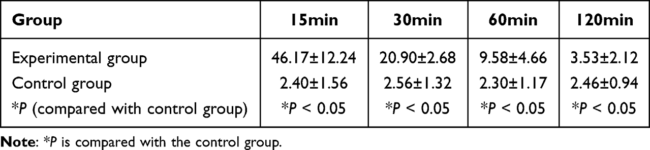 |
Table 1 HbCO Concentration of Rat Tail-Vein Blood at Each Time Point (%) |
Water Maze Test Results
The data were collected by the Morris water maze, and the results showed that the memory of the rats after poisoning was impaired. Specifically, the average latency increased, and the frequency of passing the platform decreased. The frequency of passing the platform of fewer than 3 times is indicative of DEACMP (Figure 1B).13,14 However, no obvious change was observed in the control group (Figure 1A, Additional Table).
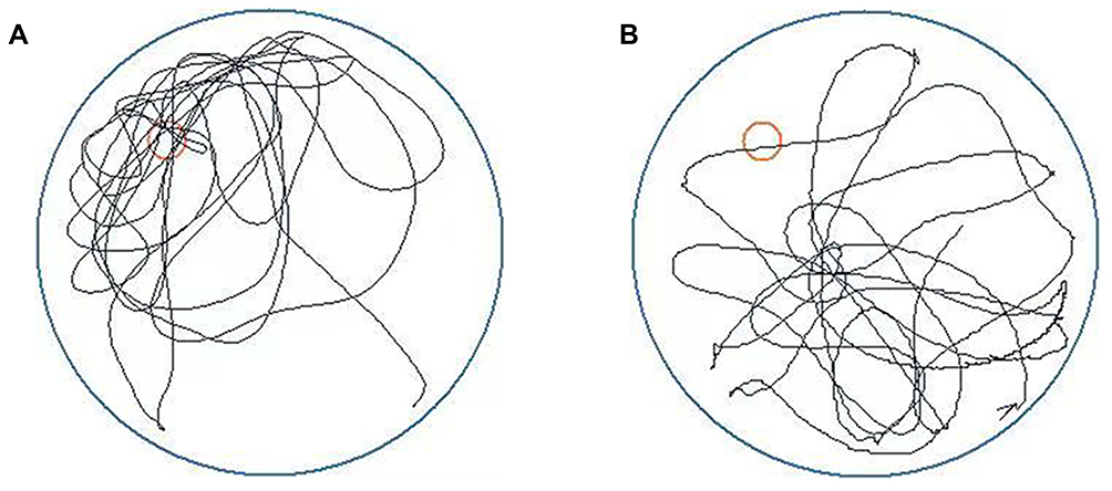 |
Figure 1 Morris water maze experiment (spatial exploration experiment), the red circle in the figure indicates the position of platform. (A) Rats of the control group. (B) DEACMP rats. |
Pathological Changes of Hippocampus of Rats
The pathological sections of rat brain tissue were stained with hematoxylin and eosin. Under the ordinary optical microscope, it can be seen that the hippocampal cells in the control group were plump, the perinuclear pale circle was obvious, and the cells were arranged orderly (Figure 2A). In the rats with delayed encephalopathy (Figure 2B), extensive damage to cells decreased cell number, thinning of the cell layer, disorder of cell structure, obvious cell shrinkage, pyknosis, and hyperchromatic nuclei, and unclear structure was observed in the hippocampus and subcortex under a microscope. However, no obvious focal necrosis was observed.
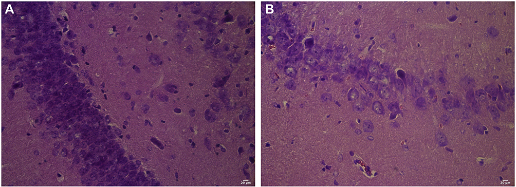 |
Figure 2 (A) Hippocampus of rats in the control group (HE). (B) Hippocampus of rats in the experimental group (HE). |
Establishment of ATP Standard Curve
The peak time of the ATP standard was measured to be 5.278 min (Figure 3). The peak values of 0, 40, 80, 120, 160, and 200 ug/mL ATP standard solution were measured, respectively, and the measured stable peak time was 5.278 min. The linear relationship was obtained via the proportional relationship, and the standard curve Y (peak area) = 0.2492X (ATP content) – 0.6363 (Figure 4).
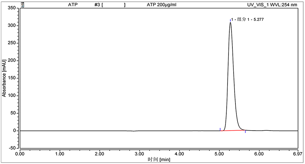 |
Figure 3 Chromatogram of ATP standard. |
 |
Figure 4 ATP standard curve. |
Results of Sample Determination
The prepared samples were analyzed, and the ATP chromatogram of the rat hippocampus was obtained (Figure 5). Compared with the control group, the ATP content of the delayed encephalopathy group model in the experimental group significantly increased, and the difference was statistically significant (*P < 0.05). The intra-group comparison was performed for the ATP content of the DEACMP group. The results showed group 21d > group 14d > group 7d, and the difference was statistically significant (#P < 0.05 is compared with 7d, &P< 0.05 is compared with 14d). The difference between group 21d and group 28d was not significant ($P > 0.05, Table 2). The results showed that hippocampal ATP in delayed encephalopathy rats was higher than that in normal rats, proving that hippocampal ATP content was related to DEACMP, and we have added the relative description in the Results.
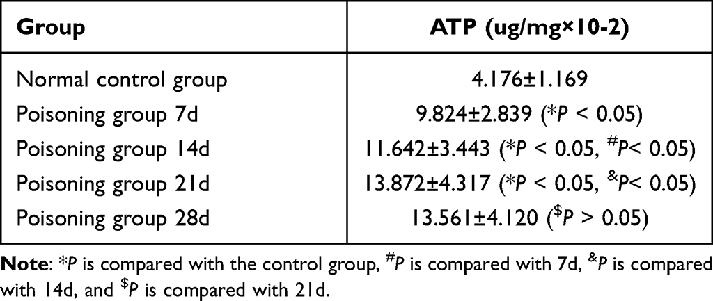 |
Table 2 ATP Content in Hippocampus of Rats in Different Experimental Groups |
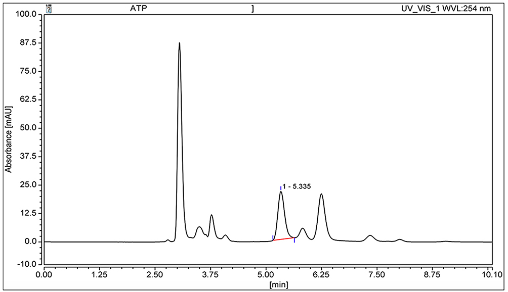 |
Figure 5 Chromatogram of sample for hippocampus of rats. |
Discussion
The pathogenesis of DEACMP is not presently clear. The establishment of an accurate and reliable animal model is the basis for further experimental research. At present, there are two main modeling methods: intraperitoneal injection and inhalation. The former features simple operation, strong controllability, and experimental gas savings. However, this method is different from the normal poisoning method in that the CO absorption method is different, and the poisoning time arising out of this method is not continuous. As a result, the amount of CO absorption is not constant and can cause many deaths in laboratory animals, which can then affect the experimental results. The CO inhalation method is more similar to that of clinical patient poisoning in that the toxic gas enters the blood in the same way. In this sense, an optimal modeling method was selected to generate the DEACMP model by the inhalation of 3000 ppm CO gas for 40 min, according to the reports of Hara et al. After the model was established, the Morris water maze test was performed for the experimental group, and the results showed that the rats with delayed encephalopathy exhibited abnormal behavior, such as increased latency and decreased frequency of passing the platform; HE staining was used to observe the pathological changes in the hippocampus of the rats. In the experimental group, the pathological manifestations of delayed encephalopathy appeared in the hippocampal area. The experimental results showed DEACMP manifestations, proving that the animal model established in this experimental study is ideal and reliable.
The pathogenesis of DEACMP is currently believed to be correlated with the apoptosis mechanism, the theory of ischemia and hypoxia, the inflammatory injury mechanism, the immune injury mechanism, and the CO/NO signal transduction mechanism.15–18 Some studies3 have confirmed that high concentrations of ATP accumulate in the surrounding area after partial central nerve injury and participate in the apoptosis or necrosis of secondary nerve cells. ATP is not only a classical energy substance but also a neuromodulator and neurotransmitter. Its receptors are widely distributed in the nervous system. At present, the ATP and its receptors have become a new research hotspot, and the role of ATP in the occurrence of neurological diseases has also drawn the attention of scholars. Studies by other scholars4,5 reported the effect of ATP on spinal cord neurons of cultured embryonic mice. The results showed that low concentrations of ATP could increase the activity of cells, while high concentrations of ATP could reduce the activity of cells and inhibit the formation of neuronal processes. For the experimental group, the precise scientific HPLC method was employed to measure the ATP content in the hippocampus of rats in different time periods in order to explore whether the ATP content in the hippocampus is correlated with the occurrence of delayed encephalopathy.
The results of this study showed that the ATP content in the hippocampus of DEACMP rats increased significantly, especially in the period from 14 to 21 days. This suggests that the ATP content in the hippocampus of rats is correlated with the occurrence of delayed encephalopathy. Through this experiment, it was clear that the content of ATP in the nervous system of rats with delayed onset encephalopathy was significantly increased, and it was also proved that delayed onset encephalopathy would affect the energy metabolism of nerve cells. However, it remains to be further studied whether ATP is increased through what pathway and whether ATP plays a protective or destructive role in the pathogenesis of tardive encephalopathy. This study was only based on the analysis of the changes in ATP content in nerve cells of rats with toxic tardive encephalopathy. The lack of specific reasons for the changes of neurotransmitters in neurons and the change principle of energy pathways requires further research and analysis. The results of this study can provide research basis for the pathogenesis and further diagnosis and treatment of delayed onset encephalopathy.
Ethical Approval
All experiments were evaluated and approved by the ethics Committee of Hulunbuir people’s Hospital and complied with the National Institutes of Health Guide for the Care and Use of Laboratory Animals.
Acknowledgments
We would like to acknowledge the hard and dedicated work of all the staff that implemented the intervention and evaluation components of the study.
Funding
Role and mechanism of P2Y12 receptor-mediated microglia activation in delayed encephalopathy caused by acute carbon monoxide poisoning(81641040).
Disclosure
The authors declare that they have no competing interests in this work.
References
1. Huang YQ, Peng ZR, Huang FL, et al. Mechanism of delayed encephalopathy after acute carbon monoxide poisoning. Neural Regener Res. 2020;15(12):2286. doi:10.4103/1673-5374.284995
2. Xiang WP, Xue H, Wang BJ. Delayed encephalopathy of acute carbon monoxide intoxication in rats: potential mechanism and intervention of dexamethasone. Pak J Pharm Sci. 2014;6(Suppl):2025–2028.
3. Xiang W, Yang Z, Xue H, et al. P2Y12 receptor-mediated microglia activation involved in delayed encephalopathy after acute carbon monoxide poisoning. Aging. 2021;13(4):6134–6143. doi:10.18632/aging.202607
4. Wang WQ, Wang SK, Sun ZY, et al. 细胞外ATP对体外培养乳鼠脊髓神经元生长的影响 [Effect of extracellular ATP on the growth of neonatal rat spinal cord neurons in vitro]. Chin J Microsurg. 2004;27(4):3. Chinese.
5. Fellin T, Sul JY, D’Ascenzo M, Takano H, Pascual O, Haydon PG. Bidirectional astrocyte-neuron communication: the many roles of glutamate and ATP. Novartis Found Symp. 2006;276:208–17;discussion 217–21, 233–7, 275–81. doi:10.1002/9780470032244.ch16
6. Hara S, Kobayash M, Kuriiwa F, et al. Gene expression in rat striatum following carbon monoxide poisoning. Genom Data. 2017;12:74–75. doi:10.1016/j.gdata.2017.03.007
7. Liao QJ, Wang J. Preparation of carbon monoxide poisoning delayed encephalopathy rat model by intraperitoneal injection. Prog Mod Biomed. 2011;2011:114.
8. Doná F, Conceição IM, Ulrich H. Variations of ATP and its metabolites in the hippocampus of rats subjected to pilocarpine-induced temporal lobe epilepsy. Purinergic Signal. 2016;12:295–302. doi:10.1007/s11302-016-9504-9
9. Wahab A, Chen JX, Jia CX, Murtaza G, Wu CH, Wang N. Molecular docking-based research on the potential anti-encephalopathy effect of gentianine. World J Tradit Chin Med. 2021;7:377–382. doi:10.4103/wjtcm.wjtcm_3_21
10. Zhang L, Pan J. Determination of ATP, ADP and AMP in mouse myocardial tissue by high performance liquid chromatography. Chin J Hosp Pharm. 2008;28(21):117.
11. Li WX, Zhang SQ, Li MM, et al. Pharmacokinetic comparison of four major bio-active components of naoxintong capsule in normal and acute blood stasis rats using ultra-performance liquid chromatography coupled with triple-quadrupole mass spectrometry. World J Tradit Chin Med. 2022;8:92–99. doi:10.4103/wjtcm.wjtcm_53_21
12. Zhou L, Fan MD, Chen YT, Zhang S. Improvement of learning and memory in rats of delayed neuropsychologic sequelae after carbon monoxide poisoning by hyperbaric oxygen and the possible machanism. Biomed Eng Clin Med. 2017;21(4):215.
13. Fu SZ, Liu Y, Yang JY, et al. The building of delayed neuropesychologic sequelae rat model by carbon monoxide peritoneum injection.China. J Crit Care Med. 2007;27(11):996–1003.
14. Zhao LY, Yu JC. Expression of heme oxygenase-1 and protein after delayed encephalopathy in carbon monoxide poisoning mice. Chin J Tissue Eng Res. 2014;18(18):2836–2840.
15. Bleecker ML. Carbon monoxide intoxication. Handb Clin Neurol. 2015;131:191–203. doi:10.1016/B978-0-444-62627-1.00024-X
16. Cao H, Tan X, Liu Z, et al. The effect of adding transcranial direct current stimulation to hyperbaric oxygen therapy in patients with delayed encephalopathy after carbon monoxide poisoning: a randomised controlled trial. Front Neurol. 2021;12:719765. doi:10.3389/fneur.2021.719765
17. Shen M, Zheng Y, Zhu K. Hydrogen gas protects against delayed encephalopathy after acute carbon monoxide poisoning in a rat model. Neurol Res. 2020;42(1):22–30. doi:10.1080/01616412.2019.1685064
18. Su C, Zhao N, Zou J, et al. TDZD-8 alleviates delayed neurological sequelae following acute carbon monoxide poisoning involving tau protein phosphorylation. Inhalation Toxicol. 2020;32:79–85. doi:10.1080/08958378.2020.1741739
 © 2023 The Author(s). This work is published and licensed by Dove Medical Press Limited. The
full terms of this license are available at https://www.dovepress.com/terms
and incorporate the Creative Commons Attribution
- Non Commercial (unported, 3.0) License.
By accessing the work you hereby accept the Terms. Non-commercial uses of the work are permitted
without any further permission from Dove Medical Press Limited, provided the work is properly
attributed. For permission for commercial use of this work, please see paragraphs 4.2 and 5 of our Terms.
© 2023 The Author(s). This work is published and licensed by Dove Medical Press Limited. The
full terms of this license are available at https://www.dovepress.com/terms
and incorporate the Creative Commons Attribution
- Non Commercial (unported, 3.0) License.
By accessing the work you hereby accept the Terms. Non-commercial uses of the work are permitted
without any further permission from Dove Medical Press Limited, provided the work is properly
attributed. For permission for commercial use of this work, please see paragraphs 4.2 and 5 of our Terms.