
Back to Journals » Journal of Pain Research » Volume 19
Comparison of Ultrasound-Guided Erector Spinae Plane Block, Thoracic Paravertebral Block, and Control for Postoperative Recovery in Video-Assisted Thoracic Surgery Patients: A Randomized Controlled Trial
Authors You Y, Zhang X, Zheng X, Zheng X
Received 9 October 2025
Accepted for publication 10 May 2026
Published 28 May 2026 Volume 2026:19 572950
DOI https://doi.org/10.2147/JPR.S572950
Checked for plagiarism Yes
Review by Single anonymous peer review
Peer reviewer comments 2
Editor who approved publication: Dr Jinlei Li
Yisheng You,1,* Xuru Zhang,1,* Xiaowei Zheng,2 Xiaochun Zheng3
1Department of Anesthesiology, Clinical Oncology School of Fujian Medical University, Fujian Cancer Hospital, Fuzhou, 350014, People’s Republic of China; 2Department of Otolaryngology, Fujian Medical University Union Hospital, Fuzhou, People’s Republic of China; 3Department of Anesthesiology, Fuzhou University Affiliated Provincial Hospital, Fuzhou, 350001, People’s Republic of China
*These authors contributed equally to this work
Correspondence: Xiaochun Zheng, Department of Anesthesiology, Fuzhou University Affiliated Provincial Hospital, 134 Dong Jie, Gulou District, Fuzhou City, Fujian Province, 350001, People’s Republic of China, Tel +86137 0505 8351, Email [email protected]
Background: The objective of this study was to examine the effect of ultrasound-guided Erector Spinae Plane Block (ESPB) and Thoracic Paravertebral Block (TPVB) on the quality of recovery (QoR) in video-assisted thoracic surgery (VATS).
Methods: A total of 129 patients were randomly assigned to three groups: ESPB group and TPVB group received ultrasound-guided nerve blocks, and Control group received multimodal analgesia protocol only. The primary outcome measure was QoR-15 scale at 24 hours postoperatively. Secondary outcome measures included visual analog scale (VAS) scores at rest and cough, intraoperative remifentanil dosage, number of postoperative patient-controlled intravenous (PCI) self-controlled compressions, and the incidence of adverse effects.
Results: The QoR-15 scores at 24 hours postoperatively were significantly higher in ESPB and TPVB group as compared to Control group. The static (at rest) and dynamic (at cough) VAS scores within 7 h of surgery, intraoperative remifentanil dosage, and number of postoperative PCI self-controlled compressions were significantly lower in ESPB and TPVB groups as compared to Control group (all P< 0.001). The occurrence of nausea, vomiting, and dizziness in ESPB and TPVB group was lower than that in Control group (P< 0.05), whereas other adverse effects were not statistically different between the three groups.
Conclusion: The ultrasound-guided ESPB may be a promising form of regional anesthesia to aid recovery after VATS surgery, which is similar to TPVB.
Trial Registration: This study has been registered at the Chinese Clinical Trial Registry (ChiCTR2500105099).
Keywords: erector spinae plane block, thoracic paravertebral block, video-assisted thoracic surgery, quality of recovery
Introduction
The video-assisted thoracoscopic surgery (VATS) is one of the most popular minimally invasive surgery techniques for performing thoracic surgery worldwide, and it is now considered the preferred resection method for lung carcinoma in the majority of medical institutions.1,2 In comparison to conventional open thoracotomy, VATS may achieve less trauma, shorter hospitalization, and better recovery of physical functioning.3–6 It is noteworthy, however, that moderate-to-severe acute pain is more commonly seen in patients undergoing VATS, along with notable occurrences of postoperative chronic pain.7–10
Thoracic epidural analgesia (TEA) and thoracic paravertebral block (TPVB) are well-validated, evidence-based regional analgesic techniques that are widely recommended for pain management following major thoracic surgery.11 Epidural anesthesia achieves comprehensive analgesia by blocking spinal nerves within the epidural space. In contrast, TPVB targets the thoracic paravertebral space to block spinal nerve roots, their branches, and the sympathetic trunk, providing analgesic efficacy comparable to that of epidural anesthesia with a relatively lower risk of hemodynamic instability.12 Although both approaches have been proven effective, they are not without limitations. Epidural anesthesia may cause hypotension and bradycardia, carries a relatively high incidence of accidental epidural spread, and requires specialized expertise for implementation.11 TPVB is associated with potential risks including pneumothorax, difficulty with catheter placement, a high failure rate of needle insertion, and inadvertent spread of local anesthetics into the epidural space, which may limit its routine clinical application.13
Erector spinae plane block (ESPB), first pioneered by Forero et al in 2016, is a novel regional analgesic technique used for anesthesia and analgesia of the thoracic and abdominal wall.14 In recent years, a growing number of clinical studies have explored the perioperative analgesic value of ESPB in thoracic surgery, cardiac surgery, and breast surgery, reporting its feasibility, safety, and potential analgesic efficacy.15–18 Current evidence suggests that compared with TEA and TPVB, ESPB may have advantages such as simpler technical operation, lower risk of hemodynamic effects, and fewer associated complication.19–21 However, it is noteworthy that most of these studies are single-center or small-sample trials, and their conclusions are often limited to short-term pain scores rather than comprehensive postoperative recovery outcomes.22
To date, high-quality randomized controlled trials (RCTs) directly comparing the perioperative analgesic efficacy, quality of recovery (QoR), and safety of ESPB, TPVB, and TEA in patients undergoing VATS remain scarce. In addition, in this clinical context, the role of conventional general anesthesia alone (without additional regional analgesia) as a control group has not been systematically evaluated in large-scale comparative studies, leaving a critical evidence gap in optimizing postoperative analgesic strategies. In the present study, we designed a three-arm randomized controlled trial aimed at objectively comparing the differences in perioperative analgesic efficacy, QoR, and safety among ultrasound-guided ESPB, TPVB, and conventional general anesthesia alone in patients with lung cancer undergoing elective VATS. The primary objective is to assess whether ESPB can provide analgesic efficacy and quality of postoperative recovery comparable to those of TPVB, while evaluating the incremental value of adding regional analgesia to general anesthesia. No superiority or inferiority was assumed for any intervention; our goal is to generate high-quality comparative evidence to provide an evidence-based basis for clinical decision-making regarding postoperative analgesia in VATS patients, rather than inferring clinical benefits based on the novelty of ESPB.
Methods
Patients
This prospective, randomized controlled study was approved and implemented by the Institutional Review Board and Ethics Committee of Clinical Oncology School of Fujian Medical University (ethics batch number: K2025-200-01). This study has been registered at the Chinese Clinical Trial Registry (ChiCTR2500105099). The authors complied with the Consolidated Standards of Reporting Trials (CONSORT) guidelines for reporting randomized controlled trials. We recruited a consecutive series of adult patients who were scheduled for lobectomy under complete VATS for lung carcinoma from July 2025 to August 2025. Inclusion criteria were age 25–75 years, Body Mass Index (BMI) 18–30kg/m2, and American Society of Anesthesiologists (ASA) physical status score I to III, and study participants have a full understanding of the purpose and significance of this trial, voluntarily participate in this clinical trial, and sign the informed consent form. Exclusion criteria were as follows: (1) Emergency or emergency surgery; (2) Those with spinal or spinal cord abnormalities who are not suitable for nerve block anesthesia; (3) Infection at the puncture site; (4) Patients with increased intracranial pressure or central nervous system injury; (5) Have a history of chronic narcotic use or alcohol abuse; (6) Those who are allergic to the components or components of ropivacaine hydrochloride that may be used in the study; (7) Those with a history of allergic diseases in the past, those who have had malignant hyperthermia in the past, and those who have a history of epilepsy in the past; (8) Known significant liver disease (Child-Pugh score B or C or presence of ascites); (9) Renal failure (serum creatinine level ≥1.5 mg/dL); (10) Significant cardiac disease (ejection fraction <50%); (11) Overt chronic obstructive pulmonary disease (forced expiratory volume in 1 second or diffusion capacity of carbon monoxide <50% of the predicted value); (12) Subjects with a history of mental illness and cognitive dysfunction; (13) Patients with hematologic diseases, coagulation disorders, or oral warfarin, new anticoagulants such as rivaroxaban and dabigatran due to other diseases; (14) Other conditions judged by the investigator that the subject is not suitable to participate in this clinical trial. The written informed consent was obtained from all participants included in this study.
After obtaining written informed consent, participants were split randomly into receiving a single shot of ESPB or TPVB at T5 and T8 levels, and the Control group received a standardized multimodal analgesia protocol only. A research anesthesiologist who was not involved in the study performed the randomization using the online program Research Randomizer (http://www.randomization.com). The follow-up interviewers and data entry personnel were blinded to the group assignment throughout.
General Anesthesia Technique
All participants were routinely fasted and water-deprived at least 8h before the operation and premedicated with penehyclidine hydrochloride 0.5mg intramuscularly 30 minutes prior to anesthesia. Upon arriving on the operation room, standard ASA monitoring and bispectral index (BIS)were utilized throughout the operations. After resting in the supine position for 5 min, systolic blood pressure (SBP), diastolic blood pressure (DBP), mean arterial pressure (MAP), and heart rates (HR) were measured as a baseline record (T1). For the purposes of this study, general anesthesia was administered in a standardized manner, utilizing sufentanil at a dosage of 0.4 μg/kg, etomidate at a dosage of 0.3 mg/kg, and rocuronium at a dosage of 0.6 mg/kg. Tracheal intubation was performed through a video laryngoscope using a double-lumen tracheal tube. The SBP, DBP, and HR were recorded at intubation time (T2), at the beginning of surgery (T3), and at extubation time (T4). Anesthesia was maintained by sevoflurane, propofol, remifentanil, and rocuronium, with the BIS held between 40 and 60. Body temperature was continuously monitored with a nasopharyngeal probe and maintained at 36 to 36.5°C. All of the VATS surgeries were conducted following standardized procedures by the same group of surgeons. The standardized postoperative pain regimen is given below. Postoperative patient-controlled intravenous(PCI) analgesia involved a solution (100 mL) containing sufentanil 2.5ug/kg, flurbiprofen axetil 200mg, and palonosetron 0.5 mg (background infusion of 2 mL/h, a single self-control dose of 2 mL, and lockout interval of 15 min). After the operation, patients were scored according to the visual analog scale (VAS) score, and when the VAS score was at least 4 points, the patient pressed the analgesic pump once autonomously.
Regional Anesthesia Technique
Ultrasound-guided ESPB and TPVB were performed by a trained anesthesiologist familiar with ultrasound-guided nerve blocks prior to the induction of general anesthesia using a portable ultrasound machine Sonosite (Sonosite, USA).
In the ESPB group, the patient was placed in a lateral position and disinfection, followed by towel spreading. The transverse process of the 5th thoracic vertebra was identified, and then to confirm the trapezius, rhomboid, and erector spinae from superficial to deep using a convex array probe (frequency 5–8 MHz) (Figure 1). Subsequently, a 22-gauge, 80 mm puncture needle was slipped into the gap between the transverse process and the erector spinal muscle via an in-plane technique after local anesthesia. If there were no blood, gas, or cerebrospinal fluid drawn back, the patients were injected with 0.5% ropivacaine 20 mL. In the T8 plane, the trapezius, latissimus dorsi, and erector spinae were sequentially identified from superficial to deep using ultrasound, and local anesthesia was injected at the same site as at T5. The block plane was assessed by thermo-sensory changes in the mid-axillary line through a cotton swab dipped in wet alcohol to confirm the efficacy of the nerve block after 15 minutes.
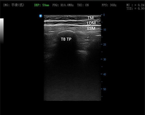 |
Figure 1 Ultrasonographic image of the pleura and T8 transverse process in the middle of the image. “Low image processing” means low smoothing mode, which preserves more image details with slightly higher noise. Depth indicates the ultrasound scanning depth is set to 59 mm. Frequency represents the probe central frequency at high level of 10.0 MHz. ON means the function is enabled. Frames per second refers to a frame rate of 36 frames per second. Mechanical index reflects the ultrasonic cavitation risk, with a value of 0.24. TIS: Thermal Index for Soft Tissue. The soft tissue thermal index is 0.90.The blue T indicates the probe orientation mark (Mark point), and the blue numerical scale 0–50 represents a depth range of 0–50 mm. Abbreviations: TP, transverse process; TM, trapezius; LDM, latissimus dorsi muscle; ESM, erector spinae muscle; IMG, Image Processing; DEP, Depth; FRQ, Frequency; THI, Tissue Harmonic Imaging; FPS, Frames Per Second; MI: Mechanical Index; TIS, Thermal Index for Soft Tissue. |
In the TPVB group, the patient was similarly in the lateral decubitus position. The ultrasound was utilized to clearly visualize the T5 spinous process, transverse process, supracostal transverse ligament, and pleura. After a 22G puncture needle pierced the superior transverse intercostal ligament., 0.5% ropivacaine 20 mL was injected, and the sign of hemizygosity of the pleura was seen after the injections (Figure 2). The same method was used for the paravertebral nerve block of T8. After 15 minutes, the block plane was measured, thus confirming that the nerve block was adequate.
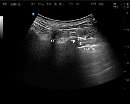 |
Figure 2 Pleura placed in the middle of the image at T8 level. “Low image processing” means low smoothing mode, which preserves more image details with slightly higher noise. Depth indicates the ultrasound scanning depth is set to 83 mm. Frequency represents the probe central frequency at high level of 4.2 MHz. ON means the function is enabled. Frames per second refers to a frame rate of 20 frames per second. Mechanical index reflects the ultrasonic cavitation risk, with a value of 0.46. The soft tissue thermal index is 0.96.The blue T indicates the probe orientation mark (Mark point), and the blue numerical scale 0–80 represents a depth range of 0–80 mm. Abbreviations: TP, transverse process; SP, spinous process; TPVS, thoracic paravertebral space; IMG, Image Processing; DEP, Depth; FRQ, Frequency; THI, Tissue Harmonic Imaging; FPS, Frames Per Second; MI, Mechanical Index; TIS, Thermal Index for Soft Tissue. |
Blinding
Due to the distinct operational characteristics of ultrasound-guided ESPB, TPVB, and the control group without regional block intervention, it was impractical to blind the anesthesiologists performing the blocks. To mitigate potential bias and uphold the validity of postoperative recovery outcome evaluations, a rigorous single-blind strategy was adopted, with blinding strictly maintained for outcome assessors, and data analysts throughout the entire trial period.
Outcomes
The primary endpoint of this study was the QoR, using the Chinese version of the QoR-15 scale at 24 hours postoperatively. The QoR-15, a 15-item questionnaire assessing the quality of patients’ postoperative recovery, which consists of five subscales: physical comfort (5 items), physical independence (2 items), emotional state (4 items), psychological support (2 items), and pain (2 items). There are 15 questions in total, and all items are rated on a 10-point scale from 0 to 10. The sum of the scores was the QoR-15 correlation score of patients, which ranges from 0 to 150, with a higher total score indicating a better quality of recovery. Patients who were recruited in this study completed the Chinese version of the QoR-15 scale items in truthfully, alone or with the help of an experienced anesthesia nurse prior to surgery (baseline), and on the first postoperative day, respectively. The secondary endpoints were VAS scores at rest, VAS scores at cough, intraoperative remifentanil dosage, number of postoperative PCI self-controlled compressions, and the incidence of adverse effects recorded postoperatively. All participants were given instructions about calculating postoperative pain assessment using the VAS scale in the preoperative visit (0 indicating no pain, 10 indicating the maximum pain imaginable). The postoperative pain assessments were performed at 1, 3, 5, 7, 24, and 48 hours as static (at rest) and dynamic (at cough) postoperatively. The total times of analgesia pump pressing were evaluated for the first postoperative 24 hours. Side effects, including nausea, vomiting, dizziness, urinary retention, and extrapleural hematoma were recorded.
Statistical Analysis
Sample size calculations were performed using NCSS PASS software (NCSS LLC, Kaysville, Utah). Based on the results of our pre-experiment research, the mean QoR-15 scale at 24 hours postoperatively for ESPB, TPVB and Control group as 138, 137, and 131, respectively. With α = 0.05, a test efficacy of 90%, the required sample size was 90 cases. After considering patient withdrawal and loss to follow-up, the planned sample size should be at least 114 patients.
All statistical analysis were conducted using SPSS software (version 22.0, SPSS, Inc, Chicago, IL). The normality test of data distribution was evaluated using the Kolmogorov–Smirnov test. Continuous variables were presented as means with standard deviation (SD) or medians with interquartile range (IQR), and categorical variables were expressed as counts with percentages. Normally distributed continuous variables were compared among groups using one-way analysis of variance (ANOVA), and post hoc comparisons were made using Tukey’s test. Non-normally distributed continuous variables were compared between the groups using the Kruskal Wallis One Way Analysis of Variance and Dunn’s Post Hoc test. The Pearson χ2-test was utilized to compare the differences between categorical variables. Statistical comparisons among the three parallel groups were performed using one-way ANOVA followed by post-hoc tests. A statistically significant result was defined as a two-sided P value less than 0.05.
Results
A total of 129 participants were initially enrolled. Of whom, one participant in the Control group and one participant in the TPVB group were excluded because of conversion to thoracotomy. During the follow-up period, two participants in the Control group and one participant in each ESPB and TPVB group died, one participant in the Control group, two participants in the ESPB group, and one participant in the TPVB group were lost to follow-up. One hundred and nineteen subjects were included in the final analysis. The participant’s inclusion flow chart is shown in Figure 3. The demographic attributes of the participants in each group are presented in Table 1. There were no differences in the baseline characteristics between each group.
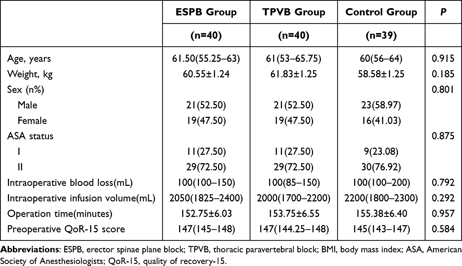 |
Table 1 Demographic Data and Baseline Characteristics of Patients |
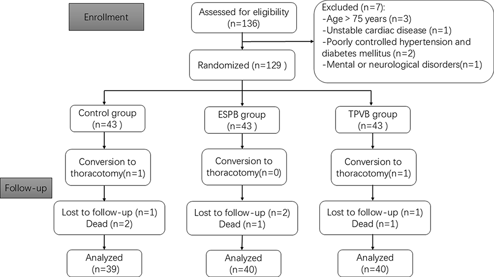 |
Figure 3 Flow chart of the randomized controlled trial. |
The comparison of hemodynamic data at different time points in the intraoperative period of patients in the three groups was shown in Figure 4. No statistically significant differences were observed in MAP and HR at the T1 time point between the three groups. At the time point from T2 to T4, the MAP and HR of the ESPB group and TPVB group were significantly lower than those of the Control group (all P<0.05), whereas the difference between the ESPB group and TPVB group was not statistically significant (Table 2).
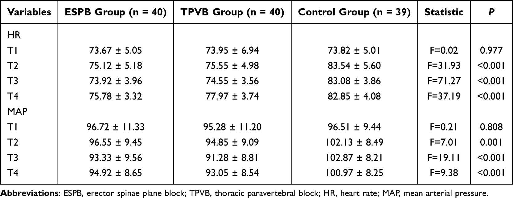 |
Table 2 Intraoperative Heart Rate and Mean Arterial Pressure of the Three Groups at Different Time Points |
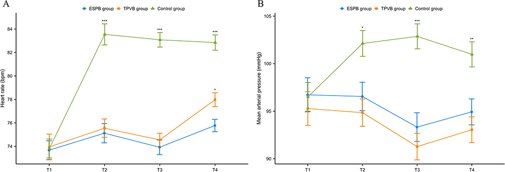 |
Figure 4 (A) Intraoperative heart rate of the three groups at different time points. (B) Intraoperative mean arterial pressure of the three groups at different time points. *P < 0.05, **P < 0.01, ***P < 0.001 vs Control group. |
The Chinese version of the QoR-15 scores at baseline and 24 postoperative hours are presented in Figure 5. The Chinese version of the QoR-15 scores in the ESPB and TPVB group were higher than that in Control group (P<0.001), while no statistical difference was observed between ESPB and TPVB groups.
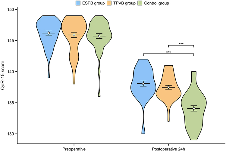 |
Figure 5 QoR-15 scores preoperatively and at 24 hours postoperatively in the three groups. ***P < 0.001 vs Control group. |
The postoperative VAS scores at rest and cough in different time points are shown in Figure 6. In intergroup comparisons at each time interval, the VAS scores both at rest and cough were significantly lower in the first 7 h postoperatively in the ESPB and TPVB group as compared to the Control group (all P<0.001), whereas there was no significant difference between the ESPB and TPVB group (all P>0.05). No significant differences in VAS scores were observed both at rest and cough between the three groups at the 24h and 48h postoperative time points (Tables 3 and 4).
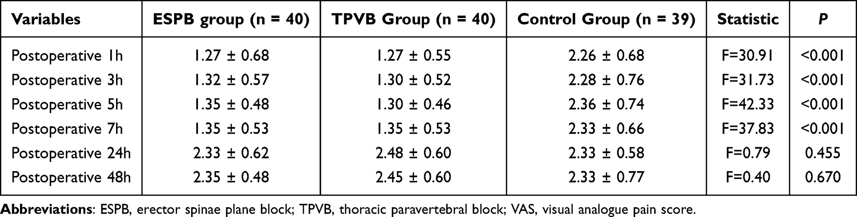 |
Table 3 The Postoperative VAS Scores at Rest in the Three Groups at Different Time Points |
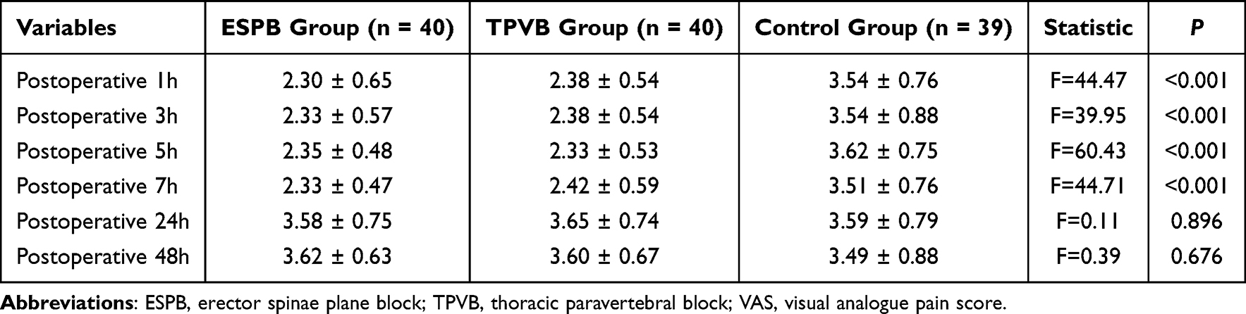 |
Table 4 The Postoperative VAS Scores at Cough in the Three Groups at Different Time Points |
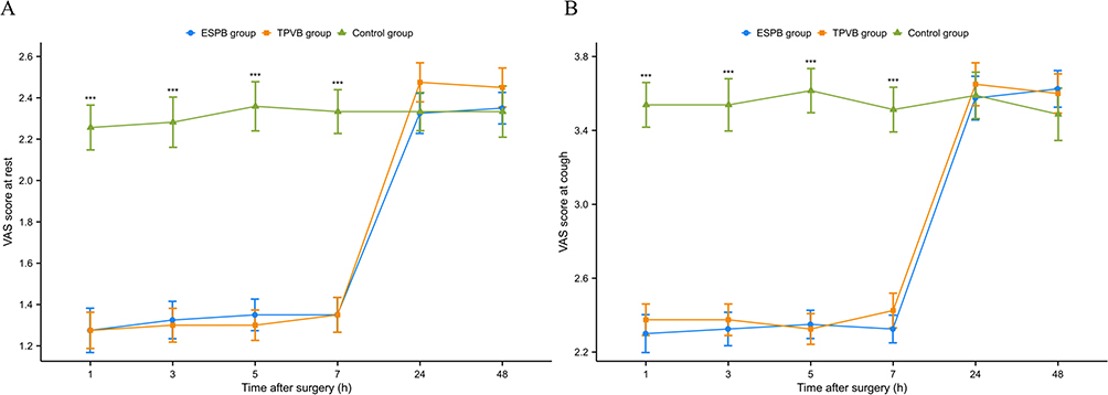 |
Figure 6 (A) The postoperative VAS scores at rest in the three groups at different time points. (B) The postoperative VAS scores at cough in the three groups at different time points. ***P < 0.001 vs Control group. Abbreviation: VAS, visual analog scale. |
Figure 7 shows the intraoperative cumulative remifentanil consumption and the number of postoperative PCI self-controlled compressions during the first 48 h after surgery in each group. The intraoperative cumulative remifentanil consumption was significantly lower in both ESPB and TPVB groups as compared to the Control group (P<0.001), and the difference was not statistically significant between the ESPB and TPVB groups. The number of postoperative PCI self-controlled compressions during the first 48 h after surgery was significantly lower in both ESPB and TPVB groups as compared to the Control group (P<0.001), and the difference was not statistically significant between ESPB and TPVB groups.
 |
Figure 7 (A) The intraoperative cumulative remifentanil consumption in the three groups. (B) The number of postoperative PCI self-controlled compressions during the first 48 h after surgery in the three groups. ***P < 0.001 vs Control group. Abbreviation: PCI, patient-controlled intravenous. |
As shown in Table 5, the incidence of nausea, vomiting, and dizziness was lower in ESPB and TPVB than in the Control group (all P<0.05). Two patients in the control group, two patients in the TPVB group, and one patient in the ESPB group reported urinary retention, respectively. Extrapleural hematoma occurred in only one case in the ESPB group.
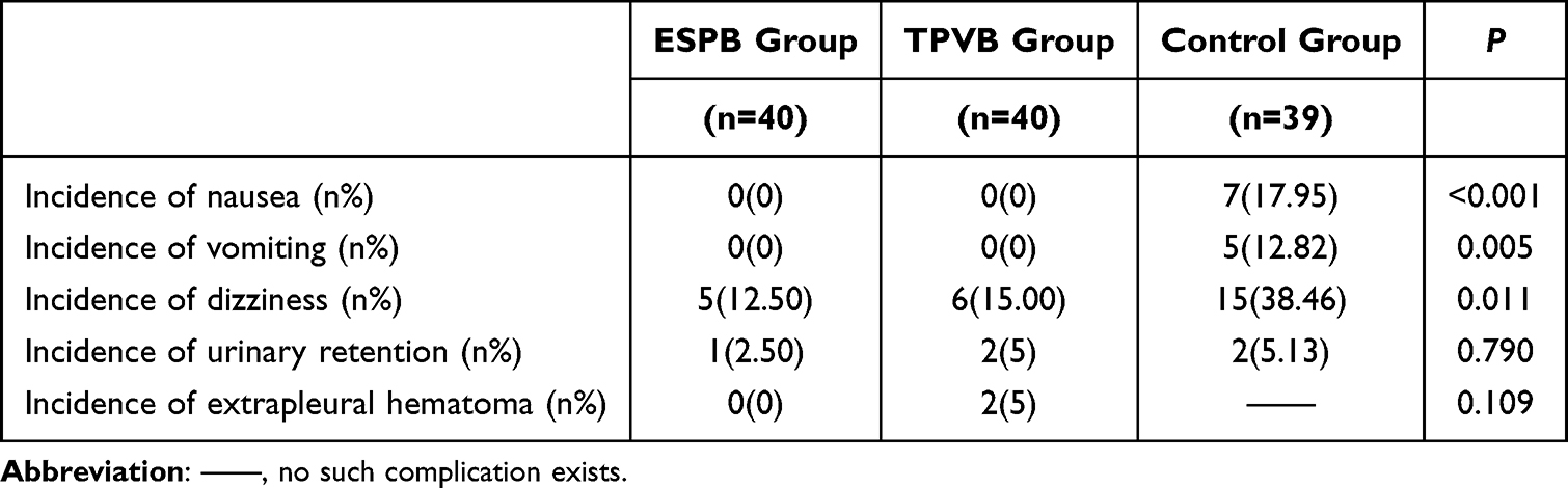 |
Table 5 The Incidence of Adverse Effects Recorded After VATS |
Discussion
The results of this single-center randomized controlled clinical trial documented that the use of preoperative ultrasound-guided ESPB can improve early postoperative quality of recovery measured by QoR-15 score compared to general anesthesia alone, together with a reduction of postoperative VAS scores at rest and cough, the intraoperative amount of remifentanil requirement, and the number of patients requiring rescue analgesia in the first 48 hours. Our findings have provided novel evidence supporting ultrasound-guided ESPB may be an effective and promising form of regional anesthesia to aid recovery after VATS surgery in patients with lung carcinoma, which is similar to TPVB.
It has long been well established that TEA and TPVB are the gold standard regional analgesia for thoracic surgery. The spinal nerves enter the paravertebral space after penetrating corresponding intervertebral foramina just below the transverse processes of the thoracic vertebrae. The achievement of postoperative analgesia is facilitated by the administration of local anesthetics into the paravertebral space through the technique known as TPVB. This procedure effectively obstructs the spinal nerve roots within this specific anatomical region. TPVB has the capacity to impede nerve conduction across multiple thoracic dermatomes, both superior and inferior to the punctured vertebral body through the diffusion of the medication. Additionally, it can selectively block the thoracic dermatomes on the ipsilateral side of the surgical intervention. In our study, the range of block was between C7 and T2 to T10 and T12 in the TPVB group, which was consistent with Marhofer et al.23 Despite the exact efficacy of TPVB that is well described in numerous previous studies, there are several factors that may have hindered its widespread adoption for postoperative analgesia in VATS, such as sophisticated technological operations and the risk of serious complications.24 New interfascial block techniques, such as ESPB, hold promise to be a safe and controllable alternative to TPVB because of the ability to provide effective analgesia while reducing the risk of injury to adjacent structures.25
The ESPB is a relatively novel technique that acts on the ventral rami of spinal nerves in the paravertebral space, and the rami communicate and sympathetic chain through both transforaminal and epidural spread, providing effective pain relief to a variety of surgical procedures, including breast, cardiac, thoracic, abdominal, and lumbar surgery.26–30 Simultaneously, there is growing evidence that ESPB also shows favorable therapeutic outcomes in complex pain syndromes, including myofascial pain syndrome, chronic low back pain, and back myofascial pain syndrome.31–33 Our study produces findings that align with a recent single-center, non-inferiority randomized controlled trial conducted by Xu et al,34 they compared the sufentanil dosage and the intensity of pain (on an 11-point scale) during the 24-hour postoperative period between ESPB and TPVB in laparoscopic nephroureterectomy, and revealed that the analgesic effect of erector spinae was comparable to that of paravertebral block. In this pilot study, we compared the intraoperative cumulative remifentanil consumption and the number of postoperative PCI self-controlled compressions during the first 48 h after surgery in ESPB and TPVB group, and demonstrated no statistical difference between the two groups.
As reported, the ESPB is currently considered an easily administered method for the perioperative analgesia, able to quickly identify anatomic landmarks on ultrasound.35,36 We revealed statistically significant difference between dizziness, nausea, and vomiting in the ESPB and Control groups, while there was no significant difference observed between the ESPB and TPVB groups. Our findings support the conclusions from recent studies indicating that there have been few side effects and complication related to ESPB in many clinical applications.37,38 ESPB and TPVB can be performed above or below the target puncture point, both favoring well spreading of the local anaesthetics without the impact of skin infection at the target puncture point, and equally achieving the desired range of block. Nonetheless, extrapleural hematoma occurred in two patients in our study, and both patients were in the TPVB group. Although these differences were not statistically significant due to the very small sample size, it still suggests that ESPB may be safer than TPVB. The advantage of ESPB over TPVB also lies in the superficial target puncture site and clearly local anatomy that the spinal nerve roots, pleura and paravertebral vessels can be avoided during the puncture, which greatly eliminates the risk of nerve injury, haemothorax, pneumothorax, and local anesthetic systemic toxicity.39 Some studies have reported that it can even be safely utilized in patients who are taking antiplatelets or anticoagulants.40 Meanwhile, the easy-to-master operation technique reduces the threshold of the clinical application of ESPB, suggesting that it may become a more advantageous method of perioperative analgesia for thoracic surgery than TPVB.
Our study has limitations in that it is a single-center small cohort study. Unfortunately, due to ethical reasons we did not include a placebo group. Participants knew whether they had received a nerve block injection or not. The placebo effect, which increases bias error, cannot be minimized. Second, we did not examine potentially more relevant outcome metrics such as escalation of postoperative care, medical economics, or analgesic properties of longer duration. Thirdly, this study has limitations in terms of mechanism exploration and long-term follow-up. In the future, through basic experiments, we can further explore the analgesic mechanism of ESPB in VATS, and conduct long-term follow-up on patients’ quality of life and the incidence of chronic pain. Fourthly, the preoperative baseline QoR-15 scores of the research subjects were relatively high, which reduced the sensitivity of identifying clinical significant differences. In the future, we can analyze the preoperative QoR-15 score data using a larger sample size, adopt appropriate statistical methods to correct the ceiling effect, and supplement sensitivity analysis to reduce the influence of the ceiling effect on the result judgment. In the future, multi-center, large-sample, double-blind, and rigorously designed clinical trials need to be conducted to supplement multiple comparison corrections and optimize the research design to avoid problems such as the ceiling effect.
Conclusion
Both ultrasound-guided ESP and TPVB improved perioperative hemodynamic stability, relieved postoperative pain, reduced intraoperative and postoperative opioid consumption, and decreased postoperative adverse reactions in patients undergoing VATS. Due to its easier operation and lower complication risk, ESPB may be a favorable alternative to TPVB for perioperative analgesia in thoracic surgery, but this conclusion should be interpreted cautiously given the study’s limitation. Our findings are preliminary, and further large-scale, multicenter, rigorously designed trials are needed to validate the potential advantages of ESPB over TPVB.
Data Sharing Statement
The datasets used and analysed during the current study are available from the corresponding author on reasonable request.
Ethical Approval and Consent to Participate
This study adhered to the Consolidated Standards of Reporting Trials statement and the Declaration of Helsinki. The study was approved by the Ethics Committee of Clinical Oncology School of Fujian Medical University (Ethics batch number: K2025-200-01). This study has been registered at the Chinese Clinical Trial Registry (ChiCTR2500105099). All participants provided written informed consent.
Acknowledgments
We would like to thank Yuanyuan Zhang for her advice and assistance with data analysis.
Author Contributions
All authors made a significant contribution to the work reported, whether that is in the conception, study design, execution, acquisition of data, analysis and interpretation, or in all these areas; took part in drafting, revising or critically reviewing the article; gave final approval of the version to be published; have agreed on the journal to which the article has been submitted; and agree to be accountable for all aspects of the work.
Funding
There is no external financial support or sponsorship of this work.
Disclosure
The authors declared no potential conflicts of interest with respect to the research, authorship, and/or publication of this article.
References
1. Maiga AW, Deppen SA, Denton J, et al. Uptake of video-assisted thoracoscopic lung resections within the veterans affairs for known or suspected lung cancer. JAMA Surg. 2019;154(6):524–13. doi:10.1001/jamasurg.2019.0035
2. Lampridis S, Maraschi A, Le Reun C, et al. Robotic versus video-assisted thoracic surgery for lung cancer: short-term outcomes of a propensity matched analysis. Cancers. 2023;15(8):2391. doi:10.3390/cancers15082391
3. Olland A, Reeb J, Sauleau E, et al. Video-assisted thoracoscopic lobectomy versus open thoracotomy conventional lobectomy for stage I non-small cell lung cancer. Cochrane Database Syst Rev. 2017. doi:10.1002/14651858.CD012641
4. Nagahiro I, Andou A, Aoe M, et al. Pulmonary function, postoperative pain, and serum cytokine level after lobectomy: a comparison of VATS and conventional procedure. Ann Thorac Surg. 2001;72(2):362–365. doi:10.1016/S0003-4975(01)02804-1
5. Bendixen M, Jørgensen OD, Kronborg C, et al. Postoperative pain and quality of life after lobectomy via video-assisted thoracoscopic surgery or anterolateral thoracotomy for early stage lung cancer: a randomised controlled trial. Lancet Oncol. 2016;17(6):836–844. doi:10.1016/S1470-2045(16)00173-X
6. Bendixen M, Kronborg C, Jørgensen OD, et al. Cost-utility analysis of minimally invasive surgery for lung cancer: a randomized controlled trial. Eur J Cardio-Thorac Surg. 2019;56(4):754–761. doi:10.1093/ejcts/ezz064
7. Rizk NP, Ghanie A, Hsu M, et al. A prospective trial comparing pain and quality of life measures after anatomic lung resection using thoracoscopy or thoracotomy. Ann Thorac Surg. 2014;98(4):1160–1166. doi:10.1016/j.athoracsur.2014.05.028
8. Bayman EO, Parekh KR, Keech J, et al. A prospective study of chronic pain after thoracic surgery. Anesthesiology. 2017;126(5):938–951. doi:10.1097/ALN.0000000000001576
9. Steegers MAH, Snik DM, Verhagen AF, et al. Only half of the chronic pain after thoracic surgery shows a neuropathic component. J Pain. 2008;9(10):955–961. doi:10.1016/j.jpain.2008.05.009
10. Furrer M, Rechsteiner R, Eigenmann V, et al. Thoracotomy and thoracoscopy: postoperative pulmonary function, pain and chest wall complaints. Eur J Cardio-Thorac Surg. 1997;12(1):82–87. doi:10.1016/S1010-7940(97)00105-X
11. Eaves GK, Ware EE, Touchet DR, et al. Efficacy and safety of thoracic epidural vs paravertebral block for analgesia in thoracotomy: a systematic review of randomized controlled trials. Curr Pain Headache Rep. 2025;29(1):69. doi:10.1007/s11916-025-01383-2
12. Zhang J-W, Feng X-Y, Yang J, et al. Ultrasound-guided single thoracic paravertebral nerve block and erector spinae plane block for perioperative analgesia in thoracoscopic pulmonary lobectomy: a randomized controlled trial. Insights Imaging. 2022;13(1):16. doi:10.1186/s13244-021-01151-x
13. Pace MM, Sharma B, Anderson-Dam J, et al. Ultrasound-guided thoracic paravertebral blockade: a retrospective study of the incidence of complications. Anesthesia Analgesia. 2016;122(4):1186–1191. doi:10.1213/ANE.0000000000001117
14. Forero M, Adhikary SD, Lopez H, et al. The erector spinae plane block: a novel analgesic technique in thoracic neuropathic pain. Regional Anesthesia and Pain Medicine. 2016;41(5):621–627. doi:10.1097/AAP.0000000000000451
15. Bonvicini D, Tagliapietra L, Giacomazzi A, et al. Bilateral ultrasound-guided erector spinae plane blocks in breast cancer and reconstruction surgery. J Clin Anesthesia. 2018;44:3–4. doi:10.1016/j.jclinane.2017.10.006
16. Gürkan Y, Aksu C, Kuş A, et al. Ultrasound guided erector spinae plane block reduces postoperative opioid consumption following breast surgery: a randomized controlled study. J Clin Anesthesia. 2018;50:65–68. doi:10.1016/j.jclinane.2018.06.033
17. Oksuz G, Bilgen F, Arslan M, et al. Ultrasound-guided bilateral erector spinae block versus tumescent anesthesia for postoperative analgesia in patients undergoing reduction mammoplasty: a randomized controlled study. Aesthetic Plastic Surg. 2019;43(2):291–296. doi:10.1007/s00266-018-1286-8
18. Krishna SN, Chauhan S, Bhoi D, et al. Bilateral erector spinae plane block for acute post-surgical pain in adult cardiac surgical patients: a randomized controlled trial. J Cardiothorac Vasc Anesthesia. 2019;33(2):368–375. doi:10.1053/j.jvca.2018.05.050
19. Adhikary SD, Prasad A, Soleimani B, et al. Continuous erector spinae plane block as an effective analgesic option in anticoagulated patients after left ventricular assist device implantation: a case series. J Cardiothorac Vasc Anesthesia. 2019;33(4):1063–1067. doi:10.1053/j.jvca.2018.04.026
20. Scimia P, Basso Ricci E, Droghetti A, et al. The ultrasound-guided continuous erector spinae plane block for postoperative analgesia in video-assisted thoracoscopic lobectomy. Region Anesthesia Pain Med. 2017;42(4):537. doi:10.1097/AAP.0000000000000616
21. Rao Kadam V, Currie J. Ultrasound-guided continuous erector spinae plane block for postoperative analgesia in video-assisted thoracotomy. Anaesthesia Intensive Care. 2018;46(2):243–245.
22. Tulgar S, Selvi O, Ozer Z. Clinical experience of ultrasound-guided single and bi-level erector spinae plane block for postoperative analgesia in patients undergoing thoracotomy. J Clin Anesthesia. 2018;50:22–23. doi:10.1016/j.jclinane.2018.06.034
23. Marhofer D, Marhofer P, Kettner SC, et al. Magnetic resonance imaging analysis of the spread of local anesthetic solution after ultrasound-guided lateral thoracic paravertebral blockade: a volunteer study. Anesthesiology. 2013;118(5):1106–1112. doi:10.1097/ALN.0b013e318289465f
24. Joshi GP, Bonnet F, Shah R, et al. A systematic review of randomized trials evaluating regional techniques for postthoracotomy analgesia. Anesthesia Analgesia. 2008;107(3):1026–1040. doi:10.1213/01.ane.0000333274.63501.ff
25. Durey B, Djerada Z, Boujibar F, et al. Erector spinae plane block versus paravertebral block after thoracic surgery for lung cancer: a propensity score study. Cancers. 2023;15(8):2306. doi:10.3390/cancers15082306
26. Chin KJ, Malhas L, Perlas A. The erector spinae plane block provides visceral abdominal analgesia in bariatric surgery: a report of 3 cases. Region Anesthesia Pain Med. 2017;42(3):372–376. doi:10.1097/AAP.0000000000000581
27. Byrne K, Smith C. Human volunteer study examining the sensory changes of the thorax after an erector spinae plane block. Region Anesthesia Pain Med. 2020;45(10):761–762. doi:10.1136/rapm-2019-101019
28. Urits I, Charipova K, Gress K, et al. Expanding role of the erector spinae plane block for postoperative and chronic pain management. Curr Pain Headache Rep. 2019;23(10):71. doi:10.1007/s11916-019-0812-y
29. Otero PE, Fuensalida SE, Russo PC, et al. Mechanism of action of the erector spinae plane block: distribution of dye in a porcine model. Region Anesthesia Pain Med. 2020;45(3):198–203. doi:10.1136/rapm-2019-100964
30. Schwartz RH, Urits I, Viswanath O, et al. Use of an erector spinae plane block for perioperative pain control in coronary artery bypass graft surgery. J Clin Anesthesia. 2020;61:109652. doi:10.1016/j.jclinane.2019.109652
31. Piraccini E, Corso RM, Maitan S. Ultrasound guided erector spinae plane block for myofascial pain syndrome. J Clin Anesthesia. 2019;57:121. doi:10.1016/j.jclinane.2019.04.016
32. Durmus IE, Surucu S, Muz A, et al. The effectiveness of erector spinae plane block in patients with chronic low back pain. Eur Rev Med Pharmacol Sci. 2023;27(1):138–143. doi:10.26355/eurrev_202301_30864
33. Guven Kose S, Kose HC, Celikel F, et al. Ultrasound-guided rhomboid intercostal block versus erector spinae plane block for unilateral dorsal back myofascial pain syndrome: a prospective, randomized trial. Minerva Anestesiologica. 2023;89(4):279–288. doi:10.23736/S0375-9393.22.16715-5
34. Xu ZZ, Li X, Chen B-L, et al. A randomised controlled trial of the non-inferiority of erector spinae plane block vs. thoracic paravertebral block for laparoscopic nephro-ureterectomy. Anaesthesia. 2023;78(4):442–448. doi:10.1111/anae.15959
35. Altıparmak B, Korkmaz Toker M, Uysal AI, et al Ultrasound-guided erector spinae plane block versus oblique subcostal transversus abdominis plane block for postoperative analgesia of adult patients undergoing laparoscopic cholecystectomy: randomized, controlled trial. J Clin Anesthesia. 2019;57:31–36. doi:10.1016/j.jclinane.2019.03.012
36. Hacibeyoglu G, Topal A, Arican S, et al USG guided bilateral erector spinae plane block is an effective and safe postoperative analgesia method for living donor liver transplantation. J Clin Anesthesia. 2018;49:36–37. doi:10.1016/j.jclinane.2018.06.003
37. Elsharkawy H, Bajracharya GR, El-Boghdadly K, et al Comparing two posterior quadratus lumborum block approaches with low thoracic erector spinae plane block: an anatomic study. Region Anesthesi Pain Med. 2019;44:549–555. doi:10.1136/rapm-2018-100147
38. Grocott HP. Erector spinae plane block for postoperative analgesia: a magic bullet too good to be true, or is it just too soon to tell? Anesthesia Analgesia. 2020;130(1):e29. doi:10.1213/ANE.0000000000004428
39. Canturk M. Ultrasound-guided lumbar erector spinae plane block: the sole anesthetic method for emergent strangulated inguinal herniorrhaphy for a patient on anticoagulant treatment. J Clin Anesthesia. 2020;61:109685. doi:10.1016/j.jclinane.2019.109685
40. Smith LM, Barrington MJ. Ultrasound-guided blocks for cardiovascular surgery: which block for which patient? Curr Opin Anaesthesiol. 2020;33(1):64–70. doi:10.1097/ACO.0000000000000818
 © 2026 The Author(s). This work is published and licensed by Dove Medical Press Limited. The
full terms of this license are available at https://www.dovepress.com/terms
and incorporate the Creative Commons Attribution
- Non Commercial (unported, 4.0) License.
By accessing the work you hereby accept the Terms. Non-commercial uses of the work are permitted
without any further permission from Dove Medical Press Limited, provided the work is properly
attributed. For permission for commercial use of this work, please see paragraphs 4.2 and 5 of our Terms.
© 2026 The Author(s). This work is published and licensed by Dove Medical Press Limited. The
full terms of this license are available at https://www.dovepress.com/terms
and incorporate the Creative Commons Attribution
- Non Commercial (unported, 4.0) License.
By accessing the work you hereby accept the Terms. Non-commercial uses of the work are permitted
without any further permission from Dove Medical Press Limited, provided the work is properly
attributed. For permission for commercial use of this work, please see paragraphs 4.2 and 5 of our Terms.
Recommended articles
Comparison of Edge of Lamina Block with Thoracic Paravertebral Block and Retrolaminar Block for Analgesic Efficacy in Adult Patients Undergoing Video-Assisted Thoracic Surgery: A Prospective Randomized Study
Gao X, Chen M, Liu P, Zhou S, Kong S, Zhang J, Cao J
Journal of Pain Research 2023, 16:2375-2382
Published Date: 13 July 2023
Effect of Intermittent Thoracic Paravertebral Block on Postoperative Nausea and Vomiting Following Thoracoscopic Radical Resection of the Lung Cancer: A Prospective Randomized Trial
Ma T, Yu Y, Cao H, Wang H, Wang M
Journal of Pain Research 2024, 17:931-939
Published Date: 7 March 2024
Thoracic Paravertebral Block with Liposomal Bupivacaine Versus Plain Bupivacaine in Patients Undergoing Thoracoscopic Lung Resection: A Randomized Controlled Study
Yang Z, Liu M, Wang C, Song Y, Wu J, Wu D, Yao M, Yang Y, Miao C, Liang C
Drug Design, Development and Therapy 2025, 19:6955-6964
Published Date: 12 August 2025