Back to Journals » Infection and Drug Resistance » Volume 15
Comparison of BP26, Omp25 and Omp31 and a Multiepitope-Based Fusion Protein in the Serological Detection of Canine Brucellosis
Authors Yao M, Liu M, Chen X, Li J, Li Y, Wei YR, Liu Y, Yang KL, Duan X, Shao W, Sun X, Fan X, Sun S, Tian L, Yin D ![]() , Sun M
, Sun M
Received 31 May 2022
Accepted for publication 5 September 2022
Published 7 September 2022 Volume 2022:15 Pages 5301—5308
DOI https://doi.org/10.2147/IDR.S374432
Checked for plagiarism Yes
Review by Single anonymous peer review
Peer reviewer comments 2
Editor who approved publication: Prof. Dr. Héctor Mora-Montes
Meixue Yao,1,* Mengda Liu,2,* Xia Chen,3 Jianjun Li,4 Yan Li,5 Yu Run Wei,2 Yong Liu,3 Kang Long Yang,3 Xiaoxiao Duan,5 Weixing Shao,2 Xiangxiang Sun,2 Xiaoxu Fan,2 Shufang Sun,2 Lili Tian,2 Dehui Yin,1 Mingjun Sun2
1Key Laboratory of Human Genetics and Environmental Medicine, School of Public Health, Xuzhou Medical University, Xuzhou, 221004, People’s Republic of China; 2Laboratory of Zoonoses, China Animal Health and Epidemiology Center, Qingdao, 266032, People’s Republic of China; 3Yubei Animal Husbandry and Aquatic Products Station, Chongqing, 401120, People’s Republic of China; 4School of Animal Science and Veterinary Medicine, Tianjin Agriculture College, Tianjin, 300384, People’s Republic of China; 5Qingdao Animal Disease Prevention and Control Center, Qingdao, 266000, People’s Republic of China
*These authors contributed equally to this work
Correspondence: Mingjun Sun, Laboratory of Zoonoses, China Animal Health and Epidemiology Center, No. 369 Nanjing Road, Qingdao, 266032, People’s Republic of China, Email [email protected] Dehui Yin, Key Laboratory of Human Genetics and Environmental Medicine, School of Public Health, Xuzhou Medical University, Xuzhou, 221004, People’s Republic of China, Email [email protected]
Background: Brucellosis is one of the most important zoonotic diseases in the world. Canine brucellosis, caused mainly by Brucella canis, is seriously neglected, and there is a lack of accurate diagnostic tools.
Methods: In this study, to compare BP26, Omp25, Omp31 and a multiepitope-based fusion protein in the serological detection of canine brucellosis, using 34 brucellosis-positive dog sera and 62 negative control sera, the Brucella outer membrane proteins Omp31, BP26, Omp25 and a multiepitope-based fusion protein were evaluated by iELISA for their potential use as antigens in the serological diagnosis of canine brucellosis.
Results: The results showed that the multiepitope-based fusion protein performed best in distinguishing brucellosis-positive and brucellosis-negative dog sera, with a positive predictive value (PPV) of 100% and a negative predictive value (NPV) of 98.41%. BP26 and Omp31 showed excellent sensitivity in detecting brucellosis-positive dog sera, but their cross reaction to sera infected with Vibrio parahaemolyticus and Listeria monocytogenes may hinder their application as diagnostic reagents. Omp25 lacked sufficient sensitivity and showed limited ability in distinguishing positive and negative dog sera.
Conclusion: The multiepitope-based fusion protein can be used as an ideal antigen for serologically diagnosing canine brucellosis currently prevalent worldwide.
Keywords: Brucella, outer membrane protein, multiepitope, canine brucellosis
Introduction
Brucellosis, caused by members of the genus Brucella, is one of the most important zoonotic diseases in the world. Of the twelve classified species in the genus Brucella, B. abortus, B. melitensis and B. suis are the main pathogenic species, causing cattle, small ruminant and pig brucellosis, respectively.1 These three species are also the main causative agents of human brucellosis. B. canis is a relatively newly identified species and is primarily infectious to dogs, but its pathogenicity to humans was also confirmed shortly after its first identification.2 Due to the huge number of dogs raised across the world, a healthy dog population free from Brucella infection would be of great significance to public health, especially for owners who have intimate contact with their companion dogs in daily life.
The prevalence of canine brucellosis differs among countries, and a higher prevalence is usually found in rural areas of developing countries.3–6 In China, canine brucellosis is recognized as a seriously neglected disease. Sporadic serological surveys suggested that the prevalence of canine brucellosis in the capital city of Beijing was 1.40%, while in the northwest city of Urumqi, the prevalence was as high as 25.5%.7,8 Human brucellosis cases caused by B. canis have been reported sporadically,9 but more cases could be overlooked due to nonspecific signs and unreliable diagnostic tools. Currently, in China, there are no reliable and commercially available diagnostic reagents for canine brucellosis. The oligopolysaccharide (OPS)-based serological diagnosis for smooth Brucella (B. abortus, B. melitensis and B. suis)-caused brucellosis is not applicable for B. canis-caused disease, as OPS is missing from B. canis. The traditional rapid slide agglutination test (RSAT and ME-RSAT) using whole cells of rough Brucella failed to show sufficient specificity.10 The recently developed molecular techniques (PCR and qPCR) have high sensitivity and specificity, but they are used in a limited manner in laboratories equipped with expensive instruments.11,12 Thus, accurate and easy-to-perform serological methods are still needed for the diagnosis of canine brucellosis.
Over the past years, studies on diagnostic methods with high sensitivity and specificity for canine brucellosis have mainly focused on the iELISA technique and using outer Brucella membrane and cytoplasmic proteins as antigens.13–15 Some functional enzymes in Brucella, such as LS and SOD, were also evaluated for their potential competency in the serological diagnosis of canine brucellosis.14,16 However, most of these antigens were prepared from Brucella cultures, which are not easily available for some kit manufacturers due to biosafety concerns. In addition, the average sensitivity and specificity of these methods were no more than 95%, and there are still improvements that need to be made. More recently, we developed a novel fusion protein containing multiple B-cell epitopes derived from Brucella main outer membrane proteins (OMPs) that displayed considerable ability in identifying smooth Brucella-infected animal sera.17 In this study, the potential use of this fusion protein in the serological diagnosis of canine brucellosis was evaluated with a collection of dog sera positive for brucellosis and control sera collected from healthy dogs. At the same time, its efficacy as a diagnostic antigen was compared to those of other important outer membrane proteins, such as Omp31, BP26 and Omp25. It is hoped that the data revealed in this paper could accelerate the development of a more accurate diagnostic kit for canine brucellosis.
Materials and Methods
Canine Sample Collection and Testing
From 2019 to 2020, we collected 1220 canine serum samples from pet clinics and kennels in three cities of Tianjin, Qingdao and Chongqing, China. First, these sera were screened for brucellosis using the RSAT method. The R-type Rose Bengal reagent was provided by the China Institute of Veterinary Drug Control (CIVDC). RSAT-positive samples (n=110) were further investigated by tracing back to their original hosts, from which blood and swab (urethral or vaginal) samples were collected for PCR detection and bacteriological isolation.11,12 Meanwhile, we used an in-house qPCR technique to determine if the dog was infected by B. canis. The primers and probe were redesigned based on the BR0952 fragment in the Brucella genome according to a paper published by V. Hinić.18 The sequence of the upper primer was 5’-CCTGCAAAAAGCAGGAACCA, that of the reverse primer was 5’-CCTCCGCCAGTCGTGAAA, and that of the probe was 5’ -ATATGGCCGGCTATCCGCGTTCG. Brucella isolation was carried out in a biosafety level III laboratory at the China Animal Health and Epidemiology Center (CAHEC). Thirty-four dogs were confirmed to be infected by Brucella by PCR and qPCR detection (n=32) and B. canis isolation (n=2). The backgrounds of 34 dogs confirmed to have Brucella infection are listed in Table S1. A total of 62 sera were chosen as negative controls from three beagle kennels (n=37) with no reported brucellosis cases in the past two years and fifteen pet-raising households (n=25) that were clinically and superficially healthy. PCR results also showed that these dogs were negative for brucellosis. These brucellosis-positive and brucellosis-negative sera were used in the following iELISA to evaluate the efficacy of protein antigens in diagnosing canine brucellosis.
Preparation of the Multiepitope-Based Fusion Protein and Main OMPs
The multiepitope-based fusion protein was prepared according to our previously described method.19 Briefly, 22 B-cell epitopes were predicted from five Brucella OMPs (BP26, Omp16, Omp2b, Omp25 and Omp31) using BepiPred Linear Epitope Prediction software. These peptides were then concatenated by a GGGGS linker to form a fusion protein with 495 amino acids. The fusion protein was expressed in BL21 cells using the pET30a vector. Whole Omp25 and BP26 proteins were expressed and purified with a prokaryotic expression system, and the detailed procedure can be found in our published paper.20 For Omp31 (WP_006133751.1), a truncated protein without a signal peptide (amino acids 1–19) was expressed in BL21 cells using the pET28a (+) vector. All proteins expressed in the prokaryotic system were purified by nickel column affinity chromatography according to the manufacturer’s instructions (Takara, China).
Canine Serum Testing
An indirect ELISA method was used to test canine sera. In the antigen coating step, 100 μL of protein solution properly diluted in carbon buffer solution (CBS, pH = 9.6) was added to each well of a 96-well plate (NUNC, Denmark) and incubated overnight at 4 °C. After washing three times with PBST (PBS containing 0.05% Tween 20), 200 μL of 5% skimmed milk powder was added to each well to block the plate. After another round of washing, 100 μL of diluted serum (1:10 in 4% BSA) was added to each well of the plate and incubated at 37 °C for 1 h. The plate was washed three times with PBST, and then HRP-labeled protein G (Bersee Science and Technology, China) was added (1:8000 dilution in PBST) to each well and incubated at room temperature for 30 min. After that, the plates were washed four times with PBST, and then 100 µL of TMB substrate (Sangon Biotech, China) was added to each well and incubated at room temperature for 15 min. The coloring reaction was terminated by adding 50 µL of stopping buffer, and the plate was measured immediately using an ELISA plate reader (BioTek, USA).
Cross-Reactivity with the Fusion Proteins Omp31, BP26 and Omp25
To verify the specificity of the fusion as diagnostic antigens, we used rabbit serum that had been immunized with pathogenic bacteria that have or may have common antigens with Brucella, such as Yersinia enterocolitica O9, Escherichia coli O157, Escherichia coli H7, Salmonella, Listeria monocytogenes, Vibrio parahaemolyticus and Vibrio cholera. These sera were purchased from Tianjin Biochip Corporation (Tianjin, China). The cross-reactions with the fusion proteins Omp31, BP26 and Omp25 were verified by the iELISA described above.
Statistical Analysis
Dot plot and receiver operating characteristic curve (ROC) analyses were performed using GraphPad Prism version 6.05. The significance of gray intensity differences was determined by Student’s t test (unpaired t test). Differences were considered statistically significant when P< 0.05.
Results
Optimization of Antigen Concentration and Serum Dilution for iELISA
The optimal conditions for antigen coating and serum dilution in the iELISA were determined by chessboard titration. The brucellosis-positive serum used in this assay was collected from a poodle confirmed by bacteriological isolation, and the negative serum was collected from a healthy beagle dog. In the experiment using Omp31 as an antigen, the maximum ratio of P/N was 17.49, under which the optimal antigen coating concentration was 0.625 μg/mL and the optimal dilution of serum was 1:50 (Table 1). For Omp25, the maximum ratio of P/N was only 3.22, with an optimal antigen coating concentration of 1.25 μg/mL. Fusion protein antigen and BP26 showed similar abilities to discern canine brucellosis-positive and brucellosis-negative sera, with maximum P/N ratios of 4.33 and 4.67, respectively. However, for fusion protein antigen, a higher concentration (2.5 μg/mL) was needed to coat the plate, and more serum (1:25) was used in the iELISA.
 |
Table 1 Optimization of Antigen Concentration and Serum Dilution for iELISA |
Comparison of BP26, Omp25 and Omp31 and Fusion Proteins in the Serological Detection of Canine Brucellosis
Using the above optimized conditions and 96 canine brucellosis-positive and brucellosis-negative sera, the efficiencies of the multiepitope-based fusion proteins Omp31, BP26 and Omp25 in serologically detecting canine brucellosis were evaluated and compared. According to the iELISA results, the average OD450 values of 34 positive sera were 1.034, 1.317, 0.935 and 0.325 for the fusion proteins Omp31, BP26 and Omp25, respectively, while the average OD450 values of 62 negative sera were 0.189, 0.300, 0.310 and 0.184, respectively. Dotplot demonstrated that the fusion proteins Omp31 and BP26 performed better than Omp25 in distinguishing canine brucellosis-positive and brucellosis-negative sera (Figure 1).
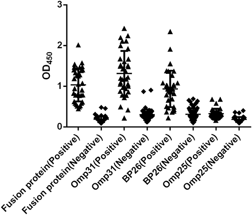 |
Figure 1 Dotplot of iELISAs using the fusion protein, Omp31, BP26 and Omp25 as antigens to detect canine brucellosis-positive and brucellosis-negative sera. |
The ROC curves were also obtained for these four antigens (Figure 2). The fusion protein had the largest area under the ROC curve (AUC=0.9991; 95% CI=0.9965 to 1.002), followed by Omp31 (AUC=0.9692; 95% CI=0.9215 to 1.017) and BP26 (AUC=0.9355; 95% CI=0.8824 to 0.9886). Omp25 showed the smallest area under the ROC curve (AUC=0.9099; 95% CI=0.8505 to 0.9692).
 |
Figure 2 ROC analysis of the fusion protein, Omp31, BP26 and Omp25 in detecting canine brucellosis. |
Based on the Youden index, the optimal cutoff value for fusion protein antigen was 0.4855, under which the sensitivity was 97.06% (95% CI=0.8467 to 0.9993) and the specificity was 100% (95% CI=0.9422 to 1.000). At the optimal cutoff value, 33 out of 34 positive samples were accurately diagnosed, and all negative samples were correctly identified. The positive predictive value (PPV) was 100%, and the negative predictive value (NPV) was 98.41%, making the fusion protein the best performing antigen in the serological diagnosis of canine brucellosis (Table 2). Correspondingly, the fusion protein antigen showed the highest accuracy (98.96%) in the serological detection of canine brucellosis. The accuracies of Omp31 and BP26 were 95.83% and 90.63%, respectively, while for Omp25, the accuracy was only 82.29%.
 |
Table 2 Predictive Values for Positive and Negative Calculated at Different Cutoff Values |
Cross-Reactivity with the Fusion Proteins Omp31, BP26 and Omp25
According to the S/N values (OD450, sample/negative) by iELISA, the fusion protein and BP26 showed no cross-reactivity to the rabbit sera immunized with Yersinia enterocolitica O9 and Escherichia coli O157, which are pathogens known to have cross-reactivity with S-type Brucella (Table 3). The fusion protein had slight cross-reactivity with Escherichia coli H7, Salmonella and Vibrio parahaemolyticus, but all the S/N values were below 2.1, displaying relatively good specificity. BP26 and Omp31 showed stronger cross-reactivity to sera infected with Listeria monocytogenes and Vibrio parahaemolyticus respectively, while Omp25 displayed the worst specificity among the four protein antigens.
 |
Table 3 Cross-Reaction of Rabbit Sera with Fusion Protein, Omp31, BP26 and Omp25 by iELISA |
Discussion
Canine brucellosis, caused primarily by B. canis, is currently endemic globally. In the southeastern USA, the seroprevalence rate of canine brucellosis has been estimated to be 7% to 8%.21 In European countries, a survey showed that 5.4% of dogs could be infected by B. canis.5 The disease has also been reported in African and Asian countries, such as Nigeria, Zimbabwe and Korea.22–24 In China, canine brucellosis was first reported in 1984, and subsequent epidemiological investigation showed that it was endemic in 20 provinces.25 Since its first identification in the 1960s, B. canis-caused human infection has aroused increasing concern regarding its zoonotic significance. Human cases reported in Japan and Argentina revealed that pet owners and laboratory workers handling Brucella cultures are the most susceptible population for B. canis infection.26–28 More recently, in the Netherlands, a patient was diagnosed with B. canis following exposure to infected dogs in a breeding facility, emphasizing that B. canis infection is an occupational disease.29 In China, a 45-year-old woman was diagnosed with B. canis infection in Zhejiang Province.9 This patient was initially diagnosed with pleurisy based on her clinical symptoms, which mainly included fever, back pain and fatigue. However, when B. canis was isolated from her blood, she was finally confirmed to be infected by Brucella. Thus, human brucellosis caused by B. canis may often be misdiagnosed due to the lack of knowledge about this pathogen and the lack of typical symptoms in patients. As there is a large number of pet dogs being raised worldwide and the prevalence of canine brucellosis is still unknown in many areas, an overall epidemiological investigation on canine brucellosis will be necessary and significant to human health.
The unknown prevalence of canine brucellosis, to a great extent, is due to the lack of accurate and commercially available testing reagents. RSAT and 2ME-RSAT using rough Brucella as an antigen were the first methods to detect canine brucellosis,30,31 and they are still popularly used worldwide.32,33 However, in China, there is no officially authorized RSAT antigen, and in-house prepared reagents are only available on a limited scale. Moreover, RSAT has poor specificity in detecting dog sera. Among the 1220 canine sera screened for this study, approximately 10% were positive, while using PCR and qPCR as confirmatory tests, the positive rate decreased to 2.78% (34/1440). In our study, RSAT has a strong cross-reactivity to rabbit serum immunized with Vibrio parahaemolyticus. Vibrio parahaemolyticus is a very popular foodborne pathogen in China.34 We isolated this pathogen from dog anal swabs, so we speculated that Vibrio parahaemolyticus was one of the main factors affecting the specificity of RSAT in detecting canine brucellosis. In addition to Vibrio parahaemolyticus, cross-reactivity was also discovered to Bordetella, Pseudomonas and Moraxella for RAST.10 Thus, RAST-positive dogs should be further tested with a more specific method.
PCR and qPCR have high sensitivity in detecting pathogen-specific nucleic acids. For canine brucellosis, PCR primer sets have been proposed to detect B. canis from blood, swab and tissue samples, including ITS66 and ITS279 targeting the 16–23S rRNA interspace region and BcSS-PCR primers targeting the B. canis species-specific fragment.35–37 In this study, we also applied these two sets of primers to determine Brucella infection. With Brucella genome DNA preserved in our laboratory, we found that the ITS66 and ITS279 primer set can amplify genome DNA of B. canis, B. melitensis, B. abortus and B. suis, while BcSS primers can identify genomes of B. canis and B. melitensis. However, dog genome fragments were often amplified with these two sets of PCR primers with similar product sizes to the targeting fragment, so all the PCR products had to be subjected to sequencing to rule out nonspecific amplification. In contrast, the qPCR used in this study demonstrated higher sensitivity and specificity, which easily ruled out all the nonspecific amplification and picked up all the positive samples confirmed by sequencing. The shortcoming of this qPCR assay is that it cannot distinguish B. canis from B. suis, although it can rule out B. melitensis and B. abortus.18 However, according to the relevant literature, no B. suis has ever been isolated from dogs. Meanwhile, the successfully isolated Brucella isolates from two Brucella-positive samples were identified as B. canis (Table S1), which also showed an MLVA16 profile (2-3-9-11-3-1-5-2-5-40-9-6-7-9-4-3) very similar to B. canis strains discovered in China.9,23 All these works demonstrate that all the positive control sera used in this study were very likely to be B. canis-infected. According to our testing data, the serological method of RAST may have good sensitivity for testing canine brucellosis, but this method lacks sufficient specificity. PCR and qPCR are very sensitive in detecting specific nucleic acid fragments and could be ideal testing methods, but the shedding of B. canis in infected dogs is fluctuating and elusive, which can lead to lower sensitivity than serological tests. Many antigens have been assessed for their potential use in serological detection for canine brucellosis.13,15,16 In this study, we also evaluated four protein antigens for their efficacy in the serological detection of canine brucellosis. According to the data, the B-cell multiepitope-based fusion protein displayed the highest accuracy, followed by Omp31 and BP26 (Table 2). The results indicated that protein antigens such as the fusion protein, Omp31 and BP26 can be used to detect not only smooth Brucella-caused brucellosis but also rough Brucella-caused brucellosis.20,38,39 Based on a cross-reactivity assay, the fusion protein and Omp31 displayed less reaction to other commonly observed pathogens; therefore, they are the best candidate antigens for serological kit development for canine brucellosis.
Conclusion
Canine brucellosis is currently a neglected zoonosis worldwide. As traditional RAST lacks sufficient sensitivity and specificity, more accurate serological methods are urgently needed. The data displayed here proved that the novel B-cell epitope-based fusion protein and Omp31 had better specificity than BP26 and Omp25 and provide sufficient sensitivity in serologically detecting canine sera. Thereafter, the fusion protein and Omp31 may be ideal antigens for developing a more accurate serological kit for canine brucellosis.
Ethics Approval
All experiments involving animal samples were fully compliant with ethical approval granted by the Animal Care and Ethics Committee of Xuzhou Medical University (approval no.: 201801W005). The methods were carried out in accordance with the Accreditation of Laboratory Animal Care International (AAALAC). The study was carried out in compliance with the ARRIVE guidelines.
Funding
This work was supported by the National Natural Science Foundation of China (Grant number 81802101) and General Administration of Customs, P.R. China (Grant number 2019HK125). The funders had no role in study design, data collection and analysis, decision to publish, or preparation of the manuscript.
Disclosure
The authors declare that they have no competing interests.
References
1. Godfroid J, Nielsen K, Saegerman C. Diagnosis of brucellosis in livestock and wildlife. Croat Med J. 2010;51(4):296–305. doi:10.3325/cmj.2010.51.296
2. Kauffman LK, Petersen CA. Canine brucellosis: old foe and reemerging scourge. Vet Clin North Am Small Anim Pract. 2019;49(4):763–779. doi:10.1016/j.cvsm.2019.02.013
3. Chinyoka S, Dhliwayo S, Marabini L, Dutlow K, Matope G, Pfukenyi DM. Serological survey of Brucella canis in dogs in urban Harare and selected rural communities in Zimbabwe. J S Afr Vet Assoc. 2014;85(1):e1–e5. doi:10.4102/jsava.v85i1.1087
4. Ayoola MC, Ogugua AJ, Akinseye VO, et al. Sero-epidemiological survey and risk factors associated with brucellosis in dogs in south-western Nigeria. Pan Afr Med J. 2016;23:29. doi:10.11604/pamj.2016.23.29.7794
5. Buhmann G, Paul F, Herbst W, et al. Canine brucellosis: insights into the epidemiologic situation in Europe. Front Vet Sci. 2019;6:151.
6. Jamil T, Melzer F, Khan I, et al. Serological and molecular investigation of brucella species in dogs in Pakistan. Pathogens. 2019;8(4):294. doi:10.3390/pathogens8040294
7. Qi H, Zhang H, Deng X, Zhang X, Cao Y. Dog and cat brucellosis epidemiology. Beijing Agr. 2012;18:98–99.
8. Wang T, Zhang Y, Wang L, et al. Epidemiological investigation of canine brucellosis in Urumqi City of Xinjiang. China Animal Health Inspection. 2018;35(6):8–11.
9. Piao D, Wang H, Di D, et al. MLVA and LPS characteristics of brucella canis isolated from humans and dogs in Zhejiang, China. Front Vet Sci. 2017;4:223. doi:10.3389/fvets.2017.00223
10. Hollett RB. Canine brucellosis: outbreaks and compliance. Theriogenology. 2006;66(3):575–587. doi:10.1016/j.theriogenology.2006.04.011
11. Keid LB, Soares RM, Vieira NR, et al. Diagnosis of canine brucellosis: comparison between serological and microbiological tests and a PCR based on primers to 16S-23S rDNA interspacer. Vet Res Commun. 2007;31(8):951–965. doi:10.1007/s11259-006-0109-6
12. Kang SI, Lee SE, Kim JY, et al. A new Brucella canis species-specific PCR assay for the diagnosis of canine brucellosis. Comp Immunol Microbiol Infect Dis. 2014;37(4):237–241. doi:10.1016/j.cimid.2014.07.003
13. Baldi PC, Wanke MM, Loza ME, Fossati CA. Brucella abortus cytoplasmic proteins used as antigens in an ELISA potentially useful for the diagnosis of canine brucellosis. Vet Microbiol. 1994;41(1–2):127–134. doi:10.1016/0378-1135(94)90142-2
14. Wanke MM, Delpino MV, Baldi PC. Comparative performance of tests using cytosolic or outer membrane antigens of Brucella for the serodiagnosis of canine brucellosis. Vet Microbiol. 2002;88(4):367–375. doi:10.1016/S0378-1135(02)00152-9
15. Barrouin-Melo SM, Poester FP, Ribeiro MB, et al. Diagnosis of canine brucellosis by ELISA using an antigen obtained from wild Brucella canis. Res Vet Sci. 2007;83(3):340–346. doi:10.1016/j.rvsc.2007.02.006
16. Tsogtbaatar G, Tachibana M, Watanabe K, Kim S, Suzuki H, Watarai M. Enzyme-linked immunosorbent assay for screening of canine brucellosis using recombinant Cu-Zn superoxide dismutase. J Vet Med Sci. 2008;70(12):1387–1389. doi:10.1292/jvms.70.1387
17. Yin D, Bai Q, Wu X, et al. A multi-epitope fusion protein-based p-ELISA method for diagnosing bovine and goat brucellosis. Front Vet Sci. 2021;8:708008. doi:10.3389/fvets.2021.708008
18. Hinic V, Brodard I, Thomann A, et al. Novel identification and differentiation of Brucella melitensis, B. abortus, B. suis, B. ovis, B. canis, and B. neotomae suitable for both conventional and real-time PCR systems. J Microbiol Methods. 2008;75(2):375–378. doi:10.1016/j.mimet.2008.07.002
19. Yin D, Bai Q, Wu X, et al. Paper-based ELISA diagnosis technology for human brucellosis based on a multiepitope fusion protein. PLoS Negl Trop Dis. 2021;15(8):e0009695. doi:10.1371/journal.pntd.0009695
20. Bai Q, Li H, Wu X, Shao J, Sun M, Yin D. Comparative analysis of the main outer membrane proteins of Brucella in the diagnosis of brucellosis. Biochem Biophys Res Commun. 2021;560:126–131. doi:10.1016/j.bbrc.2021.04.127
21. Cosford KL. Brucella canis: an update on research and clinical management. Canad Vet j. 2018;59(1):74–81.
22. Cadmus SI, Adesokan HK, Ajala OO, Odetokun WO, Perrett LL, Stack JA. Seroprevalence of Brucella abortus and B. canis in household dogs in southwestern Nigeria: a preliminary report. J S Afr Vet Assoc. 2011;82(1):56–57. doi:10.4102/jsava.v82i1.35
23. Ledwaba MB, Gomo C, Lekota KE, et al. Molecular characterization of Brucella species from Zimbabwe. PLoS Negl Trop Dis. 2019;13(5):e0007311. doi:10.1371/journal.pntd.0007311
24. Kang SI, Heo EJ, Cho D, et al. Genetic comparison of Brucella canis isolates by the MLVA assay in South Korea. J veterinar med sci. 2011;73(6):779–786. doi:10.1292/jvms.10-0334
25. Shang DQ. Investigation of B. canis infection in China. Zhonghua Liu Xing Bing Xue Za Zhi. 1989;10(1):24–29.
26. Wallach JC, Giambartolomei GH, Baldi PC, Fossati CA. Human infection with M-strain of Brucella canis. Emerg Infect Dis. 2004;10(1):146–148. doi:10.3201/eid1001.020622
27. Lucero NE, Corazza R, Almuzara MN, et al. Human Brucella canis outbreak linked to infection in dogs. Epidemiol Infect. 2010;138(2):280–285. doi:10.1017/S0950268809990525
28. Nomura A, Imaoka K, Imanishi H, et al. Human Brucella canis infections diagnosed by blood culture. Emerg Infect Dis. 2010;16(7):1183–1185. doi:10.3201/eid1607.090209
29. Kolwijck E, Lutgens SP, Visser VX, et al. First case of human Brucella canis infection in the Netherlands. Clin Infect Dis. 2022. doi:10.1093/cid/ciac425
30. George LW, Carmichael LE. A plate agglutination test for the rapid diagnosis of canine brucellosis. Am J Vet Res. 1974;35(7):905–909.
31. Badakhsh FF, Carmichael LE, Douglass JA. Improved rapid slide agglutination test for presumptive diagnosis of canine brucellosis. J Clin Microbiol. 1982;15(2):286–289. doi:10.1128/jcm.15.2.286-289.1982
32. Johnson CA, Carter TD, Dunn JR, et al. Investigation and characterization of Brucella canis infections in pet-quality dogs and associated human exposures during a 2007–2016 outbreak in Michigan. J Am Vet Med Assoc. 2018;253(3):322–336. doi:10.2460/javma.253.3.322
33. Whitten TV, Brayshaw G, Patnayak D, et al. Seroprevalence of Brucella canis antibodies in dogs entering a Minnesota humane society, Minnesota, 2016–2017. Prev Vet Med. 2019;168:90–94. doi:10.1016/j.prevetmed.2019.04.015
34. Xie T, Yu Q, Tang X, Zhao J, He X. Prevalence, antibiotic susceptibility and characterization of Vibrio parahaemolyticus isolates in China. FEMS Microbiol Lett. 2020;367(16):fnaa136. doi:10.1093/femsle/fnaa136
35. Keid LB, Soares RM, Vasconcellos SA, et al. A polymerase chain reaction for detection of Brucella canis in vaginal swabs of naturally infected bitches. Theriogenology. 2007;68(9):1260–1270. doi:10.1016/j.theriogenology.2007.08.021
36. Aras Z, Uçan US. Detection of Brucella canis from inguinal lymph nodes of naturally infected dogs by PCR. Theriogenology. 2010;74(4):658–662. doi:10.1016/j.theriogenology.2010.03.023
37. Batinga MCA, Dos Santos JC, Lima JTR, et al. Comparison of three methods for recovery of Brucella canis DNA from canine blood samples. J Microbiol Methods. 2017;143:26–31. doi:10.1016/j.mimet.2017.08.019
38. Cassataro J, Pasquevich K, Bruno L, Wallach JC, Fossati CA, Baldi PC. Antibody reactivity to Omp31 from Brucella melitensis in human and animal infections by smooth and rough Brucellae. Clin Diagn Lab Immunol. 2004;11(1):111–114. doi:10.1128/cdli.11.1.111-114.2004
39. Chaudhuri P, Prasad R, Kumar V, Gangaplara A. Recombinant OMP28 antigen-based indirect ELISA for serodiagnosis of bovine brucellosis. Mol Cell Probes. 2010;24(3):142–145. doi:10.1016/j.mcp.2009.12.002
 © 2022 The Author(s). This work is published and licensed by Dove Medical Press Limited. The
full terms of this license are available at https://www.dovepress.com/terms
and incorporate the Creative Commons Attribution
- Non Commercial (unported, 3.0) License.
By accessing the work you hereby accept the Terms. Non-commercial uses of the work are permitted
without any further permission from Dove Medical Press Limited, provided the work is properly
attributed. For permission for commercial use of this work, please see paragraphs 4.2 and 5 of our Terms.
© 2022 The Author(s). This work is published and licensed by Dove Medical Press Limited. The
full terms of this license are available at https://www.dovepress.com/terms
and incorporate the Creative Commons Attribution
- Non Commercial (unported, 3.0) License.
By accessing the work you hereby accept the Terms. Non-commercial uses of the work are permitted
without any further permission from Dove Medical Press Limited, provided the work is properly
attributed. For permission for commercial use of this work, please see paragraphs 4.2 and 5 of our Terms.