Back to Journals » International Journal of General Medicine » Volume 16
Comparing Diagnostic Efficacy of C-TIRADS Positive Features on Different Sizes of Thyroid Nodules
Authors Zhou Y ![]() , Li WM
, Li WM ![]() , Fan XF, Huang YL, Gao Q
, Fan XF, Huang YL, Gao Q
Received 28 April 2023
Accepted for publication 25 July 2023
Published 14 August 2023 Volume 2023:16 Pages 3483—3490
DOI https://doi.org/10.2147/IJGM.S416403
Checked for plagiarism Yes
Review by Single anonymous peer review
Peer reviewer comments 2
Editor who approved publication: Dr Scott Fraser
Yue Zhou,1,* Wei-Min Li,1,* Xiao-Fang Fan,1 Yan-Li Huang,2 Qi Gao3
1Department of Ultrasonography, Affiliated Hospital of Jiangnan University, Wuxi, Jiangsu, People’s Republic of China; 2Department of Special Clinic, General Hospital of Eastern Theater Command, PLA, Nanjing, Jiangsu, People’s Republic of China; 3Department of Ultrasonography, Zhongda Hospital Affiliated to Southeast University, Nanjing, Jiangsu, People’s Republic of China
*These authors contributed equally to this work
Correspondence: Xiao-Fang Fan, Department of Ultrasonography, Affiliated Hospital of Jiangnan University, Wuxi, Jiangsu, 214000, People’s Republic of China, Tel +8613861843829, Email [email protected]
Purpose: To explore the diagnostic value of positive features in the Chinese Thyroid Imaging Reporting and Data System (C-TIRADS) for thyroid nodules of different sizes.
Patients and Methods: A total of 1864 patients with 2347 thyroid nodules were selected from January 2021 to December 2022 and assessed according to C-TIRADS. According to the maximum diameter, nodules were divided into the A1 group (≤ 10 mm), A2 group (> 10 mm,< 20 mm), and A3 group (≥ 20 mm). With surgical pathology as the golden standard, the receiver operating characteristic curves (ROC) were constructed, and each group’s area under the curve (AUC) was calculated. The diagnostic value of positive features in C-TIRADS for different sizes of thyroid nodules was analyzed.
Results: In all groups, malignant thyroid nodules had a higher incidence of positive features than benign nodules (P < 0.05). In A1 group, the diagnostic efficiency of C-TIRADS positive features for thyroid nodules was vertical orientation> ill-defined/irregular margin or extrathyroidal extension> solid composition> markedly hypoechoic> microcalcifications. The AUCs were 0.718, 0.675, 0.609, 0.558, and 0.581, respectively. In A2 group, the diagnostic efficacy of each positive features for thyroid nodules was ill-defined/irregular margins or extra-thyroid invasion> solid composition> microcalcifications> markedly hypoechoic> vertical orientation. The AUCs were 0.854, 0.730, 0.719, 0.670, and 0.609, respectively. In A3 group, the diagnostic efficacy of each positive features for thyroid nodules was ill-defined/irregular margin or extrathyroidal extension> microcalcifications> solid composition> vertical orientation> markedly hypoechoic. The AUCs were 0.847, 0.778, 0.767, 0.584, and 0.560, respectively.
Conclusion: C-TIRADS positive features exhibited different diagnostic efficacy for thyroid nodules of various sizes, especially for thyroid nodules ≤ 10 mm, for which all positive features had low diagnostic efficacy.
Keywords: ultrasound, C-TIRADS, sizes, thyroid nodule
Graphical Abstract:

Thyroid nodules are one of the most prevalent endocrine system diseases. Studies have shown that about 70% of the population worldwide have thyroid nodules, most benign and malignant nodules account for approximately 10%-15%.1–3 Ultrasound imaging is simple and rapid with high resolution. Its value in detecting and evaluating thyroid nodules has gradually been recognized in clinical practice and has become the preferred examination method. However, varied sizes of thyroid nodules have distinct ultrasound assessment outcomes, which affects the diagnostic efficacy in some way.4,5
Our study analyzed ultrasonic features of thyroid nodules according to the Chinese Thyroid Imaging Reporting and Data System (C-TIRADS) and investigated the diagnostic efficacy of each positive index for different sizes of thyroid nodules.
Materials and Methods
General Information
A total of 1864 patients with 2347 nodules were prospectively selected. The patients were treated in the Affiliated Hospital of Jiangnan University, Eastern Theater General Hospital (formerly Bayi Hospital), and Zhongda Hospital Affiliated to Southeast University, from January 2021 to December 2022, confirmed by surgical pathology. There were 567 males and 1297 females, aged 15–83 years, with an average of 51.07±13.20 years. The maximum nodule diameter was 4–83 mm, averaging 12.65 ± 12.14 mm. The primary surgical indications are as follows: 1) Confirmed or suspected thyroid malignancy based on FNAB; 2) No definite FANB results but highly suspicious nodules on ultrasound for thyroid nodules >1 cm; 3) Nodules ≤10 mm with undefined fine-needle aspiration results but nodule C-TIRADS category ≥4 and the patient was very anxious and demanded surgical treatment, difficult to follow up; 4) Large-size nodule compressing surrounding tissues.
Apparatus and Methods
SonoScape S60 color Doppler ultrasound imaging system was used, and a linear array probe was selected, with a probe frequency of 7.8–15 MHz. During the examination, the patient was supine, with the bilateral neck fully exposed. The thyroid gland was scanned by combining transverse and longitudinal sections and observed from multiple sections and angles. The nodules were evaluated according to the C-TIRADS grading guidelines, including location, orientation, margin, acoustic halo, structure, echo, focal hyperechogenicity, posterior echo, size, etc. Two attending sonographers with more than 10 years of working experience and above qualifications evaluated all cases jointly. One chief physician reviewed inconsistent results. According to the maximum diameter of thyroid nodules, patients were classified into the A1 (≤10 mm), A2 (>10 mm, <20 mm), and A3 groups (≥20 mm). Ultrasound imaging features of thyroid nodules in different groups were evaluated with C-TIRADS, scored, and categorized.
Evaluation of C-TIRADS Positive Features
According to the C-TIRADS classification system, vertical orientation, solid composition, markedly hypoechoic, microcalcifications, and ill-defined/irregular margin or extrathyroidal extension were regarded as positive features.6 Vertical orientation denotes that the long axis of the nodule and the skin tend to be perpendicular when evaluating the transverse or longitudinal section, and the anterior-posterior diameter of the nodule is greater than the left-right or upper-inferior diameter. A solid nodule is composed entirely of solid tissue and contains no cystic components. Markedly hypoechoic represents that nodules have lower echogenicity than strap neck muscles. Microcalcification refers to the presence of < 1 mm punctate strong echo in the nodule, and acoustic shadow may or may not appear in the rear. Nodule margin assessment depends on clarity and regularity. Irregular margins are spiculated, angular, or micro-lobulated; ill-defined margins denote that the border of the nodule is difficult to distinguish from the surrounding thyroid parenchyma; extra-thyroidal invasion represents nodules invading the thyroid capsule, damaging the capsule. Adjacent soft tissue and/or vessels are invaded in severe cases.
Statistical Analysis
SPSS27.0 and MedCalc19.3.1 were used. Measurement data were expressed as mean ± SD. An independent sample t-test was used for comparison between two groups. Count data were expressed as frequency and percentage, and groups were analyzed using the χ2 test and Fisher’s exact probability method. Linear trends of groups were compared using the χ2 test for trend. According to surgical pathology, a receiver operating characteristic curve (ROC) was plotted. The area under the curve (AUC) and the Youden index were calculated. An AUC of 0.85–0.95 indicates good diagnostic efficacy; an AUC of 0.70–0.85 indicates moderate diagnostic efficacy; an AUC of 0.50–0.70 denotes low diagnostic efficacy. The highest Youden index represents the best cutoff scores in each group, and P<0.05 refers to statistical significance.
Results
Pathological results
According to the 2020 fifth edition of WHO Classification of Endocrine Neoplasms (Thyroid Neoplasms).7,8 A total of 2347 thyroid nodules were included in this study, including 731 benign cases (31.15%), 13 low-risk cases (0.56%), and 1603 malignant cases (68.30%). The pathological outcomes of thyroid nodules of different groups are as follows (Table 1).
 |
Table 1 Pathological Results of Thyroid Nodules of Each Group |
Ultrasound Feature Analysis
For statistical analysis, this study classified low-risk thyroid nodules as malignant. The sonographic features of thyroid nodules of different groups are as follows (Table 2). Significant differences were observed between groups (P < 0.0001). The representative images of thyroid nodules of each group are showed in Figure 1A–I.
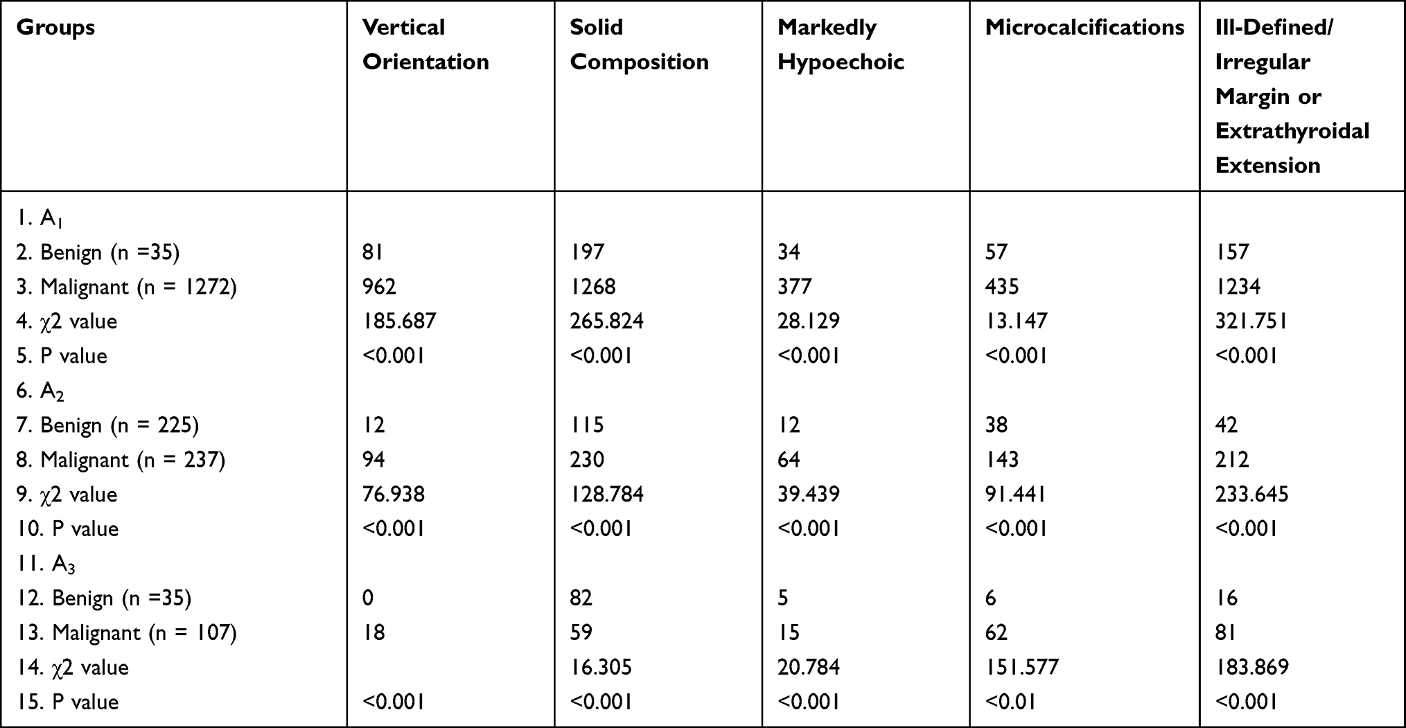 |
Table 2 C-TIRADS Positive Features in Benign and Malignant Thyroid Nodules of Each Group |
 |
Figure 1 Typical ultrasonic imaging of thyroid nodules in each groups and ROC curves of each group. Longitudinal section (A) and transverse section (B) showed a left thyroid nodule ≤10 mm, postoperative pathology showing papillary thyroid microcarcinoma (C). Longitudinal section (D) and transverse section (E) showed a right thyroid nodule >10 mm, <20 mm, postoperative pathology showing papillary thyroid microcarcinoma (F). Longitudinal section (G) and transverse section (H) showed a left thyroid nodule ≥20 mm, postoperative pathology showing follicular adenoma (I). ROC curve of A1 group (J). ROC curve of A2 group (K). ROC curve of A3 group (L). |
Diagnostic Efficacy of C-TIRADS Positive Features for Thyroid Nodules
The diagnostic efficacy of positive features is shown in Table 3. Except for the PPV of vertical orientation and NPV of solid composition, significant differences were seen in other features between groups (all P values< 0.05). Meanwhile, the sensitivity and PPV of ill-defined/irregular margin or extrathyroidal extension, markedly hypoechoic, and solid composition for thyroid cancer decreased linearly with the increase of nodule size (all P values <0.05). The specificity and NPV of vertical orientations, ill-defined/irregular margin or extrathyroidal extension, and markedly hypoechoic increased linearly (all P values <0.05). Microcalcifications showed linearly increased sensitivity, specificity, and NPV for thyroid carcinoma with increasing nodule size (all P values <0.05).
 |
Table 3 The Diagnostic Efficacy of Different Positive Features in Each Groups |
ROC Curves of Thyroid Nodule Score of Each Group
The ROC curve (Figure 1J–L) was drawn according to the benign and malignant nature of nodules and their scores. The results showed that the diagnostic efficacy of each positive index in the A1 group for thyroid nodules was vertical orientation> ill-defined/irregular margin or extrathyroidal extension> solid composition> markedly hypoechoic> microcalcifications. The AUCs were 0.718 (95% CI: 0.695–0.741), 0.675 (95% CI: 0.651–0.698), 0.609 (95% CI: 0.584–0.634), 0.581 (95% CI: 0.556–0.606), and 0.558 (95% CI: 0.533–0.583). Except for the difference between markedly hypoechoic and microcalcifications (Z = 1.690, P = 0.0910), the differences between other positive features were statistically significant (all P < 0.05). In the A2 group, the diagnostic efficacy of each positive index for thyroid nodules was ill-defined/irregular margins or extra-thyroid invasion> solid composition> microcalcifications> markedly hypoechoic> vertical orientation. The AUCs were 0.854 (95% CI: 0.818–0.885), 0.730 (95% CI:0.687–0.770), 0.719 (95% CI: 0.675–0.759), 0.670 (95% CI: 0.625–0.713), 0.609 (95% CI: 0.563–0.654), respectively. Except for the difference between solid composition and microcalcifications (Z = 1.928, P = 0.0538), differences between other positive indicators were statistically significant (all P < 0.05). In the A3 group, the diagnostic efficacy of each positive index for thyroid nodules was: ill-defined/irregular margin or extrathyroidal extension> microcalcifications> solid composition> vertical orientation> markedly hypoechoic. The AUCs were 0.847 (95% CI: 0.805–0.882), 0.778 (95% CI: 0.731–0.820), 0.767 (95% CI: 0.719–0.809), 0.584 (95% CI: 0.531–0.636), and 0.560 (95% CI: 0.507–0.612), respectively. Except for the difference between solid composition and microcalcifications, markedly hypoechoic and vertical orientation were not significant (Z values were 1.207 and 0.351, and P values were 0.2274 and 0.7253), the differences between other positive features were statistically significant (all P < 0.05).
Discussion
TI-RADS is a thyroid classification system based on breast imaging reports and the management system. It established a standard imaging dictionary and performed standardized risk classification for thyroid nodule malignancy, which evades the influence of the sonographer’s subjectivity. Currently, there are multiple TI-RADS classification systems worldwide. The TI-RADS system devised by the American College of Radiology (ACR) is the most influential. However, the ACR-TIRADS classification system aims to guide fine-needle aspiration, and categories 4 and 5 have relatively low malignancy rates, inconsistent with the current domestic clinical situation.9 Thus, Professor Zhou Jianqiao et al developed a C-TIRDS classification system according to Chinese reality. Vertical orientation, solid composition, markedly hypoechoic, microcalcifications, and ill-defined/irregular margin or extrathyroidal extension are selected as positive features.6 This system has been widely applied in China and is extensively acclaimed due to its convenient and rapid assessment of thyroid nodules. Because cytologic evaluation remains the diagnostic test of choice to distinguish benign from malignant thyroid nodules yet fails to discriminate as benign or malignant in up to one-third of cases. Although cytology is one of many clinical factors that inform decision making with regard to the management of thyroid nodules, additional risk factors to consider include: family history of thyroid malignancy, history of radiation exposure, nodule size, sonographic risk assessment, patient symptoms, and thyroid function. Diagnostic test accuracy must also be considered, with evaluation of test sensitivity, specificity, and the underlying disease prevalence in the population.10 So in this study, surgical pathology were treated as the golden standard. The ultrasound features of 2347 thyroid nodules were analyzed, showing that positive features had distinct diagnostic efficacy on varying sizes of thyroid nodules. All positive features had relatively low diagnostic efficacy on thyroid nodules ≤10 mm.
For thyroid nodules ≤10 mm, the diagnostic efficacy of positive features in C-TIRADS from high to low was vertical orientation> ill-defined/irregular or extra-thyroidal invasion> solid composition> markedly hypoechoic> microcalcifications, among which vertical orientation had medium diagnostic efficacy, and the rest had relatively low efficacy. Via analysis, the pathology of malignant thyroid nodules ≤10 mm indicated PTMC as the dominant subtype. Research about PTMC has gradually matured. Its typical characteristics are all incorporated in the C-TIRADS classification system, including solid composition, ill-defined margins, vertical orientation, markedly hypoechoic, microcalcification, etc. Moreover, ultrasound has elevated diagnostic accuracy as exploration deepens. Nevertheless, for benign nodules in the A1 group, chronic lymphocytic thyroiditis and granulomatous thyroiditis overlap considerably with thyroid microcarcinoma in sonographic features.11–13 Additionally, it is tricky to differentiate follicular adenoma from follicular carcinoma using sonography.14 Most nodular goiters have typical ultrasound features like cystic-solid, clear border, and transverse diameter greater than longitudinal diameter.15 However, nodular goiters in this study were small, and their characteristics were found to be similar to PTMC for the first time. These nodules were mostly benign and degenerative, primarily caused by hemorrhage, degeneration, infarction, and fibrosis. During hematoma absorption, degenerated nodules gradually became irregular and exhibited malignant sonographic features as nodules gradually shrank.16,17 When nodular goiters are accompanied by interstitial collagenization, calcification, and crystal formation, partial nodular goiters can display punctate strong echo with extended time. However, it is thorny to discriminate between solid thyroid micronodules and microcalcification using sonography, which affects sonographic evaluations of thyroid nodules. This may be why C-TIRADS positive features had low diagnostic efficacy on thyroid nodules ≤10 mm.
With increased nodule size, all positive features have changed diagnostic efficacy on thyroid nodules. In the A2 group, the diagnostic efficacy of each positive index for thyroid nodules from high to low: ill-defined/irregular margins or extra-thyroid invasion> solid composition> microcalcifications> markedly hypoechoic> vertical orientation. The A3 group: ill-defined/irregular margin or extrathyroidal extension> microcalcifications> solid composition> vertical orientation> markedly hypoechoic. Compared with thyroid nodules ≤10 mm, the diagnostic efficacy of all positive markers increased except for changed order and reduced diagnostic efficacy of vertical orientation. We consider the reason was that benign and malignant thyroid nodules have significant distinct sonographic features. Benign thyroid nodules of larger size are mainly nodular goiters. Nodular goiter and follicular adenoma are more common among benign thyroid nodules, and larger nodular goiter and follicular adenoma have a higher chance of developing cystic change.18,19 This reduced the scores of benign thyroid nodules. Although partial papillary thyroid cancer cases could also be accompanied by cystic alterations, the incidence of other positive features was significantly higher than benign nodules, including ill-defined margins, microcalcification, etc. Meanwhile, follicular adenoma in benign nodules often coexists with the surrounding acoustic halo, which is conducive to ultrasound assessment.18 Based on this, larger thyroid nodule size has a lower possibility of overlapping sonographic features between malignant and benign nodules, which facilitates further analysis of nodules with ultrasound imaging. This is why C-TIRADS positive features had higher diagnostic efficacy on thyroid nodules of larger sizes.
There are some limitations to this study. Although this study followed strict surgical standards, thyroid nodules ≤10 mm can choose to be followed up without biopsy. In this study, considerable thyroid nodules were ≤10 mm and underwent biopsy and surgery, which might lead to excessive treatment for thyroid microcarcinoma.
Conclusion
C-TIRADS positive features have varying diagnostic efficacy on thyroid nodules of different sizes. They had relatively low diagnostic efficacy for thyroid nodules ≤10 mm, which may increase the misdiagnosis rate of thyroid micronodules, thereby increasing overdiagnosis and overtreatment.
Ethical Statement
The authors are accountable for all aspects of the work in ensuring that questions related to the accuracy or integrity of any part of the work are appropriately investigated and resolved. All procedures performed in this study involving human participants were in accordance with the Declaration of Helsinki (as revised in 2013). The study was approved by the Ethics Committee of Affiliated Hospital of Jiangnan University. Individual consent for this retrospective analysis was waived. For the patient’s information provided by the Affiliated Hospital of Jiangnan University during the treatment period due to illness, such as name, age, gender, occupation, address, ID card, related diseases and treatment plan. Due to the privacy of patients, the Affiliated Hospital of Jiangnan University keeps the above information confidential.
Funding
Wuxi Maternal and Child Health Promotion Project (FYTG202103).
Disclosure
The authors declare that they have no conflicts of interest in this work.
References
1. Hyun MK, Kim JH, Kwon JW. Incidence of thyroid cancer and medical cost among patients with newly diagnosed thyroid nodules in Korea: a retrospective cohort study using nationwide data. J Cancer Res Ther. 2019;15:676–680. doi:10.4103/0973-1482.204895
2. Wang J, Yu F, Shang Y, et al. Thyroid cancer: incidence and mortality trends in China, 2005-2015. Endocrine. 2020;68:163–173. doi:10.1007/s12020-020-02207-6
3. Pizzato M, Li M, Vignat J, et al. The epidemiological landscape of thyroid cancer worldwide: GLOBOCAN estimates for incidence and mortality rates in 2020. Lancet Diabetes Endo. 2022;10:264–272. doi:10.1016/S2213-8587(22)00035-3
4. Chen X, Gao M, Hu L, et al. The diagnostic value of the ultrasound gray scale ratio for different sizes of thyroid nodules. Cancer Med. 2019;8:7644–7649. doi:10.1002/cam4.2653
5. Zhang S, Huang L, Huang Q, et al. The value of relative size in the ultrasound diagnosis of follicular thyroid neoplasm. Int J Gen Med. 2021;14:2321–2328. doi:10.2147/IJGM.S313468
6. Zhou J, Yin L, Wei X, et al. 2020 Chinese guidelines for ultrasound malignancy risk stratification of thyroid nodules: the C-TIRADS. Endocrine. 2020;70:256–279. doi:10.1007/s12020-020-02441-y
7. Mete O, Wenig BM. Update from the 5th edition of the world health organization classification of head and neck tumors: overview of the 2022 WHO classification of head and neck neuroendocrine neoplasms. Head Neck Pathol. 2022;16:123–142. doi:10.1007/s12105-022-01435-8
8. Baloch ZW, Asa SL, Barletta JA, et al. Overview of the 2022 WHO classification of thyroid neoplasms. Endocr Pathol. 2022;33:27–63. doi:10.1007/s12022-022-09707-3
9. Tessler FN, Middleton WD, Grant EG, et al. ACR thyroid imaging, reporting and data system (TI-RADS): white paper of the ACR TI-RADS committee. J Am Coll Radiol. 2017;14:587–595. doi:10.1016/j.jacr.2017.01.046
10. Patel SG, Carty SE, Lee AJ. Molecular testing for thyroid nodules including its interpretation and use in clinical practice. Ann Surg Oncol. 2021;28:8884–8891. doi:10.1245/s10434-021-10307-4
11. Zhang Q, Liao L, Peng Q, et al. Value of contrast-enhanced ultrasound in differentiating clinically atypical subacute thyroiditis from papillary thyroid carcinomas. Ultrasound Med Biol. 2021;47:3384–3392. doi:10.1016/j.ultrasmedbio.2021.09.001
12. Yang L, Zhao H, He Y, et al. Contrast-enhanced ultrasound in the differential diagnosis of primary thyroid lymphoma and nodular hashimoto’s thyroiditis in a background of heterogeneous parenchyma. Front Oncol. 2021;7:597–603.
13. Zhao W, Kang Q, Qian F, et al. Convolutional neural network-based computer-assisted diagnosis of hashimoto’s thyroiditis on ultrasound. J Clin Endocrinol Metab. 2022;107:953–963. doi:10.1210/clinem/dgab870
14. Liu R, Gao L, Xia Y, et al. Three ultrasound phenotypes of non-invasive follicular thyroid neoplasm with papillary-like nuclear features proposed for imaging-pathology analysis: single center experience. Gland Surg. 2021;10:307–318. doi:10.21037/gs-20-612
15. Holzer K, Bartsch DK. Struma nodosa [Nodular goiter]. Chirurg. 2020;91:712–719. doi:10.1007/s00104-020-01218-3
16. Yan Y, Zhang F, Ge H, et al. Effect of the size of benign thyroid degenerative nodules on ACR TI-RADS categories. J Med Ultrasound. 2022;49:71–76. doi:10.1007/s10396-021-01163-6
17. Huang QX, Huang XW. Comments on”Effect of the size of benign thyroid degenerative nodules on ACR TI-RADS categories. J Med Ultrasound. 2022;49:503. doi:10.1007/s10396-022-01214-6
18. Peng Y, Zhou W, Zhan WW, et al. Ultrasonographic assessment of differential diagnosis between degenerating cystic thyroid nodules and papillary thyroid microcarcinomas. World J Surg. 2017;41:2538–2544. doi:10.1007/s00268-017-4060-1
19. Xu B, Ghossein RA. Noninvasive follicular thyroid neoplasm with papillary-like nuclear features (NIFTP): an update. Head Neck Pathol. 2020;14:303–310. doi:10.1007/s12105-019-01124-z
 © 2023 The Author(s). This work is published and licensed by Dove Medical Press Limited. The
full terms of this license are available at https://www.dovepress.com/terms
and incorporate the Creative Commons Attribution
- Non Commercial (unported, 3.0) License.
By accessing the work you hereby accept the Terms. Non-commercial uses of the work are permitted
without any further permission from Dove Medical Press Limited, provided the work is properly
attributed. For permission for commercial use of this work, please see paragraphs 4.2 and 5 of our Terms.
© 2023 The Author(s). This work is published and licensed by Dove Medical Press Limited. The
full terms of this license are available at https://www.dovepress.com/terms
and incorporate the Creative Commons Attribution
- Non Commercial (unported, 3.0) License.
By accessing the work you hereby accept the Terms. Non-commercial uses of the work are permitted
without any further permission from Dove Medical Press Limited, provided the work is properly
attributed. For permission for commercial use of this work, please see paragraphs 4.2 and 5 of our Terms.
Recommended articles
Comparison of Diagnostic Values of ACR TI-RADS versus C-TIRADS Scoring and Classification Systems for the Elderly Thyroid Cancers
Huang H, Zhu MJ, Gao Q, Huang YL, Li WM
International Journal of General Medicine 2023, 16:4441-4451
Published Date: 29 September 2023
The Impact of Echogenicity Grading on the Diagnostic Performance of C-TIRADS
Huang GL, Su JY, Liang RQ, Yang XY, Xu LF
International Journal of General Medicine 2025, 18:5509-5517
Published Date: 15 September 2025
Diagnostic Performance of the ACR TI-RADS, C-TIRADS, K-TIRADS, and EU TI-RADS Classification Systems for Thyroid Micronodules: A Multicenter Prospective Study
Zhang JC, Feng HL, Li QY, Li HJ, Li WM
International Journal of General Medicine 2026, 19:610763
Published Date: 10 July 2026