
Back to Journals » Journal of Inflammation Research » Volume 19
Comparative Effects of Anti-Inflammatory Therapies on Olfactory Bulb Histopathology and Olfactory Function in a Rat Model of Experimental Allergic Rhinitis

Received 31 October 2025
Accepted for publication 17 January 2026
Published 10 February 2026 Volume 2026:19 575552
DOI https://doi.org/10.2147/JIR.S575552
Checked for plagiarism Yes
Review by Single anonymous peer review
Peer reviewer comments 3
Editor who approved publication: Dr Tara Strutt
Engin Kayıkçı,1 Hazal Kayıkçı2
1Ministry of Health, Etlik City Hospital, Department of Neurosurgery, Ankara, Turkiye; 2Division of Allergy and Clinical Immunology, Hacettepe University, Ankara, Turkiye
Correspondence: Hazal Kayıkçı, Faculty of Medicine, Division of Allergy and Clinical Immunology, Hacettepe University, Ankara, Turkiye, Tel/Fax +90 5303449286, Email [email protected]
Purpose: This study aimed to evaluate histopathological changes in the olfactory bulb and nasal mucosa, as well as olfactory outcomes, following treatment modalities in an rat model of experimental AR.
Methods: This experimental study included 49 female Sprague–Dawley rats, randomly allocated into seven groups (n = 7 per group). Allergic rhinitis was induced by ovalbumin sensitization and intranasal challenge. Study groups included negative and positive controls, and treatment groups receiving methylprednisolone, montelukast, levocetirizine, olopatadine, or fluticasone propionate between days 21 and 34. The primary outcome was histopathological evaluation of inflammatory and structural changes in the nasal mucosa and olfactory bulb at day 34. The secondary outcome was assessment of olfactory function using the food-finding latency test.
Results: All animals completed the study without mortality. Compared with the positive control group, all treatment modalities resulted in significant improvement in nasal mucosal histopathology, including vascular dilatation, goblet cell hyperplasia, inflammatory cell infiltration, plasma cell infiltration, mast cell infiltration, eosinophil infiltration, chondrocyte hypertrophy, and ciliary loss. Similarly, significant improvements were observed in most olfactory bulb parameters, including glomerular layer thickness, glomerular diameter, glomerular organization, neuronal degeneration, vascular congestion, perivascular edema, and microglial activation. No significant superiority was observed among treatment groups in histopathological outcomes. In olfactory testing, a significant reduction in food-finding latency at day 28 compared with day 21 was observed only in the methylprednisolone and olopatadine groups (p = 0.038 and p = 0.027). By day 34, significant reductions in food-finding latency compared with day 21 were observed within all treatment groups (p < 0.001 for methylprednisolone, olopatadine, and fluticasone propionate; p = 0.005 for levocetirizine; p = 0.026 for montelukast).
Conclusion: All treatment modalities improved olfactory mucosal and olfactory bulb histopathological parameters, with no clear superiority. Notably, methylprednisolone and olopatadine provided early improvement in olfactory function, highlighting their therapeutic potential.
Keywords: olfactory bulb, olfaction disorders, allergic rhinitis, anti-inflammatory agents
Introduction
Allergic rhinitis (AR) is defined as a condition characterized by sneezing, rhinorrhea, nasal congestion, and itching symptoms caused by immunoglobulin E (IgE)-mediated reactions to inhaled allergens, involving mucosal inflammation driven by T-helper 2 (Th2) cells.1 The prevalence of AR has been reported to range between 5% and 50% worldwide.2 Olfactory dysfunction is present in 21–23% of AR patients.3 Although the cause of hyposmia in AR is suggested to be related to nasal congestion and impaired odor transmission to the olfactory epithelium or inflammation in the olfactory cleft, the exact pathophysiology of olfactory dysfunction in allergic rhinitis remains unclear.2,4
Olfactory dysfunction impairs the ability to discriminate flavors, detect harmful odors from the environment, and reduces quality of life.5 Current guidelines on allergic rhinitis recommend nasal steroids, oral or nasal H1 antihistamines, and leukotriene receptor antagonists for the treatment of rhinitis.6 Systemic corticosteroids, due to their potent anti-inflammatory properties, inhibit cellular migration and cytokine release in the nasal mucosa and secretions.7 Leukotriene receptor antagonists have a stronger effect on nasal congestion compared to oral H1 antihistamines; however, their impact on symptoms and quality of life is weaker than intranasal corticosteroids.8 Nasal H1 antihistamines are more effective on AR symptoms than oral H1 antihistamines and also exhibit anti-inflammatory effects, such as mast cell stabilization, inhibition of chemokine release, and suppression of inflammatory cell chemotaxis and migration.9,10 While the effects of allergic rhinitis treatments on symptom control and inflammatory responses have been studied, data on their impact on olfactory loss remains highly limited in the literature.6 The pilot, randomized, double-blind, placebo-controlled CIRANO study suggested that levocetirizine effectively alleviates PER symptoms and provides temporary improvement in olfactory loss, primarily linked to reduced nasal inflammation rather than increased nasal patency.11 Hilberg’s study found a significant improvement in olfactory function in nasal symptom surveys after topical steroid budesonide treatment.12 No reports are available on the effects of leukotriene receptors’ antagonism on the olfactory function except for the improvement in olfaction in hyposmic patients due to the reduction in inflammatory processes.13 Beyond nasal obstruction and local epithelial inflammation, increasing experimental evidence suggests that allergic rhinitis–associated inflammation may extend along the olfactory pathway and affect central olfactory structures. Experimental animal studies have demonstrated that nasal allergic inflammation can induce structural and inflammatory changes in the olfactory bulb, including glomerular disorganization, neuronal degeneration, and microglial activation, which are accompanied by impaired olfactory signal processing and behavioral olfactory deficits.14,15 Furthermore, suppression of olfactory marker protein expression and neuroinflammatory alterations within the olfactory bulb have been shown to contribute to olfactory dysfunction in allergic rhinitis models, with partial reversibility following anti-inflammatory treatment.16 These findings support the concept that neuroimmune crosstalk between peripheral nasal inflammation and central olfactory regions may play a role in persistent olfactory dysfunction, even in the absence of marked nasal obstruction.17 However, comparative data evaluating how different anti-inflammatory treatment modalities influence olfactory bulb histopathology and functional olfactory outcomes in allergic rhinitis models remain limited. In experimental allergic rhinitis models, olfactory function can be assessed using behavioral tests such as the food-finding latency test.18
Considering the limited data on olfactory loss in allergic rhinitis treatment responses, this study aimed to investigate the histopathological effects of different anti-inflammatory treatment modalities on the olfactory bulb and nasal mucosa in a rat model of allergic rhinitis. In addition, olfactory function was evaluated using the food-finding latency test in order to correlate structural inflammatory changes with functional olfactory outcomes.
Materials & Methods
All animals were housed under a 12-hour light/dark cycle at 50% relative humidity and a constant temperature of 22 °C, and were fed a standard laboratory diet. Food and water were provided ad libitum throughout the experiment. A total of 49 female Sprague–Dawley rats (200–250 g, 8–12 weeks old) were used. The rats were randomly divided into seven groups (n = 7 per group): Negative control (Group N), Positive control (AR group), systemic methylprednisolone group (AR + MPZ), leukotriene receptor antagonist (montelukast) group (AR + LTRA), systemic H1 antihistamine (levocetirizine) group (AR + L), topical antihistamine (olopatadine) group (AR + OLO), and topical steroid (fluticasone propionate) group (AR + FLU).
The AR model was established based on standardized protocols from the literature.19,20 Except for the negative control group, all rats were sensitized by intraperitoneal injection of ovalbumin (OVA; Lofarma S.p.A., Milan, Italy; 100 μg/rat) combined with aluminum hydroxide (Al(OH)₃, 5 mg/rat) dissolved in 0.9% saline. A total of seven injections were administered on days 1, 3, 5, 7, 9, 11, and 13. Starting from day 14, animals received a daily intranasal topical application of 50 μL of 2% OVA in saline (25 μL per nostril) for 14 days.
As in previous studies, the AR + L group received intraperitoneal administration of levocetirizine (10 mg/kg) dissolved in saline, while the AR + LTRA group received intraperitoneal montelukast (10 mg/kg) dissolved in saline.21 Similarly, the AR + MPZ group was administered intraperitoneal methylprednisolone (10 mg/kg) dissolved in saline.22 These treatments were given from day 21 to day 27 alongside intranasal OVA exposure, while the control and AR groups received only saline during this period. For topical treatments, the AR + OLO group received intranasal olopatadine (0.1 mg/kg, 25 μL per nostril) once daily, and the AR + FLU group received intranasal fluticasone propionate (FP; 25 μg/nasal passage) once daily.18 Both topical treatments were administered with a micropipette to ensure precise delivery. From days 28 to 34, all treatment groups continued to receive their respective therapies without OVA exposure (Figure 1).
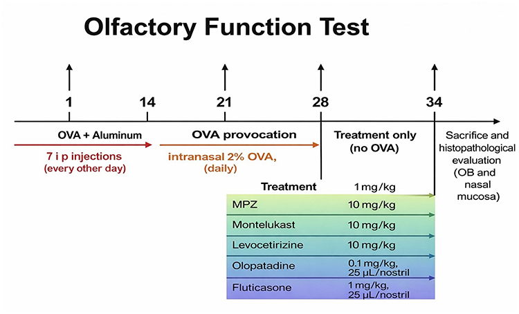 |
Figure 1 Timeline of ovalbumin (OVA)-induced allergic rhinitis rat model and treatment interventions (OVA: ovalbumin). |
Histopathology of the Olfactory System
On day 34, euthanasia was performed under deep anesthesia induced by intraperitoneal administration of thiopental sodium (50 mg/kg), followed by decapitation. Nasal mucosa and olfactory bulb tissues were collected immediately after euthanasia for histopathological evaluation. For histological evaluation of the nasal mucosa, tissues were stained with hematoxylin and eosin (HE), and vascular dilatation, goblet cell hyperplasia, inflammatory cell infiltration, plasma cell infiltration, mast cell infiltration, eosinophil infiltration, chondrocyte hypertrophy, and ciliary loss were assessed.
Olfactory bulb specimens obtained after decapitation were fixed in 10% formaldehyde for 48 h, decalcified in 10% formic acid for 36 h, washed overnight under running tap water, dehydrated through graded alcohols (50%, 75%, 96%, and 100%), cleared in xylene, and embedded in paraffin blocks. Serial sections (5 µm) were prepared using a Leica RM 2125 RT microtome; the first three and every tenth section were mounted on slides. After deparaffinization, the slides were stained with HE.
Each histopathological parameter was evaluated under light microscopy by an experienced histopathologist blinded to the treatment groups, using predefined morphological criteria. Glomerular layer thickness and diameter were assessed based on structural enlargement or reduction compared with normal architecture. Glomerular organization and neuronal degeneration were scored according to the degree of architectural disruption and neuronal loss. Vascular congestion and perivascular edema were graded based on the extent of vascular dilation and perivascular fluid accumulation. Microglial activation was assessed according to the presence and distribution of activated microglial cells characterized by increased cellular density and morphological changes. Scoring was performed at ×200 and ×400 magnification, and the severity of each parameter was graded as none (0), mild (1), moderate (2), or severe (3).19
Olfactory Function Test
Olfactory function was assessed using the “Cookie Finding Test”.19,23 Anosmia was defined as the inability to locate a hidden food pellet within 5 min. Rats were deprived of food for 12 h before testing. Each rat was placed in a cage (42 × 27×15 cm) where a food pellet was buried under wood shavings at a random location, avoiding corners or walls. The latency to find the pellet was recorded as an indicator of olfactory function.
The study was approved by the Local Ethics Committee for Animal Experiments of NESA Laboratory (Approval No: 079).
Statistical Analyses
Statistical analyses were performed using SPSS software, version 29.0 (IBM Corp., Armonk, NY, USA). Descriptive statistics were calculated, and the normality of continuous variables was assessed using the Kolmogorov–Smirnov and Shapiro–Wilk tests. Non-normally distributed ordinal variables (scoring parameters) were compared using the Mann–Whitney U-test (two groups) or the Kruskal–Wallis test (multiple groups). For normally distributed continuous variables (olfactory test results), intergroup comparisons were conducted using one-way ANOVA, followed by the Games–Howell post hoc test. Within each group, comparisons across four time points were made using the paired-sample t-test. Results are presented as median (min–max) and mean ± SD. A p-value <0.05 was considered statistically significant.
Results
All 49 rats randomized into the seven experimental groups completed the study and were included in the final analyses; no mortality or animal exclusion occurred (Supplementary Figures S1). In the positive control group, the mean cookie-finding time on day 1 was 98.1 ± 130.1 seconds, whereas on day 21 it increased to 353.9 ± 153.2 seconds. A statistically significant difference was detected in cookie-finding time before and after 21 days of OVA administration (p = 0.007). Additionally, the mean cookie-finding time on the final day of OVA administration, day 28, was 263.7 ± 233.6 seconds, which also differed significantly from day 1 (p = 0.039), confirming successful induction of allergic rhinitis and associated olfactory dysfunction.
Histopathology of the Olfactory System
Marked histological improvements were observed in both nasal mucosal samples and glomerular histopathology in the AR + MPZ treated group compared with the positive control group, except for neuronal nuclear integrity (Table 1; Figures 2 and 3; Supplementary Table S1; Supplementary Figures S2 and S3). In the leukotriene receptor antagonist–treated group, significant improvements were observed in all evaluated nasal mucosal and most olfactory bulb histopathological parameters compared with the positive control group, except for mitral cell density (Table 1; Supplementary Table S2; Supplementary Figures S2 and S3). In the levocetirizine-treated group, significant improvements were observed in all evaluated nasal mucosal parameters compared with the positive control group. In olfactory bulb histopathology, neuronal nuclear integrity and glomerular organization did not differ significantly, whereas all remaining parameters showed significant improvement (Table 1; Supplementary Table S3, Supplementary Figures S2 and S3). Similarly, in the AR + OLO group, all nasal mucosal parameters demonstrated significant improvement, while glomerular organization and neuronal nuclear integrity did not show statistically significant changes compared with the positive control group (Table 1; Supplementary Table S4, Supplementary Figures S2 and S3). In the AR + FLU group, all nasal mucosal parameters improved significantly. In olfactory bulb histopathology, significant improvements were observed in glomerular layer thickness, glomerular diameter, perivascular edema, mitral cell density, and microglial activation compared with the positive control group (p = 0.004, 0.007, 0.017, <0.001, and <0.001, respectively), whereas other parameters did not reach statistical significance (Table 1; Supplementary Table S5; Supplementary Figures S2 and S3). Comparison of all treatment groups showed no statistically significant intergroup differences in any nasal mucosal or olfactory bulb histopathological parameters, indicating comparable histopathological improvement across treatment modalities (Table 1, Figure 4, Supplementary Table S6; Supplementary Figures S4 and S5).
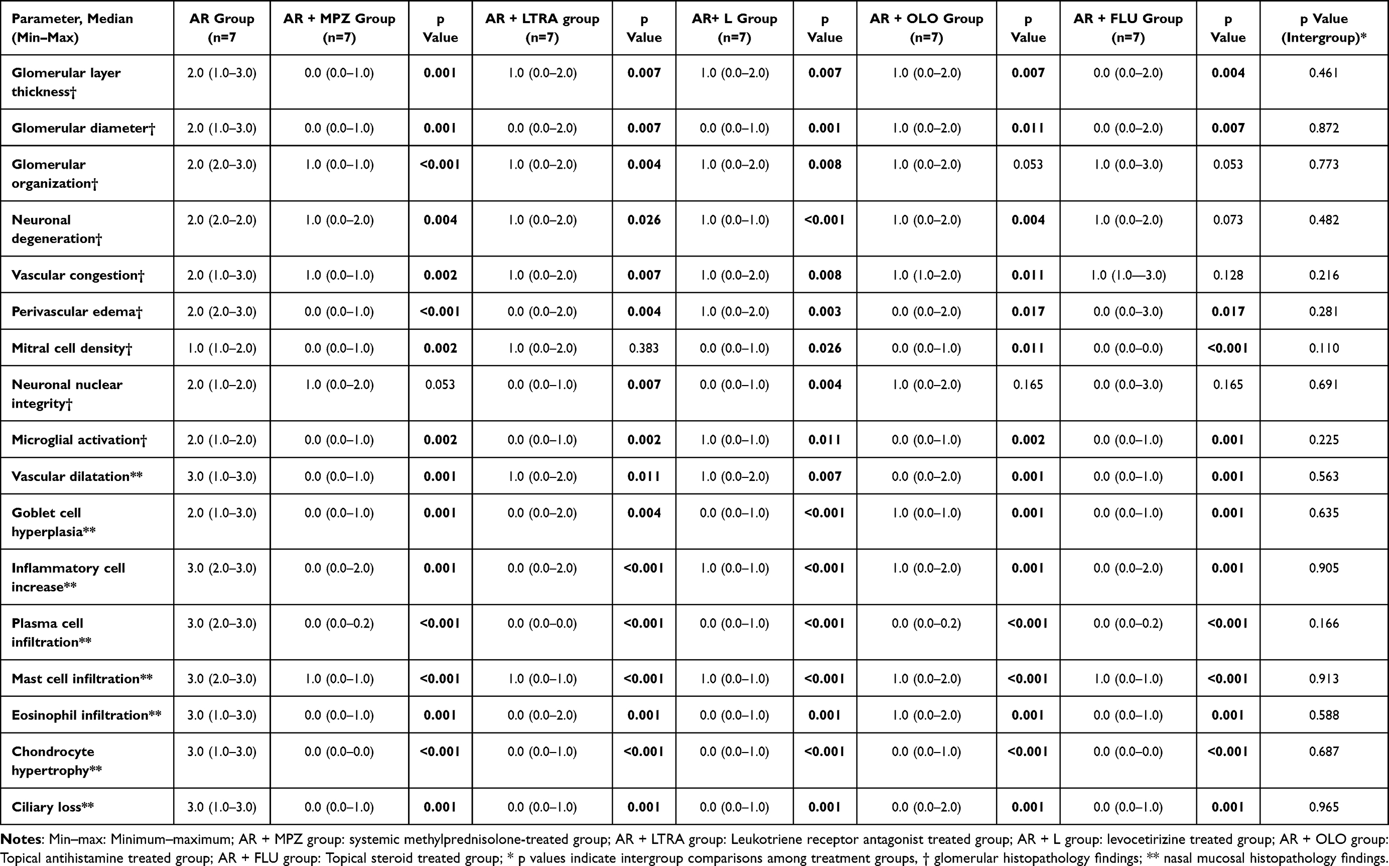 |
Table 1 Comparative Histopathological Analysis of Nasal Mucosa and Olfactory Bulb Across Treatment Modalities |
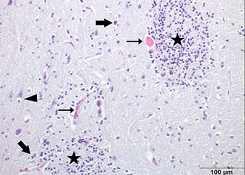 |
Figure 2 Histopathological changes in the olfactory bulb. ➔: degenerative cells; →: congestion and perivascular edema; ▲: neuronophagia; ★: severe gliosis) (H&E, ×400). |
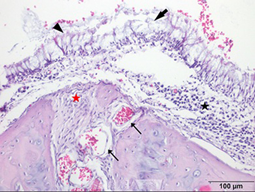 |
Figure 3 Histopathological changes in the nasal mucosal epithelium. ➔: ciliary loss; →: vascular dilatation; ▲: goblet cell hyperplasia/hypertrophy with inflammation; ★ (black): severe inflammation; ★ (red): chondrocyte inflammation; (H&E, ×400). |
 |
Figure 4 Integrated multi-domain response to treatment (radar plot). |
Olfactory Function Test
On day 1, prior to allergic rhinitis induction, and on day 21, after successful establishment of the model, olfactory test latencies did not differ significantly among the groups (p = 0.245 and p = 0.254, respectively; Table 2). At day 28 (treatment day 7), compared with day 21, a significant reduction in food-finding latency was observed only in the AR + MPZ and AR + OLO groups (p = 0.038 and p = 0.027, respectively). By day 34 (treatment day 14), these improvements persisted in the AR + MPZ and AR + OLO groups, and additional significant reductions were observed in the AR + FLU, AR + L, and AR + LTRA groups (p < 0.001, p < 0.001, p < 0.001, p = 0.005, and p= 0.026, respectively; Table 3).
 |
Table 2 Comparison of Cookie-Finding Latency in the Olfactory Test Across Groups at Baseline (Day 1) and After AR Induction (Day 21) |
 |
Table 3 Changes in Food-Finding Latency Across Treatment Groups |
Discussion
In this study, using an experimentally induced allergic rhinitis model in rats, treatment with methylprednisolone, levocetirizine, montelukast, fluticasone propionate, and olopatadine resulted in significant improvement in both nasal mucosal and olfactory bulb histopathology compared with the positive control group. However, no superiority was observed among the different treatment modalities in terms of histopathological outcomes. In addition, while late-phase improvement in olfactory function was observed across all treatment groups, early functional improvement occurred only in the methylprednisolone- and olopatadine-treated groups.
The OVA antigen has been widely used to establish animal models for evaluating the efficacy of antiallergic drugs, as it induces nasal allergic symptoms comparable to those observed in human AR.24 Accordingly, in our study, OVA administration led to a remarkable increase in cookie-finding time in the AR group, consistent with previous reports.14 These findings confirmed the successful establishment of the model.
Previous studies have demonstrated that inflammation of the olfactory epithelium can impair olfactory sensory neuron signaling and subsequently affect olfactory bulb function, particularly at the level of the glomerular layer.14 Anti-inflammatory treatments have been shown to improve nasal inflammation in allergic rhinitis models; however, data regarding their effects on olfactory bulb histopathology remain limited.14,15,22 In this context, our findings provide additional evidence that anti-inflammatory therapies may also improve olfactory bulb histopathological parameters consistent with inflammation.
Systemic corticosteroids exert potent anti-inflammatory effects through suppression of inflammatory cell migration and cytokine release.6 Shimizu et al reported that dexamethasone reduced mucus secretion and decreased eosinophil infiltration.25 In line with these findings, methylprednisolone treatment in our study resulted in significant improvement in multiple histopathological parameters in both the nasal mucosa and olfactory bulb. However, neuronal nuclear integrity did not demonstrate significant recovery, suggesting that restoration of neuronal structural integrity may require longer treatment duration or additional neuroprotective mechanisms beyond inflammation control.16
In the study by Tabaru et al, montelukast was shown to improve nasal inflammatory parameters such as goblet cell hyperplasia, vascular congestion, and eosinophilic infiltration in an AR rat model.21 Although several studies have demonstrated the effects of montelukast on nasal mucosal histopathology, to the best of our knowledge, no previous work has evaluated its impact on olfactory bulb histopathology.21,25 In our study, montelukast significantly improved olfactory bulb histopathological parameters, suggesting a potential role in mitigating olfactory loss associated with allergic rhinitis, thereby providing a novel contribution to the literature. However, no significant improvement was observed in mitral cell density; this may be related to the relatively short treatment duration or to ongoing inflammatory-like changes that may limit structural neuronal recovery following neuronal degeneration.17
Shimizu et al reported that H1 antagonists decreased eosinophil infiltration,25 and another study demonstrated that desloratadine improved olfactory function.26 In our study, levocetirizine produced significant improvements in all histopathological changes of both the olfactory bulb and the nasal mucosa, which is a noteworthy finding. Notably, these effects were observed only with levocetirizine among the H1 antihistamines, and further studies are required to determine whether similar benefits can also be achieved with other agents.
Recent data from animal models have demonstrated that short-term intranasal corticosteroid therapy can protect the olfactory sensory neuron population and preserve olfactory function against inflammation-induced damage.16 Among the available intranasal corticosteroids, fluticasone propionate was selected in the present study because it is one of the most commonly prescribed agents and is characterized by minimal systemic absorption (<1%).27 Consistent with its established anti-inflammatory effects, Jacobson et al showed that fluticasone significantly reduced nasal symptoms and inhibited seasonal infiltration of mast cells and eosinophils in both the nasal epithelium and subepithelium in patients with allergic rhinitis.28 In line with these findings, our results demonstrated significant improvement in multiple nasal mucosal and olfactory bulb histopathological parameters, whereas alterations considered partially reversible or irreversible, such as neuronal nuclear integrity, neuronal degeneration, and glomerular organization, did not show significant recovery. Although direct evidence regarding the effects of topical antihistamines on olfactory bulb inflammation is scarce, olopatadine has been shown to suppress allergen-induced nasal symptoms and reduce pro-inflammatory cytokine release in the nasal mucosa.29,30 In our study, olopatadine treatment resulted in significant improvement in nasal mucosal histopathology and in olfactory bulb histopathological parameters consistent with inflammation, supporting its potential role in mitigating olfactory dysfunction associated with allergic rhinitis.
Although inflammatory changes in the olfactory bulb have been demonstrated to be consistent with olfactory dysfunction following different anti-inflammatory treatments, studies directly comparing the effects of various treatment modalities on olfactory inflammation remain limited.14,15,22 Bozkurt et al showed that prednisolone, montelukast, and omalizumab had comparable effects in reducing inflammatory cell infiltration in nasal and sinus histopathology. In another study, montelukast and prednisolone demonstrated similar efficacy on nasal symptoms and multiple inflammatory endpoints.22 In agreement with these findings, our study demonstrated no significant differences among treatment groups in either nasal mucosal or olfactory bulb histopathology, providing additional evidence that multiple therapeutic options may achieve similar structural anti-inflammatory effects.
Previous studies suggest that although nasal obstruction due to local inflammation contributes to olfactory dysfunction in AR, the persistence of smell impairment after inflammation resolution points to additional mechanisms, such as direct effects on olfactory sensory neurons and olfactory bulb pathways.31,32 Accordingly, we compared the effects of various treatment modalities on olfactory test performance, and our results revealed both early and late differences in functional recovery. Our findings suggest that methylprednisolone and olopatadine provided early improvement in olfactory function. This may be explained by the rapid anti-inflammatory action of corticosteroids, which suppress acute inflammatory responses within hours, as well as by the fast onset of olopatadine, which has been shown in previous studies to alleviate nasal symptoms within about 30 minutes of administration.33,34 Although histopathological improvement was observed across treatment groups, correlations between individual histopathological parameters and olfactory performance were generally weak. Only limited associations were detected at specific time points, suggesting that functional olfactory recovery may not directly mirror the severıntity of individual histopathological changes and may instead reflect complex, time-dependent recovery dynamics, as previously reported in both experimental and clinical studies.35,36 This dissociation suggests that olfactory function may depend on integrated and time-dependent processes beyond individual histopathological features. Experimental and clinical studies indicate that functional olfactory recovery can occur independently of complete structural normalization and may be influenced by the timing and dynamics of anti-inflammatory effects rather than by discrete histopathological changes alone.14,16,25
From a clinical perspective, these findings suggest that olfactory dysfunction in allergic rhinitis may reflect changes beyond the nasal mucosa, including in the olfactory bulb, and that although commonly used therapies achieve comparable histopathological improvement, the earlier functional recovery observed with methylprednisolone and olopatadine may be clinically relevant in selected patients.
The main limitations of this study include the lack of inflammatory marker or cytokine measurements to support the histopathological findings. Another limitation is the relatively short treatment duration, which may have limited the assessment of neuronal recovery. Longer follow-up periods and the incorporation of inflammatory biomarkers may provide further insight into neurobiological recovery processes and underlying mechanisms. Given the exploratory nature of this preclinical study and ethical constraints on animal use, an a priori sample size calculation was not performed; however, post-hoc analyses indicated sufficient power to detect large effects, supporting the interpretation of comparable treatment efficacy. Histopathological scoring was performed by a single experienced observer, which precluded formal inter-rater reliability analysis; however, evaluations were conducted in a blinded manner using predefined morphological criteria consistent with those reported in previous experimental studies.19 Despite these limitations, the present study contributes novel evidence by demonstrating that different anti-inflammatory treatment modalities effectively reduce olfactory inflammation without clear superiority and by identifying temporal differences in functional olfactory recovery. Furthermore, our results revealed that methylprednisolone and olopatadine were the most effective treatments for early improvement of olfactory dysfunction.
Conclusion
All treatment modalities improved olfactory mucosal and olfactory bulb histopathological parameters, with no clear superiority. Notably, while late-phase olfactory functional recovery was observed with all treatment modalities, the exclusive early improvement in the methylprednisolone and olopatadine groups is a noteworthy finding.
Data Sharing Statement
The data that support the findings of this study are not publicly available due to ethical restrictions, but may be available upon reasonable request from the corresponding author.
Institutional Review Board Statement
The study was approved by the Local Ethics Committee for Animal Experiments of NESA Laboratory (Approval No: 079).
Acknowledgment
This study was conducted in accordance with the ARRIVE guidelines.
Author Contributions
Conceptualization, H. Kayikci and E. Kayikci; methodology, H. Kayikci and E. Kayikci; formal analysis, H. Kayikci and E. Kayikci; writing—original draft preparation, H. Kayikci and E. Kayikci; writing—review and editing, H. Kayikci. All authors gave final approval of the version to be published; have agreed on the journal to which the article has been submitted; and agree to be accountable for all aspects of the work.
Funding
The author(s) received no financial support for the research, authorship, and/or publication of this article.
Disclosure
The author(s) declared no potential conflicts of interest with respect to the research, authorship, and/or publication of this article.
References
1. Bousquet J, Khaltaev N, Cruz AA. et al. Allergic rhinitis and its impact on asthma (ARIA) 2008 update (in collaboration with the World Health Organization, GA²LEN and AllerGen). Allergy. 2008;63(Suppl 86):8–13. doi:10.1111/j.1398-9995.2007.01620.x
2. Wise SK, Damask C, Roland LT, et al. International consensus statement on allergy and rhinology: allergic rhinitis—2023. Int Forum Allergy Rhinol. 2023;13(4):293–859. doi:10.1002/alr.23090
3. Rydzewski B, Pruszewicz A, Sulkowski WJ. Assessment of smell and taste in patients with allergic rhinitis. Acta Otolaryngol. 2000;120(2):323–326. doi:10.1080/000164800750001189
4. Gupta N, Harit A, Taneja HC, Kumar R, Tripathi AK. Olfaction and its correlates in allergic rhinitis: a case control study. Indian J Otolaryngol Head Neck Surg. 2019;71(Suppl 3):1782–1786. doi:10.1007/s12070-017-1149-7
5. Croy I, Nordin S, Hummel T. Olfactory disorders and quality of life: an updated review. Chem Senses. 2014;39(3):185–194. doi:10.1093/chemse/bjt072
6. Brożek JL, Bousquet J, Agache I, et al. Allergic rhinitis and its impact on asthma (ARIA) guidelines—2016 revision. J Allergy Clin Immunol. 2017;140(4):950–958. doi:10.1016/j.jaci.2017.03.050
7. Mener DJ, Shargorodsky J, Varadhan R, Lin SY. Topical intranasal corticosteroids and growth velocity in children: a meta-analysis. Int Forum Allergy Rhinol. 2015;5(2):95–103. doi:10.1002/alr.21430
8. Seidman MD, Gurgel RK, Lin SY, et al. Clinical practice guideline: allergic rhinitis. Otolaryngol Head Neck Surg. 2015;152(1 Suppl):1–43. doi:10.1177/0194599814561600
9. Kaliner MA, Berger WE, Ratner PH, et al. The efficacy of intranasal antihistamines in the treatment of allergic rhinitis. Ann Allergy Asthma Immunol. 2011;106(2 Suppl):6–11. doi:10.1016/j.anai.2010.08.010
10. Yamauchi Y, Fujikura T, Shimosawa T. The effect of H1 antagonists carebastine and olopatadine on histamine-induced expression of CC chemokines in cultured human nasal epithelial cells. Allergol Int. 2007;56(2):171–177. doi:10.2332/allergolint.O-06-446
11. Guilemany JM, García-Piñero A, Alobid I, et al. The loss of smell in persistent allergic rhinitis is improved by levocetirizine due to reduction of nasal inflammation but not nasal congestion (the CIRANO study). Int Arch Allergy Immunol. 2012;158(2):184–190. doi:10.1159/000331329
12. Hilberg O. Effect of terfenadine and budesonide on nasal symptoms, olfaction, and nasal airway patency following allergen challenge. Allergy. 1995;50(8):683–688. doi:10.1111/j.1398-9995.1995.tb02586.x
13. Wilson AM, White PS, Gardiner Q, Nassif R, Lipworth BJ. Effects of leukotriene receptor antagonist therapy in patients with chronic rhinosinusitis in a real-life rhinology clinic setting. Rhinology. 2001;39(3):142–146.
14. Rodrigues J, Rocha MI, Teixeira F, et al. Structural, functional and behavioral impact of allergic rhinitis on olfactory pathway and prefrontal cortex. Physiol Behav. 2023;265:114171. doi:10.1016/j.physbeh.2023.114171
15. Lv H, Liu P, Zhou F, Gao Z, Fan W, Xu Y. TAK-242 ameliorates olfactory dysfunction in a mouse model of allergic rhinitis by inhibiting neuroinflammation in the olfactory bulb. Int Immunopharmacol. 2021;92:107368. doi:10.1016/j.intimp.2021.107368
16. Jung AY, Kim YH. Reversal of Olfactory Disturbance in Allergic Rhinitis Related to OMP Suppression by Intranasal Budesonide Treatment. Allergy Asthma Immunol Res. 2020;12(1):110–124. doi:10.4168/aair.2020.12.1.110
17. Schubert C, Schulz K, Sonner JK, et al. Neuroinflammation causes mitral cell dysfunction and olfactory impairment in a multiple sclerosis model. J Neuroinflammation. 2025;22(1):71. doi:10.1186/s12974-025-03388-5
18. Hotchkiss JA, Hilaski R, Cho H, et al. Fluticasone propionate attenuates ozone-induced rhinitis and mucous cell metaplasia in rat nasal airway epithelium. Am J Respir Cell Mol Biol. 1998;18(1):91–99. doi:10.1165/ajrcmb.18.1.2897
19. Becerik Ç, Karaca ÇT, Özcan Z, Kul S, Toros SZ. The role of substance P receptor antagonists in allergic rhinitis: ovalbumin-induced rat model. Laryngoscope. 2023;133(11):2891–2897. doi:10.1002/lary.30628
20. Kar M, Bayar Muluk N, Bafaqeeh SA, Cingi C. Consensus on the methodology for experimental studies in allergic rhinitis. Int J Pediatr Otorhinolaryngol. 2019;121:68–71. doi:10.1016/j.ijporl.2019.03.009
21. Tabaru A, Ogreden S, Akyel S, Oktay MF, Uslu K, Emre FK. Comparison of treatment efficacy of omega-3 fish oil and montelukast in ovalbumin-protease-induced allergic rhinitis model in rats. Braz J Otorhinolaryngol. 2024;90(3):101399. doi:10.1016/j.bjorl.2024.101399
22. Bozkurt MK, Tülek B, Bozkurt B, Akyürek N, Mehmet Ö, Kiyici A. Comparison of the efficacy of prednisolone, montelukast, and omalizumab in an experimental allergic rhinitis model. Turk J Med Sci. 2014;44(3):439–447. doi:10.3906/sag-1212-23
23. Hendricks SJ, Ducray A, Harding JW, Herzog MH, Otto T. Cookie finding test: a behavioral assessment of olfactory function in mice. J Neurosci Methods. 1994;55(3):201–209.
24. Senturk E, Yildirim YS, Dogan R, et al. Assessment of the effectiveness of cyclosporine nasal spray in an animal model of allergic rhinitis. Eur Arch Otorhinolaryngol. 2018;275(1):117–124. doi:10.1007/s00405-017-4786-2
25. Shimizu T, Hirano H, Majima Y, Sakakura Y. A mechanism of antigen-induced mucus production in nasal epithelium of sensitized rats: a comparison with lipopolysaccharide-induced mucus production. Am J Respir Crit Care Med. 2000;161(5):1648–1654. doi:10.1164/ajrccm.161.5.9908101
26. Li S, Zhang X, Li Z, et al. Desloratadine ameliorates olfactory disorder and suppresses AMPA receptor GluA1 expression in allergic rhinitis rats. Arch Immunol Ther Exp. 2020;68(1):6. doi:10.1007/s00005-020-00569-3
27. Allen DB. Systemic effects of intranasal steroids: an endocrinologist’s perspective. J Allergy Clin Immunol. 2000;106(4):S179–S190. doi:10.1067/mai.2000.110038
28. Jacobson MR, Juliusson S, Löwhagen O, Balder B, Kay AB, Durham SR. Effect of topical corticosteroids on seasonal increases in epithelial eosinophils and mast cells in allergic rhinitis: a comparison of nasal brush and biopsy methods. Clin Exp Allergy. 1999;29(10):1347–1355. doi:10.1046/j.1365-2222.1999.00608.x
29. Watts AM, Cripps AW, West NP, Cox AJ. Modulation of allergic inflammation in the nasal mucosa of allergic rhinitis sufferers with topical pharmaceutical agents. Front Pharmacol. 2019;10:294. doi:10.3389/fphar.2019.00294
30. Pipkorn P, Costantini C, Reynolds C, et al. The effects of the nasal antihistamines olopatadine and azelastine in nasal allergen provocation. Ann Allergy Asthma Immunol. 2008;101(1):82–89. doi:10.1016/S1081-1206(10)60839-3
31. Zhang Z, Liu J, Xie L, Cao W, Ma F, Zhang Y. Tissue eosinophils and mucous inflammatory cytokines for the evaluation of olfactory recovery after endoscopic sinus surgery in patients with nasal polyposis. Am J Otolaryngol. 2022;43(5):103561. doi:10.1016/j.amjoto.2022.103561
32. Guss J, Doghramji L, Reger C, Chiu AG. Olfactory dysfunction in allergic rhinitis. ORL J Otorhinolaryngol Relat Spec. 2009;71(5):268–272. doi:10.1159/000242429
33. Morita K, Koga T, Moroi Y, Urabe K, Furue M. Rapid effects of olopatadine hydrochloride on the histamine-induced skin responses. J Dermatol. 2002;29(11):709–712. doi:10.1111/j.1346-8138.2002.tb00207.x
34. Patel P, Roland PS, Marple BF, et al. An assessment of the onset and duration of action of olopatadine nasal spray. Otolaryngol Head Neck Surg. 2007;137(6):918–924. doi:10.1016/j.otohns.2007.08.005
35. Hummel T, Whitcroft KL, Andrews P, et al. Position paper on olfactory dysfunction. Supplement. Rhinology. 2017;54(26):1–30. doi:10.4193/Rhino16.248
36. Doty RL. Olfaction in health and disease. Handb Clin Neurol. 2019;164:1–62.
 © 2026 The Author(s). This work is published and licensed by Dove Medical Press Limited. The
full terms of this license are available at https://www.dovepress.com/terms
and incorporate the Creative Commons Attribution
- Non Commercial (unported, 4.0) License.
By accessing the work you hereby accept the Terms. Non-commercial uses of the work are permitted
without any further permission from Dove Medical Press Limited, provided the work is properly
attributed. For permission for commercial use of this work, please see paragraphs 4.2 and 5 of our Terms.
© 2026 The Author(s). This work is published and licensed by Dove Medical Press Limited. The
full terms of this license are available at https://www.dovepress.com/terms
and incorporate the Creative Commons Attribution
- Non Commercial (unported, 4.0) License.
By accessing the work you hereby accept the Terms. Non-commercial uses of the work are permitted
without any further permission from Dove Medical Press Limited, provided the work is properly
attributed. For permission for commercial use of this work, please see paragraphs 4.2 and 5 of our Terms.