
Back to Journals » Journal of Hepatocellular Carcinoma » Volume 12
Combined TACE and Microwave Ablation for Recurrent Spiegel-Lobe HCC: A Four-Case Series
Authors Guan X, Hou R  , Han Z, Piao X, Zhang S
Received 24 August 2025
Accepted for publication 6 December 2025
Published 19 December 2025 Volume 2025:12 Pages 2839—2844
DOI https://doi.org/10.2147/JHC.S558992
Checked for plagiarism Yes
Review by Single anonymous peer review
Peer reviewer comments 2
Editor who approved publication: Dr David Gerber
Xin Guan,1,* Ruosen Hou,2,* Zhezhu Han,1 Xuezhe Piao,1 Songnan Zhang1
1Department of Oncology, Yanbian University Hospital, Yanji, Jilin, People’s Republic of China; 2Department of Oncology, China Medical University, Shenyang, Liaoning, People’s Republic of China
*These authors contributed equally to this work
Correspondence: Songnan Zhang, Department of Oncology, Yanbian University Hospital, Yanji, Jilin, People’s Republic of China, Email [email protected]
Background: The Spiegel lobe, a distinct subdivision of the hepatic caudate lobe, is characterized by its unique anatomical position, with close proximity to major hilar vasculature and extrahepatic structures. Therapeutic intervention for Spiegel lobe lesions remains technically challenging, with no established treatment protocol currently available.
Case Presentation: This study reports four cases of recurrent hepatocellular carcinoma (HCC) in the Spiegel lobe treated with conventional transarterial chemoembolization (c-TACE) followed by microwave ablation (MWA) via a transhepatic left lobe approach. Postprocedural imaging assessment confirmed complete response (CR) in all patients according to mRECIST criteria. During a mean follow-up period of 33.75 months (range: 31– 37), three patients maintained disease-free status, whereas one patient developed intrahepatic recurrence at 19.2 months. This patient achieved CR after repeat TACE-MWA combined with adjuvant lenvatinib and tislelizumab therapy. The mean overall survival (OS) was 33.75 months (95% CI: 31.2– 36.3).
Conclusion: The combination of transcatheter arterial chemoembolization (TACE) and microwave ablation (MWA) for treating recurrent hepatocellular carcinoma (HCC) in the Spiegel lobe was proven feasible in this series of studies, and demonstrated favorable efficacy and safety profiles.
Keywords: Spiegel lobe, recurrent hepatocellular carcinoma, microwave ablation, transcatheter arterial chemoembolization
Introduction
Hepatocellular carcinoma (HCC) in the Spiegel lobe demonstrates a lower incidence compared to other hepatic segments, and its unique anatomical location limits the applicability of therapeutic interventions.1 Surgical resection is the treatment of choice for early-stage HCC; however, the Spiegel lobe’s complex anatomical location, adjacent to the inferior vena cava and hepatic veins, poses significant surgical challenges, including increased risks of hemorrhage and postoperative recurrence compared to non-caudate lobe resections.2 Stereotactic body radiation therapy (SBRT) has emerged as a cornerstone local treatment modality in the multidisciplinary management of HCC.3 For early-stage HCC, SBRT achieves local control rates of 85% to 95% at 2–3 years.4 However, caudate lobe HCC presents substantially higher treatment-related risks than tumors in other liver regions, such as the left or right lobes. Radioembolization is primarily indicated for patients with unresectable primary or metastatic liver cancers, particularly those with intermediate to advanced-stage hepatocellular carcinoma (HCC). Nevertheless, its high cost poses a significant barrier to its widespread adoption.5 TACE, a standard therapeutic option for HCC, is commonly employed for Spiegel lobe HCC. However, the variability, multiplicity of feeding arteries, and small caliber of the Spiegel lobe’s blood supply present challenges for TACE, including incomplete embolization and an elevated risk of recurrence.6 MWA is primarily utilized for the curative treatment of early-stage hepatocellular carcinoma. This minimally invasive technique is distinguished by its reduced invasiveness, high efficacy, and rapid recovery. It yields clinical outcomes comparable to surgical resection, thereby addressing the limitations of TACE.7 The primary challenge in thermal ablation near large blood vessels is the heat sink effect, where rapid blood flow dissipates heat from the ablation zone, thus preventing the tissue from reaching the temperature threshold required for complete coagulative necrosis of tumor cells. MWA utilizes high-frequency electromagnetic fields to agitate polar molecules and ions within tumor tissue, generating heat through dielectric hysteresis and molecular friction, which rapidly elevates tissue temperatures above 100°C. Consequently, its heating efficiency is largely unaffected by tissue conductivity or the heat-sink effect from nearby blood vessels, facilitating uniform and complete ablation of tumors adjacent to major vasculature.8 The combination of TACE and MWA has been clinically validated to overcome the limitations of monotherapy for recurrent small HCC; however, its application in Spiegel lobe HCC remains underreported. In this case series, we present four cases of recurrent HCC in the Spiegel lobe managed with a combination of TACE and MWA, all of which achieved complete remission. The 1-year and 2-year progression-free survival (PFS) rates were 100% and 75%, respectively.
Case Presentation
This study enrolled four patients with recurrent HCC in the Spiegel lobe who received combined TACE and MWA therapy at Yanbian University Hospital between October 2021 and January 2022. Preoperative laboratory evaluations revealed no abnormalities, with alpha-fetoprotein (AFP) levels within the normal range in one patient. Imaging studies demonstrated that Case 1 presented with two additional intrahepatic recurrent nodules outside the Spiegel lobe (Supplementary Figure), whereas Cases 2–4 showed solitary recurrence confined to the Spiegel lobe (Table 1). All patients received a sequential treatment regimen consisting of TACE followed by MWA. In Cases 1–3, hepatic arteriography clearly delineated the Spiegel lobe lesions, facilitating selective embolization of the tumor-feeding arteries. In Case 4, the Spiegel lobe lesion was not visualized on hepatic arteriography; therefore, a small volume of lipiodol was injected into the main hepatic artery as a radiopaque marker. MWA was conducted within eight days following TACE in all four cases. Pre-ablation computed tomography (CT) scans confirmed adequate lipiodol deposition and clear visualization of all lesions. All cases underwent percutaneous puncture and ablation under combined CT and ultrasound guidance (Figure 1). The detailed procedural techniques for TACE and MWA are described below:
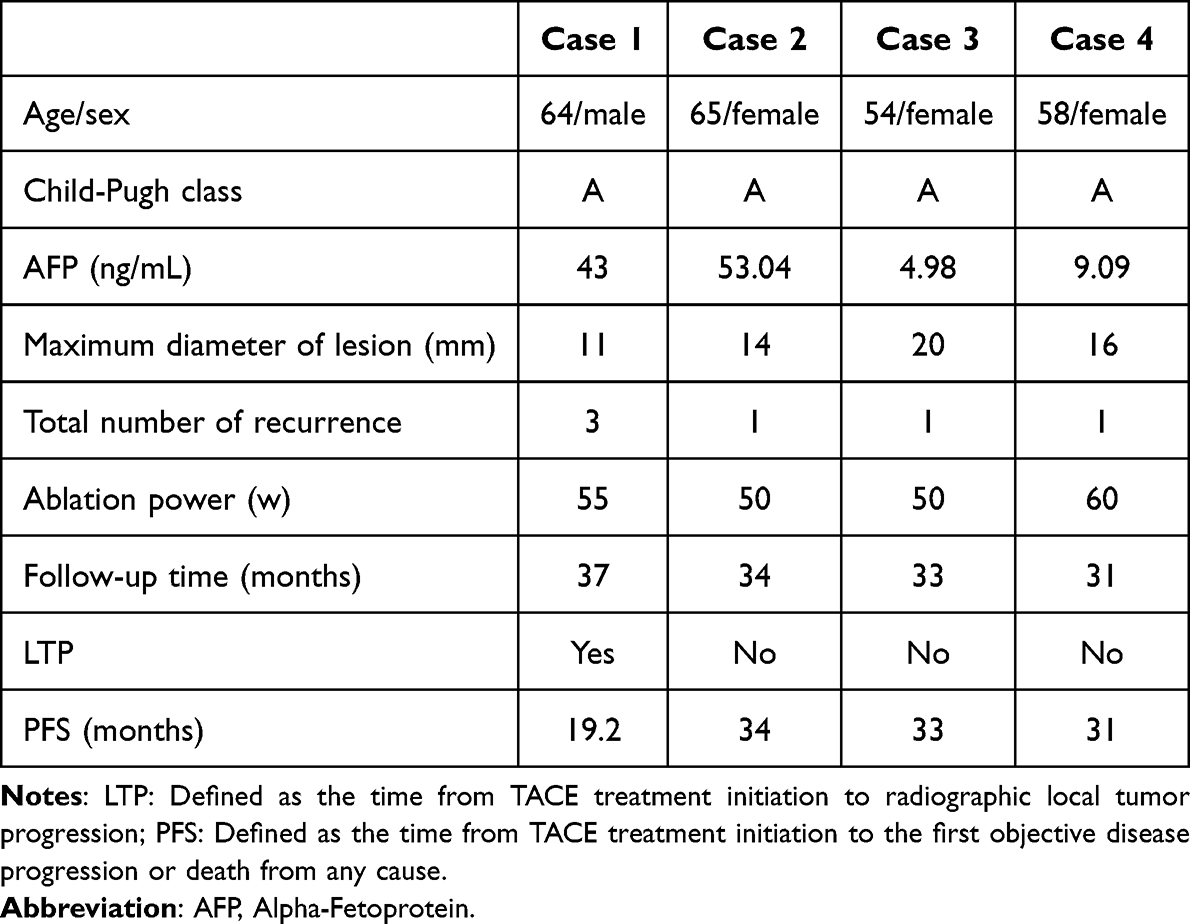 |
Table 1 Basic Characteristics of All Involved Patients |
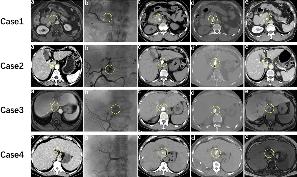 |
Figure 1 Imaging findings during combined TACE and MWA for Spiegel lobe HCC in four patients. (a) Pre-TACE lesion localization. (b) The image shows a dense shadow caused by the deposition of TACE contrast medium during the operation. In Case 4, the Spiegel lobe lesion could not be clearly seen on hepatic arteriography and therefore has not been marked in the figure. (c) Pre-ablation CT scan with the tumor delineated by the deposited iodinated oil. (d) Intraoperative plain CT scan for MWA, demonstrating precise needle placement at the lesion center. (e) One-month follow-up imaging confirms treatment response, with the ablation zone showing no enhancement and complete tumor coverage. The circle denotes the lesion location. |
TACE Procedure: Patients were placed in a supine position. After routine preparation with sterile draping and disinfection, local infiltration anesthesia with subcutaneous lidocaine was administered in the right inguinal region. The right femoral artery was punctured, and a 4F arterial sheath was inserted using the Hengrui Medical RAPIDTHRUC microcatheter guidewire system. Tumor size and vascular supply were assessed through imaging from the abdominal aorta to the hepatic artery. The microcatheter was advanced into the tumor’s blood-supplying artery within the Spiegel lobe, and embolization was carried out using epirubicin, iodized oil and polyethanol embolization microspheres.
MWA Procedure: MWA was performed using the UniFilm uCT 760 CT scanner, with guidance from a Mindray UMT-300 ultrasound device. A Vision Medical MTC-3C microwave therapeutic instrument and a Vision MTC-3CA-2 microwave ablation needle were used for ablation. Under color ultrasound guidance, a disposable microwave ablation needle was inserted through the left lobe of the liver into the Spiegel lobe lesion. Given the small lesion size (≤20 mm) in all four patients, a single ablation needle was deemed sufficient for each case, without adjunctive protective measures such as artificial ascites. Owing to the close proximity of the lesions to critical structures including the inferior vena cava, ablation power was appropriately reduced and the procedure was conducted under real-time ultrasound guidance. The ablation parameters for Cases 1, 2, 3, and 4 were set at 60W for 10 minutes, 50W for 9 minutes, 50W for 8 minutes, and 55W for 14 minutes, respectively. Color Doppler ultrasound must confirm that the ablation zone extends at least 5 mm beyond the lesion margins. The ablation antenna was then withdrawn at a constant speed (0.5 cm/s) under real-time imaging guidance. After ablation, the antenna was removed, and hemostasis was achieved by applying pressure with sterile gauze to the puncture site for five minutes.
Treatment and Follow-Up
All four patients successfully underwent the ablation procedure. One-month follow-up imaging confirmed complete remission (CR) in all cases. None of the patients received adjuvant therapy postoperatively, and follow-up evaluations using computed tomography (CT) or magnetic resonance imaging (MRI) were conducted every 3–4 months. As of January 2025, the mean follow-up duration was 33.75 months (range: 31 to 37 months). In Case 1, a 10-mm recurrent lesion was identified in the left lateral liver segment 19.2 months post-ablation. The patient was subsequently treated with repeat TACE and MWA. The MWA was performed via a left hepatic approach using a single antenna at 50 W for 7 minutes. Post-procedure assessment confirmed CR. The patient then received combined therapy with lenvatinib and tislelizumab for 17.8 months. No secondary recurrence was documented during the most recent follow-up. No recurrence was detected in the remaining patients. The mean progression-free survival (PFS) for the four patients was 28.3 months, with no mortality reported (Figure 2). The study observed no severe complications, including hemorrhage, bile duct injury, liver abscess, tumor seeding, or liver failure. Mild adverse events were, however, recorded in some cases. Specifically, Case 2 experienced abdominal pain on the first post-procedural day, which resolved with symptomatic analgesic management.
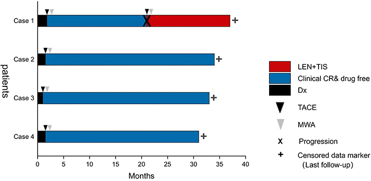 |
Figure 2 Swimmer plot of 4 patients who achieved complete response (CR). Censored data: Indicates patients who had not experienced disease progression by the end of the follow-up period. Y-axis: Individual patients. Abbreviations: MWA, microwave ablation; TACE, transarterial chemoembolization; Dx, diagnosis; LEN+TIS, lenvatinib plus Tislelizumab. |
Discussion
Current evidence indicates that TACE combined with MWA significantly improves complete tumor necrosis rates (90–100%) in recurrent small HCC. The median progression-free survival (PFS) with this sequential therapy is 16.8 months (95% CI: 14.2–19.4). Reported overall survival (OS) rates are 93.2% at 1 year, 64.5% at 3 years, and 55.0% at 5 years.9 HCC recurrence is multifactorial, involving tumor-related factors (eg, size, multifocality, differentiation grade) and host-related factors (eg, immune status, genetic/protein profiles). In this study, Over a mean follow-up duration of 33.75 months, complete ablation was achieved in all four patients after the initial ablation therapy, resulting in a 100% technical success rate and prolonged survival. The mean progression-free survival (PFS) was 28.3 months, with Cases 2–4 exhibiting PFS exceeding 29 months, surpassing the historical median PFS of 11 months for TACE monotherapy and 13.5 months for MWA monotherapy in caudate lobe HCC. The 30-month PFS rate among the four patients was 75%, exceeding the previously reported rates of 93.75%, 53.15%, and 28.13% at 6, 12, and 18 months, respectively, for caudate lobe HCC treated with MWA in earlier studies.10 In Case 1, tumor recurrence was observed at 19.2 months after ablation. This patient experienced early postoperative recurrence (RFS: 13 months) with three lesions distributed across the left, right, and caudate lobes, demonstrating a multi-lobar pattern. In contrast, the other three patients presented with solitary recurrent lesions confined to the caudate lobe. Given the lesion characteristics (multifocal and multi-lobar vs solitary and unilobar) and consistent with previous clinical studies linking multifocality and multi-lobar involvement to poorer outcomes, Case 1 was assessed to have a significantly higher recurrence risk than the other three cases.11 This observation aligns with the established literature, which identifies tumor multifocality as a key factor influencing the efficacy and prognosis of sequential TACE combined with MWA therapy.12
The caudate lobe is situated on the posterior surface of the liver, partially surrounding the retrohepatic inferior vena cava. Superiorly, it is adjacent to the confluence of the three hepatic veins; inferiorly, it borders the portal vein and bile ducts, which form the hepatic pedicle at the first porta hepatis; and posteriorly, it is adjacent to the retrohepatic inferior vena cava. The Spiegel lobe is a hepatic projection located to the left of the intrahepatic inferior vena cava.13 Owing to its distinct anatomical configuration and close proximity to major vascular structures, thermal ablation of HCC in the Spiegel lobe poses significant technical challenges. The synergistic advantages of combining TACE with MWA include. Firstly, TACE mitigates the heat-sink effect caused by adjacent arteries or the portal vein through occlusion of tumor-feeding vessels, thereby reducing the risk of tumor metastasis.14 Furthermore, lipiodol deposition following embolization induces tumor hypoxia, which enhances thermal sensitivity and improves the safety profile of ablation therapy.15 Secondly, lipiodol embolization during TACE clearly delineates the location and extent of the lesion, enabling precise visualization of its position and size on computed tomography (CT), thus facilitating accurate ablation.16 Selecting an appropriate hepatic puncture route is crucial for achieving optimal treatment outcomes and minimizing procedural complications. In the four cases presented, a left hepatic lobe puncture approach was selected. This approach provides greater flexibility in needle trajectory adjustment, a shorter puncture path, and the ability to avoid major vessels within the hepatic hilum.17 However, due to the proximity of the left hepatic lobe to the inferior vena cava and gastrointestinal organs, puncture via this route carries a risk of injury to the inferior vena cava and adjacent structures. To mitigate complications, interventional radiologists must acquire extensive puncture experience and utilize a layer-by-layer needle advancement technique under computed tomography (CT) guidance. Notwithstanding its potential clinical implications for managing recurrent HCC in the Spiegel lobe with TACE combined with MWA, this study has several limitations. First, the study’s retrospective, single-center design and small sample size (n=4) limit the generalizability of the findings. This limitation arises from two primary factors: the complex anatomy of the Spiegel lobe and the relatively low patient acceptance of this combined regimen. These constraints increase the potential for selection bias. Second, the relatively short median follow-up period precluded adequate assessment of late complications and long-term recurrence. Consequently, the long-term efficacy and safety profile of this combined regimen could not be comprehensively evaluated. Future studies should adopt a multicenter, large-sample, prospective, randomized controlled trial (RCT) design. Such a study should aim to refine inclusion and exclusion criteria, standardize safety assessments, and increase the sample size. These measures would help validate our preliminary findings and enhance the reliability of the outcomes.
Conclusion
In conclusion, the combined approach of TACE and MWA proved to be a feasible, effective, and safe treatment for recurrent hepatocellular carcinoma in the Spiegel lobe in this case series. Given the limited sample size, larger, comparative trials are warranted to validate these findings.
Ethics Statement
Details of the case reported in the article were approved by the central institution, The Affiliated Hospital of Yanbian University (Nos. 2025364).
Consent for Publication
This case series has been approved for publication by all four patients. This report and any attendant pictures were revealed with the patients’ written consent.
Acknowledgments
We give thanks to the patients and everyone involved in this study.
Funding
This research was supported by the National Natural Science Foundation of China (Nos. 82460527), Natural Science Foundation of Jilin Province (Nos. YDZJ202401136ZYTS).
Disclosure
The author(s) report no conflicts of interest in this work.
References
1. Miyayama S. Radiological vascular anatomy of the caudate lobe of the liver required for transarterial chemoembolization of hepatocellular carcinoma. Interv Radiol. 2023;8(3):118–129. PMID: 38020459; PMCID: PMC10681755. doi:10.22575/interventionalradiology.2022-0046
2. Yang B, Yuan M, Yang T, Liao Z, Wu H. Transarterial chemoembolization vs. liver resection as initial treatment for hepatocellular carcinoma occurring exclusively in caudate lobe: a retrospective propensity matching analysis. Oncol Res. 2022;30(1):23–33. PMID: 37304009; PMCID: PMC10207968. doi:10.32604/or.2022.026044
3. Sahai P, Mall A, Solanky AP, et al. Clinical outcomes with combination of stereotactic body radiation therapy in patients with hepatocellular carcinoma. J Cancer Res Ther. 2025;21(5):1013–1025. PMID: 41004290. doi:10.4103/jcrt.jcrt_2351_24
4. Apisarnthanarax S, Kim EY. SBRT: the most precise and least invasive tool in the liver cancer toolbox. Int J Radiat Oncol Biol Phys. 2025;123(4):919. PMID: 41101869. doi:10.1016/j.ijrobp.2025.08.023
5. Li ZY, Xie C, Cai HQ. Overview of Yttrium-90 radioembolization for advanced hepatocellular carcinoma: current status and future perspectives. World J Clin Oncol. 2025;16(9):109730. PMID: 41024843; PMCID: PMC12476610. doi:10.5306/wjco.v16.i9.109730
6. Miyayama S, Yamashiro M, Yoshie Y, et al. Hepatocellular carcinoma in the caudate lobe of the liver: variations of its feeding branches on arteriography. Jpn J Radiol. 2010;28(8):555–562. PMID: 20972854. doi:10.1007/s11604-010-0471-8
7. Dou JP, Yu J, Cheng ZG, et al. Ultrasound-guided percutaneous microwave ablation for hepatocellular carcinoma in the caudate lobe. Ultrasound Med Biol. 2016;42(8):1825–1833. PMID: 27126238. doi:10.1016/j.ultrasmedbio.2016.03.024
8. Crocetti L, Scalise P, Bozzi E, Candita G, Cioni R. Thermal ablation of hepatocellular carcinoma. J Med Imaging Radiat Oncol. 2023;67(8):817–831. PMID: 38093656. doi:10.1111/1754-9485.13613
9. Ji J, Yang W, Shi HB, Liu S, Zhou WZ. Transcatheter arterial chemoembolization alone versus combined with microwave ablation for recurrent small hepatocellular carcinoma after resection: a retrospective comparative study. BMC Gastroenterol. 2022;22(1):321. PMID: 35768773; PMCID: PMC9241260. doi:10.1186/s12876-022-02387-7
10. Peng Y, Cui D, Li W, et al. Ultrasound-guided percutaneous microwave ablation for hepatocellular carcinoma originating in the caudate lobe: a pilot clinical study. J Cancer Res Ther. 2021;17(3):764–770. PMID: 34269311. doi:10.4103/jcrt.JCRT_1851_20
11. Yang W, Li X, Tan X. Microwave ablation versus radiofrequency ablation for hepatocellular carcinoma: a propensity score matching and inverse probability weighting analysis. Int J Hyperthermia. 2025;42(1):2524389. PMID: 40665719. doi:10.1080/02656736.2025.2524389
12. Wang HY, Zhang GX, Fan WZ, et al. Simultaneous versus sequential transcatheter arterial chemoembolization combined with microwave ablation for hepatocellular carcinoma: a retrospective propensity score-matched analysis. Hepatobiliary Pancreat Dis Int. 2025;24(3):286–293. PMID: 40000294. doi:10.1016/j.hbpd.2025.02.001
13. Kumon M. Anatomical study of the caudate lobe with special reference to portal venous and biliary branches using corrosion liver casts and clinical application. Liver Cancer. 2017;6(2):161–170. PMID: 28275582; PMCID: PMC5340219. doi:10.1159/000454682
14. Hyun D, Cho SK, Shin SW, Rhim H, Koh KC, Paik SW. Treatment of small hepatocellular carcinoma (≤2 cm) in the caudate lobe with sequential transcatheter arterial chemoembolization and radiofrequency ablation. Cardiovasc Intervent Radiol. 2016;39(7):1015–1022. PMID: 26975761. doi:10.1007/s00270-016-1314-5
15. Zuo TY, Liu FY, Wang MQ, Chen XX. Transcatheter arterial chemoembolization combined with simultaneous computed tomography-guided radiofrequency ablation for large hepatocellular carcinomas. Chin Med J. 2017;130(22):2666–2673. PMID: 29133753; PMCID: PMC5695050. doi:10.4103/0366-6999.218002
16. Xue J, Ni H, Wang F, Xu K, Niu M. Advances in locoregional therapy for hepatocellular carcinoma combined with immunotherapy and targeted therapy. J Interv Med. 2021;4(3):105–113. PMID: 34805958; PMCID: PMC8562181. doi:10.1016/j.jimed.2021.05.002
17. Yan Y, Lin ZY, Chen J. Analysis of imaging-guided thermal ablation puncture routes for tumors of the hepatic caudate lobe. J Cancer Res Ther. 2020;16(2):258–262. PMID: 32474510. doi:10.4103/jcrt.JCRT_341_19
 © 2025 The Author(s). This work is published and licensed by Dove Medical Press Limited. The
full terms of this license are available at https://www.dovepress.com/terms
and incorporate the Creative Commons Attribution
- Non Commercial (unported, 4.0) License.
By accessing the work you hereby accept the Terms. Non-commercial uses of the work are permitted
without any further permission from Dove Medical Press Limited, provided the work is properly
attributed. For permission for commercial use of this work, please see paragraphs 4.2 and 5 of our Terms.
© 2025 The Author(s). This work is published and licensed by Dove Medical Press Limited. The
full terms of this license are available at https://www.dovepress.com/terms
and incorporate the Creative Commons Attribution
- Non Commercial (unported, 4.0) License.
By accessing the work you hereby accept the Terms. Non-commercial uses of the work are permitted
without any further permission from Dove Medical Press Limited, provided the work is properly
attributed. For permission for commercial use of this work, please see paragraphs 4.2 and 5 of our Terms.