
Back to Journals » Infection and Drug Resistance » Volume 19
Coexistence and Genetic Location of Aminoglycoside-Modifying Enzyme Genes in Clinical Escherichia coli Isolates from a Tertiary Hospital in Ouagadougou
Authors Kouta OF  , Metuor Dabire A, Bonkoungou M, Bambara LEB , Zohoncon TM
Received 11 December 2025
Accepted for publication 18 March 2026
Published 23 March 2026 Volume 2026:19 588330
DOI https://doi.org/10.2147/IDR.S588330
Checked for plagiarism Yes
Review by Single anonymous peer review
Peer reviewer comments 4
Editor who approved publication: Dr Hemant Joshi
Olawoumi Fabrice Kouta,1 Amana Metuor Dabire,1,2 Macaire Bonkoungou,1 Lionel Eliada Benoit Bambara,1 Théodora Mahoukèdè Zohoncon3,4
1Department of Biochemistry-Microbiology, Joseph KI-ZERBO University, Ouagadougou, Burkina Faso; 2Department of Biochemistry-Microbiology, Daniel OUEZZIN COULIBALY University, Dédougou, Burkina Faso; 3Department of Molecular Biology and Genetics, Annigoni Biomolecular Research Centre (CERBA), Ouagadougou, Burkina Faso; 4Department of Microbiology, Saint Camille Hospital of Ouagadougou (HOSCO), Ouagadougou, Burkina Faso
Correspondence: Olawoumi Fabrice Kouta, Email [email protected]
Introduction: Escherichia coli resistance to aminoglycosides is a major obstacle to the treatment of nosocomial infections. This resistance is often promoted by aminoglycoside-modifying enzymes (AMEs) encoded by genes such as aac(6′)-II, aac(3)-II, aph(3′)-VI, and ant(3″)-I.
Objective: To study the coexistence and genetic localization of aac(6′)-II, aac(3)-II, aph(3′)-VI, and ant(3″)-I genes in clinical isolates of E. coli obtained at Saint-Camille Hospital in Ouagadougou and exhibiting a phenotype resistant to at least one of the aminoglycosides tested.
Methodology: E. coli isolates were identified using standard microbiological methods and tested for susceptibility to aminoglycosides according to CA-SFM recommendations. Resistance genes were detected using conventional PCR, and their genetic support was evaluated by plasmid extraction and targeted amplification.
Results: E. coli accounted for 191/300 (63.66%) of the isolates analyzed. The resistance rates observed were 91 (47.6%) for streptomycin, 42 (22.0%) for kanamycin, 36 (18.8%) for gentamicin, and 3 (1.6%) for amikacin. The presence of the aac(6′)-II, aac(3)-II, ant(3″)-I, and aph(3′)-VI genes was observed in 89 (87.3%), 38 (37.3%), 33 (32.4%), and 2 (2.0%) of E. coli isolates, respectively. In addition, the aac(6’)-II and ant(3”)-I genes coexisted in 21 (20.58%) of isolates, while the aac(3)-II and aph(3’)-VI genes were simultaneously present in 2 (2.0%) of isolates. A significant correlation was observed between aac(3)-II and resistance to various aminoglycosides, as well as between aph(3’)-VI and resistance to neomycin and netilmicin (p < 0.05). The majority of aac(6’)-II genes 88 (98.88%) were located on chromosomes, while 26 (68.42%) of the aac(3)-II genes identified were found on plasmids.
Conclusion: These results highlight the importance of monitoring the evolution of antibiotic resistance and promoting the judicious use of antibiotics to limit the spread of such resistance.
Keywords: aminoglycoside, bacterial resistance, Escherichia coli, plasmid, Burkina Faso
Introduction
Antibiotic-resistant Escherichia coli bacterial infections pose a significant threat to public health systems, resulting in high morbidity and mortality rates, and may contribute to a rapid increase in global healthcare costs.1
According to data from the World Health Organization’s GLASS surveillance system, there has been an increase in the resistance of Escherichia coli strains to commonly used antibiotics, particularly aminoglycosides, with significant geographical variation. From 2018 to 2023, antibiotic resistance increased in 40% of the pathogen-antibiotic combinations monitored by GLASS, with an estimated average annual growth rate of between 5% and 15%.2
Aminoglycosides are broad-spectrum antibiotics that act by inhibiting bacterial protein synthesis and have been used to treat life-threatening infections for nearly 80 years.3 In clinical settings, aminoglycoside resistance is most often mediated by the presence of various aminoglycoside-modifying enzymes (AMEs).4,5 These AMEs (AAC acetyltransferases, APH phosphotransferases, and ANT nucleotidyl transferases) chemically inactivate aminoglycoside molecules and reduce their affinity for the ribosomal target, resulting in a loss of therapeutic activity. N-acetyltransferases (ACC) inactivate aminoglycosides by acetylation, O-nucleotidyltransferases (ANT) cause adenylation of aminoglycosides, and O-phosphotransferases (APH) can phosphorylate aminoglycosides.5,6 The clinical impact of AMEs is amplified by the coexistence of several genes encoding these enzymes within the same isolate and by the mobilization of these genes on mobile genetic elements such as plasmids, transposons, and integrons.7
In hospital settings in resource-limited countries, where empirical use of antibiotics promotes the selection and spread of multidrug-resistant isolates, the dissemination of these genes is particularly concerning. It is essential to understand not only the diversity of the genes involved, but also their modes of dissemination. This study is part of this dynamic by exploring, for the first time at the Saint Camille Hospital in Ouagadougou, the coexistence of aminoglycoside-modifying enzymes and their genetic carriers in clinically isolated Escherichia coli.
Methodology
Collection of Bacterial Strains
A total of 300 non-duplicated Gram-negative bacilli strains were collected at the microbiology laboratory of Saint Camille Hospital in Ouagadougou (HOSCO) between July 2024 and January 2025 by inoculating biological samples onto Uri Select, Hektoen Enteric agar and Brain Heart Infusion (BHI) broth for blood samples. After incubation for 24 hours at 37°C, Escherichia coli strains were identified using the API 20E gallery (BioMérieux), in accordance with the manufacturer’s recommendations, and constituted the specific sample selected for aminoglycoside susceptibility testing and molecular analysis. The selected colonies were then cultured on Muller-Hinton agar at 37°C for 24 hours to ensure their purity. Details regarding sampling are presented in Table 1.
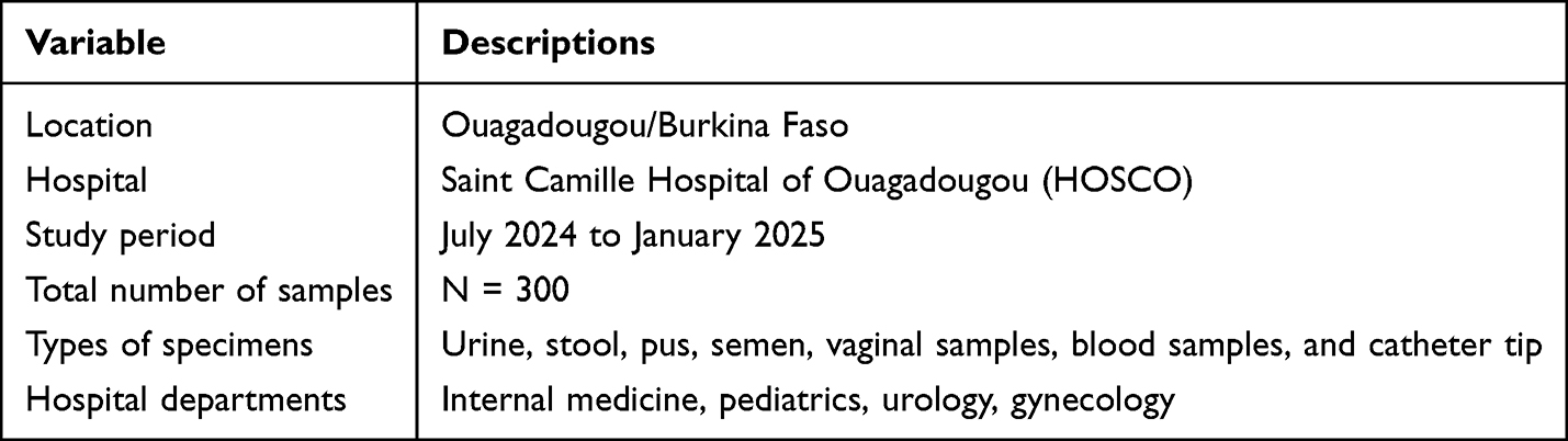 |
Table 1 Study Site and Sample Characteristics |
The sensitivity of bacterial strains to aminoglycosides was evaluated by diffusion on Mueller-Hinton (MH) agar, in accordance with the protocols of the Antibiogram Committee of the French Society of Microbiology (EUCAST/CA-SFM, 2024).
Discs impregnated with Gentamicin (10 µg), Kanamycin (30 µg), Tobramycin (10 µg), Netilmicin (10 µg), Neomycin (10 µg), Amikacin (30 µg) and streptomycin (10 µg) were tested. To validate the antibiogram, a strain of Escherichia coli ATCC 25922 provided by the microbiology laboratory at the Princess Sarah Clinic was used in accordance with the protocols of the Antibiogram Committee of the French Society of Microbiology (EUCAST/CA-SFM, 2024). Only strains resistant to at least one of the antibiotics tested were considered for molecular analysis. The isolate selection process is summarized in Figure 1.
 |
Figure 1 Flow diagram of isolate selection and analysis process. Diagram showing the selection process of bacterial isolates included in the study, from sample collection to phenotypic antimicrobial susceptibility testing and molecular detection of résistance genes. |
Inclusion and Exclusion Criteria for This Study
All patients who attended consultations at HOSCO during the study period and who were prescribed a bacteriological examination as part of their treatment were included in this study.
All isolates not identified as E. coli and duplicates from the same patient were excluded from this study.
Molecular Analysis
At the molecular level, the study focused on identifying resistance genes using conventional PCR applied to total genomic DNA extracted from the analyzed strains. In order to elucidate the genetic basis of these genes, PCR was then performed on plasmid DNA obtained from strains that had tested positive during the initial amplification. Genes identified in both genomic and plasmid DNA were considered to be carried by plasmids, while genes amplified only from genomic DNA were considered to be carried by the bacterial chromosome.
This approach allows us to infer the probable location of the detected genes, although definitive confirmation would require genomic sequencing.8
Extraction of Total Genomic DNA
Total genomic DNA extraction from bacterial strains was performed by heat shock.9 Pure colonies, cultured for 24 hours, were selected and suspended in 200 μL of sterile distilled water, then placed in properly labeled Eppendorf tubes. These tubes were then incubated in a water bath at 100°C for 15 minutes to promote the release of bacterial genetic material.
After the procedure, centrifugation was performed for 10 minutes at 13,000 rpm, allowing the supernatant containing the extracted DNA to be recovered, which was then transferred to a new Eppendorf tube.
Plasmid DNA Extraction
Plasmid DNA from the strains was extracted using the alkaline lysis method described by Birnboim.10
The selected bacterial strains were cultured in LB (Luria Bertani) broth at 37°C for 24 hours. Then, 1.5 mL of each bacterial suspension was collected in labeled cryotubes. After centrifugation at 12,000 g for 3 min, the supernatants were removed. This step was repeated once to obtain a sufficient amount of pellet containing the sedimented bacterial cells.
The pellet was recovered and placed in a sterile microtube and suspended in 150 μL of 25 mM Tris-HCl buffer, pH 8; 10 mM EDTA.
In order to lyse the bacterial cells, 300 μL of 200 mM sodium hydroxide, 1% SDS prepared fresh was added and then incubated at room temperature for 5 min. Next, 225 μL of 3 M sodium acetate, pH 5.5, was added to precipitate the chromosomal DNA initially denatured by the sodium hydroxide and the proteins complexed by the SDS detergent.
The mixture was homogenized by successive inversions (10 times) and the tubes were placed on ice for 10 min. Centrifugation at 12,000 g for 10 min was then performed to separate the genomic DNA and plasmid DNA.
The supernatant containing soluble plasmid DNA was recovered in another sterile Eppendorf tube. Two volumes of fresh absolute ethanol were added and the mixture was incubated at −20°C for 30 min to precipitate the plasmid DNA.
After incubation, the tubes were centrifuged at 12,000 g for 15 minutes and the supernatants were removed. The pellet containing the plasmid DNA was purified by adding 300 μL of 75% ethanol and centrifuging for 15 minutes at 12,000 g at 4°C.
The supernatant was removed and the pellet was dried for a few minutes at 37°C.
The plasmid DNA obtained was eluted in 50 μL of sterile distilled.
DNA quantification and purity assessment were performed using NanoDrop. Consequently, any sample with an A260/A280 absorbance ratio between 1.8 and 2 was considered pure.
Amplification of Extracts by Conventional PCR
DNA extracts were amplified by conventional PCR (polymerase chain reaction) using the GeneAmp PCR System 9700 thermocycler (Applied Biosystems, USA). This approach enabled the detection of the aac(6’)-II, aac(3)-II, aph(3’)-VI, and ant(3”)-I genes. The reaction mixture, with a total volume of 20 μL, consisted of 4 μL of Firepol® Master Mix 5X, 0.6 μL of sense primer, 0.6 μL of antisense primer, 12.8 μL of PCR water, and 2 μL of bacterial DNA from each strain.11 A negative control including the elution buffer was included to ensure that there was no contamination. Table 2 presents a summary of the primer sequences and the PCR program used.
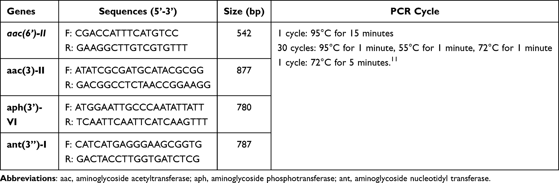 |
Table 2 Primers and PCR Program |
Agarose Gel Electrophoresis
The DNA fragments amplified by PCR were separated by electrophoresis on a 1.5% agarose gel prepared in a 1X Tris-acetate-EDTA solution with 8 μL of ethidium bromide added. A 100 bp molecular weight marker was used to estimate the size of the fragments obtained for the aac(6’)-II and ant(3”)-I genes. For the aac(3)-II and aph(3’)-VI genes, a 1 kb molecular weight marker was used. Migration was performed at a voltage of 100 V for 35 minutes. After migration, the DNA fragments were visualized under UV light using the Vilber E-Box Transilluminator, and the images were recorded.
Characterization of Resistance Genes
The DNA of strains resistant to at least one of the antibiotics tested was analyzed by conventional PCR to detect the presence of the aac(6’)-II, aac(3)-II, aph(3’)-VI, and ant(3”)-I genes. As the primer pairs used were specific to the genes sought, the presence of bands measuring 877 bp, 780 bp, 542 bp and 787 bp was defined as characteristic of the presence of the aac(3)-II and aph(3’)-VI (Figure 2), aac(6’)-II and ant(3”)-I (Figure 3), respectively.
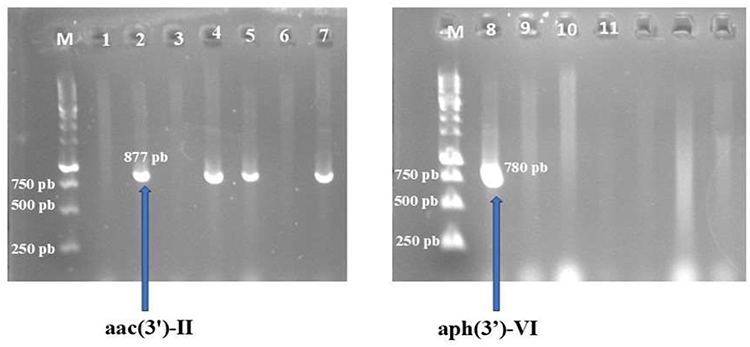 |
Figure 2 Agarose gel of PCR products aac(3)-II and aph (3’)-VI. M: 1 Kb molecular weight marker; 1: negative control 3.6: absence of the aac(3)-II gene; 2,4,5,7: presence of the aac(3)-II gene; 8: presence of the aph (3’)-VI gene; 9, 10, and 11: absence of the aph(3’)-VI gene. |
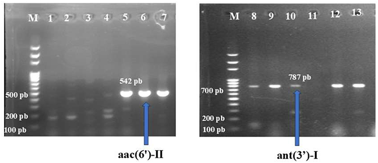 |
Figure 3 Agarose gel of PCR products aac(6’)-II and ant(3”)-I. M: molecular weight marker 100 bp; 1: negative control; 2,3: absence of the aac(6’)-II gene; 4,5,6,7: presence of the aac(6’)-II gene; 8,9,10,12,13: presence of the ant(3”)-I gene; 11: absence of the ant(3”)-I gene. |
Statistical Analysis
The collected data were entered into Excel 2019 software and then checked for consistency before statistical analysis. Antibiotic resistance rates and the prevalence of resistance genes were calculated using SPSS 21 software. The association between phenotypic resistance and the presence of resistance genes was analyzed using Fisher’s exact test. The statistical significance threshold was set at a value of p ˂ 0.05.
Results
Isolation and Identification
Out of a total of 300 Gram-negative bacilli collected, Escherichia coli accounted for 63.66% (191/300) of isolates. These E. coli strains were isolated in 134 (70.1%) of urine samples, 30 (15.7%) of stool samples, 19 (9.9%) of pus samples, and 5 (2.6%) of semen samples. Vaginal samples, blood samples, and catheter tip samples each accounted for 1 (0.5%).
Antibiotic Sensitivity Tests
Our results reveal a higher resistance rate to streptomycin 91 (47.6%) compared to kanamycin and gentamicin, which recorded resistance rates of 42 (22.0%) and 36 (18.8%), respectively (Table 3). Only 3 (1.6%) of isolates were resistant to amikacin.
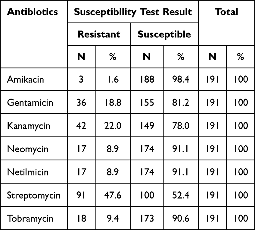 |
Table 3 Susceptibility of Isolates to Antibiotics |
Frequency of Resistance Genes
Of the 191 E. coli strains analyzed in this study, 102 (53.40%) showed resistance to at least one of the antibiotics tested and were subjected to molecular analysis.
In this study, the aac(6’)-II and aac(3)-II genes were the most frequently detected in E. coli strains, with frequencies of 89 (87.3%) and 38 (37.3%), respectively (Figure 4). The ant(3”)-I and aph(3’)-VI genes were present in 33 (32.4%) and 2 (2.0%) of isolates, respectively. In addition, 21 (20.58%) of isolates combined the aac(6’)-II and ant(3”)-I genes, and 2 (2.0%) carried both aac(3)-II and aph(3’)-VI.
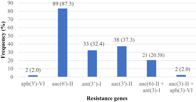 |
Figure 4 Frequency of resistance genes. aac(6’)-II + ant(3”)-I: Coexistence of the aac(6’)-II and ant(3”)-I genes in the same strain; aac(3’)-II and aph(3’)-VI: Coexistence of the aac(3’)-II and aph(3’)-VI genes in the same strain. |
Association Between Gene and Phenotypic Resistance
Fisher’s exact test showed a significant association between the presence of the aac(3)-II gene and resistance to certain antibiotics such as: Gentamicin (p ˂ 0.001), Kanamycin (p = 0.002), Netilmicin (p < 0.005) and Tobramycin (p ˂ 0.001) (Table 4).
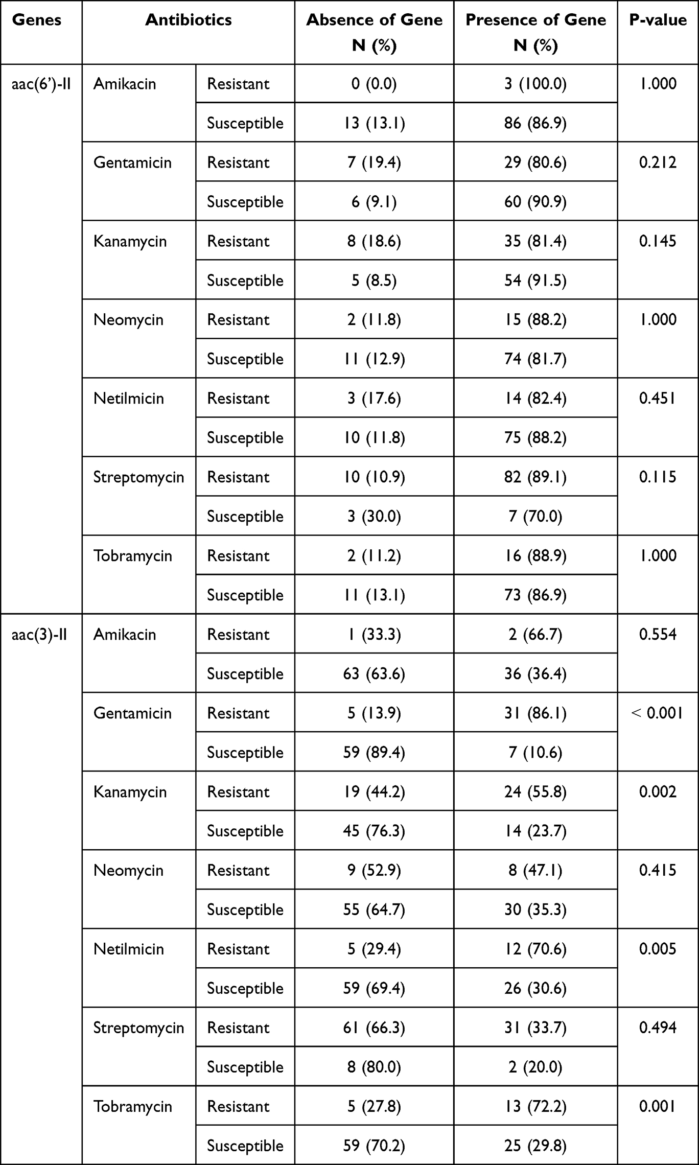 |
Table 4 Association Between the Aac(6’)-II and Aac(3’)-II Genes and Phenotypic Resistance |
In addition, the presence of the aph(3’)-VI gene was significantly associated with the resistance of E. coli strains to Netilmicin (p = 0.026) and Neomycin (p = 0.026) (Table 5). Conversely, no association was observed between the carriage of the aac(6’)-II and ant(3”)-I genes and resistance to the antibiotics tested.
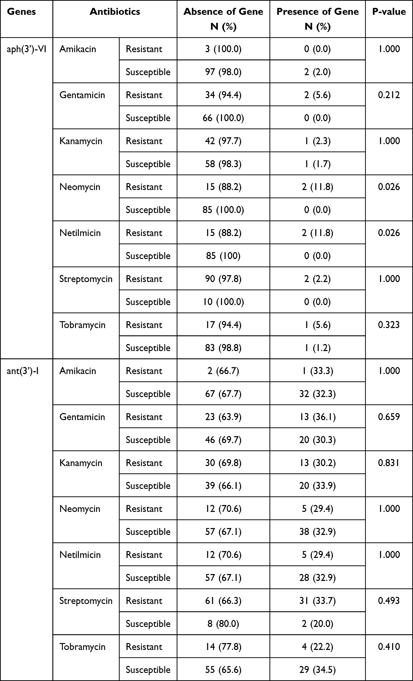 |
Table 5 Association Between the Aph(3’)-VI and Ant(3’)-I Genes and Phenotypic Resistance |
Genetic Carriers of Resistance Genes
Molecular analysis revealed that 26 (68.42%) of the aac(3’)-II genes detected in this study were carried by plasmids (Table 6). As for the ant(3”)-I genes detected, 11 (33.33%) were carried by plasmids, compared to 22 (66.67%) carried by chromosomal DNA. The majority of aac(6’)-II genes 88 (98.88%) were carried by chromosomes.
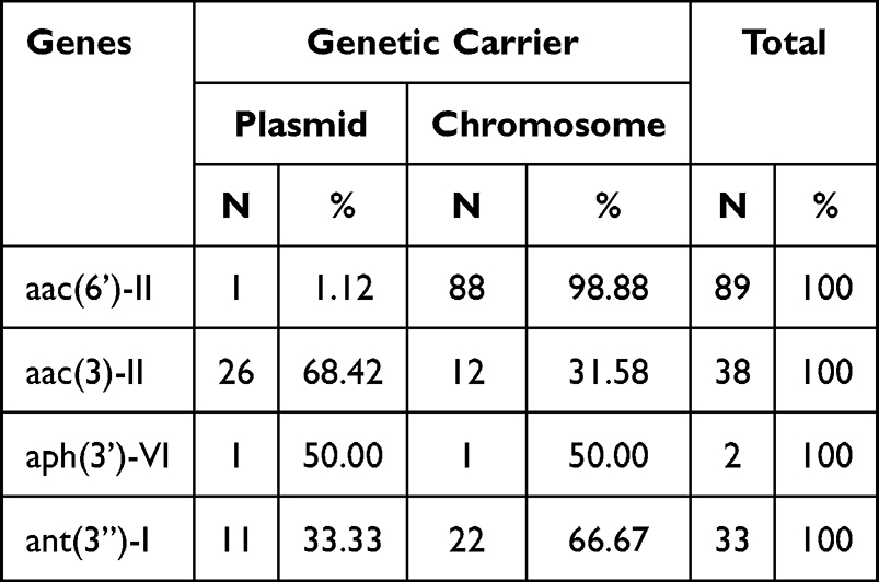 |
Table 6 Genetic Carrier of Aminoglycoside Resistance Genes |
Discussion
This study conducted at Saint-Camille Hospital in Ouagadougou focused on the coexistence and genetic location of aminoglycoside-modifying enzymes in Escherichia coli. Of the 300 Gram-negative bacilli isolates analyzed, Escherichia coli strains accounted for 191 (63.66%). This result confirms the significant presence of this bacterium among Gram-negative bacilli involved in clinical infections.
From a phenotypic point of view, 47.6% of the isolates were resistant to streptomycin, while 22% were resistant to kanamycin, 18.8% to gentamicin, and only 1.8% to amikacin. The high rate of resistance to streptomycin is probably indicative of its prolonged and widespread use, thus promoting the emergence of resistant strains. Conversely, the low rate of resistance to amikacin suggests that this antibiotic class remains relatively unaffected in our environment. These results are consistent with previous studies that have shown higher levels of resistance to older or more frequently used aminoglycosides, as well as lower rates of resistance to antibiotics that are still relatively rare or underused.12,13
At the molecular level, a predominance of the aac(6′)-II gene (87.3%) was observed in our study. This gene codes for an acetyltransferase responsible for modifying aminoglycosides, particularly gentamicin and tobramycin, and has been identified in numerous studies as a key resistance marker. In addition, the aac(3)-II gene was identified in 37.3% of cases, ant(3″)-I in 32.4%, and aph(3′)-VI in 2.0%. This diversity of aminoglycoside-modifying genes highlights the complexity of the resistance mechanisms present in our bacterial strains.
Previous research has shown that the prevalence of these genes varies according to geographical context and local antibiotic use practices.11,14 To our knowledge, no African publication mentions the presence of the aac(6′)-II gene in clinical isolates of E. coli. Therefore, ours is probably the first in the region. In Tunisia, aac(3)-II and aph(3′)-VI were identified in 42% and 32.4% of aminoglycoside-resistant isolates, respectively, while ant(3″)-I was mentioned in 32.4% of the same isolates.15 In Morocco, aac(3)-II and aph(3′)-VI were found in 27.7% and 8.3% of aminoglycoside-resistant Enterobacteriaceae.16
The coexistence of several resistance genes within the same strain was observed in our study. Indeed, the combination of aac(6′)-II and ant(3″)-I was found in 20.58% of isolates, while the combination of aac(3)-II and aph(3′)-VI was observed in 2.0% of cases. This accumulation of resistance determinants implies that the strains do not depend on a single mechanism, but acquire a significant genetic arsenal that enhances their ability to resist various aminoglycosides and potentially other classes of antibiotics. Such associations have also been observed in various clinical and veterinary settings.16,17
Our results reveal a significant correlation between the presence of the aac(3)-II gene and resistance to several aminoglycosides, including gentamicin, kanamycin, netilmicin, and tobramycin. Similarly, the aph(3′)-VI gene was associated with resistance to both netilmicin and neomycin. These results confirm the crucial role of these genes in aminoglycoside resistance in E. coli, as demonstrated in numerous previous studies. The significant correlation with gentamicin observed in this study indicates that it is a crucial molecular marker of resistance in our hospital environment. In addition, the aph(3′)-VI gene is frequently carried by plasmids, thus promoting horizontal transfer of resistance between different strains.18,19
In contrast, the lack of a notable correlation between the aac(6′)-II and ant(3″)-I genes and phenotypic resistance could indicate low gene expression or a reduced role for these mechanisms in local strains. These results suggest that other factors influence the expression of these enzymes. Sequencing analyses would be useful to identify possible point mutations or regulatory elements that could modulate gene activity.
Comparable geographical disparities have been observed in other countries, where the profile of aminoglycoside resistance genes is frequently influenced by antibiotic therapy practices.20
Study of the genetic location for these genes reveals that the aac(6′)-II gene is located on a chromosome in 98.88% of cases, indicating stable integration and fixation within the genomes of local strains.8 Conversely, the aac(3’)-II gene is mainly localized on a plasmid in 68.42% of cases, while the aph(3’)-VI and ant(3”)-I genes are associated with plasmids in 50.0% and 33.33% of cases, respectively.
Chromosomal genes are less likely to be transferred but show greater persistence within a strain, while plasmid genes facilitate their transfer between strains or species, thus promoting their dissemination. This is consistent with existing literature data, which identify plasmids, integrons, or transposons as the main vectors for horizontal resistance transfer.21
The significant presence of plasmid genes, particularly aac(3)-II, suggests a considerable risk of dissemination. In a hospital environment characterized by high antibiotic pressure and rapid patient turnover, genetics plays a crucial role. Secondly, although the aac(6′)-II gene is predominantly chromosomal, this does not solve the problem, this gene is already well established within the local bacterial population, posing a significant challenge for its eradication. The genetic location of resistance genes was determined in our study using an indirect approach based on separate amplifications performed using total genomic DNA and plasmid DNA, which limits the generalizability of the results. Although this strategy provides an indication of genetic location, approaches based on whole-genome sequencing will allow the location of these genes to be confirmed with precision.
The simultaneous presence of multiple genes within the same strain can reduce the effectiveness of aminoglycosides, posing a major therapeutic challenge, especially when these agents are the last resort. Therefore, these results highlight the urgent need for rigorous antibiotic stewardship, enhanced hygiene practices in healthcare facilities, and continuous molecular surveillance to identify strains with an increased risk of spread.
The clinical significance of this study lies in the fact that aminoglycosides are frequently used in combination with other molecules for the treatment of invasive infections, one of the main causative agents of which is Escherichia coli. Thus, the detection of a high prevalence of resistance genes in our context suggests an increased risk of failure of probabilistic regimens including these molecules. These data therefore provide essential information for adapting local treatment protocols and strengthening rational antibiotic therapy strategies. Despite the high detection of aminoglycoside-modifying genes, the high sensitivity to amikacin in this study suggests that this molecule remains an effective therapeutic option in our context. These data are essential for guiding local probabilistic antibiotic therapy and supporting strategies for the rational use of aminoglycosides.
Conclusions
This study highlights the high prevalence of genes encoding aminoglycoside-modifying enzymes, particularly aac(6′)-II and aac(3)-II, in clinical isolates of E. coli from Saint-Camille Hospital in Ouagadougou. The notable correlation between these genes and resistance to various aminoglycosides illustrates their crucial role in the phenotypic resistance observed. The presence of several genes within the same isolate, as well as their plasmid transfer, indicate an increased risk of horizontal transmission in hospital settings. These results highlight the need to strengthen molecular surveillance of resistance genes, limit the empirical use of aminoglycosides, and develop infection control strategies based on bacterial genomics. However, extending this study to other hospitals and other bacterial species would provide a broader view of the regional dynamics of aminoglycoside resistance. In addition, genomic analysis by sequencing would refine our understanding of the mechanisms of resistance and their spread.
Ethical Considerations
This study was approved by the Institutional Ethics Committee of the Saint-Camille Hospital in Ouagadougou (HOSCO) under authorization No. 2024-05-005.
Acknowledgments
We would like to thank the administrative authorities of Joseph Ki-Zerbo University and the entire team at the Laboratory of Molecular Biology and Genetics (LABIOGENE) who worked to carry out this study.
Funding
The authors received no specific grant from any funding agency.
Disclosure
The authors report no conflicts of interest in this work.
References
1. Founou RC, Founou LL, Essack SY. Clinical and economic impact of antibiotic resistance in developing countries: a systematic review and meta-analysis. PLoS One. 2017;12(12):e0189621. doi:10.1371/journal.pone.0189621
2. Global antibiotic resistance surveillance report 2025. Available from: https://www.who.int/publications/i/item/9789240116337.
3. Serio AW, Keepers T, Andrews L, Krause KM. Aminoglycoside revival: review of a historically important class of antimicrobials undergoing rejuvenation. EcoSal Plus. 2018;8(1). doi:10.1128/ecosalplus.esp-0002-2018
4. Garneau-Tsodikova S, Labby K J. Mechanisms of resistance to aminoglycoside antibiotics: overview and perspectives. MedChemComm. 2016;7(1):
5. Ramirez MS, Tolmasky ME. Amikacin: uses, resistance, and prospects for inhibition. Molecules. 2017;22(12):2267. doi:10.3390/molecules22122267
6. Yang W, Hu F. Research updates of plasmid-mediated aminoglycoside resistance 16S rRNA methyltransferase. Antibiotics. 2022;11(7):906. doi:10.3390/antibiotics11070906
7. El-Far SW, Abukhatwah MW. Prevalence of aminoglycoside resistance genes in clinical isolates of pseudomonas aeruginosa from Taif, Saudi Arabia—an emergence indicative study. Microorganisms. 2023;11(9):2293. doi:10.3390/microorganisms11092293
8. Goswami C, Fox S, Holden MTG, Connor M, Leanord A, Evans TJ. Origin, maintenance and spread of antibiotic resistance genes within plasmids and chromosomes of bloodstream isolates of Escherichia coli. Microbial Genomics. 2020;6(4):e000353. doi:10.1099/mgen.0.000353
9. Amana MD, Wend-Kuni TRY, Aminata BY, et al. Detection of multidrug-resistant enterobacteria simultaneously producing extended-spectrum -lactamases of the PER and GES types isolated at Saint Camille Hospital Center, Ouagadougou, Burkina Faso. AJMR. 2019;13(26):
10. Birnboim HC, Doly J. A rapid alkaline extraction procedure for screening recombinant plasmid DNA. Nucleic Acids Res. 1979;7(6):
11. El-Badawy MF, Tawakol WM, El-Far SW, et al. Molecular identification of aminoglycoside-modifying enzymes and plasmid-mediated quinolone resistance genes among Klebsiella pneumoniaeclinical isolates recovered from Egyptian patients. Int J Microbiol. 2017;2017:8050432. doi:10.1155/2017/8050432
12. Ghonaim MM, Mostafa RG, Alkady SS. Phenotypic and molecular characterization of aminoglycoside resistance in clinical Escherichia coli isolates from patients at Menoufia University hospitals. Egyptian J Med Microbiol. 2019;28(3):
13. Shi Y, Li C, Yang G, et al. A rapid and accurate method for the detection of four aminoglycoside modifying enzyme drug resistance gene in clinical strains of Escherichia coli by a multiplex polymerase chain reaction. PeerJ. 2020;8:e8944. doi:10.7717/peerj.8944
14. Soleimani N, Aganj M, Ali L, Shokoohizadeh L, Sakinc T. Frequency distribution of genes encoding aminoglycoside modifying enzymes in uropathogenic E. coli isolated from Iranian hospital. BMC Res Notes. 2014;7(1):842. doi:10.1186/1756-0500-7-842
15. Harbaoui S, Ferjani S, Abbassi MS, et al. Genetic background of aminoglycoside-modifying enzymes in various genetic lineages of clinical aminoglycosides-resistant E. coli and K. pneumoniae isolates in Tunisia. J Appl Microbiol. 2024;135(7):lxae164. doi:10.1093/jambio/lxae164
16. Essalhi A, Nayme K, Maaloum F, et al. Characterization of aminoglycoside-modifying enzymes in uropathogenic enterobacterales of community origin in Casablanca, Morocco. Acta Microbiol Hellenica. 2024;69(4):
17. Belaynehe KM, Shin SW, Hong-Tae P, Yoo HS. Occurrence of aminoglycoside-modifying enzymes among isolates of Escherichia coli exhibiting high levels of aminoglycoside resistance isolated from Korean cattle farms. FEMS Microbiol Lett. 2017;364(14):fnx129.
18. Krause KM, Serio AW, Kane TR, Connolly LE. Aminoglycosides: an overview. Cold Spring Harb Perspect Med. 2016;6(6):a027029. doi:10.1101/cshperspect.a027029
19. Ramirez MS, Tolmasky ME. Aminoglycoside modifying enzymes. Drug Resist Updates. 2010;13(6):
20. Poirel L, Madec JY, Lupo A, et al. Antimicrobial resistance in Escherichia coli. Microbiol Spectr. 2018;6(4). doi:10.1128/microbiolspec.ARBA-0026-2017
21. Azimi L, Armin S, Samadi Kafil H, et al. Evaluation of phenotypic and genotypic patterns of aminoglycoside resistance in the Gram-negative bacteria isolates collected from pediatric and general hospitals. Mol Cell Pediatr. 2022;9(1):2. doi:10.1186/s40348-022-00134-2
 © 2026 The Author(s). This work is published and licensed by Dove Medical Press Limited. The
full terms of this license are available at https://www.dovepress.com/terms
and incorporate the Creative Commons Attribution
- Non Commercial (unported, 4.0) License.
By accessing the work you hereby accept the Terms. Non-commercial uses of the work are permitted
without any further permission from Dove Medical Press Limited, provided the work is properly
attributed. For permission for commercial use of this work, please see paragraphs 4.2 and 5 of our Terms.
© 2026 The Author(s). This work is published and licensed by Dove Medical Press Limited. The
full terms of this license are available at https://www.dovepress.com/terms
and incorporate the Creative Commons Attribution
- Non Commercial (unported, 4.0) License.
By accessing the work you hereby accept the Terms. Non-commercial uses of the work are permitted
without any further permission from Dove Medical Press Limited, provided the work is properly
attributed. For permission for commercial use of this work, please see paragraphs 4.2 and 5 of our Terms.
Recommended articles

The First Report of Escherichia coli and Klebsiella pneumoniae Strains That Produce Both NDM-5 and OXA-181 in Jiangsu Province, China
Tao G, Tan H, Chen Q
Infection and Drug Resistance 2023, 16:3245-3255
Published Date: 24 May 2023