
Back to Journals » Journal of Inflammation Research » Volume 19
Clinical Profile of Patients with Urticarial Vasculitis in China: A Retrospective of 142 Cases
Authors Hu W  , Zhu Y, Geng S, Cao Z , Peng B
Received 7 November 2025
Accepted for publication 19 December 2025
Published 23 January 2026 Volume 2026:19 576374
DOI https://doi.org/10.2147/JIR.S576374
Checked for plagiarism Yes
Review by Single anonymous peer review
Peer reviewer comments 2
Editor who approved publication: Dr Anish R. Maskey
Weihong Hu, Yanyan Zhu, Songmei Geng, Zhiqiang Cao, Bin Peng
Department of Dermatology, Northwest Hospital, The second Affiliated Hospital of Xi’an Jiaotong University, Xi’an, Shaanxi, 710004, People’s Republic of China
Correspondence: Bin Peng, Department of Dermatology, Northwest Hospital, The Second Affiliated Hospital of Xi’an Jiaotong University, No. 157 Xiwu Road, Xi’an, Shaanxi, 710004, People’s Republic of China, Email [email protected]
Background: Normocomplementemic urticarial vasculitis (NUV) and hypocomplementemic urticarial vasculitis (HUV) share clinical features, notably wheals, yet clear differential diagnostic criteria are lacking.
Objective: To describe HUV and NUV features in Chinese inpatients for diagnosis.
Methods: Retrospective study (March 2017–June 2024, China) of NUV/HUV patients. We analyzed clinical characteristics, laboratory results, and histological features, and compared drug efficacy.
Results: Females constituted the majority of patients with urticarial vasculitis(UV). All patients present with wheals persisting for more than 24 hours, and 76.1% of the wheals exhibited partial or complete non-blanching. Angioedema was more common in patients with HUV than in NUV (44.4% vs 25.0%, p< 0.05). Systemic involvement was observed in 61.1% of HUV and 41.2% of NUV. HUV was significantly associated with autoimmune diseases (p< 0.05), decreased complement levels (C3, C4, C1q, and C1 inhibitor)(p< 0.001), and elevated erythrocyte sedimentation rate (ESR) (p< 0.01). A negative correlation between ESR and C3 levels was found in HUV, in contrast to a positive correlation in NUV. Direct immunofluorescence (DIF) test showing immunoreactants at the blood vessel were present in 50% of patients with HUV and 18.2% patients with NUV. Regarding treatment, 91.9% of NUV responded effectively to oral corticosteroids, whereas only 66.7% of HUV did; the remaining 33.3% of HUV required combination therapy with biologics.
Conclusion: Our study identified key factors associated with the occurrence of UV subtypes. For patients with recurrent wheals, complement levels, ESR, and the distribution pattern of immunofluorescence deposits can serve as basis for classifying UV subtypes and prompt earlier treatment.
Keywords: urticarial vasculitis, hypocomplementemic, epidemiology, clinical profile
Introduction
Urticarial vasculitis (UV) is an immune mediated disease with a complex etiology and pathogenesis.1 It typically presents as a persistent urticaria-like rash, which may be accompanied by systemic manifestations such as arthritis and arthralgia, and multi-organ involvement. Although the exact etiology of UV remains unclear, it is thought to involve immune complex deposition within vascular walls, complement activation, and the generation of antibodies against C1q.2,3 These processes trigger the complement cascade, resulting in the production of C3a and C5a, which promote mast cell degranulation, histamine release, vasodilation, increased vascular permeability, and ultimately urticarial lesions.4
Based on serum complement levels, UV can be subdivided into 2 main entities: normocomplementemic urticarial vasculitis (NUV) and hypocomplementemic urticarial vasculitis (HUV).1 Although the reported prevalence of HUV among UV patients ranges between 9% and 21%,5,6 HUV is associated with abnormal complement activation and a potentially more severe disease course. Pulmonary, gastrointestinal, and renal involvement are more commonly observed in HUV.7
Differentiating HUV from NUV represents a significant diagnostic challenge in clinical practice.8 This difficulty stems largely from the substantial overlap in their core clinical presentations, primarily characterized by urticarial lesions persisting for more than 24 hours, which may resolve with post-inflammatory hyperpigmentation. Consequently, HUV is frequently subject to delayed diagnosis.9–11 Such misclassification carries important therapeutic implications. Patients with HUV are often initially managed according to treatment algorithms established for NUV, which typically involve non-steroidal anti-inflammatory drugs or short courses of corticosteroids. However, HUV often necessitates earlier and more aggressive intervention with immunosuppressants or biologic agents to control systemic inflammation and prevent involvement of critical organs.12 Therefore, diagnostic inaccuracy may result in suboptimal treatment and adversely affect patient prognosis.
Globally, the current understanding of the epidemiology, distinct clinical spectra, and long-term outcomes of different UV subtypes remains limited. This knowledge gap is particularly evident for Chinese populations, for whom comprehensive data are scarce.
To improve the differentiation between NUV and HUV and to reduce diagnostic delay in HUV, this research comprehensively describes the epidemiological and clinical profiles of UV and explores potential discriminators between HUV and NUV applicable to clinical practice in China.
Materials and Methods
Study Design and Patients
This retrospective study was conducted at the Second Affiliated Hospital of Xi’an Jiaotong University. Patients diagnosed with urticarial vasculitis (UV) between March 2017 and June 2024 were included, with data collection and analysis performed from July to September 2025. The study followed the STROBE guidelines for reporting observational research. The Institute Research Committee approved the study. Patient consent was not required by the ethical board.
A total of 371 patients were initially diagnosed with UV. 145 patients met the inclusion criteria of UV; however, three were excluded, as their serum complements were not available. The remaining 142 patients were further categorized to NUV and HUV based on complement levels (Figure 1). HUV was defined as complement C3 < 70 mg/dL (normal range: 70–140mg/dL) or C4 < 10mg/dL (normal range: 10–40mg/dL).1,13 The demographic data, clinical features, histopathological findings and medication treatment of the patients were reviewed.
 |
Figure 1 Flowchart showing the selection of cases. |
Inclusion Criteria: recurrent urticarial lesions lasting more than 24 hours, accompanied by symptoms such as pruritus or burning sensation; histopathological findings showing vasculitis changes (Figure 2A). Exclusion Criteria: a history of receiving medications such as glucocorticoids within the 4 weeks prior to study enrollment; concurrent hepatic or renal dysfunction; pregnancy or lactation; diagnosis of rheumatoid arthritis or other forms of vasculitis; concomitant malignancy.
 |
Figure 2 Histopathological findings of UV. (A) Mild dermal edema with perivascular inflammatory infiltrates in the superficial and mid dermis (hematoxylin and eosin stain, x100). The region magnified x400, showing infiltrates dominated by eosinophils, neutrophils, and lymphocytes with erythrocyte extravasation. (B) DIF showing granular deposition of C3 within capillary walls and basement membrane zone (red arrow). |
Clinical and Laboratory Assessment
The collected data included gender, age, disease duration, manifestations of skin lesions, systemic symptoms, as well as involvement of the rheumatologic, pulmonary, renal, gastrointestinal, ophthalmic, cardiac, and nervous systems.
Involvement of specific organ systems was defined as follows: renal involvement was defined by the presence of persistent proteinuria (qualitative urine protein ≥ ++ or 24-hour urine protein > 0.5 g) and/or hematuria (urine sediment red blood cells > 5 per high-power field); joint involvement was defined by documented arthritis or arthralgia; gastrointestinal involvement was defined as unexplained abdominal pain, nausea, or vomiting, confirmed by imaging studies or gastroenterology consultation; pulmonary involvement was determined based on chest imaging (X-ray or CT) reports and pulmonology consultation findings.
Laboratory evaluations encompassed the determination of serum complement components (C3, C4, C1q, C1 inhibitor), antinuclear antibody (ANA), anti-double-stranded DNA antibody, anti-extractable nuclear antigen antibody, antineutrophil cytoplasmic antibody (ANCA), inflammatory markers (ESR) and inflammatory cytokines (IL-1, IL-6).14–16 Therapeutic regiments were also documented. All enrolled patients underwent histopathological review of skin biopsy specimens by a dermatopathologist to confirm the diagnosis. Skin lesions with a disease duration of less than 48 hours were preferentially selected as samples. Among the 142 cases, 39 additionally underwent direct immunofluorescence (DIF) testing for human IgG, IgM, IgA, and C3,which aided in differentiating UV from other vasculitic or autoimmune disorders.
Statistical Analysis
Statistical analysis was performed with GraphPad Prism version 8.3.0. In the descriptive analysis, frequencies and percent ages for categorical variables and mean and standard deviation were used for quantitative variables. Differences between groups were analysed by Student’s t test, Mann Whitney U-test and x2 test, and data were presented as the mean and S.D or the median when appropriate. For variables with missing data (accounting for <5% of the total dataset), we adopted a complete case analysis approach. Prior to conducting independent-samples/paired-samples t-tests, we assessed the normality of all continuous variables. Among these analytical variables, some satisfied the normality assumption (p>0.05), while others failed to meet this assumption. For variables that did not conform to the normality assumption, we employed a nonparametric test (Mann–Whitney U-test) for analysis. A value of p<0.05 was considered significant.
Results
Patients and Disease Characteristics
This study ultimately included 142 patients with urticarial vasculitis (UV) for analysis. The cohort was predominantly female (61.3%), with a median age at diagnosis of 46.5 years. All patients presented with skin lesions persisting for more than 24 hours, accompanied by pruritus, and 15.5% reported a burning sensation (Table 1). Regarding systemic involvement, 24 patients (16.9%) had pulmonary involvement, 2 patients (1.4%) had autoimmune disease and 7 patients (4.9%) had thyroid dysfunction.
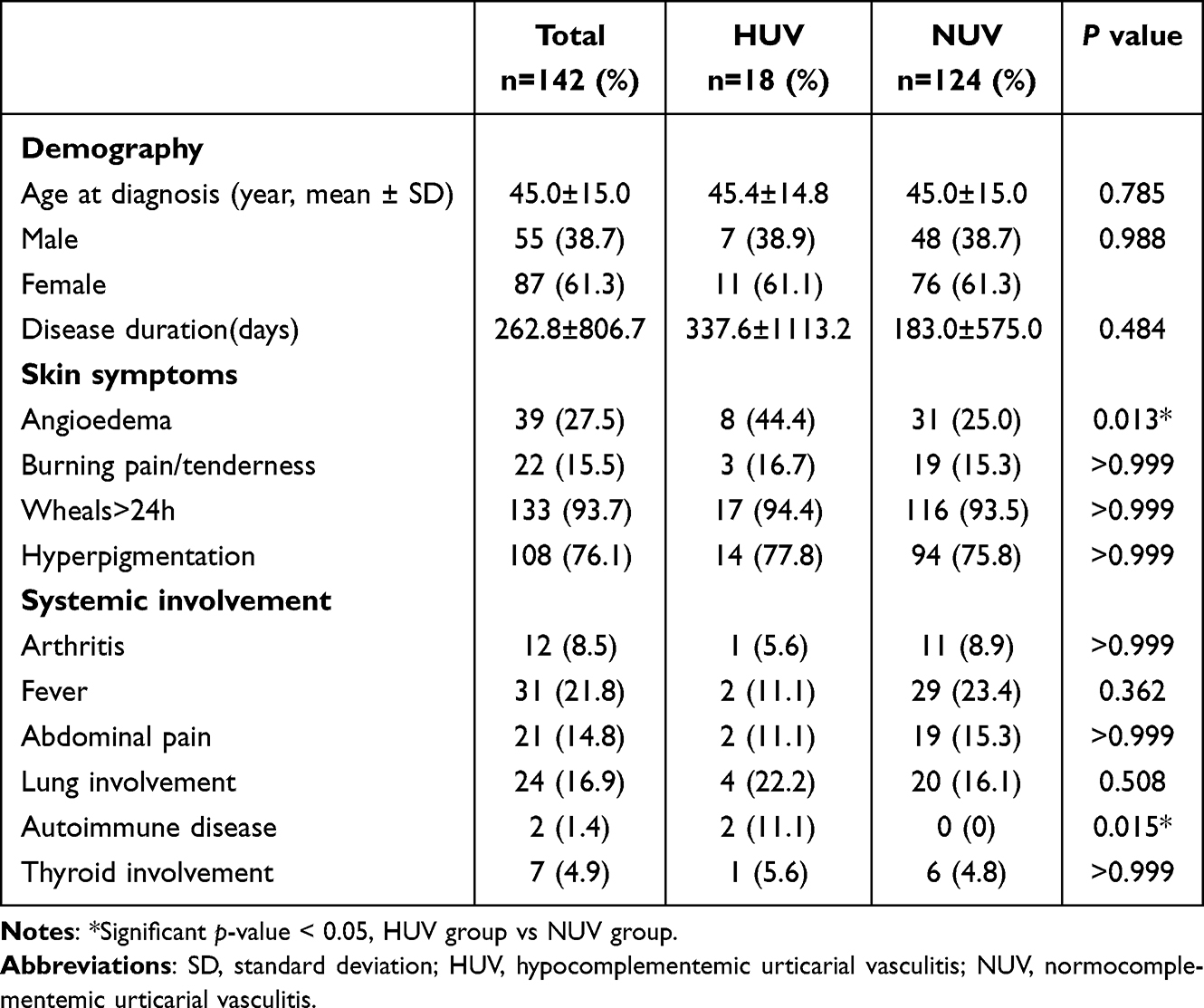 |
Table 1 Patient and Disease Characteristics |
Among the 142 patients, 124 were classified as NUV and 18 as HUV. Statistically significant differences were observed in certain clinical features between the two groups. Angioedema was more common in HUV patients than in NUV patients (44.4% vs 25.0%, p=0.013; Table 1), and autoimmune diseases were exclusively observed in HUV patients (1 case of lupus nephritis(LN) and 1 case of Sjögren’s syndrome(SS)), with this difference also reaching statistical significance (p=0.015; Table 1).
Despite a numerically longer disease duration in HUV (337.6 ± 1113.2days) compared to NUV (183.0±575.0days), the difference was not statistically significant (p=0.484; Table 1). Additionally, the prevalence of arthralgia, arthritis, fever, and abdominal pain did not differ significantly between the two groups.
Laboratory Examination
This study analyzed routine laboratory findings and key immunological parameters in 142 patients with UV. Several laboratory parameters differed significantly between the HUV and NUV groups (Table 2 and Figure 3). As expected, complement levels (C3, C4, C1q, and C1 inhibitor) were significantly lower in HUV patients compared to NUV patients (p < 0.001; Table 2). And the ESR was significantly higher in the HUV group (p < 0.01; Figure 3A). Notably, the correlation between C3 levels and ESR varied distinctly by group: In HUV patients, C3 levels were negatively correlated with ESR (r = 0.694; Figure 3D), whereas a positive correlation was observed in NUV patients (r = 0.600; Figure 3E).
 |
Table 2 Laboratory Investigations |
 |
Figure 3 Correlation of complement and ESR, and inflammatory/cytokine in HUV and NUV. (A) C3 was negatively correlated with ESR in HUV (r=0.694, p=0.04); (B) C3 was positively correlated in NUV (r=0.600, p<0.01). (C) ESR was elevated in the HUV (p<0.01); (D) IL-6 did not statistically significant in both groups (p=0.35); (E) IL-1 did not statistically significant in both groups (p=0.85). **p < 0.01, ns indicates no significant difference. Abbreviations: ESR, erythrocyte sedimentation rate; HUV, hypocomplementemic urticarial vasculitis; NUV, normocomplementemic urticarial vasculitis. |
Regarding cytokine levels, no statistically significant differences were observed between the two groups in the serum levels of the cytokines IL-6 and IL-1 (Figure 3B and C). DIF was performed on specimens from 39 patients (HUV, n=6; NUV, n=33), immunoglobulins or C3 was commonly observed in both groups (Table 2 and Figure 2B).
Therapeutic Regimens
Table 3 summarizes the treatment for UV. The results showed that corticosteroid treatment was effective (defined as complete resolution of wheals, alleviation of associated symptoms such as arthralgia and fever, and no recurrence within 4 weeks of follow-up) in 91.9% of NUV patients compared to 66.7% of HUV patients, a difference that was statistically significant (p=0.007; Table 3). Furthermore, 33.3% of HUV patients required combination therapy with biologics to achieve symptom relief, a significantly higher proportion than the 3.2% observed in NUV patients (p<0.001; Table 3). Of note, antihistamines were effective in only 4.8% of NUV patients and were ineffective in HUV patients.
 |
Table 3 Drug Treatment of UV |
Discussion
Urticarial vasculitis (UV) is a distinct clinicopathological syndrome. Its cutaneous lesions exhibit urticarial features, with wheals persisting for more than 24 hours, and in some cases even 3 to 5 days without resolution. After regression, these lesions often leave pigmented macules or ecchymoses, and are frequently accompanied by pain.17 Consistent with prior reports, UV condition more commonly affects women and peaks in frequency during the fourth and sixth decades of life.12,18 While our results align with the demographic pattern described in the literature, the underlying reasons for the female predominance warrant further investigation in larger cohorts.
The prevalence of HUV in UV has been reported variably in the literature. In our cohort, 18 out of 142 patients were HUV, which is a higher proportion than that reported by Kulthanan (9.7%) and Loricera J (9.5%).19,20 This discrepancy could be attributed to differences in patient referral patterns (eg, tertiary center bias), regional genetic or environmental factors.
Typically, HUV is associated with more frequent systemic symptoms (eg, fever, arthralgia, glomerulonephritis, chronic obstructive pulmonary disease(COPD)) and occurs predominantly in females.21,22 In our study, there were two patients with HUV with lupus nephritis (LN) and Sjögren’s syndrome(SS), while 4 had pulmonary diseases, which were consistent with the literature reports. Notably, while smoking may contribute to pulmonary disease, previous reports have documented COPD in immune complex-mediated disorders, which could provide context for the pulmonary findings in our HUV cases.23 Some studies note that HUV is often linked to connective tissue diseases (particularly systemic lupus erythematosus(SLE)), with approximately 50% of HUV patients potentially progressing to SLE.24,25 Within our small subgroup of patients with HUV, the identification of concurrent autoimmune conditions (LN, SS) points to a potential clinical overlap between HUV and systemic immune dysregulation. However, given the limited sample size (n=2), this observed association between HUV and autoimmune disorders merits cautious interpretation. Accordingly, these preliminary observations call for validation in larger-scale, multi-center prospective studies. Collectively, these data imply that HUV may constitute a vasculitic manifestation secondary to underlying systemic immune dysfunction, rather than an exclusively cutaneous disorder.
HUV patients in our study exhibited a significantly elevated erythrocyte sedimentation rate(ESR) and a demonstrable negative correlation between C3 levels and ESR. This inverse relationship suggests the coexistence of a heightened systemic inflammatory state and complement consumption in HUV.26,27 Mechanistically, these observations are consistent with the hypothesis that complement depletion in HUV may be driven by activation of the classical pathway, likely secondary to immune complex deposition and subsequent inflammation. However, as an observational study, our data cannot confirm this pathogenetic mechanism. C1q was reduced in the majority of HUV patients, consistent with previous studies.28,29 The lack of correlation between C1 and C1q suggests that complement depletion is unlikely due to C1 esterase deficiency but is more compatible with classical pathway activation via immune complex deposition.30,31
This immune complex-mediated hypothesis finds some support in our direct immunofluorescence(DIF) results, the rate of perivascular C3 appeared higher in HUV than in NUV, whereas NUV showed a trend towards more frequent basement membrane zone (BMZ) deposition. Although not all differences were statistically significant, this pattern may reflect divergent pathogenic mechanisms—HUV being predominantly mediated by circulating immune complexes,32 and NUV potentially involving local complement activation or other non-immune complex pathways.33
Despite the overall favorable response to glucocorticoids in both groups, HUV patients showed no meaningful clinical improvement (in terms of no complete wheal resolution, symptom alleviation, or 4-week recurrence-free status) with antihistamine monotherapy, suggesting a pathologic mechanism that extends beyond mast cell degranulation.34,35
The present study has several limitations. First, the small sample size of the HUV may affect the statistical validity of the subgroup analyses. Second, the time point of complement detection was not standardized (acute phase vs remission), which could obscure the dynamic nature of complement consumption and potentially lead to an underestimation of its fluctuation. Finally, the lack of long-term follow-up data prevented us from assessing the prognostic value of complement levels or tracking the potential evolution of HUV into well-defined autoimmune diseases over time. Future multicenter prospective studies are needed to validate the clinical significance of complement-based classification and to explore the utility of complement activation-specific biomarkers in disease surveillance.
Conclusion
Our analysis further delineates several specific parameters associated with UV subtype distinction: complement levels provide the quantitative basis for classifying HUV versus NUV; ESR is markedly elevated in HUV and demonstrates a negative correlation with C3, suggesting a greater systemic inflammatory burden in this subtype; and DIF reveals distinct immunofluorescence profile—implying potential divergence in underlying immunopathogenesis. Owing to the retrospective design and the limited sample size of the HUV subgroup, our findings indicate that serum complement, and ESR, may serve as adjunctive tools for subtyping and initial therapeutic stratification. Nevertheless, their definitive clinical utility and prognostic relevance warrant confirmation through large-scale prospective studies.
Data Sharing Statement
The data for this retrospective cohort study at the Second Affiliated Hospital of Xi’an Jiaotong University cannot be publicly shared due to ethical, privacy, and institutional data policy. Sufficient details on data collection methods, including patient selection criteria and variables, are provided.
Ethics Committee Approval
This retrospective study was conducted in accordance with the ethical principles of the Declaration of Helsinki. This study was reviewed and approved by the Ethics Committee of the Second Affiliated Hospital of Xi’an Jiaotong University (2025-096). Due to the anonymity of patient data, the requirement for informed consent was waived in this study.
Author Contributions
Weihong Hu: Writing - original draft, Writing - review & editing, Methodology
Yanyan Zhu: Conceptualization, Data curation, Formal analysis
Songmei Geng: Software, Supervision, Validation, Data curation
Zhiqiang Cao: Methodology, Supervision
Bin Peng: Writing - review & editing, Project administration, Funding acquisition.
All authors took part in drafting, revising or critically reviewing the article; gave final approval of the version to be published; have agreed on the journal to which the article has been submitted; and agree to be accountable for all aspects of the work.
Funding
This work was sponsored by the Natural Science Basic Research Program of Shaanxi Province (grant number 2023-JC-QN-0864 and S2025-JC-QN-2428).
Disclosure
The authors report no conflicts of interest in this work.
References
1. Wang RX, Newman SA. Urticarial Vasculitis. Immunol Allergy Clin North Am. 2024;44(3):483–9. doi:10.1016/j.iac.2024.03.006
2. McDuffie FC, Sams WM, Maldonado JE, et al. Hypocomplementemia with cutaneous vasculitis and arthritis: possible immune complex syndrome. MayoClin Proc. 1973;48:340–348. doi:10.1016/j.mehy.2006.12.051
3. Mehregan DR, Gibson LE. Pathophysiology of Urticarial Vasculitis. Arch Dermatol. 1998;134(1):88–89. doi:10.1001/archderm.134.1.88
4. Jenny G, Seelen MAJ, Van DMBA, et al. Complement activation in inflammatory skin diseases. Front Immunol. 2018;9:639. doi:10.3389/fimmu.2018.00639
5. Davis MD, Daoud MS, Kirby B, Gibson LE, Rogers RS. Clinicopathologic correlation of hypocomplementemic and normocomplementemic urticarial vasculitis. J Am Acad Dermatol. 1998;38(6 Pt 1):899–905. doi:10.1016/S0190-9622(98)70154-9
6. Dincy CV, George R, Jacob M, Mathai E, Pulimood S, Eapen EP. Clinicopathologic profile of normocomplementemic and hypocomplementemic urticarial vasculitis: a study from South India. J Eur Acad Dermatol Venereol. 2008;22(7):789–794. doi:10.1111/j.1468-3083.2007.02641.x
7. Singh SK, Gupta M, Rani S, Singh P. Hypocomplementemic urticarial vasculitis syndrome: a rare form of vasculitis. Cureus. 2025;17(1):e78227. doi:10.7759/cureus.78227
8. Puhl V, Bonnekoh H, Scheffel J, et al. A novel histopathological scoring system to distinguish urticarial vasculitis from chronic spontaneous urticaria. Clin Transl Allergy. 2021;11:e12031. doi:10.1002/clt2.12031
9. Micheletti RG. Cutaneous small vessel vasculitis: a practical guide to diagnosis and management. Am J Clinical Dermatol. 2023;24:89–95. doi:10.1007/s40257-022-00736-6
10. Gu SL, Jorizzo JL. Urticarial vasculitis. Int J Womens Dermatol. 2021;7:290–297. doi:10.1016/j.ijwd.2021.01.021
11. Mehregan DR, Hall MJ, Gibson LE. Urticarial vasculitis: a histopathologic and clinical review of 72 cases. J Am Acad Dermatol. 1992;26:441–448. doi:10.1016/0190-9622(92)70069-R
12. Jachiet M, Flageul B, Deroux A, et al. The clinical spectrum and therapeutic management of hypocomplementemic urticarial vasculitis: data from French nationwide study of fifty-seven patients. Arthritis Rheumatol. 2015;67:527–534. doi:10.1002/art.38956
13. Jennette JC, Falk RJ, Bacon PA, et al. 2012 revised International Chapel Hill Consensus Conference Nomenclature of Vasculitides. Arthritis Rheum. 2013;65(1):1–11. doi:10.1002/art.37715
14. Kamyab K, Ghodsi SZ, Ghanadan A, et al. Eosinophilic infiltration: an underreported histological finding in urticarial vasculitis. Int J Dermatol. 2019;58(7):825–829. doi:10.1111/ijd.14402
15. Bettuzzi T, Deroux A, Jachiet M, et al. Dramatic but suspensive effect of interleukin-1 inhibitors for persistent urticarial vasculitis: a French multicentre retrospective study. Ann Rheum Dis. 2019;78(10):1446–1448. doi:10.55563/clinexprheumatol/aa7zgn
16. Krause K, Mahamed A, Weller K, et al. Efficacy and safety of canakinumab in urticarial vasculitis: an open-label study. J Allergy Clin Immunol. 2013;132(3):751–754. doi:10.1016/j.jaci.2013.04.008
17. Lozano Masdemont B, Horcajada Reales C, Munoz L G-R, et al. Purpuric component features that differentiate urticarial vasculitis and urticaria without vasculitis. Reumatol Clin. 2018;14:53–55. doi:10.1016/j.reuma.2016.08.006
18. Sjöwall C, Mandl T, Skattum L, Olsson M, Mohammad AJ. Epidemiology of hypocomplementaemic urticarial vasculitis (anti-C1q vasculitis). Rheumatology. 2018;57(8):1400–1407. doi:10.1093/rheumatology/key110
19. Kulthanan K, Cheepsomsong M, Jiamton S, et al. Urticarial vasculitis: etiologies and clinical course. Asian Pac J Allergy Immunol. 2009;27:95–102. doi:10.1111/j.1398-9995.2009.01999.x
20. Loricera J, Calvo-Río V, Mata C, et al. Urticarial vasculitis in Northern Spain: clinical study of 21 cases. Medicine. 2014;93:53–60. doi:10.1097/MD.0000000000000013
21. Rothermel ND, Vera Ayala C, Gonçalo M, et al. Managing urticarial vasculitis: a clinical decision-making algorithm based on expert consensus. Am J Clin Dermatol. 2025;26:61–75. doi:10.1007/s40257-024-00902-y
22. Leznoff A, Josse RG, Denburg J, et al. Association of chronic urticaria and angioedema with thyroid autoimmunity. Arch Dermatol. 1983;119:636–640. doi:10.1001/archderm.1983.01650320010007
23. Collins RL, Turner RA, Johnson AM, et al. Obstructive pulmonary disease in rheumatoid arthritis. Arthritis Rheum. 1976;19:623–628. doi:10.1002/art.1780190316
24. Aydogan K, Karadogan SK, Adim SB, et al. Hypocomplementemic urticarial vasculitis: a rare presentation of systemic lupus erythematosus. Int J Dermatol. 2006;45:1057–1061. doi:10.1111/j.1365-4632.2006.02847.x
25. Mehta JP, Jang CQH, Fahim P, et al. Hypocomplementemic urticarial vasculitis syndrome masquerading as systemic lupus erythematosus: a case report. Glomerular Dis. 2022;2:189–193. doi:10.1159/000528169
26. Chao C, Aksoy M, Varma N, et al. Rare presentation of hypocomplementaemic urticarial vasculitis syndrome (HUVS) overlapping with systemic erythematosus lupus (SLE) and small lymphocytic lymphoma (SLL). BMJ Case Rep. 2025;18(2):e262329. doi:10.1136/bcr-2024-262329
27. Balsam L, Karim M, Miller F, et al. Crescentic glomerulonephritis associated with hypocomplementemic urticarial vasculitis syndrome. Am J Kidney Dis. 2008;52:1168–1173. doi:10.1053/j.ajkd.2008.07.024
28. Corthier A, Jachiet M, Bertin D, et al. Biopsy-proven kidney involvement in hypocomplementemic urticarial vasculitis. BMC Nephrol. 2022;23:67. doi:10.1186/s12882-022-02701-1
29. Martín-Nares E, Nuñez-álvarez CA, Hernández-Molina G. Anti-C1q antibodies in IgG4-related disease are common and associated with renal involvement and cutaneous small-vessel vasculitis. Rheumatology. 2024;keae348. doi:10.1093/rheumatology/keae348
30. Umemoto D, Nishioka H. Hypocomplementemic Urticarial Vasculitis Syndrome. J Clin Rheumatol. 2023;29:e15–e16. doi:10.1097/RHU.0000000000001921
31. Kallenberg G. Anti-C1q autoantibodies. Autoimmun Rev. 2008;7:612–615. doi:10.1016/j.autrev.2008.06.006
32. Jachiet M, Flageul B, Bouaziz JD, et al. Hypocomplementemic urticarial vasculitis. Rev Med Interne. 2018;39:90–98. doi:10.1016/j.revmed.2017.07.009
33. Marzano AV, Maronese CA, Genovese G, et al. Urticarial vasculitis: clinical and laboratory findings with a particular emphasis on differential diagnosis. J Allergy Clin Immunol. 2022;149:1137–1149. doi:10.1016/j.jaci.2021.11.012
34. Kolkhir P, Bonnekoh H, Kocatürk E, et al. Management of urticarial vasculitis: a worldwide physician perspective. World Allergy Organ J. 2020;13:100107. doi:10.1016/j.waojou.2020.100107
35. Moreno-Suarez F, Pulpillo-Ruiz A, Dorado TZ, et al. Urticarial vasculitis: a retrospective study of 15 cases. Actas Dermosifiliogr. 2013;104:579–585. doi:10.1016/j.adengl.2012.12.005
 © 2026 The Author(s). This work is published and licensed by Dove Medical Press Limited. The
full terms of this license are available at https://www.dovepress.com/terms
and incorporate the Creative Commons Attribution
- Non Commercial (unported, 4.0) License.
By accessing the work you hereby accept the Terms. Non-commercial uses of the work are permitted
without any further permission from Dove Medical Press Limited, provided the work is properly
attributed. For permission for commercial use of this work, please see paragraphs 4.2 and 5 of our Terms.
© 2026 The Author(s). This work is published and licensed by Dove Medical Press Limited. The
full terms of this license are available at https://www.dovepress.com/terms
and incorporate the Creative Commons Attribution
- Non Commercial (unported, 4.0) License.
By accessing the work you hereby accept the Terms. Non-commercial uses of the work are permitted
without any further permission from Dove Medical Press Limited, provided the work is properly
attributed. For permission for commercial use of this work, please see paragraphs 4.2 and 5 of our Terms.