Back to Journals » Neuropsychiatric Disease and Treatment » Volume 19
Clinical Features, Brain-Structure Changes, and Cognitive Impairment in Basal Ganglia Infarcts: A Pilot Study
Authors Zuo L, Dong Y ![]() , Hu Y, Xiang X, Liu T, Zhou J, Shi J, Wang Y
, Hu Y, Xiang X, Liu T, Zhou J, Shi J, Wang Y
Received 16 August 2022
Accepted for publication 22 February 2023
Published 11 May 2023 Volume 2023:19 Pages 1171—1180
DOI https://doi.org/10.2147/NDT.S384726
Checked for plagiarism Yes
Review by Single anonymous peer review
Peer reviewer comments 2
Editor who approved publication: Dr Yu-Ping Ning
Lijun Zuo,1 YanHong Dong,2 Yang Hu,1 Xianglong Xiang,3 Tao Liu,4 Jianxin Zhou,5 Jiong Shi,1 Yongjun Wang1
1Department of Neurology, Beijing Tiantan Hospital, Capital Medical University, Beijing, People’s Republic of China; 2Alice Lee Centre for Nursing Studies, Yong Loo Lin School of Medicine, National University of Singapore, 117597 Singapore; 3China National Clinical Research Center for Neurological Diseases, Beijing Tiantan Hospital, Capital Medical University, Beijing, People’s Republic of China; 4Beijing Advanced Innovation Center for Biomedical Engineering, School of Biological Science and Medical Engineering, Beihang University, Beijing, People’s Republic of China; 5Department of Critical Care Medicine, Beijing Tiantan Hospital, Capital Medical University, Beijing, People’s Republic of China
Correspondence: Yongjun Wang, Department of Neurology, Beijing Tiantan Hospital, Capital Medical University, No. 119, South Fourth Ring West Road, Fengtai District, Beijing, 100070, People’s Republic of China, Tel +86-010-59978350, Fax +86-010-59973383, Email [email protected]
Introduction: Stroke has been considered to raise the risk of dementia in several studies, but the relationship between brain structural changes and poststroke cognitive impairment (PSCI) is unclear.
Methods: In this study, 23 PSCI patients with basal ganglia infarcts after 2 weeks and 29 age-matched controls underwent magnetic resonance imaging measuring cortical thickness and volume changes, as well as neuropsychological tests. CI was derived from a performance score < 1.5 standard deviations for normally distributed scores. We compared Z scores in different cognitive domains and cortical thickness and volumes in two groups. Multiple linear regressions were used to investigate the relationship between cortical thickness and volumes and neuropsychological tests.
Results: A majority of PSCI patients were in their 50s (55.19± 8.52 years). PSCI patients exhibited significantly decreased Z scores in multiple domains, such as memory, language, visuomotor speed, and attention/executive function. The volumes of the middle posterior corpus callosum, middle anterior corpus callosum, and hippocampus in PSCI patients were markedly lower than controls. The thickness of the right inferior temporal cortex and insula were significantly smaller than controls. It found that the reduced right hippocampus was related to executive dysfunction. Hippocampus dysfunction may be involved in language impairment (p< 0.05) in PSCI patients with basal ganglia infarcts.
Conclusion: These findings demonstrated that brain structure changed after ischemic stroke, and different gray-matter structural changes could lead to specific cognitive decline in PSCI patients with basal ganglia infarcts. Atrophy of the right hippocampus potentially serves as an imaging marker of early executive function of PSCI.
Keywords: hippocampus volume, poststroke cognitive impairment, structural changes
Introduction
Poststroke cognitive impairment (PSCI) is frequent, carries a poor prognosis, and remains underdiagnosed. Our previous study showed that it could affect up to 59% of stroke patients and that half the cases might develop dementia.1 Stroke patients with basal ganglia infarct may have good physical recovery, but usually suffer long-term CI. The most impacted cognitive domains after stroke are executive functions/attention and memory, which are mostly related to stroke severity and territory.2 The prevalence of memory impairment is 23%–55% at 3 months.3 The human memory system can be classified into long-term memory and short-term memory and various brain regions involved in memory processes, including the medial temporal lobe (eg, hippocampus), prefrontal cortex, inferior and lateral temporal lobes, basal ganglia, and cerebellum.2,4
Basal ganglia serve as a basic unit of complicated cognitive tasks.5,6 The basal ganglia region is rich in blood supply and is vulnerable to infarction or ischemic injury. Stroke in the basal ganglia is associated with the dysfunction of executive/attention, memory, learning and visuospatial skills7 (Figure 1). Despite this connection between basal ganglia infarction and CI,8 its underlying neural mechanism remains unclear. One study reported that this cognitive decline was primarily related to the dysfunction of cortico–basal ganglia–thalamocortical loops.9 Furthermore, basal ganglia injury may lead to dysfunction of other cortical regions and functional networks. Stroke with basal ganglia might result in complex functional impairments not constrained to just local functional abnormalities.10 Another study demonstrated that CI after basal ganglia stroke resulted not only from the site of direct ischemia or infarction but also from the functional abnormalities of distant functionally connected areas.11 Basal ganglia stroke might lead to the dysfunction of the default-mode network, which is one of the important networks involved in cognition. Basal ganglia infarction might disrupt the basal ganglia–cortex circuits and lead to impairment of multifunctional networks in the whole brain, and is associated with CI.12
 |
Figure 1 MRI of a new infarction in basal ganglia regions. |
Many studies on the relationship between structural MRI and cognition have focused on hippocampal volume in mild CI (MCI) and Alzheimer’s disease.13 Hippocampal atrophy is also associated with poststroke dementia.14 Other studies showed prominent volume reduction in the temporal lobes, as well as subregions in temporal lobes in MCI patients, such as inferior, medial, and posterior temporal gyrus, temporal pole, and parahippocampus.15 Studies exploring the link between brain-structure changes and the development of PSCI have shown inconsistent results. However, in these studies, cognitive evaluation was evaluated using the Montreal Cognitive Assessment (MoCA) test.16 One study that included patients with lacunar stroke in basal ganglia showed significant CI.17 Perivascular spaces of the basal ganglia in community-dwelling individuals free of stroke and dementia can lead to dementia over a long period.18 The different cognitive domains after an ischemic stroke and their relationship with structural MRI changes remain less clear. Most studies have focused on cognitive function of stroke patients in the chronic phase. There are few studies that have investigated cognition after stroke with basal ganglia infarcts in the acute phase. The goal of this study, therefore, was to investigate changes in cortical thickness and volume in PSCI patients with basal ganglia infarcts at 2 weeks compared to healthy controls and to evaluate the relationship between distinct structural changes and CI.
Methods
PSCI Patients
A total of 23 mild ischemic stroke patients were enrolled, as well as 29 healthy controls with age, sex, and education matched. All the patients were first-ever stroke and recruited consecutively from the Department of Neurology, Beijing Tiantan Hospital at Capital Medical University between August 2015 and November 2016. The study population comprised patients with an ischemic stroke within 7 days, with infarction of basal ganglia regions, with first-ever stroke, with National Institute of Health Stroke Scale (NIHSS) score ≤3, aged 35–65 years.
We excluded patients with stroke mimics (ie, seizures, migraine) obvious demylination or silent infarction on MRI, illiteracy, history of psychosis (documented in medical records) or major depression on the Hamilton Depression Rating Scale (HDRS; score ≥17), delirium and preexisting dementia on the Informant Questionnaire on Cognitive Decline in the Elderly (IQCODE; score >3.38),19 and other factors that interfere with cognitive evaluation, eg, severe aphasia defined as NIHSS item 9 >2, consciousness disorders defined as NIHSS item 1a>1 or 1b>1, hearing or visual impairment, severe unilateral neglect, or dyslexia.
Of the 90 consecutive patients with basal ganglia stroke recruited, 50 were excluded. The reasons for exclusion were NIHSS score >3 (n=14), not first stroke (n=12), contraindications to MRI or dropout during MRI (n=8), severe aphasia or dysarthria (n=10), severe hearing impairment (n=4), and illiteracy (n=2). The present analyses included 40 basal ganglia mild-stroke patients. Based on the neurological tests, 23 patients with PSCI were recruited. Fourteen had lesions in the left basal ganglia and nine in the right basal ganglia (Figure 1).
Control Subjects
A total of 29 healthy controls from the community matched for age, sex, and education were selected. The control subjects had no history of neuropsychiatric disorders, myelination, or lacunar infarction on MRI. All procedures were approved by the Beijing Tiantan Hospital Ethics Review Board. Written informed consent was obtained from all participants.
Data Collection and Clinical Assessment
The demographic details of different subjects are presented in Table 1. Basic daily functioning and complex function were assessed by the basic activities of daily living (ADL) scale20 and instrumental ADL scale,21 respectively. Depression condition was assessed based on the Hamilton Depression Scale (HDRS).22 The diameter and locations of lesions were checked by two radiologists.
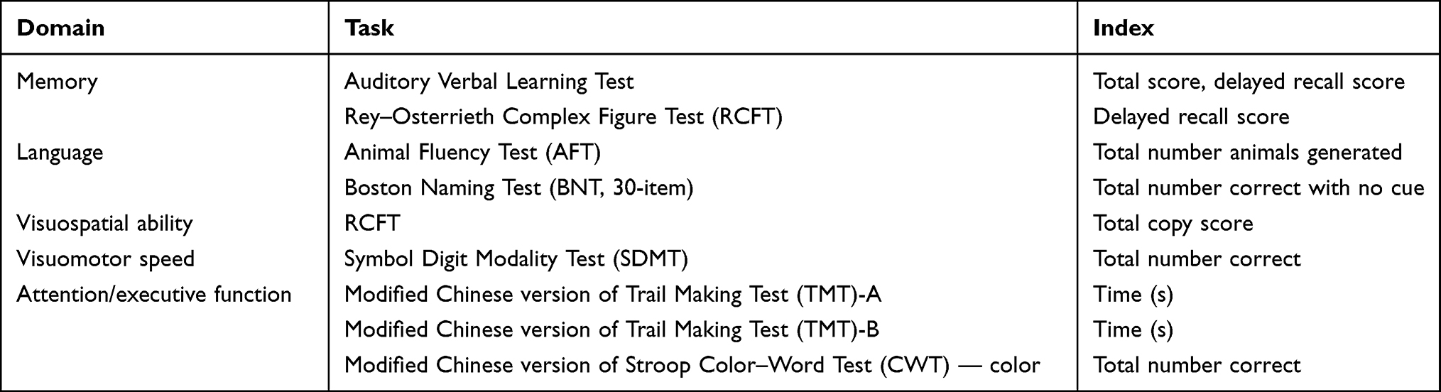 |
Table 1 Cognitive tests for each of the domains assessed |
Cognitive Assessment
Global cognitive function was assessed using the Chinese version of the MoCA scale.23 One point was added to the MoCA score for those with education <12 years.24 The neuropsychological test battery examined five cognitive domains (see Table 1), and was completed within 10 (IQR 2) days of admission. The interval between the neuropsychological assessment and MRI scan was 12 hours. The Z score was defined as onee that fell within the distribution of scores for controls, and was calculated for the neuropsychological tests. CI was defined as a score of 1.5 standard deviations below the mean on any neuropsychological test of the normative study. Final diagnoses of CI subjects were assigned based on the Diagnostic and Statistical Manual of Mental Disorders, fourth edition.25
Image Acquisition
T1-weighted images were obtained using a 3 T Prisma device (Siemens Healthcare, Erlangen, Germany). The parameters were TR = 2300 ms, TE = 2.3 ms, TI = 900 ms, scanning field 240×240 mm, matrix 256×256, layer thickness 1 mm, and interlayer spacing 1 mm. The FreeSurfer image analysis suite (http://surfer.nmr.mgh.harvard.edu) were used for cortical reconstruction and volumetric segmentation (version 6.0.0). Cortical thickness and volume were quantified. Briefly, this procedure included motion correction, averaging of multiple volumetric T1-weighted images, non-brain tissue removal, automated Talairach transformation, segmentation of subcortical white-matter and deep gray-matter volumetric structures, intensity normalization, tessellation of the gray-matter and white-matter boundary, automated topology correction, and surface deformation following intensity gradients to optimally place the gray matter–white matter and gray matter–cerebrospinal fluid borders at the location where the greatest shift in intensity defined the transition to the other tissue class.
Statistical Analysis
Statistical analyses were conducted with SPSS 20.0. Significance was set at p<0.05. Continuous variables are expressed as means ± SD or medians (IQR) according to their distribution (normal or skewed). Discrete variables were compared by X2 tests. Spearman correlation analyses were performed between the Z score of cognitive function and cortical thickness or volume. We evaluated the association between cognitive function and cortical thickness or volume using further multiple linear analyses adjusted for potential confounders, including average age, years of education, and IADL score.
Results
Characteristics and Clinical Profile of Patients
The PSCI group had significantly less education and higher instrumental ADL scores than the control group (p<0.05). There were no significant differences in age, sex, scores on the HDRS, basic ADL, or IQCODE, prevalence of hypertension, diabetes, hyperlipidemia, current or ever drinking, and current or ever smoking between the groups. Median (IQR) NIHSS and Modified Rankin Scale score were 3 (4) and 1 (1), respectively. Most patients had small-artery occlusion (n=19, 82.61%) and three large atherothrombotic infarction (13.04%) strokes. The average diameter of lesions was 17.57±8.81 mm (Table 2).
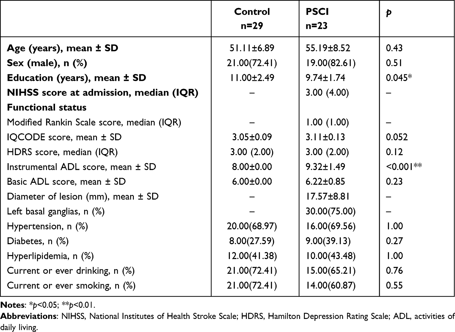 |
Table 2 Demographic information for control and PSCI groups |
Comparison of Neuropsychological Tests
The PSCI group showed significantly lower Z scores than the control group in the cognitive domains of memory, visuomotor speed, language, and attention/executive function (p<0.05, Table 3 and Figure 2).
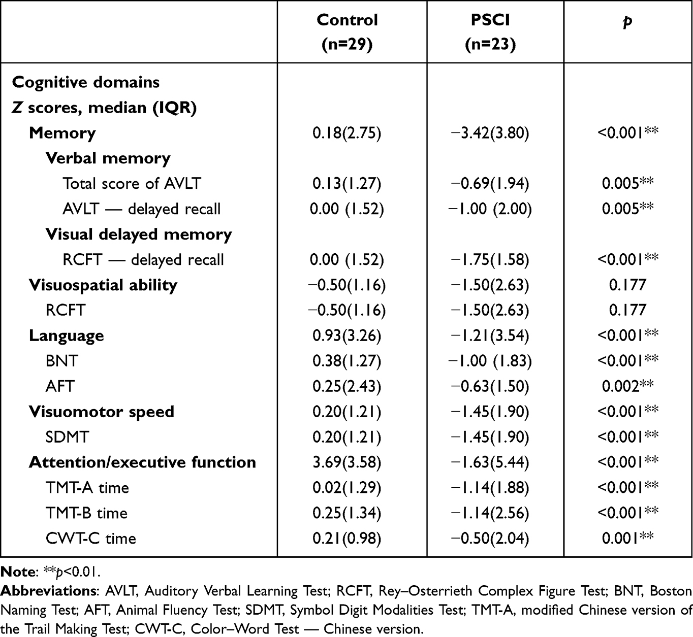 |
Table 3 Comparison of Z scores in every cognitive domain between control and PSCI groups |
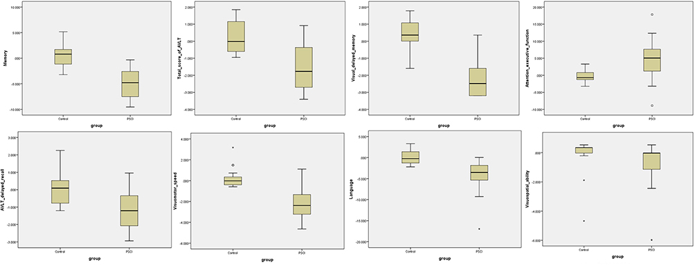 |
Figure 2 Comparison of neuropsychological tests between control group and PSCI group. *Extreme outliers (>3 times interquartile range); ○mild outliers (>1.5 times interquartile range). |
Comparison of Cortical Thickness and Brain Volume
The thicknesses of the right inferior temporal cortex and insula in the PSCI group were significantly less than controls (p<0.05). The volume of the middle posterior corpus callosum, middle anterior corpus callosum, and hippocampus in PSCI patients was remarkably less than controls (p<0.05) (Table 4). There were no significant difference in bilateral volumes of amygdala, caudate, putamen, or pallidum between the control and PSCI groups (Supplemental Table 1).
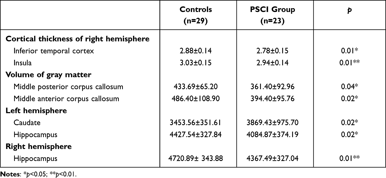 |
Table 4 Comparison of cortical thickness and brain volume between control and PSCI groups |
Correlation Analyses Between Cortical Thickness or Brain Volume and Z Scores of Cognitive Scales
Z scores for executive function were significantly and positively correlated with thickness of the right insula cortex (r=0.298, p<0.05). Z scores for executive function were significantly and positively correlated with volumes of the hippocampus (left r=0.028, right r=0.42; p<0.05) and middle posterior corpus callosum (r=0.533, p<0.05; Table 5). Z scores for language were significantly and positively correlated with thickness of the right inferior temporal cortex (r=0.360, p<0.05), while Z scores for language were significantly and positively correlated with volumes of the hippocampus (left r=0.445, right r=0.471; p<0.05) and middle posterior corpus callosum (r=0.383, p<0.05; Table 5).
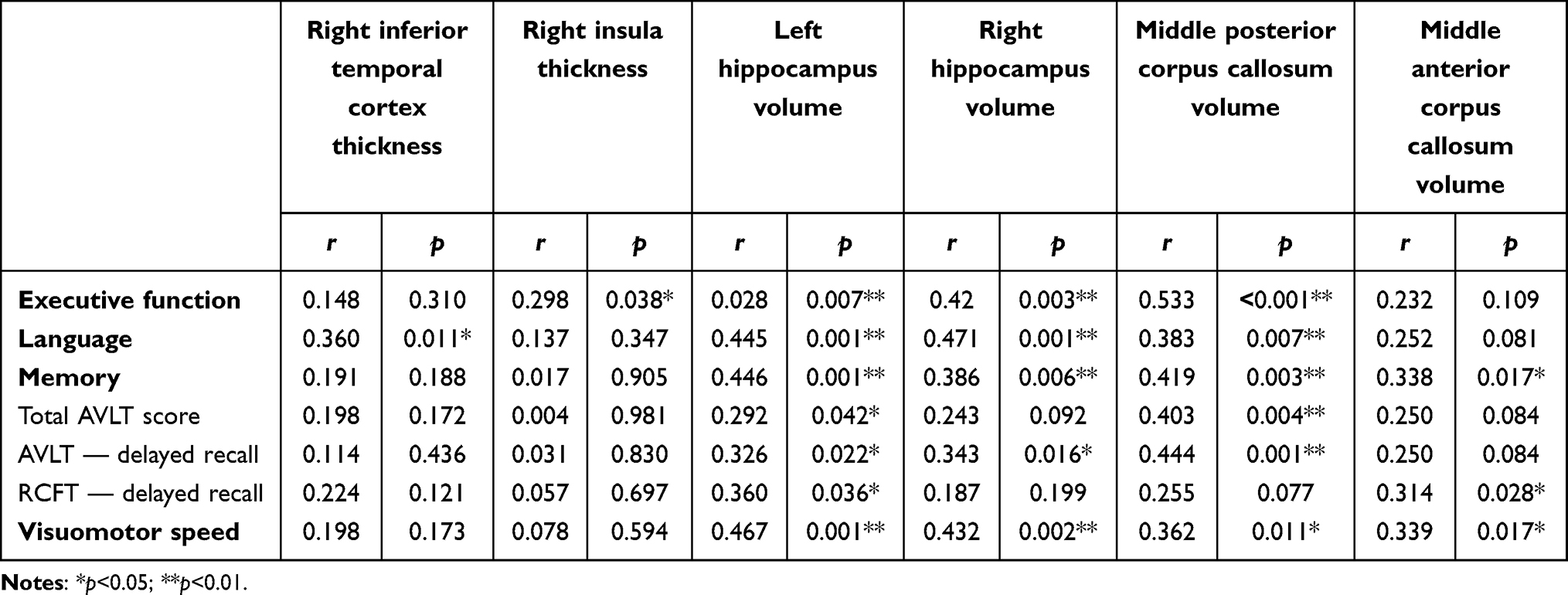 |
Table 5 Spearman correlations between cortical thickness and Z scores on cognitive scales |
Z scores for memory were significantly and positively correlated with volumes of the hippocampus (left r=0.446, right r=0.386; p<0.05), middle posterior corpus callosum, and middle anterior corpus callosum (posterior r=0.419, anterior r=0.338; p<0.05; Table 5). Z scores for verbal memory (total AVLT score) were significantly and positively correlated with volumes of the left hippocampus (r=0.292, p<0.05) and middle posterior corpus callosum (r=0.403, p<0.05; Table 5). Z scores for delayed verbal memory (AVLT delayed recall score) were significantly and positively correlated with volumes of the left hippocampus (r=0.326, p<0.05); and middle posterior corpus callosum (r=0.444, anterior r=0.26; p<0.05; Table 5). Z scores for delayed visual memory (RCFT delayed recall score) were significantly and positively correlated with volumes of the left hippocampus (r=0.36, p<0.05) and middle anterior corpus callosum (r=0.314, p<0.05; Table 5). Z scores for visuomotor speed were significantly and positively correlated with volumes of the hippocampus (left r=0.467, right r=0.432; p<0.05), middle posterior corpus callosum, and middle anterior corpus callosum (posterior r=0.362, anterior r=0.339; p<0.05; Table 5).
Multiple Linear Regression Analyses Between Cognitive Score and Cortical Thickness/Volume
Multiple linear regression analyses indicated that executive function scores (β=0.333, p=0.036) were significantly and negatively correlated with right hippocampus volume after adjustment for age, education, and instrumental ADL score. Language scores were significantly and positively correlated with hippocampus volume after adjustment for age, education, and instrumental ADL score (Table 6).
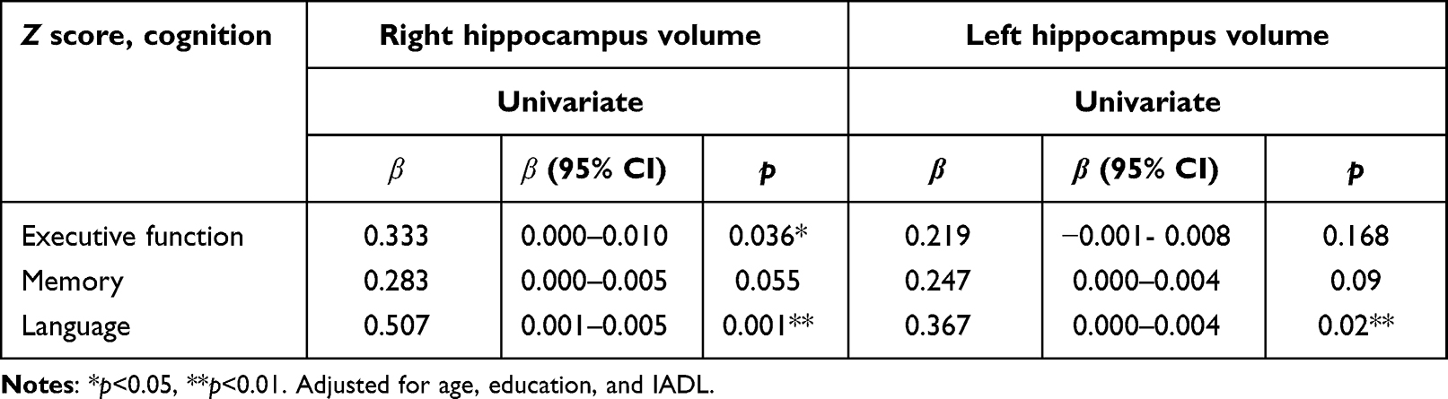 |
Table 6 Multiple linear regression analyses of cortical thickness/volume and related factors |
Discussion
The current study investigated the characteristics of PSCI patients with basal ganglia infarction. Data showed that PSCI patients exhibited significantly lower Z scores in all cognitive domains than controls: including verbal memory, visual memory, visuospatial ability, visuomotor speed, language, and attention/executive function. There is evidence that memory and executive function are affected early in vascular CI.26 PSCI is also associated with language and visuospatial dysfunction.27 As for the memory domain, PSCI patients with basal ganglia infarcts have both verbal and visual memory decline. Studies have proved the crucial role of basal ganglia in the verbal memory regulation.28,29 The functional connectivity of the basal ganglia has been modeled as two second-level modulatory connections that control projections from sensory cortices to the prefrontal cortex and from the hippocampus and medial temporal lobe to the prefrontal cortex.30 Memory formation includes encoding, storage, and retrieval. A recent study reported that subcortical region dysfunction was related to impaired information retrieval, which could finally lead to visual memory dysfunction.31 It might explain why patients with basal ganglia infarction exhibited both verbal and visual memory decline.
We explored the brain structural changes and potential mechanism of PSCI with basal ganglia infarcts by measuring cortical thickness and volume. The results showed that the PSCI group had reduced cortical thickness and brain volume across several regions than the control group, such as thickness of inferior temporal cortex and insula cortex and volume of middle posterior corpus callosum, middle anterior corpus callosum, and hippocampus. However, there was no significant difference in volumes of basal ganglia regions, such as the amygdala, caudate, putamen, or pallidum, between the two groups. Our results suggest that brain atrophy is an important substrate for CI in PSCI patients with basal ganglia infarcts. These findings seem to imply that lesions in basal ganglia not only affect local regions but also expand to functionally connected regions. Another study on basal ganglia infarction patients showed altered connectivity of the left inferior temporal gyrus.12 Cerebral ischemia and reduced cerebral blood flow might disrupt energy metabolism and lead to metabolic stress. Cells that undergo severe ischemia may die within minutes of the insult or exhibit delayed vulnerability. Cytotoxic edema promotes both a reduction of extracellular volume fraction and changes in membrane permeability.32 These events eventually damage functions of local regions because of ischemia, but connected regions remotely, because of loss of afferent synaptic input from distally or retrograde axonal degeneration.33 The hippocampus is rather sensitive to ischemia. It is possible that preexisting hippocampal vulnerability in this study left subjects sensitive to the short-term negative effects of stroke.34 In multiple linear regression analyses, we found that right hippocampus volume was significantly and positively correlated with executive function. The hippocampus is well known to participate in episodic memory processes. A previous study showed that the hippocampus is associated with subcortical regions and the prefrontal cortex to play an important role in executive functions.35,36 This functional connectivity and axonal projections enable and enhance learning–behavior translation, and partly explain why hippocampal dysfunctions result in executive deficits. Another study also reported that executive tasks required the coactivation of prefrontal and hippocampal networks.37 Stroke lesions in basal ganglia might damage the link among the hippocampus, subcortical regions, and the prefrontal cortex and lead to executive dysfunction.
We also find that hippocampus volume was related to language dysfunction. Memory and language share a common neural mechanism. The hippocampus is a part of the language network and contributes to language processing.38 One structural MRI study showed that hippocampus volume is related to language-processing deficits.39 Another functional MRI study showed that language rehabilitation is related to enhanced functional connectivity recovery among frontal, temporal, and parietal lobes with the hippocampus.40 Both support our study findings. Patients with basal ganglia infarcts might have remote structural damage and consequently CIs.
However, this research has several limitations. Firstly, although we selected basal ganglia stroke, it is a challenge to achieve stroke homogeneity in a given sample. Secondly, given the small sample, further research should be done on larger samples to verify the current findings. Thirdly, this study was cross-sectional. The relationship between brain-structure changes and cognition was obtained in the acute phase. We agree that our findings might not be suitable for all stroke patients, especially for those in the chronic phase. We will continue to investigate relationships between brain-structure integrity and cognition at 6 months or 1 year after stroke to determine whether it affects long-term poststroke cognitive function.
This study was conducted firstly to explore the cognitive function of stroke patients with basal ganglia lesions and identify the correlation between brain-structure integrity and cognitive functions in the acute phase. The results suggest that reduced hippocampus volume is associated with language and executive function in stroke patients with basal ganglia at 2 weeks.
Data Sharing
There are no additional data.
Ethics
This trial was reviewed and approved by the Beijing Tiantan Hospital Ethics Review Board (KY-2015-001-01). Written informed consent was obtained from all participants. This study was performed according to the guidelines of Capital Medical University, which abides by the Helsinki Declaration on ethical principles for medical research involving human subjects.
Acknowledgment
We gratefully acknowledge all participants for their dedication. We also thank Professor Xingao Wang from Beijing Tiantan Hospital, Capital Medical University for his modification and review of the manuscript.
Author Contributions
All authors made a significant contribution to the work reported, whether in the conception, study design, execution, acquisition of data, analysis and interpretation, or all these areas, took part in drafting, revising, or critically reviewing the article, gave final approval to the version to be published, have agreed on the journal to which the article has been submitted, and agree to be accountable for all aspects of the work.
Funding
This work was supported by the National Key Research and Development Program of China (2020YFC2004800) and the Beijing Excellent Talents Training Program (2018000021469G237).
Disclosure
The authors report no conflicts of interest in this work.
References
1. Zuo L, Dong Y, Zhu R, et al. Screening for cognitive impairment with the Montreal cognitive assessment in Chinese patients with acute mild stroke and transient ischaemic attack: a validation study. BMJ Open. 2016;6:e011310. doi:10.1136/bmjopen-2016-011310
2. Jung J, Laverick R, Nader K, et al. Altered hippocampal functional connectivity patterns in patients with cognitive impairments following ischaemic stroke: a resting-state fMRI study. NeuroImage Clin. 2021;32:102742. doi:10.1016/j.nicl.2021.102742
3. Snaphaan L, Rijpkema M, van Uden I, Fernández G, de Leeuw FE. Reduced medial temporal lobe functionality in stroke patients: a functional magnetic resonance imaging study. Brain. 2009;132:1882–1888. doi:10.1093/brain/awp133
4. Thompson RF, Kim JJ. Memory systems in the brain and localization of a memory. Proc Natl Acad Sci U S A. 1996;93:13438–13444. doi:10.1073/pnas.93.24.13438
5. Huijts M, Duits A, Staals J, Kroon AA, de Leeuw PW, van Oostenbrugge RJ. Basal ganglia enlarged perivascular spaces are linked to cognitive function in patients with cerebral small vessel disease. Curr Neurovasc Res. 2014;11:136–141. doi:10.2174/1567202611666140310102248
6. Caligiore D, Pezzulo G, Miall RC, Baldassarre G. The contribution of brain sub-cortical loops in the expression and acquisition of action understanding abilities. Neurosci Biobehav Rev. 2013;37:2504–2515. doi:10.1016/j.neubiorev.2013.07.016
7. Narasimhalu K, Wiryasaputra L, Sitoh YY, Kandiah N. Post-stroke subjective cognitive impairment is associated with acute lacunar infarcts in the basal ganglia. Eur J Neurol. 2013;20:547–551. doi:10.1111/ene.12032
8. Rockwood K, Wentzel C, Hachinski V, Hogan DB, MacKnight C, McDowell I. Prevalence and outcomes of vascular cognitive impairment. Vascular cognitive impairment investigators of the Canadian study of health and aging. Neurology. 2000;54:447–451. doi:10.1212/WNL.54.2.447
9. Lim SJ, Fiez JA, Holt LL. How may the basal ganglia contribute to auditory categorization and speech perception? Front Neurosci. 2014;8:230. doi:10.3389/fnins.2014.00230
10. Larivière S, Ward NS, Boudrias MH. Disrupted functional network integrity and flexibility after stroke: relation to motor impairments. NeuroImage Clin. 2018;19:883–891. doi:10.1016/j.nicl.2018.06.010
11. Yao G, Li J, Liu S, et al. Alterations of functional connectivity in stroke patients with basal ganglia damage and cognitive impairment. Front Neurol. 2020;11:980. doi:10.3389/fneur.2020.00980
12. Li Z, Hu J, Wang Z, You R, Cao D. Basal ganglia stroke is associated with altered functional connectivity of the left inferior temporal gyrus. J Neuroimaging. 2022;32:744–751. doi:10.1111/jon.12978
13. Kliper E, Ben Assayag E, Korczyn AD, et al. Cognitive state following mild stroke: a matter of hippocampal mean diffusivity. Hippocampus. 2016;26:161–169. doi:10.1002/hipo.22500
14. Pohjasvaara T, Mäntylä R, Salonen O, et al. MRI correlates of dementia after first clinical ischemic stroke. J Neurol Sci. 2000;181:111–117. doi:10.1016/S0022-510X(00)00437-8
15. Zhang H, Sachdev PS, Wen W, et al. Gray matter atrophy patterns of mild cognitive impairment subtypes. J Neurol Sci. 2012;315:26–32. doi:10.1016/j.jns.2011.12.011
16. Werden E, Cumming T, Li Q, et al. Structural MRI markers of brain aging early after ischemic stroke. Neurology. 2017;89:116–124. doi:10.1212/WNL.0000000000004086
17. Tang J, Zhong S, Chen Y, et al. Aberrant white matter networks mediate cognitive impairment in patients with silent lacunar infarcts in basal ganglia territory. J Cereb Blood Flow Metab. 2015;35:1426–1434. doi:10.1038/jcbfm.2015.67
18. Romero JR, Pinheiro A, Aparicio HJ, DeCarli CS, Demissie S, Seshadri S. MRI visible perivascular spaces and risk of incident dementia: the Framingham heart study. Neurology. 2022;99:e2561–e2571. doi:10.1212/WNL.0000000000201293
19. Jorm AF, Jacomb PA. The informant questionnaire on cognitive decline in the elderly (iqcode): socio-demographic correlates, reliability, validity and some norms. Psychol Med. 1989;19:1015–1022. doi:10.1017/S0033291700005742
20. Lawton MP, Brody EM. Assessment of older people: self-maintaining and instrumental activities of daily living. Gerontologist. 1969;9:179–186. doi:10.1093/geront/9.3_Part_1.179
21. Katz S, Ford AB, Moskowitz RW, Jackson BA, Jaffe MW. Studies of illness in the aged. The index of adl: a standardized measure of biological and psychosocial function. JAMA. 1963;185:914–919. doi:10.1001/jama.1963.03060120024016
22. Hamilton M. A rating scale for depression. J Neurol Neurosurg Psychiatry. 1960;23:56–62. doi:10.1136/jnnp.23.1.56
23. Yu J, Li J, Huang X. The Beijing version of the Montreal cognitive assessment as a brief screening tool for mild cognitive impairment: a community-based study. BMC Psychiatry. 2012;12:156. doi:10.1186/1471-244X-12-156
24. Nasreddine ZS, Phillips NA, Bedirian V, et al. The Montreal cognitive assessment, moca: a brief screening tool for mild cognitive impairment. J Am Geriatr Soc. 2005;53:695–699. doi:10.1111/j.1532-5415.2005.53221.x
25. American psychiatric association. Diagnostic and Statistical Manual of Mental Disorders.
26. Looi JC, Sachdev PS. Differentiation of vascular dementia from ad on neuropsychological tests. Neurology. 1999;53:670–678. doi:10.1212/WNL.53.4.670
27. Sachdev PS, Brodaty H, Valenzuela MJ, et al. The neuropsychological profile of vascular cognitive impairment in stroke and tia patients. Neurology. 2004;62:912–919. doi:10.1212/01.WNL.0000115108.65264.4B
28. Vakil E, Blachstein H, Soroker N. Differential effect of right and left basal ganglionic infarctions on procedural learning. Cogn Behav Neurol. 2004;17:62–73. doi:10.1097/01.wnn.0000094553.44085.25
29. Seidel UK, Gronewold J, Wicking M, Bellebaum C, Hermann DM. Vascular risk factors and diseases modulate deficits of reward-based reversal learning in acute basal ganglia stroke. PLoS One. 2016;11:e0155267. doi:10.1371/journal.pone.0155267
30. Wapstra NJ, Ketola M, Thompson S, et al. Increased basal ganglia modulatory effective connectivity observed in resting-state fMRI in individuals with Parkinson’s disease. Front Aging Neurosci. 2022;14:719089. doi:10.3389/fnagi.2022.719089
31. Carota A, Neufeld H, Calabrese P. Memory profiles after unilateral paramedian thalamic stroke infarction: a comparative study. Case Rep Med. 2015;2015:430869. doi:10.1155/2015/430869
32. Bramlett HM, Dietrich WD. Pathophysiology of cerebral ischemia and brain trauma: similarities and differences. J Cereb Blood Flow Metab. 2004;24:133–150. doi:10.1097/01.WCB.0000111614.19196.04
33. Kuceyeski A, Kamel H, Navi BB, Raj A, Iadecola C. Predicting future brain tissue loss from white matter connectivity disruption in ischemic stroke. Stroke. 2014;45:717–722. doi:10.1161/STROKEAHA.113.003645
34. Takehara-Nishiuchi K. Entorhinal cortex and consolidated memory. Neurosci Res. 2014;84:27–33. doi:10.1016/j.neures.2014.02.012
35. Ren J, Huang F, Zhou Y, et al. The function of the hippocampus and middle temporal gyrus in forming new associations and concepts during the processing of novelty and usefulness features in creative designs. NeuroImage. 2020;214:116751. doi:10.1016/j.neuroimage.2020.116751
36. Yang G, Li C, Wang W, et al. Risk factors for cognitive impairment in patients with first-time ischemic stroke. Am J Transl Res. 2021;13:1884–1889.
37. Hartung H, Brockmann MD, Pöschel B, De Feo V, Hanganu-Opatz IL. Thalamic and entorhinal network activity differently modulates the functional development of prefrontal-hippocampal interactions. J Neurosci. 2016;36:3676–3690. doi:10.1523/JNEUROSCI.3232-15.2016
38. Covington NV, Duff MC. Expanding the language network: direct contributions from the hippocampus. Trends Cogn Sci. 2016;20:869–870. doi:10.1016/j.tics.2016.10.006
39. Honea RA, Holsen LM, Lepping RJ, et al. The neuroanatomy of genetic subtype differences in prader-willi syndrome. Am J Med Genet B Neuropsychiatr Genet. 2012;159b:243–253.
40. Chen Q, Shen W, Sun H, et al. Effects of mirror therapy on motor aphasia after acute cerebral infarction: a randomized controlled trial. NeuroRehabilitation. 2021;49:103–117. doi:10.3233/NRE-210125
 © 2023 The Author(s). This work is published and licensed by Dove Medical Press Limited. The
full terms of this license are available at https://www.dovepress.com/terms
and incorporate the Creative Commons Attribution
- Non Commercial (unported, 3.0) License.
By accessing the work you hereby accept the Terms. Non-commercial uses of the work are permitted
without any further permission from Dove Medical Press Limited, provided the work is properly
attributed. For permission for commercial use of this work, please see paragraphs 4.2 and 5 of our Terms.
© 2023 The Author(s). This work is published and licensed by Dove Medical Press Limited. The
full terms of this license are available at https://www.dovepress.com/terms
and incorporate the Creative Commons Attribution
- Non Commercial (unported, 3.0) License.
By accessing the work you hereby accept the Terms. Non-commercial uses of the work are permitted
without any further permission from Dove Medical Press Limited, provided the work is properly
attributed. For permission for commercial use of this work, please see paragraphs 4.2 and 5 of our Terms.