Back to Journals » Infection and Drug Resistance » Volume 16
Clinical Efficacy and Diagnostic Value of Metagenomic Next-Generation Sequencing for Pathogen Detection in Patients with Suspected Infectious Diseases: A Retrospective Study from a Large Tertiary Hospital
Authors Xiao YH , Liu MF , Wu H, Xu DR, Zhao R
Received 16 December 2022
Accepted for publication 17 March 2023
Published 29 March 2023 Volume 2023:16 Pages 1815—1828
DOI https://doi.org/10.2147/IDR.S401707
Checked for plagiarism Yes
Review by Single anonymous peer review
Peer reviewer comments 2
Editor who approved publication: Prof. Dr. Héctor Mora-Montes
Yang-Hua Xiao,1,2 Mei-Fang Liu,1 Hongwen Wu,1,3 De-Rong Xu,1 Rui Zhao1
1Department of Clinical Laboratory, Medical Center for Burn and Plastic Surgery, The First Affiliated Hospital of Nanchang University, Nanchang, People’s Republic of China; 2School of Public Health, Nanchang University, Nanchang, People’s Republic of China; 3Department of Medical Instruments, The First Affiliated Hospital of Nanchang University, Nanchang, People’s Republic of China
Correspondence: De-Rong Xu; Rui Zhao, Email [email protected]; [email protected]
Purpose: Metagenomic next-generation sequencing (mNGS) is a powerful yet unbiased method to identify pathogens in suspected infections. However, little is known about its clinical effectiveness. The present study aimed to assess the efficacy of mNGS in routine clinical practice.
Patients and Methods: In this single-center retrospective cohort study, 518 patients with suspected infectious diseases were assessed for inclusion. Among them, each patient had undergone mNGS testing; 407 patients had undergone both microbial culture and mNGS testing. The result of mNGS testing was compared to microbial culture performed concurrently. The diagnostic performance of mNGS was evaluated using the comprehensive clinical diagnosis as the reference standard.
Results: There was a significant difference in the positive detection rates of pathogens between mNGS and culture (331/407, 81.3% vs 79/407, 19.4%, P < 0.001). The sensitivity of mNGS was much higher than the culture method (79.5% vs 21.3%, P < 0.001), especially in sample types of sputum and bronchoalveolar lavage fluid (BALF). Notably, the sensitivity of blood mNGS was relatively lower than other sample types (67.4% vs 88.9– 93.8%). Pathogen cfDNA load based on standardized stringently mapped read number at the species level of microorganisms (SDSMRN) was significantly lower in blood than in other sample types from the same patient (P = 0.0003). Importantly, mNGS directly led to a change of treatment regimen in 142 (27.4%) cases, including antibiotic escalation (15.3%), antibiotic de-escalation (9.1%), and early definitive diagnosis to initiate appropriate treatment (3.1%).
Conclusion: Our in-house mNGS platform significantly improved the sensitivity for the diagnosis of infectious diseases. mNGS has the potential to improve clinical outcomes by optimizing antimicrobial therapy.
Keywords: mNGS, infections, diagnostic, outcome
Introduction
Infections are the major cause of human deaths worldwide and present a rising threat to public security.1 Antimicrobial resistance is a major concern that is threatening our ability to treat bacterial infections.2 Delayed administration of antibiotics significantly increases the in-hospital mortality rate.3 The tragic outcome of infection highlights the importance of timely diagnosis and appropriate antibiotic treatment of advanced diseases. Conventional microbiologic culture and microscopy remain the gold standard for diagnostics. Full identification of fastidious microorganisms is usually reserved for cases where an isolate is in pure culture.4,5 However, it is a laborious and time-consuming procedure requiring several days to accomplish. Additionally, the sensitivity of the culture method depends largely on the duration of the infection and whether the patient has previously received antibiotic treatment.6
For these reasons, rapid and accurate identification of unknown pathogenic microorganisms is critical to guide clinical decision-making about diagnosis and treatment. Molecular methods, such as polymerase chain reaction (PCR), are an alternative method when culture-based microbial detection methods fail.7,8 However, the preselected target pathogens greatly limit the application of multiplex PCR in identifying infections caused by mixed pathogens.9 PCR-based analyses may fail to detect unexpected, rare, or novel pathogens unless multiplexed PCRs covering all pathogenic microorganisms are employed. The clinical symptoms and etiologies of multiple infections are complex and diverse, so it is difficult to diagnose using conventional microbial methods.10,11 This may complicate further treatment (eg, delay antimicrobial treatment, lead to antibiotics overuse, prolong hospitalizations, and increase healthcare costs). Accurate and timely diagnosis is essential for aggressive antimicrobial adjustment. These barriers are not insurmountable obstacles to treating infectious diseases.
Metagenomic next-generation sequencing (mNGS) is an untargeted molecular approach that can theoretically detect all potential pathogens in a single experiment. As a promising novel technique, mNGS is especially suitable for difficult and atypical infectious diseases.12 Given the untargeted nature of mNGS, the major limitation is human host background interference and the efficiency of nucleic acids extraction.13 Thus, interpretation of the results should be cautious and adequate clinical trial experience is required. Previous studies relevant to clinical application and impact of mNGS have largely been limited to single case reports or cohort studies with relatively small populations.11,14–17
To address this gap, we performed a single-center, multiple sample types, and retrospective study to assess the clinical efficacy of mNGS testing for pathogen detection in a large comprehensive tertiary hospital. The pathogens detection rate of mNGS testing was compared to conventional culture performed concurrently. The diagnostic performance of mNGS was evaluated using the final clinical diagnosis as the reference standard. The clinical efficacy of mNGS was decided by the treating team based on clinical outcomes as well as the extent to which mNGS results affect treatment decisions.
Methods
Study Population
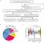
This is a retrospective cohort study that collected the clinical data for 518 patients with suspected infection who were hospitalized at the First Affiliated Hospital of Nanchang University (Jiangxi Province, China), from December 2020 to October 2021. This hospital is a 6100-bed tertiary-care teaching hospital that treats a variety of diseases. The study inclusion criteria were described below: (1) Patients with either highly suspected infectious clinical manifestations or infectious etiology; (2) The clinical symptoms of the patient have some relief after broad-spectrum antibiotic treatment, but the pathogen remains unknown; (3) The patients (or for unconscious patients their families) agree to carry out mNGS testing. Patients were excluded if they have been diagnosed with noninfectious disease before testing. The study flow chart is depicted in Figure 1A. This study was approved by the institutional review board of the First Affiliated Hospital of Nanchang University. As this was a retrospective cohort study based on the results of previous clinical diagnoses and treatment, the ethics committee granted exempt status for this study. To this end, we state this study was in conformity with the ethical guidelines of the Declaration of Helsinki and that patient-related data are strictly confidential.
Collection of Patient Samples
Based on our inclusion/exclusion criteria, 518 samples were included for analysis. The same eligible sample was divided to undergo mNGS and culture testing, which was defined as having paired mNGS and culture testing in our study. To compare the clinical efficacy of mNGS and CMT in diagnosing suspected infection diseases, the 518 samples were categorized into 2 groups defined as “mNGS with paired culture testing” and “mNGS without paired culture testing”. As shown in Figure 1A, 407 samples were performed mNGS testing and culture in parallel. Moreover, considering the possibility of repeated mNGS testing for a different period or reasons in the same patient (ie, patient monitoring, sampling from different body sites, and suspected false-negative results for the first testing), the comparison of diagnostic performance and clinical efficacy was evaluated according to the first mNGS testing per patient.
mNGS and Analyses
The Tiangen Magnetic DNA Kit (Tiangen Biotech, DP316) was used to extract the genomic DNA from all samples following the manufacturer’s manual. The extracted nucleic acid samples were used for the generation of DNA libraries using the Nextera XT kit (Illumina) according to the manufacturer’s operational manual. Libraries were sequenced using the Illumina NextSeq-550Dx sequencer. Sterile nuclease-free deionized water was extracted together with samples to serve as negative control. Commercial suspension of bacteria in the assay kit served as positive control. The sequencing depth of each sample was ≥ 20 million reads. High-quality sequencing data were generated by removing low-quality reads, duplicate reads, and shorter reads (<35 bp). Then the human host reads were subtracted using Burrows-Wheeler Aligner software to map to a human reference genome (hg19). The remaining data were aligned with the microbial genome database. Matching microbial reference genomes were downloaded from NCBI (ftp://ftp.ncbi.nlm.nih.gov/genomes/). The overall turn-around time of mNGS (from nucleic acid extraction to give results to clinicians) is 16~24 h.
Interpretation of mNGS Results
We used stringent criteria to exclude ambiguous alignments by excluding any mismatches and all sequences that align non-uniquely to the microbial genome. Only standardized stringently mapped read number at the species level of microorganisms (SDSMRN) was considered. In the report interpretation process, a colonizing microorganism database was used to filter out background noise signals. The positive result criteria of mNGS are as follows: (1) Bacteria (excluding mycobacteria) and fungi: SDSMRN≥3. (2) Parasite: SDSMRN≥50; (3) Mycobacterium tuberculosis, Brucella, Nocardia: SDSMRN≥1. Due to the difficulty of extracting nucleic acid and the low likelihood of contamination, mycobacteria were considered positive when at least 1 SDSMRN was present.
Conventional Microbiologic Tests
Routine microbiologic tests included gram staining and aerobic and anaerobic cultures. All samples (including blood, bronchoalveolar lavage fluid (BALF), sputum, cerebrospinal fluid (CSF), peritoneal fluid, bone marrow, pleural fluid, and other body fluids) were collected by the corresponding clinicians and sent to the laboratory within 1 hour for relevant tests. Aerobic and anaerobic cultures were performed for the blood samples. The other samples were inoculated onto sheep blood agar, MacConkey agar, Sabouraud agar, and chocolate agar plate. Culturing was performed at 37°C for 24 hours both aerobically and anaerobically. Bacterial species were identified using Vitek-2 Compact Instrument (BioMerieux, Balmes-les-Grottes, France). Apart from routine microbial culture, other pathogen detection methods (ie, routine laboratory staining, real-time PCR assay, enzyme-linked immunospot assay, galactomannan test, serum (1-3)-β-D-glucan test, GeneXpert, T-spot, and imaging examination) were only performed for patients with highly suspected associated infections. Two reference standards were applied in diagnosis: a clinical gold standard of culture and a composite standard that incorporated all results from other pathogen detection methods. Diagnoses were ultimately made by each doctor-in-charge based on all microbiological test results and a longitudinal review of the patient’s clinical characteristics.
Statistical Analysis
Comparative analysis was conducted by Pearson χ2 test. Discrete variables were analyzed with Fisher’s exact test when appropriate. The sensitivity and specificity of different methods were assessed. Diagnostic analysis was performed by receiver operating characteristics (ROC) curve based on SDSMRN from mNGS results and the area under the curve (AUC) was determined. The results were presented with 95% confidence interval (CI). Paired samples of mNGS were compared using the Wilcoxon signed-rank test. Data analyses were conducted using SPSS 25.0 software (SPSS Inc, Chicago, IL, USA). Two-tailed P-value ≤ 0.05 was considered significant.
Results
Patient and Sample Characteristics
The clinical and demographic features of the study patients are presented in Table 1. A total of 518 patients were included, including 341 (65.8%) male and 177 (34.2%) female patients. Overall, 139 patients (26.8%) were admitted to intensive care units during their hospitalization. The most common clinical symptoms of all patients were fever (47.3%) and cough (20.7%). The main underlying diseases were hypertension in 125 patients (24.1%), and diabetes in 53 patients (10.2%). For the final clinical diagnosis, 490 patients were ultimately diagnosed with infection. Most of our sample types are blood, with 38.6% from blood, 29.7% from BALF, 12.0% from CSF, 5.6% from sputum, 5.6% from peritoneal, 3.3% from bone marrow, 1.4% from pleural, and 3.8% from other body fluids (Figure 1B). To compare the pathogen cfDNA load from the different samples, the ratio of microbial reads was calculated by the reads of microbial divided by the total number of reads. As shown in Figure 1C, the ratio of microbial reads in sputum samples and BALF samples was higher than those in the other samples of type (eg, blood, CSF, peritoneal, marrow, and pleural fluid).The difference was statistically significant between BALF and all samples (P < 0.0001), but there was no significant difference between sputum and all samples (P = 0.15).
 |
Table 1 Patient and Sample Characteristics |
Comparison of Pathogen Detection Between mNGS and Culture
To compare the pathogens detection rate of mNGS and culture, 407 samples with paired microbial culture and mNGS testing were included for further study. There was a significant difference in the positive detection rates of pathogens between mNGS and culture (331/407, 81.3% vs 79/407, 19.4%, P < 0.001). As shown in Figure 2A, mNGS and conventional culture were both positive in 78 of 407 (19.2%) cases and were both negative in 75 of 407 (18.4%) cases. A total of 253 samples were detected as positive only by mNGS (negative by culture), and 1 was detected as positive only by culture (negative by mNGS). However, mNGS produced false-positive results of the virus in 24 cases, but none of them had clinical features of viral infection and were ultimately diagnosed as non-viral infections. For double-positive samples (both mNGS and culture positive), 61 results were found to be partly matched. We interpret this to mean that at least one pathogen overlaps when the multi-pathogens result was detected. Of the remaining 17 double-positive results, results of mNGS and culture were completely matched in eight cases and were mismatched in nine cases. The distribution of pathogens detected in this study was presented in Figure 2B. The pathogen spectrum detected by mNGS was much broader than that of the culture method. Of these pathogens, Klebsiella pneumoniae (n = 60) and Candida (n = 65) were the most frequently detected bacteria and fungi, respectively. In general, the detection rates of bacteria and fungi (except for candida) by mNGS were higher than that of the culture method (odds ratio [OR] ≥ 2). Furthermore, most of the pathogens detected by the culture method were also detected by mNGS. These results highlight the high sensitivity and great advantage of mNGS in identifying polymicrobial infections.
Diagnostic Performance of mNGS
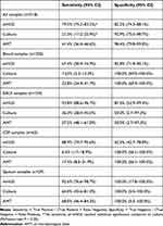
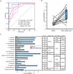
The performance of mNGS and traditional methods in diagnosis of infectious disease are shown in Table 2. The sensitivity and specificity of mNGS (all samples) for diagnosing infection were 79.5% and 82.2%, respectively. As for the culture method, the sensitivity and specificity of diagnosing infection were 21.3% and 92.9%, respectively. As expected, mNGS increased the sensitivity rate by approximately 60% in comparison with that of culture (79.5% vs 21.3%, P < 0.05), while not significantly different in specificity (82.2% vs 92.9%, P > 0.05). Notably, however, the sensitivity of blood mNGS in diagnosing infectious disease was relatively lower than other sample types (67.4% versus 88.9–93.8%). Excluding blood, the sensitivity of mNGS was comparable overall among BALF, CSF, and sputum samples (sensitivity ranged from 88.9% to 93.8%). ROC curves analysis of mNGS (all samples) for the diagnosis of infectious disease yielded an AUC of 0.882 (95% CI, 0.845–0.919) (Figure 3A). The area of the AUC for blood and CSF is 0.812 (95% CI, 0.746–0.876) and 0.788 (95% CI, 0.671–0.905), respectively. Not unexpectedly, sputum-mNGS and BLAF-mNGS with a greater area under the ROC curve (sputum, AUC=0.963 (95% CI, 0.778–1.143); BLAF, AUC=0.959 (95% CI, 0.892–1.026)).
 |
Table 2 Diagnostic Performance of Infectious Disease for mNGS and Traditional Methods |
Comparison of Relative Pathogen Reads of mNGS Testing from Blood Samples versus Other Sample Types
Only a handful of studies have reported the comparison of different sample types from the same patient of mNGS testing. To investigate which types of samples exhibited high potential diagnostic value, we selected 16 patients who underwent both blood and other sample types of mNGS testing for comparison. As shown in Figure 3B, compared to the blood samples, other sample types (including BALF, peritoneal, CSF, urine, and sputum) had a high level of SDSMRN in identifying the same pathogen (P = 0.0003). The details were shown in Figure 3C. Of the sixteen patients who underwent two or more mNGS testing, mNGS of the other sample types detected more SDSMRN than blood mNGS in 14 (87.5%) patients. However, in patient No.353, blood mNGS identified more P. aeruginosa sequences than peritoneal mNGS. Additionally, in patient No.514, the sequences of Aspergillus flavus detected by sputum mNGS was higher than blood mNGS, but blood mNGS identified E. coli sequences which not detected by sputum mNGS, then E. coli was confirmed by culture.
Efficacy of mNGS Results on Clinical Diagnosis and Treatment
In this study, clinical efficacy was evaluated based on the decisions made by the treating team after interpreting the mNGS results and whether this resulted in better outcomes. A total of 518 mNGS tests were evaluated. The positive detection rate was 436/518 (84.2%). When examining the impact of mNGS on clinical diagnosis and patient management, mNGS results exerted a positive impact on 162 (31.3%) and showed no impact in 297 (57.3%) cases (Table 3). Of the 162 cases identified to have a positive clinical impact, 79 cases were classified as antibiotic escalated, 47 cases as avoiding unnecessary antibiotic treatment (broad-spectrum antibiotics were discontinued or changed to a narrower range), and 20 cases as ruling out infection diseases, whereas 16 cases were classified as early definitive diagnosis. Unnecessary antibiotic treatment was observed in 4 (0.8%) cases. Of the 297 cases defined as having no impact, 105 cases were classified as empirical treatment continued. mNGS results were considered false or insignificant in 122 cases, of whom 47 did not find any additional pathogens than the culture method, and 75 had detected new pathogens but deemed no significance. The remaining 70 cases that had improved before the mNGS results were available were also defined as having no effect.
 |
Table 3 Clinical Efficacy of mNGS Result |
In terms of antibiotic treatment, mNGS directly led to a change of treatment regimen in 27.4% (142/518) patients. Almost all patients (118/142) experienced significant improvement, whereas the remaining patients were still seriously ill due to serious comorbidities despite appropriate treatment. Notably, mNGS testing facilitated early definite diagnosis in 16 patients infected with atypical pathogens encompassing Mycobacterium tuberculosis (n=10), Chlamydia psittaci (n=2), Coxiella burnetiid (n=2), Kaposi sarcoma virus (n=1), Mycobacterium shinjukuense (n=1). These cases highlight the power of mNGS in detecting rare and atypical pathogens. To further elaborate on the clinical impact of mNGS testing, we selectively enrolled representative 10 cases with clinically suspected infection but negative in culture. The details of these cases were shown in Table 4. Multiple presumptive causative pathogens including bacteria (P. aeruginosa and M. tuberculosis), fungi (Cryptococcus, Aspergillus, and P. jirovecii), atypical bacteria (c. psittaci), and viruses (HRV and EB) were detected in 8 of 10 cases using mNGS, highlighting the superiority of mNGS when diagnosing the complexed etiology of infection in certain cases. However, mNGS detected additional pathogens, and inevitably led to unnecessary antibiotic treatment in the remaining 2 cases. Of these two cases, one was presumably due to the contamination of the colonizer of the human respiratory tract, and the other was likely due to the viral nucleic acid fragments remnant from previous infections.
 |
Table 4 Case Series of mNGS Testing in Patients with Probable Infection but Negative in Culture |
Discussion
mNGS had been recently used in clinical practice due to its satisfactory diagnostic performance. Although many studies have evaluated the performance of mNGS in different types of infections, most of these studies are limited to a single type of sample and may lead to biased conclusions if generalized to a broader scale of clinical infectious disease.18,19 Therefore, this study aimed to address this gap and analyze the practical clinical efficacy of mNGS in a large comprehensive tertiary hospital. A total of 518 patients suspected of infection were eventually included in this study. The wide types of samples meaning the result reflects the actual clinical efficacy of mNGS in the real world to some extent. Furthermore, the local availability of the sequencing platform on-site at our hospital increased performance and reduces the turnaround time from bedside to bench.
In this study, we systematically compared the detection of mNGS and culture in a pairwise manner. The overall detection rates of mNGS were significantly higher than that of the culture method (331/407, 81.3% vs 79/407, 19.4%, P < 0.001). This result is at odds with a previous study reporting that the sensitivity of mNGS is not better than that of culture for recognizing bacteria.20 However, several other reports have reached similar conclusions to ours.21–23 The differences between studies may have been driven by discrepancies in the study population, the site of sampling, and the overall level of microbiology laboratory service. In our study, the majority of patients (76.8%) received empirical antibiotics before sampling, which may result in false-negative culture results, whereas mNGS is less affected by prior antibiotic exposure.20,24,25 Our study confirmed the unique advantage of mNGS in identifying nontuberculous mycobacteria, M. tuberculosis, C. psittaci, O. tsutsugamushi, Aspergillus, and P. jirovecii. Most species of mycobacteria are difficult to culture due to their long growth cycles, low sensitivities, and susceptibility to contamination.26 C. psittaci and O. tsutsugamushi were classified as atypical pathogens and were also difficult to detect by conventional testing. P. jirovecii is a pathogenic fungus that can cause Pneumocystis pneumonia in immunocompromised patients.27 Due to the laborious, cumbersome, and time-consuming conventional detection process, the diagnosis is often delayed or missed. Fortunately, mNGS has greatly improved the detection rate of these pathogens, highlighting the strength of mNGS for the identification of mycobacteria, atypical pathogens, and fungi.
Previous studies have demonstrated mNGS was also significantly superior to conventional microbiology tests for the detection of virus.28 However, in the present study, mNGS produced false-positive results of the virus in 24 cases, but none of them were ultimately diagnosed as viral infections. The clinical symptoms of these patients were not in agreement with the virus detected. Sample contamination was ruled out by negative control from healthy persons and sterile deionized water. We speculate that the 24 cases of the “mNGS false-positive” virus detected may be cfDNA remnants from previous infections or micro-colonization of the virus in blood. Almost all these cases were cured without antiviral treatment, confirming our speculation. Dead microorganism doss does not cause disease, but they may remain secret detectable small nucleic acid fragments. Microbial DNA from dead microbes may complicate the results of mNGS and warrants interpretation with extreme caution. In addition, the possibility of commensal/colonizers should also be considered. Further studies will be needed to further optimize mNGS to obtain more reliable results.
In terms of infectious diseases diagnosis, the sensitivity of mNGS was much higher than that of the culture method (79.5% vs 21.3%, P < 0.05), while the specificity of the culture method was better than mNGS (92.9% vs 82.2%). These results were similar to the previously reported by others.29–31 However, we found mNGS testing showed different sensitivities in different sample types. The sensitivity of mNGS in BALF and sputum samples was relatively high (92.6% and 93.8%), while the sensitivity of blood mNGS was only 67.4% (Table 2). Furthermore, there was a lower level of SDSMRN of the same pathogen in blood samples than in the paired other samples (P = 0.0003, Figure 3B). The primary explanation for this is the low pathogen cfDNA load in blood samples compared with other types of specimens. Higher levels of pathogen load in the sample can increase the credibility of mNGS result. This may suggest that BALF and sputum are among the most valuable samples for mNGS testing. However, for patients with fever and unclear infection site, blood-mNGS can be used as a supplementary experiment to exclude infection. Additionally, since respiratory tract was inhabited by a polymicrobial community, sputum, and BALF samples may be contaminated with oral normal flora, commensal organisms, and colonizers, leading to a relatively lower purity than other sample types.32 Considering the costs, adverse effects, and resistance of antimicrobial therapies, possible pathogens should be comprehensively judged by professionals based on the patient’s clinical background and other laboratory examination results. Thus, it should be noted the types of samples when interpreting the results of mNGS. Wrong mNGS data interpretation will inevitably lead to unnecessary expansion and prolongation of antibiotic therapy, and thus increased antibiotic resistance. Further studies are needed to distinguish causative pathogens and microbial colonizers in mNGS results.
Overall, the application of mNGS had a positive impact on 162 (31.3%) patients, including antibiotics escalation and de-escalation, ruling out active infection, and early definitive diagnosis to initiate appropriate treatment. Our result differs from recently published in USA where blood mNGS added little value when conducted simultaneously with conventional testing in the majority of cases.14 This inconsistency is possibly due to differences in sample types. The present study, together with other published studies, indicates that the blood mNGS testing had a relatively low sensitivity.33 Moreover, the indication for testing also had an impact. Since mNGS can cost as much as $500/sample, it has been used in the past as the last test resort for critically ill patients.34 However, the clinical outcome is still poor for critically ill patients despite mNGS testing may be useful in guiding appropriate antibiotic use. Therefore, we recommended that mNGS be used as the first-line test for sicker patients and patients with suspected atypical pathogen infection. Additional research is required to further explore which sample of types and patient populations are more favorable for performing mNGS testing.
The limitations of this study include the following. First, since mNGS testing is relatively expensive, not all suspect infected patients opt to do this, which could lead to a selection bias in our research. Second, the majority of patients received empiric antimicrobial therapy before sampling, we did not conduct a longitudinal assessment of the effects of antimicrobial therapy on mNGS and culture results since this study is a retrospective medical record review. Third, viruses were excluded from the detection performance due to the lack of routine clinical testing for viruses. Nonetheless, this study fully evaluated the clinical utility of mNGS at a tertiary care academic hospital on the diagnosis of suspected infectious diseases and patient outcomes.
Conclusion
mNGS has a broader pathogen spectrum and a better pathogen detection rate than that of the culture method, especially for rare and atypical pathogens. Additionally, mNGS have shown great advantages in diagnosing suspected infections. However, the sensitivity of blood mNGS is relatively limited by comparison with other sample types. Blood mNGS may be a supplementary test when higher diagnostic efficacy is required. In general, mNGS is valuable for identifying infections and optimizing antimicrobial therapy.
Abbreviations
mNGS, Metagenomics next-generation sequencing; PCR, polymerase chain reaction; SDSMRN, standardized stringently mapped read number at the species level; BALF, bronchoalveolar lavage fluid; CSF, cerebrospinal fluid; CI, confidence interval; ROC, Receiver Operating Characteristics; AUC, area under the curve.
Data Sharing Statement
The datasets of this study are available from the corresponding author upon reasonable request. Sequencing was obtained via the Hugobiotech (Beijing, China) NGS commercial assay, and the sequencing data is therefore not available for public access due to proprietary concerns.
Ethics Statement
Ethical approval was approved by the ethical committee of the First Affiliated Hospital of Nanchang University (Jiangxi Province, China). The need for informed patient consent was waived because of the retrospective nature of the study and the data were analyzed anonymously.
Consent to Participate
Informed consent was obtained from all individual participants included in the study.
Acknowledgments
The authors thank all the investigators who contributed to this study.
Funding
This work was supported by the National Natural Science Fund of China [grant number 82260085].
Disclosure
The authors declare no conflicts of interest.
References
1. Lozano R, Naghavi M, Foreman K, et al. Global and regional mortality from 235 causes of death for 20 age groups in 1990 and 2010: a systematic analysis for the Global Burden of Disease Study 2010. Lancet Lond Engl. 2012;380(9859):2095–2128. doi:10.1016/S0140-6736(12)61728-0
2. Howard DH, Scott RD, Packard R, Jones D. The global impact of drug resistance. Clin Infect Dis. 2003;36(Suppl 1):S4–10. doi:10.1086/344656
3. Moehring RW, Sloane R, Chen LF, et al. Delays in appropriate antibiotic therapy for gram-negative bloodstream infections: a multicenter, community hospital study. PLoS One. 2013;8(10):e76225. doi:10.1371/journal.pone.0076225
4. Rea B, Maisel JR, Glaser L, Alby K. Identification of clinically relevant mycobacterial species after extended incubation times in the BACTEC MGIT system. Am J Clin Pathol. 2019;151(1):63–67. doi:10.1093/ajcp/aqy086
5. Huang H, Deng J, Qin C, Zhou J, Duan M. Disseminated coinfection by Mycobacterium fortuitum and Talaromyces marneffei in a non-HIV case. Infect Drug Resist. 2021;14:3619–3625. doi:10.2147/IDR.S316881
6. Lin LN, Zhou JS, Hua CZ, Bai GN, Mi YM, Zhou MM. Epidemiological and clinical characteristics of pertussis in children and their close contacts in households: a cross-sectional survey in Zhejiang Province, China. Front Pediatr. 2022;10:976796. doi:10.3389/fped.2022.976796
7. Zhang H, Han Y, Jin Z, et al. Detection of viruses by multiplex real-time polymerase chain reaction in bronchoalveolar lavage fluid of patients with nonresponding community-acquired pneumonia. Can Respir J. 2020;2020:8715756. doi:10.1155/2020/8715756
8. Peri AM, Harris PNA, Paterson DL. Culture-independent detection systems for bloodstream infection. Clin Microbiol Infect. 2022;28(2):195–201. doi:10.1016/j.cmi.2021.09.039
9. Ahmed A, Rushworth JV, Hirst NA, Millner PA. Biosensors for whole-cell bacterial detection. Clin Microbiol Rev. 2014;27(3):631–646. doi:10.1128/CMR.00120-13
10. Gu W, Deng X, Lee M, et al. Rapid pathogen detection by metagenomic next-generation sequencing of infected body fluids. Nat Med. 2021;27(1):115–124. doi:10.1038/s41591-020-1105-z
11. Yan G, Liu J, Chen W, et al. Metagenomic next-generation sequencing of bloodstream microbial cell-free nucleic acid in children with suspected sepsis in pediatric intensive care unit. Front Cell Infect Microbiol. 2021;11:665226. doi:10.3389/fcimb.2021.665226
12. Chiu CY, Miller SA. Clinical metagenomics. Nat Rev Genet. 2019;20(6):341–355. doi:10.1038/s41576-019-0113-7
13. Miller S, Naccache SN, Samayoa E, et al. Laboratory validation of a clinical metagenomic sequencing assay for pathogen detection in cerebrospinal fluid. Genome Res. 2019;29(5):831–842. doi:10.1101/gr.238170.118
14. Hogan CA, Yang S, Garner OB, et al. Clinical impact of metagenomic next-generation sequencing of plasma cell-free DNA for the diagnosis of infectious diseases: a multicenter retrospective cohort study. Clin Infect Dis. 2021;72(2):239–245. doi:10.1093/cid/ciaa035
15. Shi L, Xia H, Moore MD, et al. Metagenomic next-generation sequencing in the diagnosis of HHV-1 reactivation in a critically ill COVID-19 patient: a case report. Front Med. 2021;8:715519. doi:10.3389/fmed.2021.715519
16. Cao J, Cai Q, Su W, et al. Case report: metagenomic next-generation sequencing confirmed a case of central nervous system infection with Brucella melitensis in non-endemic areas. Front Med. 2021;8:723197. doi:10.3389/fmed.2021.723197
17. Chen Y, Feng W, Ye K, et al. Application of metagenomic next-generation sequencing in the diagnosis of pulmonary infectious pathogens from bronchoalveolar lavage samples. Front Cell Infect Microbiol. 2021;11:541092. doi:10.3389/fcimb.2021.541092
18. Xu H, Hu X, Wang W, et al. Clinical application and evaluation of metagenomic next-generation sequencing in pulmonary infection with pleural effusion. Infect Drug Resist. 2022;15:2813–2824. doi:10.2147/IDR.S365757
19. Shi Y, Wu J, Liu T, et al. Analysis of metagenomic next-generation sequencing results of 25 Pus samples. Infect Drug Resist. 2022;15:6515–6524. doi:10.2147/IDR.S385925
20. Miao Q, Ma Y, Wang Q, et al. Microbiological diagnostic performance of metagenomic next-generation sequencing when applied to clinical practice. Clin Infect Dis. 2018;67(suppl_2):S231–S240. doi:10.1093/cid/ciy693
21. Yang A, Chen C, Hu Y, et al. Application of Metagenomic Next-Generation Sequencing (mNGS) using Bronchoalveolar Lavage Fluid (BALF) in diagnosing pneumonia of children. Microbiol Spectr. 2022;10(5):e01488–22. doi:10.1128/spectrum.01488-22
22. Qian YY, Wang HY, Zhou Y, et al. Improving pulmonary infection diagnosis with metagenomic next generation sequencing. Front Cell Infect Microbiol. 2021;10:567615. doi:10.3389/fcimb.2020.567615
23. Lin P, Chen Y, Su S, et al. Diagnostic value of metagenomic next-generation sequencing of bronchoalveolar lavage fluid for the diagnosis of suspected pneumonia in immunocompromised patients. BMC Infect Dis. 2022;22:416. doi:10.1186/s12879-022-07381-8
24. Goldberg B, Sichtig H, Geyer C, Ledeboer N, Weinstock GM. Making the leap from research laboratory to clinic: challenges and opportunities for next-generation sequencing in infectious disease diagnostics. mBio. 2015;6(6):e01888–15. doi:10.1128/mBio.01888-15
25. Zhang M, Wang Z, Wang J, et al. The value of metagenomic next-generation sequencing in hematological malignancy patients with febrile neutropenia after empiric antibiotic treatment failure. Infect Drug Resist. 2022;15:3549–3559. doi:10.2147/IDR.S364525
26. Wallace E, Hendrickson D, Tolli N, et al. Culturing mycobacteria. Methods Mol Biol Clifton. 2021;2314:1–58. doi:10.1007/978-1-0716-1460-0_1
27. Alsayed AR, Al-Dulaimi A, Alkhatib M, Al Maqbali M, Al-Najjar MAA, Al-Rshaidat MMD. A comprehensive clinical guide for Pneumocystis jirovecii pneumonia: a missing therapeutic target in HIV-uninfected patients. Expert Rev Respir Med. 2022;16:1167–1190. doi:10.1080/17476348.2022.2152332
28. Lu H, Ma L, Zhang H, et al. The comparison of metagenomic next-generation sequencing with conventional microbiological tests for identification of pathogens and antibiotic resistance genes in infectious diseases. Infect Drug Resist. 2022;15:6115–6128. doi:10.2147/IDR.S370964
29. Wang J, Han Y, Feng J. Metagenomic next-generation sequencing for mixed pulmonary infection diagnosis. BMC Pulm Med. 2019;19(1):252. doi:10.1186/s12890-019-1022-4
30. Huang J, Jiang E, Yang D, et al. Metagenomic next-generation sequencing versus traditional pathogen detection in the diagnosis of peripheral pulmonary infectious lesions. Infect Drug Resist. 2020;13:567–576. doi:10.2147/IDR.S235182
31. Duan H, Li X, Mei A, et al. The diagnostic value of metagenomic next⁃generation sequencing in infectious diseases. BMC Infect Dis. 2021;21(1):62. doi:10.1186/s12879-020-05746-5
32. Qu Y, Ding W, Liu S, et al. Metagenomic next-generation sequencing vs. traditional pathogen detection in the diagnosis of infection after allogeneic hematopoietic stem cell transplantation in children. Front Microbiol. 2022;13. doi:10.3389/fmicb.2022.868160
33. Blauwkamp TA, Thair S, Rosen MJ, et al. Analytical and clinical validation of a microbial cell-free DNA sequencing test for infectious disease. Nat Microbiol. 2019;4(4):663–674. doi:10.1038/s41564-018-0349-6
34. Wilson MR, Sample HA, Zorn KC, et al. Clinical metagenomic sequencing for diagnosis of meningitis and encephalitis. N Engl J Med. 2019;380(24):2327–2340. doi:10.1056/NEJMoa1803396
 © 2023 The Author(s). This work is published and licensed by Dove Medical Press Limited. The full terms of this license are available at https://www.dovepress.com/terms.php and incorporate the Creative Commons Attribution - Non Commercial (unported, v3.0) License.
By accessing the work you hereby accept the Terms. Non-commercial uses of the work are permitted without any further permission from Dove Medical Press Limited, provided the work is properly attributed. For permission for commercial use of this work, please see paragraphs 4.2 and 5 of our Terms.


