")
Back to Journals » Infection and Drug Resistance » Volume 16
Clinical and Microbiological Characteristics of Klebsiella pneumoniae Co-Infections in Pulmonary Tuberculosis: A Retrospective Study
Authors Liu J, Zhang Y, Cai J, Shao L, Jiang X, Yin X, Zhao X, Wang S
Received 16 May 2023
Accepted for publication 21 September 2023
Published 8 November 2023 Volume 2023:16 Pages 7175—7185
DOI https://doi.org/10.2147/IDR.S421587
Checked for plagiarism Yes
Review by Single anonymous peer review
Peer reviewer comments 2
Editor who approved publication: Professor Suresh Antony
Jun Liu,1,* Yi Zhang,2,* Jianpeng Cai,2,* Lingyun Shao,2 Xiufeng Jiang,3 Xiaohong Yin,4 Xinguo Zhao,4 Sen Wang2,5
1Department of Laboratory medicine, Wuxi Fifth People’s Hospital Affiliated to Nanjing Medical University, Wuxi, People’s Republic of China; 2Department of Infectious Diseases, Shanghai Key Laboratory of Infectious Diseases and Biosafety Emergency Response, National Medical Center for Infectious Diseases, Huashan Hospital, Shanghai Medical College, Fudan University, Shanghai, People’s Republic of China; 3Department of Respiratory and Critical Care Medicine, Wuxi Fifth People’s Hospital Affiliated to Nanjing Medical University, Wuxi, People’s Republic of China; 4Department of Tuberculosis, Wuxi Fifth People’s Hospital Affiliated to Nanjing Medical University, Wuxi, People’s Republic of China; 5Huashen Institute of Microbes and Infections, Shanghai, People’s Republic of China
*These authors contributed equally to this work
Correspondence: Sen Wang; Xinguo Zhao, Email [email protected]; [email protected]
Background: Klebsiella pneumoniae (K. pneumoniae) is one of the most common pathogens leading to pulmonary tuberculosis (PTB) co-infection, but the data of co-infections is scarce. This research aimed to study the clinical and microbiological characteristics of K. pneumoniae co-infections in pulmonary tuberculosis cases.
Methods: Clinical manifestations and examination results of PTB cases co-infected by K. pneumoniae were retrospectively collected from the medical record database of a tertiary teaching hospital in China between November 2019 and October 2021. The K. pneumoniae strains isolated from the patients were sent for whole-genome sequencing. Statistical analyses were conducted using Stata v.14.0.
Results: A total of 80 strains were collected from 76 PTB patients with K. pneumoniae co-infections (two strains were isolated from each of the four patients at different time points), including 37 primary and 39 retreated TB cases. Among these, 29 (36.3%) of the K. pneumoniae isolates were extended-spectrum β-lactamase (ESBL)-producing strains, and seven (8.8%) were determined as carbapenem-resistant Enterobacteriaceae (CRE) strains. We found that patients in the multidrug resistance (MDR)-group received more respiratory support than the non-MDR group (40.6% vs 18.2%, P= 0.031) and possessed higher elevated C-reactive protein (62.6% vs 41.8%, P=0.008) and lower haemoglobin (87.5% vs 47.7%, P=0.001). We found that 80.3% (61/76) patients had lung lesions and 57.8% (44/76) patients were immunocompromised within one month. The most common K. pneumoniae strain sequence type was ST23 (15%), followed by ST15 (12.5%) and ST273 (7.5%). Among the strains, 26.25% were classically hypervirulent K1/K2 K. pneumoniae, and all carried salmochelin and rmpA.
Conclusion: This study demonstrated the important clinical features, phenotypic and genomic characteristics of isolated strains of PTB patients with K. pneumoniae co-infection. These data suggested a special attention for multidrug resistant K. pneumoniae infections with more obvious inflammatory responses which calls for more respiratory support and timely clinical management.
Keywords: Klebsiella pneumoniae, tuberculosis, co-infection, manifestation, multidrug resistant
Introduction
Tuberculosis (TB), caused by Mycobacterium tuberculosis (M. tuberculosis), is a prominent issue in the field of global public health, particularly in lower-middle-income-countries. Nearly two-thirds of global TB cases come from eight countries, including China.There are 780,000 new TB cases in China every year.1
TB is characterized by increased risk of co-infection due to immune dysfunction. Destructive alterations of the parenchyma, bronchiectasis, cicatrisation, and scarring of the lungs influence normal pulmonary function.2 Additionally, there were several immunopathological factors that could decrease protective immunity during active TB.3 These reports indicate that M. tuberculosis may increase patients’ risk of infection with other bacteria by directly destroying lung tissue and inhabiting immune response, leading to poor treatment outcome and high mortality.4
Klebsiella pneumoniae is an encapsulated gram-negative organism that can cause infections at multiple sites, including the lungs, urinary tract, bloodstream, and brain, as well as in wounds and at surgical sites.5 Klebsiella spp. are ubiquitous opportunistic pathogens residing in soil and water, with the ability to colonise onto medical devices and in health care settings.6 Infections caused by K. pneumonia are more likely to occur in people with pre-existing health conditions, which are common among paediatric wards and elderly and immunocompromised individuals within the healthcare environment.7,8 K. pneumoniae was found to be the predominant species isolated among presumptive cases of TB.4,9–14 K. pneumoniae has emerged as a major pathogen of international concern owing to the increasing incidences of carbapenem-resistant strains. In China, 15.79% of examined K. pneumoniae strains were found to be extended-spectrum β-lactamase (ESBL) producers,15 and their resistance rates to imipenem and meropenem were 23.1% and 24.4%, respectively. Therefore, the treatment has become more complex, with increased side effects and the need for prolongation, causing increased morbidity and mortality.5,16
Although various studies have reported the severity of K. pneumoniae and M. tuberculosis co-infections, further generalisations of the clinical manifestations are much limited, and whether these K. pneumoniae came from community or nosocomial infection remains unclear. This lack of information could lead to delayed diagnosis and inadequate treatment, resulting in prolonged morbidity and increased associated mortality. Thus, we conducted this study to reveal the K. pneumoniae co-infections clinical features and bacteria sources. The aim of the study was to study the clinical manifestations of K. pneumoniae co-infections in PTB cases, especially the influences of differed microbiological characteristics, and figured out the potential sources of co-infected K. pneumoniae.
Materials and Methods
Study Design
A hospital-based, retrospective study was conducted among pulmonary TB (PTB) patients at Wuxi No. 5 People’s Hospital in East China region. The hospital is a TB-designated tertiary-level centre that provides TB prevention and treatment services to a population of over 7.5 million people. We enrolled all PTB patients with K. pneumoniae coinfection from the hospital from November 2019 to October 2021.
Patients and Data Collection
The inclusion criteria for this analysis were hospitalised patients with laboratory or clinically confirmed pulmonary TB. TB diagnosis was according to WHO guidelines: a bacteriologically confirmed TB case was defined as any presumptive TB patient with a positive culture, smear microscopy (acid-fast bacilli smear) or GeneXpert MTB/RIF. A clinically diagnosed TB case was defined as a presumptive TB patient diagnosed by a clinician or medical practitioner, usually based on abnormal chest radiograph, extrapulmonary cases, suggestive histology and clinical signs like chronic cough, fever, night sweats and weight loss, but not bacteriologically confirmed. Sputum acceptable for bacterial culture growing a potentially pathogenic (non-mycobacterial) bacterial organism was considered to harbour bacterial respiratory infection (as opposed to colonisation).
We collected both clinical and examinations to analyze the baseline information of these patients from medical records and laboratory system. We recorded the results of the AFB staining, mycobacterial culture, and Xpert MTB/RIF (Cepheid, Sunnyvale, CA, USA) assays on sputum specimens from the laboratory records. To identify co-infections, Gram stain and bacterial culture results on sputum specimens from the bacterial microbiology laboratory were recorded. Whether the isolated microorganisms were normal flora or not was determined by doctors. Additionally, we obtained the corresponding demographic information and clinical data (encompassing age, sex, hospitalisation days, prior surgery, prior immunosuppressant use, prior pulmonary TB, smoking history, respiratory symptoms, and laboratory examination) and antimicrobial treatment data from patients’ medical charts. The K. pneumoniae infection status was evaluated by clinical physicians using clinical manifestation (fever, respiratory symptoms including cough, expectoration, shortness of breath and etc.) and CT examinations.
Isolates and Whole-Genome Sequencing
All fresh samples from suspected respiratory co-infections of pulmonary TB were sent for culturing. The colonies were stored in glycerol in −80 °C freezer. We included 80 K. pneumoniae strains from enrolled patients. Total bacterial DNA was extracted from a single colony of clinically obtained carbapenem-resistant K. pneumoniae using the TIANAmp Micro DNA kit (Tiangen, Beijing, China) according to the manufacturer’s instructions. Qualified DNA libraries were sequenced using an Illumina NovaSeq 6000 Platform (Illumina, San Diego, CA, USA) with the pair-end 150-base pair strategy. SPAdes (v3.11.1) was used to perform de novo assembly of sequencing data with default parameters. We obtained data on the sequence type, antibiotic resistance gene, virulence gene, and K and O type from Kleborate.17 The K. pneumoniae strains were considered as MDR when the isolate was nonsusceptible to at least one agent in ≥ 3 antimicrobial categories.
Phylogenetic Analysis
To further learn the genetic similarity among ST23 and ST15 strains, we constructed detailed phylogenetic trees. The K. pneumoniae strains NTUH-K2044 (GenBank NC_012731.1) and HS17-142 (reported before)18 were used as reference genomes for ST23 and ST15 K. pneumoniae, respectively. Bowtie2 (v.2.3.3.1) was used for read mapping, and candidate SNPs were identified using SAM tools (v.1.9). ST23 SNP analysis was constructed among 13 strains, while the ST15 phylogenetic tree was generated by five strains carrying KPC-2. Molecular Evolutionary Genetics Analysis (MEGA) X with maximum-likelihood estimation and the General Time Reversible (GTR) nucleotide substitution model were used to create all of the trees.
Statistical Analyses
Clinical characteristic data are presented as the mean ± SD when Shapiro–Wilk normality was satisfied, and as the median with interquartile range (IQR) when not. Continuous variables were compared using Wilcoxon’s rank-sum test, and categorical variables were compared using Pearson’s chi-squared test or Fisher’s exact test. A p-value less than 0.05 was considered statistically significant. Statistical analyses and figures were conducted using Stata v.14.0 and GraphPad Prism v.8.0.
Results
Baseline of Enrolled K. pneumoniae Co-Infections of TB Cases
862 patients were diagnosed with pulmonary TB, and 271 patients (31.4% overall) had TB and bacterial co-infection. Among them, K. pneumoniae and M. tuberculosis co-infections were confirmed in 76 patients. The characteristics of the 76 patients are shown in Table 1. Among the patients, 62 (81.6%) were male, and the median age was 56.8 years. 37 (48.7%) were diagnosed with primary TB, and 51.3% (n = 39) with retreated TB. Moreover, 21 patients (27.6%) had received breathing support treatment during their hospitalisation. The number of patients with sputum smear or culture positive (bacteriologically positive) results was 37 (48.7%). The common comorbidities were hypertension (32.9%), diabetes (26.3%) and respiratory disease (48.7%). The most common predominant symptoms included cough or sputum (73.7%) followed by shortness of breath (25.0%).
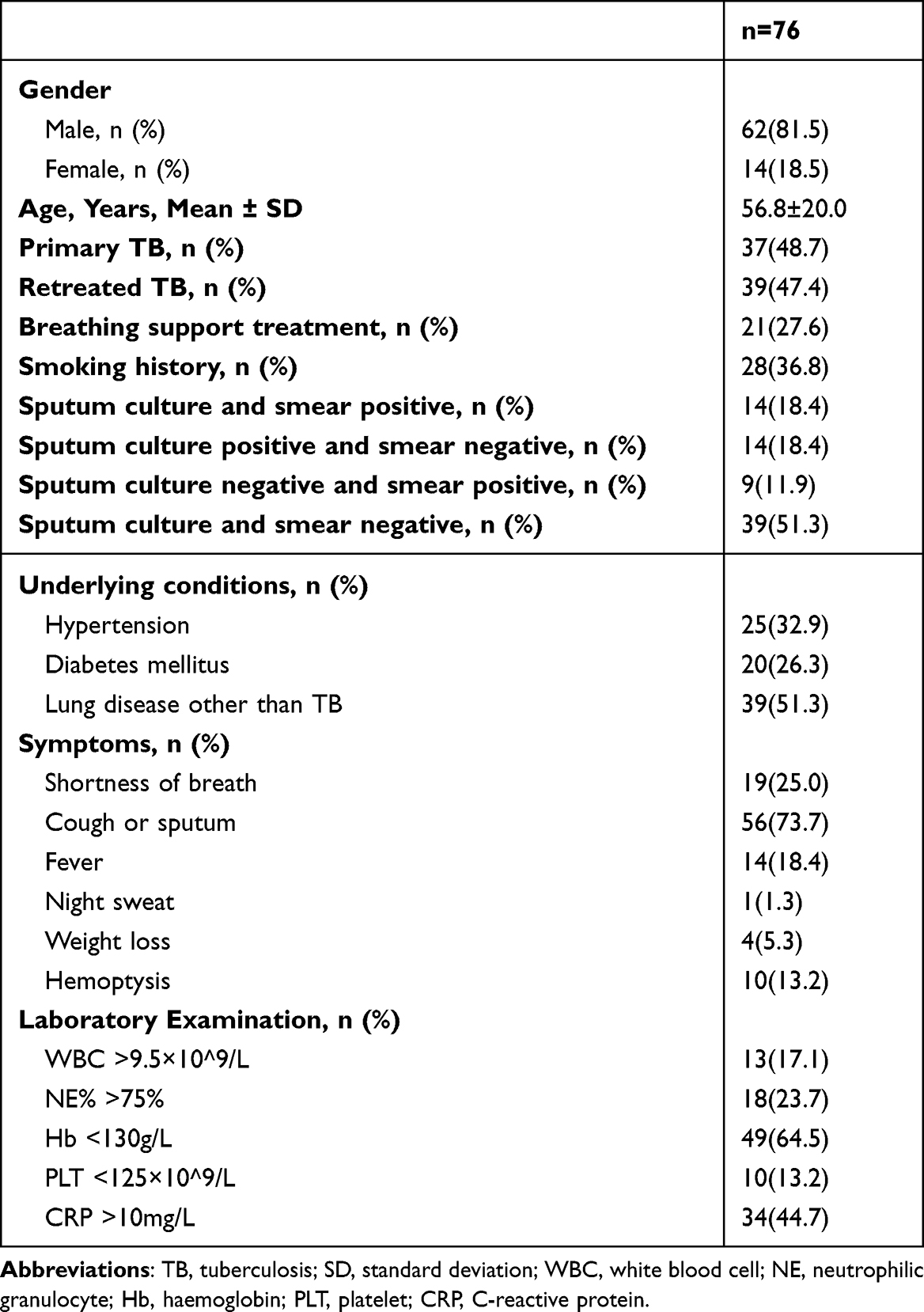 |
Table 1 Characteristics of Klebsiella pneumoniae Co-Infections in Pulmonary Tuberculosis (TB) |
A total of 80 K. pneumoniae strains were isolated from the 76 patients and identified as co-infections (two strains were isolated from each of the four patients at different time points). Of these, 32 K. pneumoniae strains isolated from patients (32/76; 42.1%) were multidrug resistant (MDR). Among these 32 patients, 26 were infected by ESBL-producing strains, 4 were infected by carbapenem-resistant Enterobacteriaceae (CRE) strains, and 2 were infected by both ESBL-producing and CRE strains. We then divided these patients into MDR (n = 32) and non-MDR groups (n = 44) according to whether the isolated strains were MDR or not, and the differences in characteristics between the two groups were examined (Table 2). The patients in the MDR group were older (61.1 ± 20.0 years old vs 53.6 ± 19.5 years old; p = 0.027), and more likely to have retreated TB (68.8% vs 38.6%; p = 0.010). Patients who received respiratory support during hospitalisation accounted for a significantly larger proportion of the MDR group than the non-MDR group (40.6% vs 18.2%; p = 0.031). The MDR group also had a higher prevalence of lung disease other than TB (68.8% vs 38.6%; p = 0.012). In terms of laboratory examinations, the proportion of patients with low haemoglobin (87.5% vs 47.7%; p = 0.001) or elevated C-reactive protein levels (62.6% vs 31.8%; p = 0.008) was greater in the MDR group than in the non-MDR group.
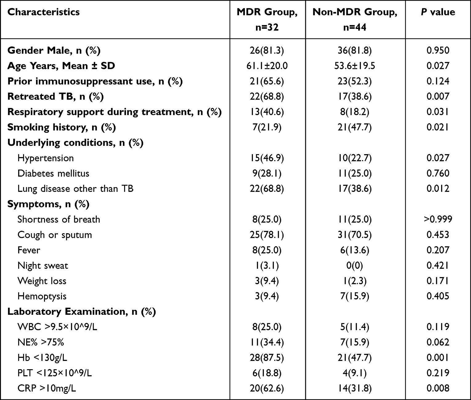 |
Table 2 Epidemiologic and Clinical Characteristics of Tuberculosis (TB) Patients with Klebsiella pneumoniae Co-Infection Stratified by Extended-Spectrum β-Lactamase (ESBL) Positivity |
Among the 76 patients, 22 (28.9%) also had co-infections of bacteria and fungi other than K. pneumoniae. The most common detected pathogens were Candida albicans (n = 8, 10.5%) and Pseudomonas aeruginosa (n = 8; 10.5%). Others included Enterobacter cloacae (n = 2; 2.6%), Acinetobacter baumannii (n = 2; 2.6%), Staphylococcus aureus (n = 2; 2.6%), Escherichia coli (n = 1; 1.3%), Staphylococcus haemolyticus (n = 1; 1.3%), Candida glabrata (n = 1; 1.3%), Candida tropicalis (n = 1; 1.3%), and Stenotrophomonas maltophilia (n = 1; 1.3%), as well as two fungal cases (filamentous fungi; 2.6%).
Clinical Manifestation of Acquiring Co-Infections
Among these TB co-infection cases, 55.26% (42/76) patients acquired K. pneumoniae infection within 48 h of admission, while the remaining patients were infected 48 h after hospitalisation. The median infection occurrence time was 2 d (IQR, 1–6 d) after admission. Since all strains were cultured from respiratory samples, we next determined whether these patients received respiratory support and analysed their lung examination data. We found that 16 patients adopted high flow oxygen therapy and 12 received invasive respiratory support including tracheal intubation and tracheostomy.
The important clinical features of K. pneumoniae co-infections in TB cases are summarised in Figure 1a and b. Among the 76 patients, 80.3% (61/76) had lung lesions, including 30.3% (23/76) with a pulmonary cavity, 22.4% (17/76) with bronchiectasis, 11.8% (9/76) with interstitial lung disease, and 27.6% (21/76) with chronic obstructive pulmonary disease.
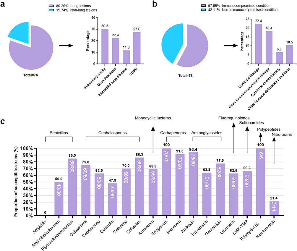 |
Figure 1 Clinical features of K. pneumoniae co-infections and phenotypic antimicrobial tests in TB cases. (a) Lung lesions in TB and K. pneumoniae co-infections cases. (b) Immune condition in TB and K. pneumoniae co-infections cases. (c) Phenotypic antimicrobial tests of enrolled strains. |
In addition, we found that 57.8% (44/76) of the patients showed an immunocompromised condition within one month, including 22.4% (17/76) that received corticoid therapy (daily prednisone ≥ 20 mg, more than 2 weeks of use), 18.4% (14/76) that received immunosuppressive therapies other than corticoids, 6.6% (5/76) that received cytotoxic chemotherapy, and 10.5% (8/76) that had other immune deficiency conditions validated by the principal investigator.
Genomic Characteristics of K. pneumoniae Strains
Traditional antimicrobial susceptibility tests showed that all 80 K. pneumoniae isolates were susceptible to polymyxin and resistant to amoxicillin. The susceptibility of isolates was 50.0% to the ampicillin/sulbactam and 85.0% to piperacillin/tazobactam. The proportion of isolates susceptible to cephalosporins ranged from 47.8% to 86.3% (47.8% to cefazolin, 62.5% to ceftriaxone, 70.0% to cefepime, 76.3% to ceftazidime, and 86.3% to cefotetan). The susceptibilities of isolates to aztreonam, levofloxacin, sulfamethoxazole/trimethoprim, and nitrofurantoin were 68.8%, 62.5%, 66.3%, and 21.4%, respectively. The susceptibilities to tobramycin, gentamycin, and amikacin were 63.8%, 77.5%, and 95.0%, separately. The resistance rate against imipenem was 8.8% (7/80) (Figure 1c). To sum up, 29 (36.3%) of the 80 K. pneumoniae isolates were ESBL-producing strains, and seven (8.8%) were determined as CRE strains.
Among the 80 K. pneumoniae isolates, 43 kinds of sequence types were identified (Figure 2a). ST23 accounted for the most (15.00%, 12/80), followed by ST15 (12.50%, 10/80), and ST273 (7.50%, 6/80). We found a distinguished distribution of sequence types of K. pneumoniae isolates between primary (n = 39) and retreated TB (n = 41) cases. Primary TB cases had more ST23 strains than retreated ones (28.2% vs 2.43%; p = 0.001) (Figure 2b). ST1764 and ST86 only existed in the primary TB group, while ST25, ST273, and ST65 were detected in the retreated TB group (Figure 2b and c).
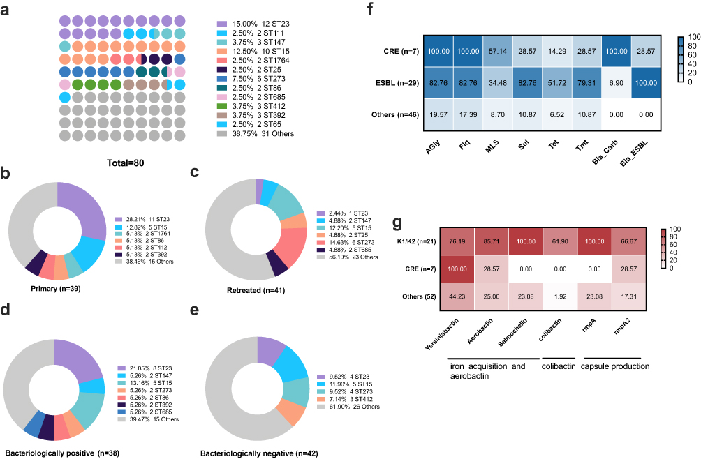 |
Figure 2 Sequence type distribution and resistance and virulence profiles. (a) Sequence type distribution in all strains. (b) Sequence type distribution in primary TB cases. (c) Sequence type distribution in retreated TB cases. (d) Sequence type distribution in bacteriologically positive TB cases. (e) Sequence type distribution in bacteriologically negative TB cases. (f) Resistance gene profile in CRE, ESBL and other strains. Agly: (aminoglycosides), Flq (fluoroquinolones), MLS (macrolides), Sul (sulphonamides), Tet (tetracyclines), Tmt (trimethoprim), Bla_ESBL (extended-spectrum β-lactamases), and Bla_Carb (carbapenemase). (g) Virulence gene profile in K1/K2, CRE, and other strains. |
A similar phenomenon was observed in strains from bacteriologically positive (n = 38) and bacteriologically negative (n = 42) pulmonary TB patients as well. Among the seven sequence types (ST23, ST147, ST273, ST86, ST685, and ST392), there were more than two strains in each type in the bacteriologically positive group, while four sequence types (ST23, ST15, ST273, and ST412) were mostly found in bacteriologically negative TB cases (Figure 2d and e).
We next depicted the resistance gene and virulence gene profiles of K. pneumoniae and found a high proportion of classic hypervirulent strains. Seven strains were confirmed to have carbapenem-resistant genes, including six blaKPC-2 and one blaOXA-232. The proportion of aminoglycoside and fluoroquinolone resistance genes of CRE (n = 7) and ESBL (n = 29) strains all surpassed 80% (Figure 2f). More than half of the ESBL strains had resistance genes of sulphonamides, tetracyclines, and trimethoprim (81.5%, 51.9%, and 77.8%, respectively), while the proportion of CRE was 28.57%. A considerable proportion of strains (21/80, 26.25%) were confirmed as classic hypervirulent K1/K2 K. pneumoniae (n = 21), with 100.00% harboured salmochelin and rmpA (Figure 2g). The existence of yersiniabactin, aerobactin, colibactin, and rmpA2 in K1/K2 strains were 76.19%, 85.17%, 61.90%, and 66.67%, separately. Yersiniabactin was found at higher levels in CRE strains compared to K1/K2 strains, while the other virulence-related genes were rare in CRE (Figure 2g).
Four patients (Pt 1–4) contributed more than one K. pneumoniae strain. Pt 1 had two different sequence type strains, ST273 and ST147, with a 20-day detection interval. Pt 2 had two ST685 strains with different β-lactams: LEN-24 and AmpH (C07), AmpH and SHV-187 (C08). Pt 3 had two ST23 strains with the same resistance and virulence genes while their phenotypic antimicrobial tests for ampicillin/sulbactam and imipenem were different (C46 and C47). Pt 4 had two ST15 strains: one had the resistance gene MphA for macrolides (C61) whereas the other did not (C62).
K. pneumoniae Transmission Event Identification
Since ST23 and ST15 occupied the most cases among the strains, we constructed phylogenetic trees to determine their genetic relationships. A total of 2367 SNPs were identified among the 13 strains, with pairwise SNP distances ranging from 16–382 SNPs. The strains C46 and C47 had a 16-SNP distance and were both cultured from Pt 1 (Figure 3a).
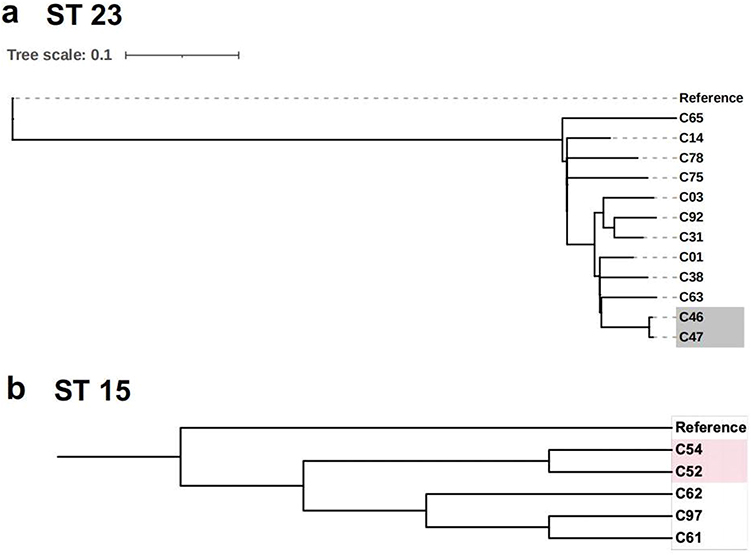 |
Figure 3 Phylogenetic tree of enrolled strains and clusters of ST23 and ST15 strains. (a) Phylogenetic tree of 13 ST23 strains. (b) Phylogenetic tree of five ST15-KPC-2 strains with similar gene profiles. |
One transmission event was identified in the ST15 group (Figure 3b). Only 34 SNPs were found in these five ST15-KPC-2 strains with similar resistance and virulence gene profiles. C52 and C54 were considered as a possible transmission event without a direct epidemiological link.
Discussion
In this retrospective study, we analyzed the clinical and microbiological characteristics of K. pneumoniae co-infections in pulmonary tuberculosis. We found that patients in MDR-group received more respiratory support than the non-MDR group and possessed higher elevated C-reactive protein and lower hemoglobin. Among the K. pneumoniae strains, 26.25% were classically hypervirulent K1/K2 K. pneumoniae, and all carried salmochelin and rmpA. One nosocomial transmission event was identified in the ST15 group.
We here identified several characteristics among these K. pneumoniae co-infections. The patients in MDR-group received more respiratory support than the non-MDR group and exhibited more obvious inflammatory response. Previous studies have validated that carbapenem resistant K. pneumoniae infections could induce a higher mortality rate compared to carbapenem sensitive K. pneumoniae infections.19–21
Our data show that traditional pneumonia antibiotics, such as β-lactams and respiratory quinolones, are effective in the treatment of pulmonary tuberculosis complicated by pulmonary infection. Most patients had positive treatment outcomes after being treated with piperacillin/sulbactam, piperacillin/tazobactam, cefonicid, or moxifloxacin. Ishikawa et al have reported that pulmonary tuberculosis patients co-infected with other microorganisms have poorer treatment effects and higher mortality rates than TB-only patients; the mortality rates with and without microorganisms were 39.8% (51/128) and 10.2% (59/580), respectively.4 However, in our study, only three patients failed treatment, which is much lower than number reported in the study by Ishikawa et al. This may be related to our focus only on TB and K. pneumoniae. We further found that all three patients who failed treatment also had respiratory failure. Thus, respiratory failure may be a risk factor for poor treatment outcomes in TB and K. pneumoniae co-infection.
Previous studies have reported a 9%–28% proportion of K. pneumoniae co-infections in pulmonary tuberculosis.13,14 It was noted that the proportion of primary and retreated TB cases in co-infection were similar, and more computed tomographic manifestations, including consolidation, patchy shadow, ground glass opacity and computed tomography reports suggestive of infection, existed in co-infection occurrence more than 48 h after admission, which may be correlated with microbiota dysbiosis and should be investigated in the future. It was notable that nearly half of the patients had other lung disease, including chronic obstructive pulmonary disease, bronchiectasis, and lung cancer. Previous studies have demonstrated that chronic lung disease is one of the risk factors of bacterial pneumonia.22,23 Therefore, TB patients with chronic lung disease are more likely to develop K. pneumoniae co-infection. The most common clinical symptoms of co-infection in our patients were cough, sputum, and shortness of breath, which was similar to the clinical manifestations of TB and other bacterial co-infections.13,14
Combined with the co-infection occurrence time and genomic profile, most of the strains may have originated from community or host pulmonary microbiota. ST23, ST86, ST65, and ST25 are common sequence types of hypervirulent strains that mostly circulate within communities.24,25 Though approximately half of the patients developed co-infections after 48 h of admission, genetic analyses identified nosocomial infections, except for C52 and C54. Additionally, nosocomial K. pneumoniae strains in China were mostly caused by multidrug-resistant strains, including ST11 strains.26
Previous studies have reported microbiota dysbiosis in TB patients;27 treated patients showed a microbiota composition that was different to that of non-treated patients.28,29 K. pneumoniae accounted for 56.04% of the total bronchoalveolar lavage fluid microbiota in pulmonary TB patients.27 Though approximately half of the patients obtained co-infections after 48 h of admission, their strains were mostly unique, indicating that the sources may be host pulmonary microbiota. The introduction of M. tuberculosis and administration of anti-tuberculosis drugs might select specific strains as dominant strains to be detected.30,31 We observed within-host microevolution in Pt 4; C62 was cultured 3 days after C61 and was missing the MphA gene.
Whole-genome sequencing (WGS) can help track the transmission route of infection and identify index cases. Here, we found two cases without a confirmed epidemiologic link, which might be disregarded. WGS retrospectively identified transmission clusters, shedding light on the importance of infection control, and can be therefore adopted as a surveillance tool in the future.32–34
Our study had a few limitations. First, we only conducted analyses of K. pneumoniae co-infections in pulmonary TB without co-infections by other pathogens. We would perform the analyses of single TB and co-infections in the future large cohort studies. Though the sample size is relatively small, the limited studies on TB and K. pneumoniae co-infection make it important to depict the clinical manifestation and sources of theses co-infections. Further investigations involving larger sample sizes and more centers and comprising samples of pulmonary TB with co-infections by other pathogens are required to gain comprehensive insights into the occurrence and prevalence of pulmonary TB. Second, we combined sequence type and admission type to infer sources of K. pneumoniae, which should be validated using pulmonary samples in the future.
Conclusions
The study explored the clinical and microbiological characteristics of K. pneumoniae infection among pulmonary TB patients, indicating a relatively high proportion of K. pneumoniae co-infection among pulmonary TB patients. A considerable part of strains was multi-drug resistant, patients in MDR-group received more respiratory support than the non-MDR group and possessed higher elevated C-reactive protein and lower haemoglobin, suggesting a special attention for multidrug resistant K. pneumoniae infections with more obvious inflammatory responses which calls for more respiratory support and timely clinical management. ST23 accounted for the most sequence type of K. pneumoniae strains, and transmission events existed. Further studies are warranted to explore the mechanism and access to the treatment of K. pneumoniae co-infection among pulmonary TB patients.
Abbreviations
PTB, Pulmonary tuberculosis; ESBL, Extended-spectrum β-lactamase; CRE, Carbapenem-resistant Enterobacteriaceae; TB, Tuberculosis; MDR, Multidrug resistant; WGS, Whole-genome sequencing; SD, Standard deviation.; NE, Neutrophilic granulocyte; Hb, Haemoglobin.; PLT, Platelet; CRP, C-reactive protein; IQR, Interquartile range.
Data Sharing Statement
The datasets used and/or analyzed during the current study are available from the corresponding author on reasonable request.
Ethics Approval and Informed Consent
This study was conducted based on Principles of Declaration of Helsinki and was approved by the Ethics Committee of Wuxi No. 5 People’s Hospital (WX2021-021-1). The requirement for informed consent was waived because of the retrospective nature of the study. Further, patient data were deidentified before data access and analysis.
Acknowledgments
The study investigators gratefully acknowledge all study participants.
Funding
This work was supported by Shanghai Municipal Science and Technology Major Project (HS2021SHZX001), Shanghai Science and Technology Committee (20dz2210400, 20dz2260100), Major projects of Wuxi Municipal Health Commission (Z202111), and Wuxi Taihu Lake Talent Plan.
Disclosure
The authors declare that this research was conducted in the absence of any commercial or financial relationships that could be construed as a potential conflict of interest.
References
1. Organization WH. Global tuberculosis report 2021; 2022; Available from: https://www.who.int/publications/i/item/9789240037021.
2. Hsu D, Irfan M, Jabeen K, et al. Post tuberculosis treatment infectious complications. Int J Infect Dis. 2020;92S:S41–S45.
3. Hernandez-Pando R, Orozco H, Aguilar D. Factors that deregulate the protective immune response in tuberculosis. Arch Immunol Ther Exp (Warsz). 2009;57(5):355–367. doi:10.1007/s00005-009-0042-9
4. Ishikawa S, Igari H, Yamagishi K, Takayanagi S, Yamagishi F. Microorganisms isolated at admission and treatment outcome in sputum smear-positive pulmonary tuberculosis. J Infect Chemother. 2019;25(1):45–49. doi:10.1016/j.jiac.2018.10.005
5. Chang D, Sharma L, Dela Cruz CS, Zhang D. Clinical Epidemiology, Risk Factors, and Control Strategies of Infection. Front Microbiol. 2021;12:750662. doi:10.3389/fmicb.2021.750662
6. Bengoechea JA, Sa Pessoa J. Klebsiella pneumoniae infection biology: living to counteract host defences. FEMS Microbiol Rev. 2019;43(2):123–144. doi:10.1093/femsre/fuy043
7. Magill SS, Edwards JR, Bamberg W, et al. Multistate point-prevalence survey of health care-associated infections. N Engl J Med. 2014;370(13):1198–1208. doi:10.1056/NEJMoa1306801
8. Udeani TKC, Moses J, Uzoechina A, Okwori AEJ, Okwosa CN. Microbial aetiologic agents associated with pneumonia in immunocompromised hosts. Afr J Infect Dis. 2010;4(1):1–6. doi:10.4314/ajid.v4i1.55084
9. Buchera FS, Silago V, Japhet G, et al. Predominance of Other Pathogenic Bacteria among Presumptive Tuberculosis Cases Attending Tuberculosis Clinics in Mwanza, Tanzania: a Cross-Sectional Laboratory-Based Study. Microorganisms. 2022;10(4):4. doi:10.3390/microorganisms10040703
10. Rafailidis PI, Kapaskelis A, Christodoulou C, Galani E, Falagas ME, Concurrent M. tuberculosis, Klebsiella pneumoniae, and Candida albicans infection in liver metastasis of bowel carcinoma. Eur J Clin Microbiol Infect Dis. 2008;27(8):753–755. doi:10.1007/s10096-008-0488-4
11. Arora AA, Krishnaswamy UM, Moideen RP, Padmaja MS. Tubercular and bacterial coinfection: a case series. Lung India. 2015;32(2):172–174. doi:10.4103/0970-2113.152645
12. Iliyasu G, Mohammad AB, Yakasai AM, Dayyab FM, Oduh J, Habib AG. Gram-negative bacilli are a major cause of secondary pneumonia in patients with pulmonary tuberculosis: evidence from a cross-sectional study in a tertiary hospital in Nigeria. Trans R Soc Trop Med Hyg. 2018;112(5):252–254. doi:10.1093/trstmh/try044
13. Attia EF, Pho Y, Nhem S, et al. Tuberculosis and other bacterial co-infection in Cambodia: a single center retrospective cross-sectional study. BMC Pulm Med. 2019;19(1):60. doi:10.1186/s12890-019-0828-4
14. Kim SB, Lee W-Y, Lee J-H, et al. A variety of bacterial aetiologies in the lower respiratory tract at patients with endobronchial tuberculosis. PLoS One. 2020;15(6):e0234558. doi:10.1371/journal.pone.0234558
15. Wang Y, Zhang Q, Jin Y, Jin X, Yu J, Wang K. Epidemiology and antimicrobial susceptibility profiles of extended-spectrum beta-lactamase-producing Klebsiella pneumoniae and Escherichia coli in China. Braz J Microbiol. 2019;50(3):669–675. doi:10.1007/s42770-019-00081-7
16. Rahmat Ullah S, Majid M, Rashid MI, Mehmood K, Andleeb S. Immunoinformatics Driven Prediction of Multiepitopic Vaccine Against and Coinfection and Its Validation via In Silico Expression. Int J Pept Res Ther. 2021;27(2):987–999. doi:10.1007/s10989-020-10144-1
17. Lam MMC, Wick RR, Watts SC, Cerdeira LT, Wyres KL, Holt KE. A genomic surveillance framework and genotyping tool for Klebsiella pneumoniae and its related species complex. Nat Commun. 2021;12(1):4188. doi:10.1038/s41467-021-24448-3
18. Zhang Y, Chen C, Wu J, et al. Sequence-Based Genomic Analysis Reveals Transmission of Antibiotic Resistance and Virulence among Carbapenemase-Producing Klebsiella pneumoniae Strains. mSphere. 2022;7(3):e0014322. doi:10.1128/msphere.00143-22
19. Xu L, Sun X, Ma X. Systematic review and meta-analysis of mortality of patients infected with carbapenem-resistant Klebsiella pneumoniae. Ann Clin Microbiol Antimicrob. 2017;16(1):18. doi:10.1186/s12941-017-0191-3
20. Zarkotou O, Pournaras S, Tselioti P, et al. Predictors of mortality in patients with bloodstream infections caused by KPC-producing Klebsiella pneumoniae and impact of appropriate antimicrobial treatment. Clin Microbiol Infect. 2011;17(12):1798–1803. doi:10.1111/j.1469-0691.2011.03514.x
21. Tumbarello M, Trecarichi EM, De Rosa FG, et al. Infections caused by KPC-producing Klebsiella pneumoniae: differences in therapy and mortality in a multicentre study. J Antimicrob Chemother. 2015;70(7):2133–2143. doi:10.1093/jac/dkv086
22. Rello J, Rodriguez A, Torres A, et al. Implications of COPD in patients admitted to the intensive care unit by community-acquired pneumonia. Eur Respir J. 2006;27(6):1210–1216. doi:10.1183/09031936.06.00139305
23. Arancibia F, Bauer TT, Ewig S, et al. Community-acquired pneumonia due to gram-negative bacteria and pseudomonas aeruginosa: incidence, risk, and prognosis. Arch Intern Med. 2002;162(16):1849–1858. doi:10.1001/archinte.162.16.1849
24. Russo TA, Marr CM. Hypervirulent Klebsiella pneumoniae. Clin Microbiol Rev. 2019;32(3):3. doi:10.1128/CMR.00001-19
25. Tang M, Kong X, Hao J, Liu J. Epidemiological Characteristics and Formation Mechanisms of Multidrug-Resistant Hypervirulent. Front Microbiol. 2020;11:581543. doi:10.3389/fmicb.2020.581543
26. Gu D, Dong N, Zheng Z, et al. A fatal outbreak of ST11 carbapenem-resistant hypervirulent Klebsiella pneumoniae in a Chinese hospital: a molecular epidemiological study. Lancet Infect Dis. 2018;18(1):37–46. doi:10.1016/S1473-3099(17)30489-9
27. Xiao G, Cai Z, Guo Q, et al. Insights into the Unique Lung Microbiota Profile of Pulmonary Tuberculosis Patients Using Metagenomic Next-Generation Sequencing. Microbiol Spectr. 2022;10(1):e0190121. doi:10.1128/spectrum.01901-21
28. Ding L, Liu Y, Wu X, et al. Pathogen Metagenomics Reveals Distinct Lung Microbiota Signatures Between Bacteriologically Confirmed and Negative Tuberculosis Patients. Front Cell Infect Microbiol. 2021;11:708827. doi:10.3389/fcimb.2021.708827
29. Kateete DP, Mbabazi MM, Nakazzi F, et al. Sputum microbiota profiles of treatment-naïve TB patients in Uganda before and during first-line therapy. Sci Rep. 2021;11(1):24486. doi:10.1038/s41598-021-04271-y
30. Murray AK, Zhang L, Yin X, et al. Novel Insights into Selection for Antibiotic Resistance in Complex Microbial Communities. mBio. 2018;9(4):4. doi:10.1128/mBio.00969-18
31. Chen X, He G, Lin S, et al. Analysis of Serial Multidrug-Resistant Tuberculosis Strains Causing Treatment Failure and Within-Host Evolution by Whole-Genome Sequencing. mSphere. 2020;5(6):6. doi:10.1128/mSphere.00884-20
32. Carlos CC, Masim MAL, Lagrada ML, et al. Genome Sequencing Identifies Previously Unrecognized Klebsiella pneumoniae Outbreaks in Neonatal Intensive Care Units in the Philippines. Clin Infect Dis. 2021;73(Suppl_4):S316–S324. doi:10.1093/cid/ciab776
33. Onori R, Gaiarsa S, Comandatore F, et al. Tracking Nosocomial Klebsiella pneumoniae Infections and Outbreaks by Whole-Genome Analysis: small-Scale Italian Scenario within a Single Hospital. J Clin Microbiol. 2015;53(9):2861–2868. doi:10.1128/JCM.00545-15
34. Chen C, Zhang Y, Yu S-L, et al. Tracking Carbapenem-Producing Outbreak in an Intensive Care Unit by Whole Genome Sequencing. Front Cell Infect Microbiol. 2019;9:281. doi:10.3389/fcimb.2019.00281
 © 2023 The Author(s). This work is published and licensed by Dove Medical Press Limited. The full terms of this license are available at https://www.dovepress.com/terms.php and incorporate the Creative Commons Attribution - Non Commercial (unported, v3.0) License.
By accessing the work you hereby accept the Terms. Non-commercial uses of the work are permitted without any further permission from Dove Medical Press Limited, provided the work is properly attributed. For permission for commercial use of this work, please see paragraphs 4.2 and 5 of our Terms.
© 2023 The Author(s). This work is published and licensed by Dove Medical Press Limited. The full terms of this license are available at https://www.dovepress.com/terms.php and incorporate the Creative Commons Attribution - Non Commercial (unported, v3.0) License.
By accessing the work you hereby accept the Terms. Non-commercial uses of the work are permitted without any further permission from Dove Medical Press Limited, provided the work is properly attributed. For permission for commercial use of this work, please see paragraphs 4.2 and 5 of our Terms.