
Back to Journals » Journal of Inflammation Research » Volume 18
Characterization of a Long-Acting Anti-Human MASP-2 Antibody for the Treatment of Complement-Related Diseases
Authors Han X  , Yang C, Wang Z, Fan K, Liu K, Ye H, Zhang W, Zhang J, Hu X , Lin H, Liao C
Received 13 February 2025
Accepted for publication 7 November 2025
Published 19 November 2025 Volume 2025:18 Pages 16247—16260
DOI https://doi.org/10.2147/JIR.S522657
Checked for plagiarism Yes
Review by Single anonymous peer review
Peer reviewer comments 2
Editor who approved publication: Professor Ning Quan
Xiangli Han,1,2,* Changyong Yang,1,2,* Zhijun Wang,1,2,* Ke Fan,1,2 Kaili Liu,1,2 Hao Ye,1,2 Wei Zhang,1,2 Jiawei Zhang,1,2 Xiaotong Hu,1,2 Hongda Lin,1,2 Cheng Liao1,2
1Shanghai Shengdi Pharmaceutical Co., Ltd, Shanghai, 200100, People’s Republic of China; 2Jiangsu Hengrui Pharmaceuticals Co., Ltd, Lianyungang, Jiangsu, 222000, People’s Republic of China
*These authors contributed equally to this work
Correspondence: Cheng Liao, Jiangsu Hengrui Pharmaceuticals Co., Ltd, Lianyungang, Jiangsu, 222000, People’s Republic of China, Tel/Fax +86 021-61063826, Email [email protected]
Purpose: The complement system is an important component of the innate immune system, which is essential for orchestrating host defense and regulating inflammation. However, overactivation of the complement system plays a pathogenic role in the onset and progression of various inflammatory diseases and rare diseases. Central to the lectin pathway (LP) among three complement activation routes, dysregulation of mannan-binding lectin-associated serine protease-2 (MASP-2) is associated with conditions ranging from IgA nephropathy (IgAN) to hematopoietic stem cell transplantation-associated thrombotic microangiopathy (HSCT-TMA). Thus, we tend to develop an anti-MASP-2 antibody to treat relevant diseases.
Methods: Herein, we developed SHR-2010, a novel fully humanized monoclonal antibody targeting MASP-2. SHR-2010 was generated through mouse immunization with recombinant human MASP-2 protein. The binding affinities of SHR-2010 were determined using surface plasmon resonance (SPR). Then, a mouse surrogate antibody ER011-11165 was developed and subsequently evaluated in the lipopolysaccharide (LPS)-induced acute kidney injury murine model. Additionally, the pharmacokinetic (PK), pharmacodynamics (PD) and safety profiles of SHR-2010 were conducted in monkeys.
Results: SHR-2010 exhibited strong inhibition of lectin pathway activation in human and rhesus serum. Administration of ER011-11165 significantly mitigated LPS-induced nephrotoxicity in acute kidney injury mouse model. PK profiling in non-human primates (NHPs) revealed high subcutaneous bioavailability and long half-life of SHR-2010, supporting a potential subcutaneous administration route and long dosing interval in clinic. Notably, PD markers demonstrated sustained LP inhibition, with 65.8– 80.1% inhibitory effect on serum LP persisting for 408 hours following a single intravenous dose of 5 mg/kg SHR-2010. The nonclinical toxicology evaluation demonstrated that SHR-2010 exhibits a proper safety profile.
Conclusion: Preclinical studies demonstrated that SHR-2010 exhibited superior pharmacokinetics and sustained lectin pathway inhibition compared to OMS721. When coupled with optimized trial design strategies, SHR-2010 could be a promising therapeutic candidate for lectin pathway-driven diseases, including IgAN.
Keywords: MASP-2, IgA nephropathy, lectin pathway, long-acting
Introduction
The complement system is an enzyme cascade involving more than 30 plasma and membrane-bound proteins, serving as a frontline against infection via activation of local inflammatory responses.1,2 Complements in the inactive form could be sequentially activated through enzymatic cascade reactions.3–5 There are three pathways of complement activation, i.e. the classical pathway, the lectin pathway, and the alternative pathway.1,2 Although differing in triggering mechanisms, all pathways converge on generating C3 convertase—the pivotal enzyme cleaving C3 into bioactive fragments.6–12 Then, the C3 convertase activates C3, followed by C5 convertase activates C5, resulting in the formation of the membrane attack complex (MAC), leading to direct pathogen lysis or inflammatory tissue damage.6–12 Previous studies have implicated that the hyperactivation of the lectin pathway could potentially contribute to multiple diseases including IgA nephropathy (IgAN) and hematopoietic stem-cell transplantation–associated thrombotic microangiopathy (HSCT-TMA).13,14
IgAN, the most common primary glomerulopathy worldwide, displays marked regional prevalence with annual incidence reaching 45 cases per million in Japan.15 It is characterized by the deposition of IgA immunoglobulins in the glomerular mesangium coupled with uncontrolled activation of the lectin pathway.16,17 The incidence and progression of IgAN are highly associated with the complement system, as evidenced by the colocalization of IgA with C3 in about 90% IgAN patients.18 Moreover, depositions of C3 and C4 were often closely related to renal pathological damages and local inflammation in patients with IgAN.18,19 Current therapeutic strategies targeting the complement system for IgAN primarily focus on the alternative and lectin pathway.20 Novartis’ iptacopan, a specific factor B inhibitor in the alternative pathway, has been approved for IgAN. However, as another important driver of IgAN, the lectin pathway still lacks effective targeted therapies. Therapies targeting the lectin pathway for treating IgAN may synergize with iptacopan, or be used to treat patients who are insensitive to iptacopan.
Mannan-binding lectin-associated serine protease-2 (MASP-2) is the key initiating enzyme to activate the lectin pathway and the complement cascade.21,22 Upon binding to a variety of pattern recognition molecules (PRMs), such as mannose-binding lectins (MBLs), MASP-2 undergoes autoactivation, thereby promoting downstream lectin pathway activation.23,24 However, aberrant MASP-2 activation could lead to multiple diseases. Previous studies have shown that the deposition of pattern recognition molecules collection-11 and subsequent MASP-2 activation could trigger renal epithelial injury.25 Studies in MASP-2-deficient mice have shown that MASP-2-mediated lectin pathway activation could contribute to ischemia-reperfusion injury after renal transplantation.23 Furthermore, studies have linked MASP-2 hyperactivation to IgAN susceptibility.26 Therefore, inhibiting MASP-2 could be a potential therapeutic approach for diseases caused by the activation of lectin pathway. OMS721 (narsoplimab) is a monoclonal antibody targeting MASP-2 developed by Omeros. As the fastest-advancing MASP-2-targeted drug in clinical development, this antibody is currently undergoing a Phase 2 trial for HSCT-TMA and has completed a Phase 3 trial for IgAN. The success of phase 2 clinical trials for HSCT-TMA have preliminary demonstrated the clinical potential of MASP-2 inhibition.
Here, we developed a novel fully humanized anti-MASP-2 monoclonal antibody, SHR-2010. In this study, we thoroughly characterized the antibody, including pharmacologic activity, pharmacodynamic (PD), pharmacokinetic (PK) and safety assessment. We obtained SHR-2010 by immunizing the mice with human MASP-2 recombinant protein, followed by antibody humanization. Then, SHR-2010 was characterized through systematic in vitro assays. SHR-2010 could efficiently bind to MASP-2 and inhibit lectin-mediated complements activation. Furthermore, MASP-2 inhibition with E011-11165, mouse surrogate antibody of SHR-2010, could mitigate acute kidney injury induced by LPS in mouse model. Essentially, SHR-2010 displayed a long half-life and sustained LP inhibition in NHPs. The nonclinical toxicology evaluation demonstrated that SHR-2010 exhibits a proper safety profile. Altogether, these studies demonstrated that SHR-2010 is potential candidate for treating complement-mediated inflammatory diseases by inhibiting lectin pathway activity.
Materials and Methods
Animal Welfare and Ethical Statement
The pharmacokinetic study protocols and animal care were approved by the Committee for the Management and Use of Experimental Animals of Hubei Topgene Xinsheng Biotechnology Co., Ltd. (Permit Number: IACUC-XS-2021-007), and strictly adhere to the requirements of the Laboratory Animal Care and Use Committee. The laboratory animal usage license number was SYXK-E-2020-0113. The toxicity study protocols and animal care followed regulations and guidelines that adhere to the Guide for Care and Use of Laboratory Animals (Institute of Laboratory Animal Research, National Research Council, 2011) and Regulations on the Administration of Laboratory Animals (Ministry of Science and Technology of the People’s Republic of China, 2013). The laboratory animal usage license number was SYXK-SU-2018-0050.
Production of Full Length and Truncated MASP-2 Proteins
Recombinant full-length human MASP-2 was prepared for mouse immunization and antibody screening. To reduce the autocatalysis of the protein and improve the protein stability, S633A and S632A mutations were introduced to generate human and monkey MASP-2 S633A mutant, mouse MASP-2 S632A mutant, respectively. For epitope identification, different truncated MASP-2 proteins including human MASP-2 CCP1, human MASP-2 CCP1-CCP2-SP/S633A, and human MASP-2 CCP2-SP/S633A were designed and produced with a His-tag at either C-terminus or N-terminus for affinity purification through HisTrap Excel columns. Full-length MASP-2 and human MASP-2 CCP1 were expressed in CHO-S cells, while CCP1-CCP2-SP/S633A) and CCP2-SP/S633A were recombinantly expressed in E. coli BL21.
Screening of Anti-MASP-2 Antibodies from Hybridomas and Humanization
Recombinant full-length human MASP-2/S633A was used for mice immunization. In brief, BALB/c mice were intraperitoneally injected with human MASP-2/S633A emulsified in Freund’s complete adjuvant (Cat. 344289, Sigma) at a prime dose of 50 μg, after which, two to three boost doses (25 μg of human MASP-2/S633A) were given at a 2-week interval. A final boost was given 3 days before fusion. Afterwards, the mice were sacrificed, and spleen cells were homogenized and fused with SP 2/0 myeloma cells. Then the mixture was seeded into 96-well plates at an appropriate cell density. 7–10 days after fusion, supernatants from each well were subjected to protein binding tests against human and monkey MASP-2 by ELISA, using an optimal coating concentration of 1 µg/mL. Positive clones were subcloned, screened, and sequenced for further characterization.
Clone 77H11 was selected for further humanization via complementarity-determining region (CDR) grafting. Briefly, the CDRs from the heavy and light chains of 77H11 were grafted onto the framework regions of the human germline sequences IGKV1-27*01 and IGKV4-30-4*01, respectively. Following structural prediction of the murine monoclonal antibody by homology modeling, back mutations were introduced at positions potentially critical for antibody-antigen binding.27,28 The nucleotide sequences encoding the variable regions of the humanized antibody were then cloned into a pcDNA3.4 vector containing the constant region sequences for the human heavy chain (IgG4 S228P variant) and light chain (kappa). This expression construct was transfected into CHOK1 cells for antibody production, and the resulting antibody was purified by protein A chromatography.
Binding Specificity of Anti-MASP-2 Antibody
The anti-MASP-2 antibodies raised against full-length MASP-2 protein were tested for their binding ability to different domains of MASP-2 by ELISA. In brief, 96 well plates were coated with the full-length and truncated MASP-2 proteins including human MASP-2 CCP1, human MASP-2 CCP1-CCP2-SP/S633A, and human MASP-2 CCP2-SP/S633A. After blocking with 3% BSA/PBST, antibodies at 100 μg/mL were added, and incubated at room temperature for 1 h. The plate was washed and HRP-labeled goat anti-human IgG Fc secondary antibody was added. After another hour of incubation, the wells were washed and TMB substrate was added. The reaction was terminated after 10 minutes by adding 1 M HCl, and the absorbance was measured at 450 nm with the microplate reader.
SPR Analysis
The interactions of SHR-2010 and MASP-2 proteins derived from human, monkey, and mouse were analyzed using an SPR (Biacore 8K, GE Healthcare) method. SHR-2010 was immobilized on the Flow cell2 by Protein A (29139121-AB, Cytiva) capture, and then MASP-2 at concentrations of 0.390625 to 50 nM were injected sequentially into the chamber in running buffer (HBS-EP + buffer (10X)) at 25°C. The binding and disassociation times were 60 seconds and 600 seconds, respectively, and the flowrate was 30 μL/min. The chip was regenerated with Glycine 1.5 subsequently. SPR signals were collected and stored using Biacore 8K Control Software, and then processed using Biacore Insight Evaluation 3.0 analysis software. The corrected signal curve was plotted by subtracting the signal value of the corresponding reference channel (Fc1) from the signal value of the detection channel (Fc2). Using double subtraction, a zero-concentration cycle with the same parameter was added before the sample cycle, and then the zero-concentration cycle from the sample cycle was subtracted to obtain the signal curve after secondary correction. The affinity kinetic curves were fitted according to a 1:1 Langmuir binding model and KD values were calculated.
The similar experimental procedure was used to analyze the interaction of OMS721 biosimilar (MA15MA3002, Sino Biological), as synthesized according to WO2012151481A,29 with MASP-2 derived from human, monkey, and mouse. OMS721 biosimilar was fixed on the Flow cell2 by capture-coupling, and then MASP-2 at concentrations of 0.78125 to 100 nM were injected sequentially into the chamber in running buffer. The binding time and disassociation time were 180 seconds and 528 seconds, respectively.
To analyze the interaction of ER011-11165 (20210926H03, Biointron) with human and mouse MASP-2, ER011-11165 was fixed on the Flow cell2 by capture-coupling, and then MASP-2 at concentrations of 0.390625 to 50 nM was injected sequentially into the chamber in running buffer. The binding and disassociation times were 60 seconds and 600 seconds, respectively. The methods used were otherwise identical to those described above.
Serum Complement C4 Deposition Assay
The in vitro inhibitory activity of the test substances on the serum complement lectin pathway of human, monkey, rat, and mouse was evaluated by using the serum complement lectin pathway specific C4 deposition assay. Mannan (Sigma-M7504-1G, Sigma), an activating moiety for the complement lectin pathway, was coated onto 384-well high binding plates overnight. After TBST washing, BSA blocking solution containing 5 mM Ca2+ was added to block for 2 hours at room temperature. At the same time, the antibody to be tested was diluted in a gradient manner and mixed with human, rhesus monkey, rat, and mouse serum at final concentration of 90%, 5%, 90% and 90%, respectively. The mixtures were then incubated at 4°C for 30 minutes. The blocked plate was washed with TBST and buffer, and the incubated antibody-serum mixture was added and incubated at 4°C for 1 hour. The chicken anti-human C4c antibody (Agrisera, IMS01-031-305) was conjugated with biotin. After three times of TBST washes, the biotin-conjugated anti-human C4c antibody at the final concentration of 3 μg/mL was added and incubated at room temperature for 1 hour. After washing, the SA-HRP (Invitrogen, 434323) was added at a 1:10,000 dilution and incubated for 30 minutes at room temperature. The absorbance at 450nm was then examined with the TMB developer.
Serum Complement Classical Pathway and Alternative Pathway Assay
The WIESLAB COMPL 300 complement assay kit was equilibrated to room temperature. SHR-2010 was mixed with human serum at a 1:9 ratio and incubate at 4°C for 30 minutes. The final concentration of SHR-2010 was 3.2, 16, 80, 400, and 2000 nM, with a final serum concentration of approximately 5.6%. The incubated antibody-serum mixture was added to CP and AP specific coated plates, respectively, and incubated at 37°C for 60 minutes. Wash the plates three times with washing buffer. Conjugate was added and incubated at room temperature for 30 minutes. The plates were washed three times and substrate solution was added and incubated at room temperature for 30 minutes before reading the OD405.
Mouse Kidney Injury Model and Assessment of Renal Function
Male C57BL/6 wildtype mice (8–9 weeks old) were randomly divided into five groups and administered via intraperitoneal injection of saline or LPS (10 mg/kg) at day 2 and a single IV dose of 3, 10 mg/kg of ER011-11165 at day 0 and day 2. Blood and urine samples were collected at 24 h after LPS injection to evaluate the renal function. Blood samples were obtained via orbital puncture. Blood urea nitrogen (BUN) and serum creatinine were measured using an automatic analyzer. Urine albumin was determined using Urine microalbumin analyzer. Urine albumin/creatine (A/C) was calculated by dividing albumin concentration in milligrams by creatinine concentration in grams.
Pharmacokinetics and Pharmacodynamics Study in Rhesus Monkeys
A total of 12 female and 12 male monkeys were randomly assigned to four groups (3/sex/group) and administered with SHR-2010 with a single intravenous (IV) dose at 0.5, 1.5, or 5 mg/kg or with a single subcutaneous (SC) dose at 5 mg/kg. Blood samples (~3 mL) for PK study were collected from each animal via the subcutaneous vein and processed for serum. Blood samples of the IV group were collected on at pre-dose, 0.083 h, 0.25 h, 1 h and 4 h post-dose on Day 0, and on Days 1, 2, 3, 4, 7, 10, 14, 17, 21, 25, 28, 31 and 35, and blood samples of the SC groups were collected at pre-dose, 0.25 h, 1 h, 4 h and 8 h post-dose at Day 0, and on Days 1, 2, 3, 4, 7, 10, 14, 17, 21, 25, 28, 31 and 35. The concentration of SHR-2010 was quantified using ELISA. In brief, microplates were coated with an anti-SHR-2010 capture antibody, followed by detection of serum SHR-2010 using anti-human IgG antibody conjugated with HRP and TMB substrate. Pharmacokinetic data were calculated using a non-compartmental model with Phoenix WinNonlin 7.0 software. To evaluate the PK/PD relationship over a broad time range, blood samples for PD study were collected at PK sampling times. The C4 deposition assay was used to specifically detect the complement lectin pathway activity in serum to assess the efficacy of SHR-2010. Mannan (Sigma-M7504-1G, Sigma) was coated onto 384-well high binding plates overnight. After TBST washing, BSA blocking solution containing 5 mM Ca2+ was added to block for 2 hours at room temperature, followed by washing. The serum separated from the blood samples was diluted to 90% and then added and incubated at 4°C for 1 hour. After three times of TBST washes, the biotin-conjugated anti-human C4c antibody at the final concentration of 3 μg/mL was added and incubated at room temperature for 1 hour. After washing, the SA-HRP at a 1:10,000 dilution was added and incubated for 30 minutes at room temperature. The absorbance at 450nm was then examined with the TMB developer.
Pharmacokinetics/Pharmacodynamic Modelling
A population PK/PD (2-compartment) model was applied to analyze the data of SHR-2010 IV and SC single dose regimens used in rhesus monkey studies. PK-PD relationships between serum concentrations of SHR-2010 and lectin pathway activity inhibition levels were also explored. Pop PK/PD analysis was performed using the nonlinear mixed effects modeling software (NONMEM, version 7.5, ICON plc, Ellicott City, MD, United States). R (version 4.2.1) was used to perform graphical diagnosis and statistical analysis.
Multiple Dose Toxicity Study in Non-Human Primates
A total of 46 male and female monkeys were administered with SHR-2010 at SC dose of 5, 20, 100 mg/kg or IV dose of 20 mg/kg, or with vehicle once a week for a total of 27 doses over the 26-week dosing period. Animals from the vehicle, 20, 100 mg/kg SC, and 20 mg/kg IV groups were recovered for additional 4 weeks to assess the reversibility of treatment-related effects. Assessment of toxicities was based on clinical observations, hematology, clinical chemistry, complements and immune complex (C3, C4 and CIC), lymphocyte sub-sets (CD3+, CD3+CD4+, CD3+CD8+, CD3+CD4+/CD3+CD8+, CD40+B, CD20+B and CD16+ NK), cytokines (IL-2, IL-4, IL-5, IL-6, IL-8, TNF-α, IFN-γ), anti-drug antibody (ADA), bone marrow smear, gross pathology, organ weight measurement, and histopathology. Before necropsy, the monkeys were anesthetized by intravenous injection. The amount of anesthetic was calculated based on body weight measured in the past one week. Following anesthesia, the monkeys were euthanized through exsanguination via the femoral artery. During the necropsy, the organs were collected from the monkeys and fixed for histopathological examinations.
Results
Discovery and the in vitro Affinity Determination of SHR-2010
Mice were immunized with human MASP-2 S633A mutant to generate anti-MASP-2 antibodies. Both human and monkey MASP-2 were applied to screen anti-MASP-2 antibodies, with a total of 151 antibodies were acquired. Potential antibodies were further screened by examining the inhibitory effect on lectin pathway activation. Finally, clone 77H11 was selected and subjected to humanization to generate SHR-2010.
The inhibitory effect of SHR-2010 on lectin pathway activation was evaluated in human, rhesus monkey serum. SHR-2010 exhibited more potent to inhibit lectin pathway than OMS721 biosimilar in human serum, with IC50 of 6.44 nM and 18.83 nM, respectively (Figure 1A). In addition, SHR-2010 and OMS721 biosimilar demonstrated a comparable inhibitory potency on lectin pathway activation in monkey serum, with IC50 of 144.2 nM and 177.6 nM, respectively (Figure 1B). ELISA binding results with different truncated MASP-2 proteins showed that SHR-2010 and OMS721 biosimilar both targeted to MASP-2 CCP1 domain (Table S1), which is responsible for substrate binding.
 |
Figure 1 Inhibitory effect of anti-MASP2 antibodies on lectin pathway activity in human and rhesus monkey serum. Serial diluted SHR-2010 and OMS721 biosimilar were pre-incubated with human or monkey serum. The mixture was added to mannan coated plate, followed by incubation with biotinylated chicken anti-C4c antibody. SA-HRP was then added for incubation. Last, OD450 was detected by adding TMB substrate. (A) Inhibitory effect of anti-MASP2 antibodies on lectin pathway activity in human serum. (B) Inhibitory effect of anti-MASP2 antibodies on lectin pathway activity in rhesus monkey serum. |
In conclusion, we have generated and identified SHR-2010 as a novel anti-MASP-2 neutralizing antibody with an inhibitory mechanism similar to OMS721 biosimilar but more potent inhibitory effect on human lectin pathway activation.
SPR assays were performed to determine the affinities of SHR-2010 for human, rhesus monkey and mouse MASP-2. The results showed that SHR-2010 exhibited high affinity for human (KD = 3.49 × 10⁻¹⁰ M) and rhesus monkey (KD = 1.38 × 10⁻¹⁰ M) MASP-2 (Figure 2A and B; Table 1). Parallel evaluation of OMS721 biosimilar revealed approximately 11-fold weaker affinities (human: KD = 3.86 × 10⁻⁹ M; rhesus monkey: KD = 1.52 × 10⁻⁹ M) than SHR-2010 (Figures 2C-D; Table 1). SHR-2010 and OMS721 biosimilar bound to mouse MASP-2 with affinities in the nanomolar range Table 1). The findings demonstrated that SHR-2010 had a strong affinity for binding to human and rhesus monkey MASP-2, while a weak affinity to mouse MASP-2.
 |
Table 1 Binding Affinities and Kinetics of SHR-2010 and OMS721 Biosimilar for Human, Rhesus Monkey, and Mouse MASP-2 |
 |
Figure 2 In vitro binding affinities of SHR-2010, OMS721 biosimilar and ER011-11165. (A and B) SPR results of human and rhesus monkey MASP-2 binding with SHR-2010. (C and D) SPR results of human and rhesus monkey MASP-2 binding with OMS721 biosimilar. (E and F) SPR results of human and mouse MASP-2 binding with ER011-11165. Analysis was performed using a 1:1 binding interaction model. |
Given the weak affinity of SHR-2010 to mouse MASP-2, its surrogate antibody ER011-11165 was developed through phage display screening of a human synthetic antibody library to enable pharmacodynamics study in mice. SPR results showed that ER011-11165 had high affinity (KD = 1.60 × 10⁻¹⁰ M) for mouse MASP-2 (Figures 2E and F; Table S2). Besides, epitope binning by SPR indicated that ER011-11165 shared similar epitope with SHR-2010 (data not shown), supporting its utility as a surrogate to evaluate the efficacy in mouse models.
Selective Blockage of Lectin Pathway by SHR-2010 in Serum
Consistent with prior efficacy assessments, SHR-2010 exhibited lectin pathway inhibition comparable to OMS721 biosimilar (Figure 1A-B and 3A-B; Table 2). However, SHR-2010 had negligible effect on lectin pathway in rat or mouse serum (Table S3). Besides, SHR-2010 had no inhibition on the alternative pathway or classical pathway, indicating the specificity of the antibody. As a positive control, Eculizumab (TM-Ecul-03212020, Shanghai TheraMabs Bio-technology Co., LTD), a humanized monoclonal antibody against C5, showed strong inhibition on these two pathways (Figure 3C and D; Table S4).
 |
Table 2 In vitro Activities on the Lectin Pathway in Human and Monkey Serum |
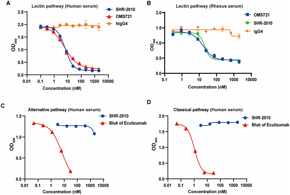 |
Figure 3 Selective blockage of lectin pathway by SHR-2010. The in vitro inhibition activity was evaluated by C4 deposition assay. (A and B) The inhibitory effect on human and rhesus monkey serum lectin pathway of SHR-2010, OMS721 biosimilar and IgG4. (C) The inhibitory effect on human alternative pathway of SHR-2010 and Eculizumab. (D) The inhibitory effect on human classical pathway of SHR-2010 and Eculizumab. |
Pharmacologic Activity of SHR-2010 Surrogate Antibody ER011-11165 Both in vitro and in vivo
As a surrogate of SHR-2010, ER011-11165 was evaluated for its efficacy in mouse models. The results showed that ER011-11165 effectively inhibited the lectin pathway activation in human serum, with the IC50 value of 6.17 nM, which was comparable to that of SHR-2010 (IC50= 6.44 nM) (Figure 4A). The inhibitory effect of ER011-11165 on mouse lectin pathway activation was slightly weaker than SHR-2010 on human lectin pathway activation, with IC50 values of 49.04 nM and 9.2 nM (Figure 4B and Table 2). Consequently, the in vitro efficacy of ER011-11165 is comparable to that of SHR-2010, further suggesting that ER011-11165 could be a murine surrogate of SHR-2010. To evaluate the in vivo efficacy of ER011-11165, a mouse model of acute kidney injury induced by LPS was established (Figure 4C). The LPS-induced kidney injury was strongly associated with innate immune activation, especially the activation of lectin pathway.30 Compared to the control group, the injection of LPS at 10 mg/kg/day resulted in significant increase of serum creatinine, urine albumin and urine albumin/creatinine ratio, suggesting the successful establishment of the mice kidney injury model. Intravenous administration of ER011-11,165 (3 and 10 mg/kg) dose-dependently mitigated LPS-induced nephrotoxicity. At 24 hours post-LPS challenge, ER011-11165 treated mice showed reduced serum creatinine, urinary albumin and albumin/creatinine ratio compared to untreated controls (Figures 4D-F). These results demonstrated that ER011-11165 could ameliorate lectin pathway-mediated renal injury in mice, providing indirect evidence for the in vivo efficacy of SHR-2010.
 |
Figure 4 The efficacy of ER011-11165 in LPS-induced acute kidney injury mouse model. (A and B) In vitro activity of ER011-11165 and SHR-2010 on human and mouse lectin pathway. (C) Schematic of pharmacologic study of acute kidney injury mice. (D–F) Analysis of serum creatinine, urine creatinine, urine albumin and albumin-creatinine-ratio. Statistical significance of the differences between control group, model group and ER011-11165-treated groups was determined by two-tailed t-test. n = 5 for control group; n = 10 for model group, 3 mg/kg and 10 mg/kg group, respectively. |
Pharmacokinetics (PK) and Pharmacodynamics (PD) Properties of SHR-2010 in Rhesus Monkeys
In rhesus monkeys, OMS721 exhibited a short half-life of approximately 3 to 8 days, leading to intravenous dosing in clinical practice.6 Considering this pharmacokinetic profile and the potential patient compliance challenges posed by IV delivery, we aimed to develop an antibody suitable for subcutaneous injection. The pharmacokinetics characterization of SHR-2010 in non-human primates was then performed through single subcutaneous (5 mg/kg) or intravenous (0.5, 1.5, 5 mg/kg) administration. The results showed that SHR-2010 possessed favorable pharmacokinetics profiles. Long half-life (140.4 to 226.3 h) was observed after being intravenously administered (Figure 5A and Table 3). Moreover, results showed that subcutaneous administration of SHR-2010 achieved completed bioavailability (104.5%) with long half-life (142.8 h) (Figure 5B and Table 3).
 |
Table 3 Key PK Parameters in Monkeys Following Single IV and SC Injection of SHR-2010 (Mean ± SD, n = 6) |
 |
Figure 5 Pharmacokinetics properties of SHR-2010 and its PK/PD correlation in non-human primates following a single intravenous dose of SHR-2010. The rhesus monkeys were administrated with SHR-2010 intravenously at 0.5, 1.5 and 5 mg/kg or subcutaneously at 5 mg/kg. (A) The serum concentrations of SHR-2010 were determined and plotted against sampling times after intravenous injection. (B) The serum concentrations of SHR-2010 were determined and plotted against sampling times after subcutaneous injection. (C) The PK-PD relationship of SHR-2010 in rhesus monkeys was determined following intravenous administration at 5 mg/kg. Drug concentrations in the serum were tested using ELISA, and lectin pathway activity were tested using C4 deposition assay. Data represent mean ± SD, n = 6 in each group. |
To investigate the relationship between serum concentrations and efficacy of SHR-2010, the C4 deposition was additionally evaluated as the PD marker after single intravenous administration of 5 mg/kg SHR-2010 in rhesus monkeys. The results showed that SHR-2010 could dose-dependently inhibit the lectin pathway in rhesus monkey serum, with maximum inhibition at 1 h after administration, indicating the rapid onset. Besides, the inhibitory effect on serum lectin pathway could maintain from 5 min to 408 h after administration (Figure 5C).
Collectively, PK/PD results demonstrated that the serum concentration of above 30000 ng/mL and inhibitory effect on lectin pathway of above 65% could be maintained through 408 hours post administration (Figure 5A-C). These findings indicated the concentration-dependent long-lasting inhibitory effects of SHR-2010 on lectin pathway.
Discussion
The lectin pathway is one of the three pathways responsible for activating the complement system. Besides the role in defending against pathogens, the lectin pathway has been shown to play a crucial role in various renal diseases and during kidney replacement therapy.31 Consequently, specific blockers targeting the lectin pathway are also being evaluated in treating renal disorders such as IgAN and lupus nephritis (LN). By using a series of in vitro models, researchers have identified that MASP-2 could directly activate endogenous complement component 3 (C3), independent of C4 and/or C2, in the lectin pathway activation process. The overactivation of lectin pathway has been demonstrated to be associated with various diseases, such as MN, IgAN, LN, membranoproliferative glomerulonephritis (MPGN), and focal segmental glomerular sclerosis (FSGS).32 These findings further suggested that targeting MASP-2 might have the potential as a novel and promising strategy for the treatment of various renal diseases.
Here, we developed SHR-2010, a novel fully humanized IgG4 monoclonal antibody targeting MASP-2. In a series of preclinical studies, we thoroughly evaluated the in vitro pharmacologic properties of SHR-2010. Our data showed that the binding affinity for human MASP-2 of SHR-2010 was 10-fold higher than that of OMS721 biosimilar (Table 1), with comparable lectin pathway inhibition activity in serum, implicating better therapeutic potential for lectin pathway dysregulation diseases. However, although SHR-2010 exhibits superior binding capacity to OMS721 biosimilar, it did not demonstrate significantly better activity than OMS721 biosimilar in in vitro experiments. There are some possible explanations: first, tighter binding is not strictly required for enhancing inhibitory efficacy; second, functional inhibition relies more heavily on sustained effective binding, yet SHR-2010’s Koff is only ~1.5-fold slower than OMS721 (2.81E-04 s−1 vs 4.23E-04 s−1); third, SPR measures binary interactions between purified proteins, whereas the C4 deposition assay is a functional test in a cellular environment, which may involve the influence of multiple components. We will further validate the underlying mechanisms in subsequent experiments.
Pan-complement inhibitors such as eculizumab could contribute to clinical infection risks.33 SHR-2010 demonstrated selective lectin pathway inhibition via C4 deposition, with no interference in classical/alternative pathways at a concentration up to 2000 nM. Therefore, clinical infection risks of SHR-2010 were expected to decrease compared with pan-complement inhibitors.
To investigate the effect of SHR-2010, we developed LPS-induced acute kidney injury mice model, in which LPS binds to collectin-11,30 MBL and subsequently activates MASP-2. We observed an elevation of serum creatinine and urine albumin, which indicated kidney injury. ER011-11165, the murine surrogate of SHR-2010, could ameliorated LPS-induced acute kidney injury, indicating that the kidney injury model was lectin pathway-dependent and SHR-2010 could have potential therapeutic effect on complement-mediated kidney diseases.
The pharmacokinetic profile critically determines drug disposition and therapeutic outcomes, where extended half-life not only sustains target engagement but also enables dosing regimens with improved compliance. Since SHR-2010 showed similar affinity to human and monkey MASP-2, while no binding to rodents, monkey was selected for subsequent PK/PD study and toxicity assessment. SHR-2010 exhibited dose-proportional PK in monkeys, outperforming OMS721 in half-life (6–9.4 vs 3–8 days).6 Predictive modeling suggested extended terminal half-life of SHR-2010 compared with OMS721 (10–20 vs 8.25 days),34 supporting monthly dosing versus weekly regimens. In addition, SHR-2010 showed high subcutaneous bioavailability (104.5%), supporting the potential application of subcutaneous administration in the clinic, which could achieve better patient compliance than OMS721 via intravenous administration.
We developed final population PK model in monkeys comprising linear 2-compartment distribution and elimination, together with a non-linear elimination component as described by a Michaelis–Menten term. Critical parameters (V1) were consistent with typical monoclonal antibody characteristics, indicating that SHR-2010 was distributed in the blood and hydrophilic extravascular space. Total clearance (CL) of SHR-2010 was concentration-dependent, with Km values estimated to be approximately 5.68 μg/mL. The terminal half-life (t1/2) in humans was predicted to be 10–20 days using the allometric scaling approach. PK-PD relationship was modeled via a direct-link mixed-effects Emax analysis. The EC50 and EC90 values indicated that concentrations of SHR-2010 were maintained above EC50 throughout the proposed clinical dosing interval (data not shown), ensuring meaningful C4b inhibition correlating with clinical response rate.
Moreover, following intravenous dosing at 5 mg/kg in rhesus monkeys, SHR-2010 exhibited sustained lectin pathway inhibition (above 80%) for 408 hours, exceeding that of OMS721 (approximately 336 hours).35 These results demonstrated that SHR-2010 was an effective and long-lasting inhibitor of the lectin pathway, which indicating a promising therapy for complement-mediated inflammatory diseases.
To assess the safety of SHR-2010, we conducted a GLP-compliant evaluation of the toxicities of SHR-2010 in rhesus monkeys. Following 26 weeks of SHR-2010 subcutaneous or intravenous administration (once a week for a total 27 times), no SHR-2010-related abnormalities in clinical observation, body temperature, respiration, ECG examination, blood pressure, ophthalmologic examination, hematology, urine, gross anatomy or histopathology were observed. Besides, no abnormalities were found about lymphocytes (CD3+, CD3+CD4+, CD3+CD8+, CD3+CD4+/CD3+CD8+, CD20+B, CD16+NK), cytokines (IL-2, IL-4, IL-5, IL-6, IL-8, TNF-α, IFN-γ), anti-drug antibody (ADA), complements (C3, C4), immune complexes (CIC), etc, indicating that SHR-2010 had no significant immunotoxicities following repeated administration. In this study, the no observed adverse effect level (NOAEL) for SHR-2010 was 100 mg/kg (SC) and 20 mg/kg (IV), the highest dose tested.
Both the alternative pathway and the lectin pathway significantly promote the progression of IgAN. Currently, Iptacopan, which targets factor B in the alternative pathway, has demonstrated promising efficacy in treating IgAN.36 However, there remains a lack of therapeutic agents targeting the lectin pathway for IgAN. We believe that targeting the lectin pathway could play a complementary role to the alternative pathway, and may even hold promise for treating patients who are insensitive to Iptacopan.
OMS721 is the most clinically advanced MASP-2 inhibitor to date. In pivotal clinical trials, OMS721 exhibited 61% of response rate and 68% of 100-day survival in HSCT-TMA patients.37 However, OMS721 was announced failure in Phase III clinical trial of IgA Nephritis. Here, we proposed several potential shortages in clinical trial design and could be improved. For example, the treatment duration of OMS721 was relatively short and the time gap between primary outcome and post-intervention could be long to diminish clinical efficacy. OMS721 was only administered from week 1 to week 12, and an additional 6 weeks dosing was then added depending on the decrease in urine protein. However, primary endpoint was detected in week 36. In addition, the primary endpoint at week 36 was set as 24 h urine protein excretion (UPE) instead of urine protein: creatinine ratio (UPCR), which might be a more robust and stable clinical endpoint. IgAN patients confirmed by renal biopsy within 8 years prior to enrollment were included, which probably increased the variations in baseline conditions and the subsequent response. Moreover, no concomitant medication was restricted including SGLT2 inhibitors etc, which have been fully proved to be protective in CKD and may have increased the reduction of UPE in the placebo group. Despite its temporary setbacks in IgAN trial, OMS721 met its pivotal primary endpoint in a phase 3 trial enrolling patients with TA-TMA. OMS721-treated patients had an over 3-fold reduction in risk of mortality compared to similarly at-risk patients without treatment. Preclinical studies demonstrated that SHR-2010 exhibited superior pharmacokinetics and sustained lectin pathway inhibition compared to OMS721. When coupled with optimized trial design strategies, SHR-2010 could be a promising therapeutic candidate for lectin pathway-driven diseases, including IgAN.
Conclusions
In conclusion, we have discovered and characterized a fully humanize anti-MASP-2 monoclonal antibody, SHR-2010, thoroughly in a series of in vitro and in vivo assays. By selective binding with MASP-2, SHR-2010 could inhibit lectin-induced complement activation without affecting the classical or alternative pathway activation. MASP-2 inhibition with surrogate showed significant efficacy in LPS-induced acute kidney injury model. SHR-2010 possessed favorable PK and safety profiles in monkeys following subcutaneous injection. Altogether, these data provided support for further clinical development of SHR-2010 as a potential best-in-class therapy for complement activation-driven renal diseases. Currently, a Phase II clinical trial to evaluate the safety and efficacy of SHR-2010 in IgAN patients via subcutaneous administration has been initiated in China.
Acknowledgments
The authors thank the colleagues that from the Department of Translational Medicine who participated in the studies. This work was funded by Jiangsu Hengrui Pharmaceuticals, Co., Ltd.
Disclosure
All authors are or have been employed by corporations engaged in the development of SHR-2010. The authors report no other conflicts of interest in this work.
References
1. Garred P, Tenner AJ, Mollnes TE, Levy FO. Therapeutic targeting of the complement system: from rare diseases to pandemics. Pharmacol Rev. 2021;73(2):792–827. doi:10.1124/pharmrev.120.000072
2. Thurman JM, Le Quintrec M. Targeting the complement cascade: novel treatments coming down the pike. Kidney Int. 2016;90(4):746–752. doi:10.1016/j.kint.2016.04.018
3. Tauber AI, Chernyak L. The birth of immunology. 2. Metchnikoff and his critics. Cell Immunol. 1989;121(2):447–473. doi:10.1016/0008-8749(89)90043-9
4. Schifferli JA, Ng YC, Peters DK. The role of complement and its receptor in the elimination of immune-complexes. New Engl J Med. 1986;315(8):488–495. doi:10.1056/NEJM198608213150805
5. Nesargikar PN, Spiller B, Chavez R. The complement system: history, pathways, cascade and inhibitors. Eur J Microbiol Immunol. 2012;2(2):103–111. doi:10.1556/EuJMI.2.2012.2.2
6. Dudler T, Yaseen S, Cummings WJ. Development and characterization of narsoplimab, a selective MASP-2 inhibitor, for the treatment of lectin-pathway-mediated disorders. Front Immunol. 2023;14:1297352. doi:10.3389/fimmu.2023.1297352
7. Ricklin D, Lambris JD. Complement in immune and inflammatory disorders: therapeutic interventions. J Immunol. 2013;190(8):3839–3847. doi:10.4049/jimmunol.1203200
8. Merle NS, Church SE, Fremeaux-Bacchi V, Roumenina LT. Complement system part i - molecular mechanisms of activation and regulation. Front Immunol. 2015;6:262. doi:10.3389/fimmu.2015.00262
9. Kishore U, Ghebrehiwet B. Editorial: c1q: a molecular bridge to innate and adaptive immunity. Front Immunol. 2020;11:417. doi:10.3389/fimmu.2020.00417
10. Matsushita M, Fujita T. Activation of the classical complement pathway by mannose-binding protein in association with a novel C1s-like serine protease. J Exp Med. 1992;176(6):1497–1502. doi:10.1084/jem.176.6.1497
11. Matsushita M, Thiel S, Jensenius JC, Terai I, Fujita T. Proteolytic activities of two types of mannose-binding lectin-associated serine protease. J Immunol. 2000;165(5):2637–2642. doi:10.4049/jimmunol.165.5.2637
12. Thiel S, Vorup-Jensen T, Stover CM, et al. A second serine protease associated with mannan-binding lectin that activates complement. Nature. 1997;386(6624):506–510. doi:10.1038/386506a0
13. Gavriilaki E, Ho VT, Schwaeble W, et al. Role of the lectin pathway of complement in hematopoietic stem cell transplantation-associated endothelial injury and thrombotic microangiopathy. Exp Hematol Oncol. 2021;10(1):57. doi:10.1186/s40164-021-00249-8
14. Mahmoudjafari Z, Alencar MC, Alexander MD, Johnson DJ, Yeh J, Evans MD. Hematopoietic stem cell transplantation-associated thrombotic microangiopathy and the role of advanced practice providers and pharmacists. Bone Marrow Transplant. 2023;58(6):625–634. doi:10.1038/s41409-023-01951-3
15. Schena FP, Nistor I. Epidemiology of IgA Nephropathy: a Global Perspective. Semin Nephrol. 2018;38(5):435–442. doi:10.1016/j.semnephrol.2018.05.013
16. Rizk DV, Maillard N, Julian BA, et al. The emerging role of complement proteins as a target for therapy of IgA nephropathy. Front Immunol. 2019;10:504. doi:10.3389/fimmu.2019.00504
17. Roos A, Rastaldi MP, Calvaresi N, et al. Glomerular activation of the lectin pathway of complement in IgA nephropathy is associated with more severe renal disease. J Am Soc Nephrol. 2006;17(6):1724–1734. doi:10.1681/ASN.2005090923
18. Hou X, Liang Y, Zhang W, Li R. The clinical and pathological effects of serum C3 level and mesangial C3 intensity in patients with IgA nephropathy. Anal Cell Pathol. 2024;2024(1):8889306. doi:10.1155/2024/8889306
19. Kim SJ, Koo HM, Lim BJ, et al. Decreased circulating C3 levels and mesangial C3 deposition predict renal outcome in patients with IgA nephropathy. PLoS One. 2012;7(7):e40495. doi:10.1371/journal.pone.0040495
20. Fontán FC, Gutiérrez E, Sevillano ÁM, Praga M. Targeting complement in IgA nephropathy. Clin Kidney J. 2023;16(Suppl 2):ii28–ii39. doi:10.1093/ckj/sfad198
21. Asgari E, Farrar CA, Lynch N, et al. Mannan-binding lectin-associated serine protease 2 is critical for the development of renal ischemia reperfusion injury and mediates tissue injury in the absence of complement C4. FASEB J. 2014;28(9):3996–4003. doi:10.1096/fj.13-246306
22. Talsma DT, Poppelaars F, Dam W, et al. MASP-2 is a heparin-binding protease; identification of blocking oligosaccharides. Front Immunol. 2020;11:732. doi:10.3389/fimmu.2020.00732
23. Ip WK, Takahashi K, Ezekowitz RA, Stuart LM. Mannose-binding lectin and innate immunity. Immunol Rev. 2009;230(1):9–21. doi:10.1111/j.1600-065X.2009.00789.x
24. Kalia N, Singh J, Kaur M. The ambiguous role of mannose-binding lectin (MBL) in human immunity. Open Med. 2021;16(1):299–310. doi:10.1515/med-2021-0239
25. Farrar CA, Tran D, Li K, et al. Collectin-1 detects stress-induced L-fucose pattern to trigger renal epithelial injury. J Clin Invest. 2016;126:1911–1925 doi:10.1172/JCI83000.
26. Endo M, Ohi H, Ohsawa I, Fujita T, Matsushita M, Fujita T. Glomerular deposition of mannose-binding lectin (MBL) indicates a novel mechanism of complement activation in IgA nephropathy. Nephrol Dial Transpl. 1998;13(8):1984–1990. doi:10.1093/ndt/13.8.1984
27. Kurella VB, Gali R. Structure guided homology model based design and engineering of mouse antibodies for humanization. Bioinformation. 2014;10(4):180–186. doi:10.6026/97320630010180
28. Kuramochi T, Igawa T, Tsunoda H, Hattori K. Humanization and simultaneous optimization of monoclonal antibody. Methods Mol Biol. 2019;1904:213–230 doi:10.1007/978-1-62703-586-6_7.
29. Dudler T, Gombotz W, Parent R, et al. Inventors. Compositions for inhibiting MASP-2 dependent complement activation. PLoS Pathogens. 2012;8(7). doi:10.1371/journal.ppat.1002793.
30. Ali YM, Lynch NJ, Shaaban AA, et al. Inhibition of the lectin pathway of complement activation reduces LPS-induced acute respiratory distress syndrome in mice. Front Immunol. 2023;14:1192767 doi:10.3389/fimmu.2023.1192767.
31. Flyvbjerg A. The role of the complement system in diabetic nephropathy. Nat Rev Nephrol. 2017;13(5):311–318. doi:10.1038/nrneph.2017.31
32. Poppelaars F, Thurman JM. Complement-mediated kidney diseases. Mol Immunol. 2020;128:175–187. doi:10.1016/j.molimm.2020.10.015
33. Barnum SR. Therapeutic inhibition of complement: well worth the risk. Trends Pharmacol Sci. 2017;38(6):503–505. doi:10.1016/j.tips.2017.03.009
34. Pullman W, Facius A, Lahu G, Smyth B The 47th Annual meeting of the European society for blood and marrow transplantation: Physicians – poster Session (P142). Bone Marrow Transplant. 2021;56(S1):300–301. doi:10.1038/s41409-021-01343-5
35. Freeman J, Cummings J, Chuidian M, Dudler T. Development of pharmacodynamic assays to assess ex vivo Masp-2 inhibition and their use to characterize the pharmacodynamics of narsoplimab (OMS721) in humans and monkeys. Blood. 2020;136(Supplement 1):26–27. doi:10.1182/blood-2020-142208
36. Perkovic V, Barratt J, Rovin B, et al; for the APPLAUSE-IgAN Investigators. Alternative complement pathway inhibition with iptacopan in IgA nephropathy. N Engl J Med. 2025;392(6):531–543. doi:10.1056/NEJMoa2410316
37. Khaled SK, Claes K, Goh YT, et al. Narsoplimab, a mannan-binding lectin-associated serine protease-2 inhibitor, for the treatment of adult hematopoietic stem-cell transplantation-associated thrombotic microangiopathy. J Clin Oncol. 2022;40(22):2447–2457. doi:10.1200/JCO.21.02389
 © 2025 The Author(s). This work is published and licensed by Dove Medical Press Limited. The
full terms of this license are available at https://www.dovepress.com/terms
and incorporate the Creative Commons Attribution
- Non Commercial (unported, 4.0) License.
By accessing the work you hereby accept the Terms. Non-commercial uses of the work are permitted
without any further permission from Dove Medical Press Limited, provided the work is properly
attributed. For permission for commercial use of this work, please see paragraphs 4.2 and 5 of our Terms.
© 2025 The Author(s). This work is published and licensed by Dove Medical Press Limited. The
full terms of this license are available at https://www.dovepress.com/terms
and incorporate the Creative Commons Attribution
- Non Commercial (unported, 4.0) License.
By accessing the work you hereby accept the Terms. Non-commercial uses of the work are permitted
without any further permission from Dove Medical Press Limited, provided the work is properly
attributed. For permission for commercial use of this work, please see paragraphs 4.2 and 5 of our Terms.