
Back to Journals » Clinical Ophthalmology » Volume 19
Brillouin Metrics of the Crystalline Lens Comparison Between Low-Medium Myopia and High Myopia
Authors Bao Y, Ma Y, Cao J, Yu Y, Fukuyama T, Zhang Z, Niu L  , Yao P, Wang X, Zhou X , Zhao J
Received 20 July 2025
Accepted for publication 17 October 2025
Published 29 October 2025 Volume 2025:19 Pages 4001—4009
DOI https://doi.org/10.2147/OPTH.S555070
Checked for plagiarism Yes
Review by Single anonymous peer review
Peer reviewer comments 2
Editor who approved publication: Dr Scott Fraser
Yongle Bao,1– 4,* Yong Ma,1,2,* Jian Cao,5,* Yanze Yu,1,2 Teruko Fukuyama,1,2 Zhe Zhang,1,2 Lingling Niu,1,2 Peijun Yao,1,2 Xiaoying Wang,1,2 Xingtao Zhou,1,2 Jing Zhao1,2
1Department of Ophthalmology, Eye and ENT Hospital, Fudan University, Shanghai, People’s Republic of China; 2Key Laboratory of Myopia and Related Eye Diseases, Chinese Academy of Medical Sciences, NHC, Shanghai, People’s Republic of China; 3Department of Ophthalmology, Zhangye People’s Hospital, Hexi University, Zhangye, Gansu, People’s Republic of China; 4Clinical Medical College, Hexi University, Zhangye, Gansu, People’s Republic of China; 5State Key Laboratory of Ophthalmology, Zhongshan Ophthalmic Center, Sun Yat-Sen University, Guangzhou, People’s Republic of China
*These authors contributed equally to this work
Correspondence: Jing Zhao, Department of Ophthalmology, Eye & ENT Hospital, Fudan University, 83 Fenyang Road, Shanghai, 200031, People’s Republic of China, Tel +86021-64377134, Email [email protected] Xingtao Zhou, Department of Ophthalmology, Eye & ENT Hospital, Fudan University, 83 Fenyang Road, Shanghai, 200031, People’s Republic of China, Tel +86021-64377134, Email [email protected]
Purpose: To investigate the impact of myopia on the Brillouin biomechanics and morphology of crystalline lenses in Chinese adults.
Methods: Patients with myopia were enrolled and divided into low-medium (spherical equivalent [SE] ≥ − 5.75 D, N = 66) and high (SE ≤ – 6.0 D, N = 72) myopia groups. All patients underwent routine ophthalmic examinations, including SE, axial length, and anterior chamber metrics (Pentacam) measurements. The Brillouin-related metrics of the crystalline lenses were measured using a Brillouin microscopy. The morphological parameters included the Width of Top Plateau (WTP), Width of Bottom Plateau (WBP), Bottom-Top (B-T), Slope of Anterior Cortex (SAC), and Slope of Posterior Cortex (SPC). The height of Plateau was a biomechanical parameter.
Results: The study included 138 eyes (138 patients); the mean age was 28.43 ± 8.31 years. WBP, B-T, SAC, and SPC significantly differed between the high and low-medium myopia groups (all P< 0.05). WBP, B-T, and SPC significantly correlated with SE in the high myopia group (Pearson’s r = 0.365, 0.287, and 0.294, respectively; all P< 0.05) but not in the low-medium group. The SPC significantly decreased for all participants based on the SE values (P< 0.05). The biomechanics and thickness of the nucleus displayed no discrepancies with SE in all participants (all P > 0.05).
Conclusion: An increase in the degree of myopia could be associated with posterior cortex thickening while maintaining stable biomechanics and morphology of the crystalline lens nucleus. Brillouin microscopy can serve as an effective technique for multidisciplinary lens biomechanical and morphological imaging.
Keywords: Brillouin microscopy, crystalline lens, myopia, morphology, biomechanics
Introduction
The crystalline lens, comprising a central nucleus and surrounding cortex, exhibits distinct biomechanical and morphological characteristics within its components.1 Accurate measurement of the biomechanical and geometric changes in these lens components is crucial for understanding physiological and pathological alterations, such as those associated with accommodation, cataract formation, and intraocular refractive and cataract surgery.2–4 Moreover, with the escalating prevalence of myopia and high myopia, considerable attention has been given to understanding myopic-related pathological changes in the crystalline lens nucleus.5,6
Previous studies have used Scheimpflug or optical coherence tomography techniques to evaluate the optical characteristics of the crystalline lens, dividing the components based on the distribution of optical gray pixels.4,7 However, the optical density and gray levels do not represent biomechanical properties. In contrast, a novel optical coherence elastography technique can assess the biomechanics of the crystalline lens in vivo.8,9 However, employing an external air puff to indirectly induce stress on the lens results in a relatively weak stress-strain correlation, as it affects the cornea initially rather than the lens.8,9 Moreover, the air puff-induced deformation of the cornea affects light propagation in the eye.8 The comprehensive impact of these factors necessitates further investigation into the in vivo analysis of the biomechanics of the crystalline lens.
Based on Brillouin light inelastic scattering, Brillouin microscopy (BM) has recently become a prominent technique owing to its noncontact and all-optical characteristics, enabling the in vivo evaluation of the intrinsic biomechanical properties of the crystalline lens and the differentiation of its components based on their biomechanical variations.10–13 In our previous study, we illustrated that Brillouin morphological metrics of the crystalline lens exhibit significant age-related alterations, but the biomechanics and thickness of the lens nucleus remain constant.14 However, considering the correlation between lens thickness and refractive power, correlations may exist between the spherical equivalent (SE) and Brillouin metrics.15,16
Therefore, this study used Brillouin microscopy to evaluate the effects of SE on Brillouin parameters to comprehensively assess the biomechanical and morphological parameters of crystalline lenses with different degrees of myopia.
Methods
Patients
This cross-sectional comparative study was conducted in accordance with principles outlined in the Declaration of Helsinki. The Eye & ENT Hospital (EENT) ethics board at Fudan University approved this study (No. 2020530). The evaluation of all relevant metrics was conducted at the EENT hospital from August 2023 to March 2024. All participants provided written informed consent. Individuals with myopia were included. Individuals with a medical history of ocular refractive surgery, or any condition other than refractive errors, were excluded. Individuals with presbyopia or cataracts were also excluded after carefully routine slit-lamp scans and manifest refraction including presbyopia evaluation.
The participants were divided into low-medium myopia (SE: –5.75 diopters [D] to –0.5 D) and high myopia (SE: <–6.0 D) groups based on the SE value.
Ocular Metrics
The participants received thorough ophthalmic examinations, and their demographic information was collected, including age, gender, and medical history. Manifest refraction was utilized to measure spherical and cylindrical D-values. SE was calculated as the spherical value plus half of the cylinder value. Tomographic and topographic parameters of the anterior chamber, including the anterior chamber depth (ACD) and anterior chamber volume (ACV), were measured using a Pentacam (Oculus Optikgeräte, Wetzlar, Germany). Axial length (AL) measurements were taken using an IOLMaster 500 (Carl Zeiss Meditec, Jena, Germany). A Canon Full-Auto Tonometer TX-F (Canon Inc., Tokyo, Japan) was used to measure the intraocular pressure (IOP).
Brillouin Microscopy and Metrics
The mechanical and Brillouin biomechanical properties have been comprehensively summarized in our previous publication.14 Briefly, the Brillouin Optical Scan System (Intelon Optics, Boston, USA) quantifies the density fluctuations of photonics within biological materials, enabling conversion into biomechanical metrics with units of GPa (or 109 Pascal).11 Given the continuous biomechanic variation across the crystalline lens, from the anterior cortex to the nucleus and posterior cortex, the Brillouin microscopy technique divides the lens into three distinct regions based on their differing biomechanical characteristics.10,14 These regions are then characterized using specific Brillouin metrics.
We classified the Width of Top Plateau (WTP), Width of Bottom Plateau (WBP), and the differences between the WBP and WTP (Bottom-Top, B-T) as thickness-related metrics and the slope of anterior and posterior cortexes (SAC and SPC, respectively) as slope-related metrics; both sets of metrics are morphology-related. The Height of Plateau (Height), a biomechanical parameter, indicated the viscoelasticity of the crystalline lens nucleus. The standardized report and Brillouin metrics in the application interface have been presented in our previous study.14 Supplementary Table 1 defines these parameters. Notably, SAC and SPC are considered morphological parameters despite their units being GPa per millimeter, owing to their association with increasing or decreasing stiffness in the anterior and posterior cortex.
Brillouin Measurements
Participants were instructed to remain in a relaxed state, and the examination was performed prior to other routine cycloplegic examination. A BM examination was conducted by the same experienced doctor (JC) in a dimly lit room (average illuminance ≤ 5 lux) with at least 5 minutes for dark adaption, as previously stated. Each measurement, including pre-measurement and pre-examination corrections, lasted for 1 minute. In the “Lens 1 pt” mode, BM measurements were taken at the central area of the pupil. The system automatically discarded the data if the subjects blinked during critical moments. Only data with a quality index of “OK” were finally included in the analyses.
Statistical Analyses
All statistical analyses were conducted using SPSS (version 26.0; IBM Corp., Armonk, NY, USA). Descriptive data are presented as means ± standard deviations and [ranges]. Only data from one eye of each patient were included in the analyses. A Student’s t-test was used to compare the ocular and Brillouin metrics between the low-medium and high myopia groups. A linear regression model was used to assess the correlation between Brillouin metrics and SE after verifying the homogeneity of variances, with only significant linear correlations expressed in the equations. Mixed linear models were used to mitigate age-related effects on Brillouin metrics and analyze their linear correlation with ocular metrics. All statistical analyses were performed with 95% confidence intervals. Statistical significance was defined as P <0.05.
Results
Metric Comparisons for Low-Medium and High Myopic Lenses
Table 1 compares the ocular and Brillouin metrics for lenses with low-medium and high myopia. The study enrolled 138 patients (138 eyes); 66 and 72 eyes were included in the low-medium (mean age: 27.58 ± 8.28 years) and high myopia (mean age: 29.22 ± 8.30 years) groups, respectively. Age did not differ between the groups (P >0.05). SE and AL were significantly higher in the high myopia group than in the low myopia group (P <0.001), but other ocular metrics (eg, IOP, ACD, and ACV) did not differ between the groups (all P >0.05). WBP, B-T, SAC, and SPC (all Brillouin morphological metrics) significantly differed between the groups (P <0.01), but WTP and Height did not (P >0.05).
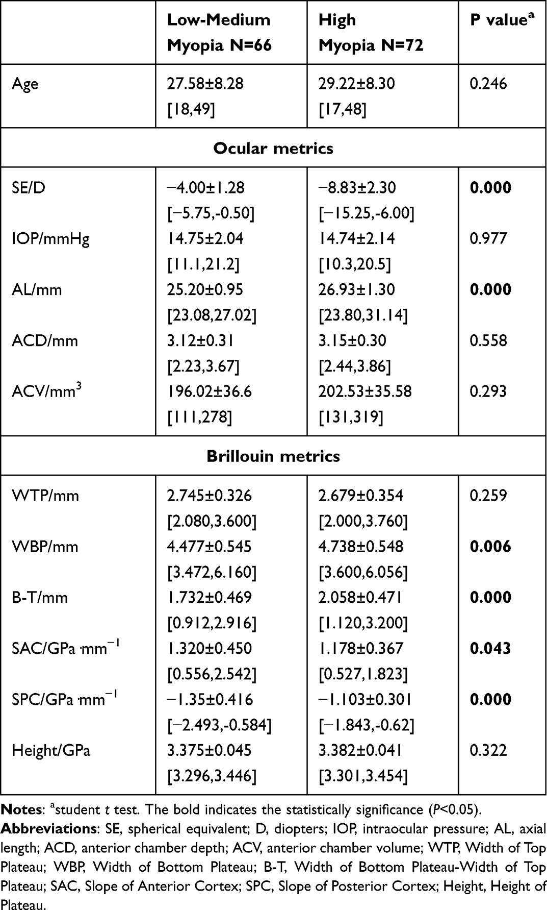 |
Table 1 Comparisons of Metrics for Low-Medium and High Myopic Lenses |
Linear Regression Models of Brillouin Metrics and SE
Figure 1A–C presents the correlations between the Brillouin metrics and SE for all participants. SAC significantly positively correlated with SE (slope: 0.026 GPa·mm−1/D, P <0.05). Other metrics significantly negatively correlated with SE, including WBP, B-T, and SPC (slopes: –0.061 mm/D, –0.058 mm/D, and –0.042 GPa·mm−1/D, respectively; all P <0.01). WTP and Height did not correlate with SE (both P >0.05).
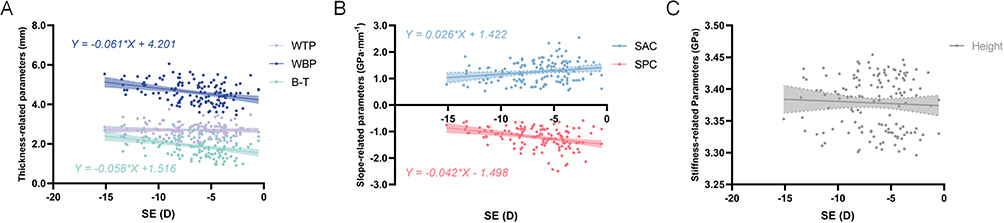 |
Figure 1 Linear regression models between the Brillouin metrics and SE. Linear regression models depict SE-related changes in Brillouin morphological (thickness- and slope-related) and biomechanical (stiffness-related) metrics of the lenses in all participants. (A) Variations of the WTP, WBP, and B-T with SE; (B) Variations of the SAC and SPC with SE; (C) Variation of the Height with SE. The shadowed zone represents the 95% standard deviation. Only statistically significant linear correlations are expressed as equations. Abbreviations: B-T, bottom-top; Height, Height of Plateau; SAC, slope of anterior cortex; SE, spherical equivalent; SPC, Slope of Posterior Cortex; WBP, Width of Bottom Plateau; WTP, Width of Top Plateau. |
Figure 2A–F present the intergroup correlations between the Brillouin metrics and SE. Correlations between the Brillouin metrics and SE did not exist in the low-medium myopia group (all P >0.05). In contrast, WBP, B-T, and SPC significantly correlated with SE in the high myopia group (slopes: –0.087 mm/D, –0.059 mm/D, and –0.039 GPa·mm−1/D, respectively, all P <0.05). WTP, SAC, and Height did not correlate with SE (all P >0.05).
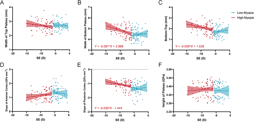 |
Figure 2 Intergroup correlations between the Brillouin metrics and SE. Linear regression analysis illustrates the SE-related tendency in the Brillouin metrics of the lenses in the low-medium and high myopia groups. (A–F) Comparisons of the Width of Top Plateau, Width of Bottom Plateau, Bottom-Top, Slope of Anterior Cortex, Slope of Posterior Cortex, and Height of Plateau. The shadowed zone represents the 95% standard deviation. Only statistically significant linear correlations are expressed as equations. Abbreviation: SE, spherical equivalent. |
Correlations Between Brillouin Metrics and Other Factors
Table 2 presents the Pearson correlations between the Brillouin metrics and ocular metrics. Age significantly correlated with all Brillouin morphological parameters except Height. The anterior chamber metrics (ACD and ACV) significantly correlated with WBP, B-T, SAC, and SPC (all P <0.05).
 |
Table 2 The Correlation Between Brillouin Metrics and Other Factors |
Considering the influence of age on crystalline lenses, a multivariate linear regression analysis between the Brillouin metrics and SE was conducted after adjusting for age (Table 3). SE remained significantly correlated with WBP, B-T, and SPC (β = –0.038, –0.043, and –0.028, respectively, all P <0.01), but SE and SAC no longer correlated (P >0.05).
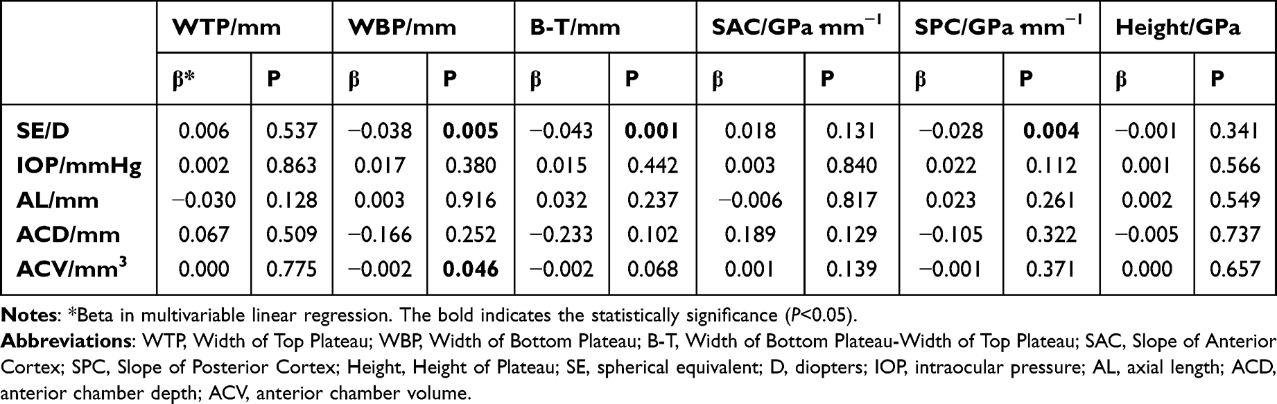 |
Table 3 The Multivariable Linear Regression Models Adjusted for Age |
Discussion
This study assessed the impact of myopia on Brillouin lenticular metrics in participants with high and low-medium myopia and investigated alterations in specific Brillouin metrics associated with the degree of myopia. Overall, we identified significant morphological variations in the thickening of the posterior cortex as the degree of myopia increased.
In our previous study, we observed significant age-related alterations in Brillouin morphological metrics, but correlations between SE and the Brillouin metrics of the crystalline lens were not found.14 In the present study, a correlation between SE and Brillouin metrics did not exist in the low-medium myopia group, which is consistent with our previous results.14 However, when we expanded the patient recruitment parameters and the range of the degree of myopia, we identified a significant correlation between the Brillouin morphological metrics and SE. Considering the potential correlation between lenticular refractive power and SE, further investigations into alterations in the components associated with the degree of myopia based on their biomechanical characteristics are warranted.15,17 Additionally, since we and Besner et al demonstrated that age affects Brillouin metrics, participant age was controlled to ensure statistically significant differences did not exist between the low-medium and high myopia groups.10,14 Since BM inherently minimizes confounding factors, such as CCT and IOP, this study primarily focused on potential influencing factors, such as AL, ACD, and ACV.14,18,19
BM provides a novel approach for the assessment of the biomechanics of the living crystalline lens, specifically the Height parameter. Therefore, we investigated potential stiffening or increased viscoelasticity of the crystalline lens in individuals with high myopia.20–22 In our study, we conducted a comparison of biomechanical metrics of the lenticular nucleus between the high myopia (3.381 ± 0.041 GPa) and low-medium myopia (3.375 ± 0.045 GPa) groups, finding no significant difference (P = 0.328). We hypothesized that the degree of myopia would not significantly affect the biomechanical properties of the crystalline lens. However, due to the strong scattering properties of cataractous lenses, the current version of Brillouin microscopy in the present study is limited in its application to the biomechanical assessment of cataractous lenses, despite its potential for future investigation into early cataract screening based on possible changes in viscoelasticity.23–25
The morphological Brillouin metrics of the lens cortex, including the WBP, B-T, SPC, and SAC, exhibited significant SE-related discrepancies and alterations. For instance, SPC negatively correlated with SE but positively correlated with the degree of myopia. This suggests that as the degree of myopia increased, the SPC also increased, indicating an increased thickness of the posterior cortex. With lower SE values, thickening of the posterior cortex and the total lens may occur; however, no correlation was observed between the WTP and SE. Previous studies have reported that the entire lens thickens as the degree of myopia increases.15,26,27 However, with aging, the remaining anterior segment (from the cornea to the posterior lens surface) undergoes changes such as thickening of the posterior cortex and an anterior displacement of the lens center.28,29 Therefore, we hypothesized that the degree of myopia may contribute to posterior cortical thickening and anterior lens movement. Our study demonstrated that the lens thickening associated with myopia was mainly manifested by an increase in posterior cortical thickness, but the nuclear thickness did not change. Given the current application of biomechanical stratification in lens analysis, future research could focus on longitudinal changes in the posterior cortex of the lens.23
We also found that the degree of myopia may contribute to the discrepancy in the anterior cortex based on the SAC measurements (Table 1). Although a significant correlation between SAC and the degree of myopia was observed, a significant correlation between SE and SAC was not observed after adjusting for age, suggesting that age primarily influenced the anterior cortex and further affected SE. Moreover, cortex growth is linear throughout life, and increasing age is associated with a slight forward movement of the lens center, which may also be associated with the thickening of the anterior cortex.30,31 Our previous study highlighted that age, ACD, and ACV comprehensively influence SAC, whereas ACD and ACV are also affected by age.14 Therefore, we propose that age may exert a more substantial influence on the thickening of the anterior cortex than SE, leading to SAC disparities.
Given the rapid rise in myopia prevalence in China, early screening and regular monitoring of both emmetropic and myopic individuals are essential.32 Brillouin microscopy holds promise for these applications; however, studies on children’s lens characteristics using this technique remain limited, primarily due to lengthy scanning durations. This study reveals relatively stable biomechanical properties of the lens nucleus and progressive thickening of the posterior cortex, findings that may enhance our understanding of refractive development and guide future research.
This study has some limitations. First, we did not recruit older participants because of concerns about the potential impact of presbyopia and cataracts on accommodation, which may affect the lenticular biomechanics and morphology measurements.33 Second, given the significant increase in the biomechanic metrics for cataract lenses, our study focused on transparent lenses or possible alterations in lenticular biomechanics induced by high myopia. Additional research is required to investigate the biomechanical and morphological properties of Brillouin aging-related or cataract lenses.34
Conclusion
This study compared the Brillouin metrics of the crystalline lens in participants with low-to-medium and high myopia, revealing SE-related thickening of the posterior cortex and the entire lens. However, significant biomechanical or morphological discrepancies related to the crystalline lens nucleus were not observed. Age plays a crucial role in Brillouin analysis of the crystalline lens, particularly regarding anterior cortex thickening.
Data Sharing Statement
The datasets generated and/or analyzed during the current study are not publicly available due to funding requirement but are available from the corresponding author (Jing Zhao, [email protected]) on reasonable request.
Consent for Publish
Written informed consent was obtained from the patients for the publication of this paper. Patients’ names are not applicable.
Author Contributions
All authors made a significant contribution to the work reported, whether that is in the conception, study design, execution, acquisition of data, analysis and interpretation, or in all these areas; took part in drafting, revising or critically reviewing the article; gave final approval of the version to be published; have agreed on the journal to which the article has been submitted; and agree to be accountable for all aspects of the work.
Funding
This work was supported by National Natural Science Foundation of China (Grant No. 82271119); Shanghai Rising-Star Program (23QA1401000); Healthy Young Talents Project of Shanghai Municipal Health Commission (2022YQ015); Project of Shanghai Science and Technology (Grant No.20410710100, 21Y11909800). The funding agencies had no role in study design, data collection and analysis, interpretation of data, or writing the manuscript.
Disclosure
The authors declare no conflicts of interest in this work.
References
1. Bassnett S, Šikić H. The lens growth process. Prog Retinal Eye Res. 2017;60:181–200. doi:10.1016/j.preteyeres.2017.04.001
2. Wang K, Pierscionek BK. Biomechanics of the human lens and accommodative system: functional relevance to physiological states. Prog Retinal Eye Res. 2019;71:114–131. doi:10.1016/j.preteyeres.2018.11.004
3. Yang W, Zhao J, Zhao J, et al. Changes in anterior lens density after implantable collamer lens V4c implantation: a 4-year prospective observational study. Acta Ophthalmol. 2021;99(3):326–333. doi:10.1111/aos.14584
4. Ye Y, Zhao J, Niu L, Shi W, Wang X, Zhou X. Long-term evaluation of anterior lens density after implantable collamer lens V4c implantation in patients with myopia over 40 years old. Br J Ophthalmol. 2022;106(11):1508–1513. doi:10.1136/bjophthalmol-2021-319205
5. Haarman AEG, Enthoven CA, Tideman JWL, Tedja MS, Verhoeven VJM, Klaver CCW. The complications of myopia: a review and meta-analysis. Invest Ophthalmol Vis Sci. 2020;61(4):49. doi:10.1167/iovs.61.4.49
6. Chong EW, Mehta JS. High myopia and cataract surgery. Curr Opin Ophthalmol. 2016;27(1):45–50. doi:10.1097/icu.0000000000000217
7. Wang L, Li C, Li J, Chen Z, Liu W, Lu P. Association of lens density quantified by IOLMaster 700 with lenticular myopia in nuclear cataract. Graefes Arch Clin Exp Ophthalmol. 2022;260(5):1565–1572. doi:10.1007/s00417-021-05495-4
8. Maczynska E, Rzeszewska-Zamiara J, Jimenez Villar A, Wojtkowski M, Kaluzny BJ, Grulkowski I. Air-puff-induced dynamics of ocular components measured with optical biometry. Investigat Opthalmol Vis Sci. 2019;60(6). doi:10.1167/iovs.19-26681
9. Li Y, Zhu J, Chen JJ, et al. Simultaneously imaging and quantifying in vivo mechanical properties of crystalline lens and cornea using optical coherence elastography with acoustic radiation force excitation. APL Photonics. 2019;4(10). doi:10.1063/1.5118258
10. Besner S, Scarcelli G, Pineda R, Yun S-H. In vivo brillouin analysis of the aging crystalline lens. Investigat Opthalmol Vis Sci. 2016;57(13). doi:10.1167/iovs.16-20143
11. Prevedel R, Diz-Muñoz A, Ruocco G, Antonacci G. Brillouin microscopy: an emerging tool for mechanobiology. Nature Methods. 2019;16(10):969–977. doi:10.1038/s41592-019-0543-3
12. Vaughan JM, Randall JT. Brillouin scattering, density and elastic properties of the lens and cornea of the eye. Nature. 1980;284(5755):489–491. doi:10.1038/284489a0
13. Bailey ST, Twa MD, Gump JC, Venkiteshwar M, Bullimore MA, Sooryakumar R. Light-scattering study of the normal human eye lens: elastic properties and age dependence. IEEE Trans Biomed Eng. 2010;57(12):2910–2917. doi:10.1109/tbme.2010.2052393
14. Ma Y, Cao J, Yu Y, et al. A Brillouin microscopy analysis of the crystalline lenses of Chinese adults with myopia. Graefes Arch Clin Exp Ophthalmol. 2024;262(10):3243–3252. doi:10.1007/s00417-024-06510-0
15. Wang X, Zhu C, Hu X, et al. Changes in dimensions and functions of crystalline lens in high myopia using CASIA2 optical coherence tomography. Ophthalmic Res. 2022;65(6):712–721. doi:10.1159/000526246
16. Zhang Y, Zhang J, Jin A, et al. Interocular difference in crystalline lens morphology in children and adolescents with unilateral high myopia. Asia Pac J Ophthalmol. 2024;13(1):100001. doi:10.1016/j.apjo.2023.100001
17. Muralidharan G, Martínez-Enríquez E, Birkenfeld J, Velasco-Ocana M, Pérez-Merino P, Marcos S. Morphological changes of human crystalline lens in myopia. Biomed Opt Express. 2019;10(12):6084–6095. doi:10.1364/boe.10.006084
18. Yun SH, Chernyak D. Brillouin microscopy. Curr Opin Ophthalmol. 2018;29(4):299–305. doi:10.1097/icu.0000000000000489
19. Scarcelli G, Yun SH. Confocal brillouin microscopy for three-dimensional mechanical imaging. Nat Photonics. 2007;2:39–43. doi:10.1038/nphoton.2007.250
20. Zhu XJ, Zhou P, Zhang KK, Yang J, Luo Y, Lu Y. Epigenetic regulation of αA-crystallin in high myopia-induced dark nuclear cataract. PLoS One. 2013;8(12):e81900. doi:10.1371/journal.pone.0081900
21. Nishimoto S, Kawane K, Watanabe-Fukunaga R, et al. Nuclear cataract caused by a lack of DNA degradation in the mouse eye lens. Nature. 2003;424(6952):1071–1074. doi:10.1038/nature01895
22. Du Y, Meng J, He W, Qi J, Lu Y, Zhu X. Complications of high myopia: an update from clinical manifestations to underlying mechanisms. Adv Ophthalmol Pract Res. 2024;4(3):156–163. doi:10.1016/j.aopr.2024.06.003
23. Cao J, Yu Y, Ma Y, Zhou X, Zhao J. Brillouin microscopy: an emerging tool for biomechanical analysis in ophthalmology. J Refract Surg. 2025;41(7):e731–e746. doi:10.3928/1081597x-20250513-02
24. Ambekar YS, Singh M, Zhang J, et al. Multimodal quantitative optical elastography of the crystalline lens with optical coherence elastography and brillouin microscopy. Biomed Opt Express. 2020;11(4):2041–2051. doi:10.1364/boe.387361
25. Edrei E, Gather MC, Scarcelli G. Integration of spectral coronagraphy within VIPA-based spectrometers for high extinction Brillouin imaging. Opt Express. 2017;25(6):6895–6903. doi:10.1364/oe.25.006895
26. Shen L, Wei C, Yang W, et al. Analysis of the relationship between lens morphology and aberrations in patients with myopia: a cross-sectional study. Int Ophthalmol. 2023;43(12):4911–4919. doi:10.1007/s10792-023-02894-w
27. Wang YH, Zhong J, Li XM. Age-related changes of lens thickness and density in different age phases. Int J Ophthalmol. 2022;15(10):1591–1597. doi:10.18240/ijo.2022.10.05
28. Cook CA, Koretz JF, Pfahnl A, Hyun J, Kaufman PL. Aging of the human crystalline lens and anterior segment. Vision Res. 1994;34(22):2945–2954. doi:10.1016/0042-6989(94)90266-6
29. Díez-Montero C, López-de la Rosa A, López-Miguel A, Maldonado MJ. Relationship between the main components of the crystalline lens and the anterior chamber depth after cataract formation. Graefes Arch Clin Exp Ophthalmol. 2023;261(10):2853–2861. doi:10.1007/s00417-023-06080-7
30. Augusteyn RC. On the contribution of the nucleus and cortex to human lens shape and size. Clin Exp Optom. 2018;101(1):64–68. doi:10.1111/cxo.12539
31. Jonas JB, Iribarren R, Nangia V, et al. Lens position and age: the Central India Eye and Medical Study. Invest Ophthalmol Vis Sci. 2015;56(9):5309–5314. doi:10.1167/iovs.15-16796
32. Morgan IG, Ohno-Matsui K, Saw SM. Myopia. Lancet. 2012;379(9827):1739–1748. doi:10.1016/s0140-6736(12)60272-4
33. de la Hoz A, Martinez-Enriquez E, Marcos S. Estimation of crystalline lens material properties from patient accommodation data and finite element models. Invest Ophthalmol Vis Sci. 2023;64(11):31. doi:10.1167/iovs.64.11.31
34. Scarcelli G, Kim P, Yun Seok H. In vivo measurement of age-related stiffening in the crystalline lens by brillouin optical microscopy. Biophys. J 2011;101(6):1539–1545. doi:10.1016/j.bpj.2011.08.008
 © 2025 The Author(s). This work is published and licensed by Dove Medical Press Limited. The
full terms of this license are available at https://www.dovepress.com/terms
and incorporate the Creative Commons Attribution
- Non Commercial (unported, 4.0) License.
By accessing the work you hereby accept the Terms. Non-commercial uses of the work are permitted
without any further permission from Dove Medical Press Limited, provided the work is properly
attributed. For permission for commercial use of this work, please see paragraphs 4.2 and 5 of our Terms.
© 2025 The Author(s). This work is published and licensed by Dove Medical Press Limited. The
full terms of this license are available at https://www.dovepress.com/terms
and incorporate the Creative Commons Attribution
- Non Commercial (unported, 4.0) License.
By accessing the work you hereby accept the Terms. Non-commercial uses of the work are permitted
without any further permission from Dove Medical Press Limited, provided the work is properly
attributed. For permission for commercial use of this work, please see paragraphs 4.2 and 5 of our Terms.
Recommended articles
Relationship Between Lifestyle During School Years, Ocular Biomechanics and Refractive Status in Adult Myopes
Baptista PML, Marques JH, Santos GF, Alves MJ, Freitas T, Abreu AC, Monteiro S, Pinto MDC, Ambrósio Jr R, Menéres P, Beirão JM
Clinical Ophthalmology 2026, 20:571074
Published Date: 26 March 2026