")
Back to Journals » Neuropsychiatric Disease and Treatment » Volume 17
Black Bamboo Rhizome Extract Improves Cognitive Dysfunction by Upregulating the Expression of Hippocampal BDNF and CREB in Rats with Cerebral Ischaemia-Reperfusion Injury
Authors Yi CA, Jiang YH, Wang Y , Li YX, Cai SC, Wu XY, Hu XS, Wan XG
Received 10 April 2021
Accepted for publication 25 June 2021
Published 12 July 2021 Volume 2021:17 Pages 2257—2267
DOI https://doi.org/10.2147/NDT.S314162
Checked for plagiarism Yes
Review by Single anonymous peer review
Peer reviewer comments 2
Editor who approved publication: Dr Yuping Ning
Chuan-An Yi,1,* Yu-Hong Jiang,1,* Ye Wang,2 Yu-Xian Li,3 Shi-Chang Cai,4 Xiu-Yu Wu,4 Xiang-Shang Hu,4 Xing-Guang Wan4
1Medical Morphology Experimental Center, Hunan University of Medicine, Hunan, People’s Republic of China; 2Key Laboratory of Dong Medical Research Hunan Province, Hunan, People’s Republic of China; 3Department of Neurology, Hunan University of Medicine, Hunan, People’s Republic of China; 4Department of Anatomy, Hunan University of Medicine, Hunan, People’s Republic of China
*These authors contributed equally to this work
Correspondence: Chuan-An Yi
Medical Morphology Experimental Center, Hunan University of Medicine, Huaihua, 418000, Hunan Province, People’s Republic of China
Tel +86-18674537258
Email [email protected]
Xing-Guang Wan
Department of Anatomy, Hunan University of Medicine, Hunan, People’s Republic of China
Tel +86 4752382481
Email [email protected]
Introduction: The study aimed to explore the effects of treatment with black bamboo rhizome extracts on learning and memory and determine the underlying mechanisms in rats with cerebral ischaemia-reperfusion injury.
Methods: Sprague-Dawley rats were randomly divided into the following four groups: control, middle cerebral artery occlusion (MCAO), low-dose drug, and high-dose drug groups. Rats underwent MCAO using a suture method before drug treatment. Then, neurological impairment was assessed using the Longa scoring method, and triphenyl tetrazolium chloride staining was used to analyse the cerebral infarction area. The Elliott formula was used to calculate water content in the brain tissue. A Morris water maze (MWM) was used to assess changes in learning and memory abilities, and Western blotting was used to detect cyclic adenosine phosphate response element-binding protein (CREB) and brain-derived neurotrophic factor (BDNF) expression in the hippocampus of MCAO rats.
Results: After treatment with black bamboo rhizome extracts, the neurological dysfunction score was lower in the drug groups than in the MCAO group, and a significant difference was observed between the high-dose drug and MCAO groups (P< 0.05). Additionally, the cerebral infarction area was significantly smaller in the drug groups than in the MCAO group (P< 0.01), and the effect was more obvious in the high-dose drug group than in the low-dose drug group. There was also a significant difference in water content between the high-dose drug and MCAO groups, and cerebral oedema was significantly reduced in the high-dose drug group (P< 0.05). In the MWM, the incubation period was significantly reduced, the number of platform crossings was significantly increased, and the search time was prolonged in the drug groups compared with those in the MCAO group (P< 0.05). Moreover, the expression of BDNF and CREB was significantly increased in the drug groups compared to that in the MCAO group, and the increase was more obvious in the high-dose group than in the low-dose group (P< 0.05).
Discussion: Black bamboo rhizome extracts significantly improved cognitive dysfunction, reduced cerebral oedema, decreased the cerebral infarction area, and improved the neurological function score and learning and memory abilities in rats with cerebral ischaemia-reperfusion injury.
Keywords: rhizome of black bamboo root, cerebral ischaemia-reperfusion injury, cognitive function, CREB/BDNF, mechanism of action
Introduction
Cerebral ischaemia-reperfusion injury is caused by decreased blood perfusion and ischaemic damage to cells. Moreover, when blood reperfusion occurs, the structural damage of brain cells and functional metabolism disorders get aggravated. Dong medicine is widely used in the treatment of nerve injury in the Wuling mountain area of China and has demonstrated curative effects, especially in cardiovascular and cerebrovascular diseases. However, chemical compositions, pharmacological actions, and mechanisms of several Dong medicines have not been reported in detail. Therefore, it is of great significance to investigate the efficiency of Dong medicine in prevention and treatment of cerebral ischaemia-reperfusion injury.
The rhizome of black bamboo root (BBR) comes from the rhizome of Phyllostachys nigra (Lodd. ex Lindl.) Munro plant, known in traditional Chinese medicine as Phyllostachys nigra root or Phyllostachys varioauriculata root. It is rarely used in traditional Chinese medicine1 and is believed to dispel wind and dampness and promote blood circulation and detoxification. It is mainly used for rheumatism, heat arthralgia, muscle and bone aches, amenorrhea, and rabies bite.2 However, there are few reports on its chemical constituents and pharmacological activity. Recent studies3,4 have demonstrated that purple bamboo root extracts have strong antioxidative and antiviral effects, effectively inhibiting viral activity. However, the efficiency of purple bamboo root in the prevention and treatment of cerebral ischaemia injury remains unclear. Therefore, to further determine the value and use of purple bamboo root, we established a focal cerebral ischaemia-reperfusion injury model to explore the therapeutic efficacy of the purple bamboo root, and the neurological impairment score, brain infarct size, water content of the brain tissue, learning and memory ability, and cyclic adenosine phosphate response element-binding protein (CREB) and brain-derived neurotrophic factor (BDNF) expression were determined. Our findings may shed light on the potential applications of purple bamboo root in cerebral ischaemia-reperfusion injury.
Materials and Methods
Main Instruments and Reagents
The following instruments and reagents were used in this study: a grinder, a stereo microscope (Nanjing Jiangnan Yongxin Optics Co., Ltd., SZ6100), Momofilaments (Beijing Cinonthch Co., Ltd., 1620), a water maze (Shanghai XinRuan Information Technology Co., Ltd.), a SuperMaze processing system (Shanghai Xin Soft, China), SDS-PAGE electrophoresis equipment (Bio-Rad, Mini Protean 3), a wet transfer membrane instrument (Bio-Rad, 170–3930), a decolorisation shaker (Jintan Medical Instruments, TY-80R), a chemiluminescence detection system (Tanon, 5200), a pathology microtome (Shanghai Leica Instruments Co., Ltd. RM2235), Pelltobarbitalum Natricum (Shanghai Longsheng Chemical Co., Ltd., CAS: 57-33-0), triphenyltetrazolium chloride (TTC) (Sigma, T8877), a protein marker (Solarbio, PR1910), a protease inhibitor cocktail (Sigma, P8340), polyvinylidene fluoride (Millipore, ISEQ00010), an electrochemiluminescence imaging system (Tanon, 180–5001), a BCA protein assay kit (Beyotime, P0010), CREB (Cell signaling, 9197s), BDNF (Abcam, Ab108319), and HRP anti-rabbit antibody (Beyotime, A0208). Anti-Bcl-2 (Abcam, ab59348), Anti-Cleaved Caspase-3 (Abcam ab2302), Anti-Bax (RabMab EPR18283).
Medicine Preparation
The rhizomes of black bamboo roots (100 g) were ground and then decocted as follows: decocted with water for 2 h and filtrated, decocted with water for 1.5 h and filtrated, and decocted with water for 1 h and filtrated. The three filtrates were combined, concentrated to 200 mL, and stored at 4 °C for further use, and the final concentration was 0.5 g/mL.
Animals and Grouping
A total of 144 SD rats (age, 8 weeks; weight, 220–250 g) were purchased from Nanjing Qinglongshan animal breeding farm (Rat accreditation number, SCXK (su) 2017-0001). The rats were randomly divided into four groups (36 rats/each group). According to whether the rats underwent ischaemia-reperfusion surgery, they were divided into the sham, model, low-dose drug, and high-dose drug groups. In brief, after the middle cerebral artery occlusion (MCAO) model was established, the rats were scored for neurological function and then grouped according to the scores; subsequently, these rats were subjected to intragastric administration. All experiments were performed in accordance with the guidelines for the Care and Use of Experimental Animals (National Research Committee, 1996) and were approved by the Ethics Committee for the use of experimental animals of Hunan University of Medicine (2020A101201).
TTC Staining
TTC powder (0.2 g) was dissolved in 10 mL PBS to make 2% TTC dye solution. The rats in each group were decapitated, and the brain tissues were collected and frozen at −20 °C for 30 min. Coronal slices were made from the frontal lobe to the back of the hemisphere (2 mm). The slices were immersed in the TTC dye solution, incubated in a 37 °C incubator for 20 minutes, and turned once every 5 minutes to protect from light. After washing, the slices were fixed in a 10% formaldehyde solution for 5–10 minutes, the excess liquid was absorbed, and the slices were placed on a blue background to take pictures.
Establishment of a MCAO Rat Model
The rats were anaesthetised by intraperitoneal injection of 10% chloral hydrate (0.3 mL/ 100 g) and fixed in a supine position, and a median cervical incision was made. The right common carotid artery (CCA), the right internal carotid artery (ICA), and the right external carotid artery (ECA) were exposed and separated in turn, with a 4–0 line for standby. After ligating the CCA and ECA, a nylon thread treated with paraffin was inserted into the ICA at the bifurcation of the ICA and the ECA. The insertion length was about 18–20 mm from the bifurcation to the micro sensory resistance so that the head end of the thread passed through the beginning of the middle cerebral artery (MCA). The ICA was then ligated, and the nylon thread was fixed and sutured.
After 2 h of ischaemia, the rats were fixed, and the nylon thread was pulled out from the bifurcation of the ICA and ECA to achieve reperfusion. The model establishment was confirmed to be successful if the Longa score was greater than 1. Only the carotid artery was exposed and sutured in the rats in the sham group. The drug dosage was calculated according to the Pharmacopoeia of the People’s Republic of China and the weight of the rat. After successful modelling, the low-dose (1.35 g/kg) and high-dose (4.5 g/kg) drug groups were treated with black bamboo rhizome extract (BBRE) via gavage for 10 days.
Assessment of Neurological Function
The Longa scoring method was used to score neurological function.5 The scoring standard was as follows: 0, no neurological deficit; 1, the left forelimb could not be fully extended; 2, left in a circle; 3, dumped to the left when walking; 4, unable to walk on their own or loss of consciousness; 5, death. A score of 1–3 points indicated successful model induction in the rats.
Determination of the Cerebral Infarction Area
After successful modelling, the rats were decapitated, and their brains were removed immediately after the last dose of the BBRE for 60 min. After removing the olfactory bulb, cerebellum, and lower brain stem, the remaining brain tissue was placed in a refrigerator at −20 °C for 20 min. Coronal slices were taken at an interval of 2 mm, usually seven slices were created; these slices were and then placed in 1% TTC phosphate buffer and incubated for 30 min at 37 °C in the dark. Normal tissues would stain red, and infarcts would stain white. Brain slices were then fixed with 4% polyoxymethylene and photographed with a digital camera. Image J was used to analyse the infarct area and total area, and the percentage of the infarct area to the total area was recorded (Infarct area percentage (%) = pale area/brain slice area).6
Detection of Brain Water Content
Rats were euthanised with 10% chloral hydrate, decapitated, and the skull was carefully peeled off with forceps. The whole brain was removed, weighed (wet weight), baked in a foil oven at 105 °C for 24 h, and then weighed (dry weight). Subsequently, the dry and wet weights were used to calculate brain tissue water content according to the Elliott formula7 as follows: brain water content (%) = (wet weight – dry weight)/wet weight × 100%.
Haematoxylin and Eosin Staining
The rats were maintained for 10 days after the operation and were euthanized. The brains were quickly removed and fixed with 4% paraformaldehyde for over 24 hours. The samples were then dehydrated, embedded in paraffin, and sectioned at 4-μm thickness. These slices were dried in an oven at 65 °C for 2 h and then placed in xylene I for 20 min, xylene II for 20 min, anhydrous ethanol I for 10 min, anhydrous ethanol II for 10 min, and 95% ethanol, 90% ethanol, 80% ethanol, and 70% ethanol for 5 min each. After haematoxylin and eosin (HE) staining, the slides were mounted on neutral resin, and tissue morphology and pathological changes were observed under a microscope.
Learning and Memory Assessment
Changes in the learning and memory abilities of rats were assessed after treatment with BBRE. After administration, the rats were subjected to Morris water maze training for five days and tested on the sixth day to evaluate their learning memory ability.
Positioning navigation test (training): This experiment lasted for five days. Rats were trained once a day at a fixed time. They were placed in the water in the c, d, a, and b quadrants in turn. The time for rats to climb onto the platform within 120 sec after entering the water was recorded as the incubation period of the search platform in this quadrant. If the rats could not climb onto the platform within 120 sec, they were led to the platform, and the incubation period was recorded as 120 sec. After all rats had completed the first quadrant training, they began the next quadrant training in turn until all rats had completed training in all the quadrants. At the end of training, the rats climbed on or were led to the platform, allowed to rest for 30 sec, and then returned to their home cage. The average of the four-quadrant latencies was recorded as the escape latency of the training.
Space exploration experiment (test): The platform was removed on the 6th day after the positioning navigation experiment, and the rats were placed in the opposite quadrant of the platform from quadrant c (fourth quadrant). The amount of time spent in each quadrant and the number of times the rats crossed the quadrant where the platform was located were recorded within 120 sec. Quadrant a was the target quadrant, and quadrants b, c, and d were other quadrants.
Western Blotting Analysis
Rats were sacrificed by decapitation, and the hippocampus tissue was removed with scissors and placed into a homogeniser. One mg of tissue was added to 10 mL of lysis solution, and the homogenate was placed on ice for 30 min. The lysed protein was transferred into a 1.5 mL centrifuge tube at 4 °C and centrifuged at 10,000 r/min for 15 min. The supernatant was transferred to an Eppendorf tube, and the extracted protein was mixed with the loading buffer at a volume of 1:4 and then placed in a metal bath for denaturation at 100 °C for 10 min. After 10% SDS-PAGE electrophoresis, the protein bands were transferred to a PVDF membrane. The membrane was blocked with 5% milk blocking buffer at room temperature for 1 h, washed three times with tris-buffered saline and Tween 20 (TBST) for 5 min each time, and then incubated with anti-BDNF or CREB antibodies (1:1000) at 4 °C overnight. After washing with TBST three times (5 min each), the membrane was incubated with the corresponding secondary antibody (which was diluted with the secondary antibody diluent (1:5000) according to the manufacturer’s instructions) for 1 h at room temperature. After washing with TBST three times (5 min each), the membrane was incubated with ECL detection reagents. Images were taken using a chemiluminescence imager. Image J software was used to quantitatively analyse the optical density of protein bands.
Statistical Analysis
Data from three independent experiments are presented as mean ± standard deviation. They were analysed using SPSS 23.0 software. The differences among groups were analysed using an analysis of variance (ANOVA). Tukey multiple comparison test was used for comparison between groups. For P<0.05, the difference was considered statistically significant.
Results
Treatment with BBRE Improved Nerve Function Deficits
The Longa score method was used to score neurological deficits in rats 10 days after drug administration. The neurological deficit score was lower in the drug groups than in the MCAO group, and a significant difference was observed between the high-dose drug group and the MCAO group (P<0.05). However, there were no significant differences between the remaining groups (Figure 1). These findings indicate that treatment with BBRE can improve nerve function defects.
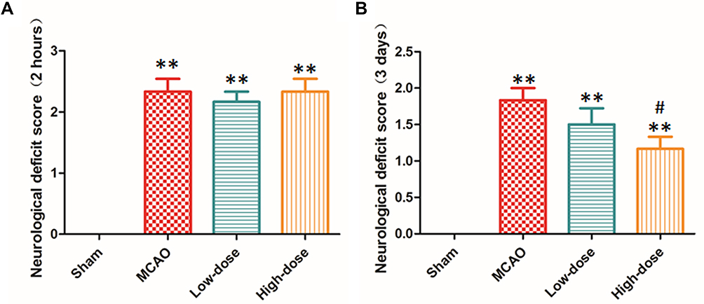 |
Figure 1 Effect of black bamboo rhizome extract on neurological deficits in MCAO rat models. (A) The neurological deficit score in MCAO rats after 2 hours of treatment with rhizome extract of black bamboo. (B) The neurological deficit score in MCAO rats after 10 days of treatment with rhizome extract of black bamboo. **P<0.01, vs sham group; #P<0.05, vs MCAO group. Abbreviation: MCAO, middle cerebral artery occlusion. |
Treatment with BBRE Reduced the Cerebral Infarction Area
To explore the effect of treatment with BBRE on cerebral infarction, Image J was used to analyse and record the infarct area and total area of MCAO rats. The white area on the TTC stained images indicated the infarct area. The model group had the largest infarct area, and the high-dose group had the smallest infarct area.
Compared to the sham group, the MCAO model group showed a significantly increased cerebral infarction area (P<0.01). After administration of BBRE for 10 days, the cerebral infarction area was significantly reduced in the drug groups compared with that in the MCAO group; the reduction was more remarkable in the high-dose drug group than in the low-dose drug group (P<0.01, Figure 2).
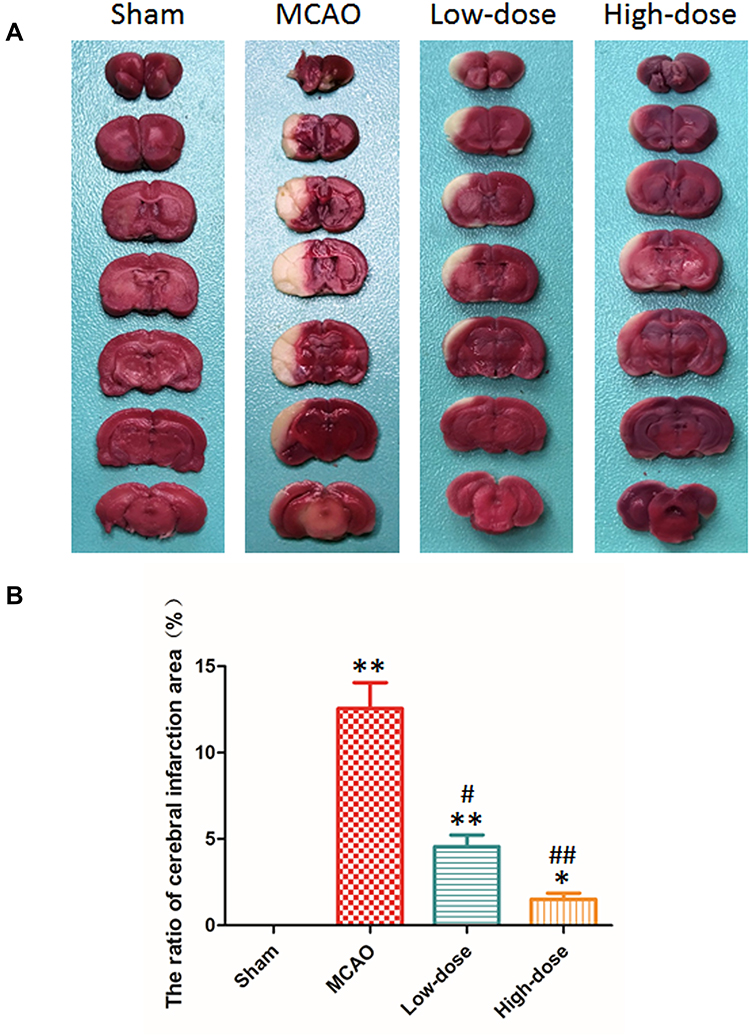 |
Figure 2 Effect of black bamboo rhizome extract on the cerebral infarction area in the MCAO rat models. (A) TTC staining results. (B) The ratio of cerebral infarction area (%). *P<0.05, **P<0.01, vs sham group; #P<0.05, ##P<0.01, vs MCAO group. Abbreviations: MCAO, middle cerebral artery occlusion; TTC, triphenyl tetrazolium chloride. |
Treatment with BBRE Reduced Brain Oedema
To verify the effect of treatment with BBRE on brain oedema, HE staining was performed, and brain tissue water content was calculated using the Elliott formula. Compared with the MCAO group, the high-dose drug group showed significantly reduced cerebral oedema (P<0.05). However, the reduction was not statistically significant in the low-dose drug group (Figure 3A). HE staining showed that there were more neurons in the hippocampus, and the structure was normal and neatly arranged in the control group. However, neurons in the MCAO group were obviously degenerated and necrotic. Notably, the number of neurons was increased, and neuronal degeneration and necrosis were decreased in the drug treatment groups. The decrease in neuronal degeneration and necrosis was more obvious in the high-dose drug group than in the low-dose group (Figure 3B).
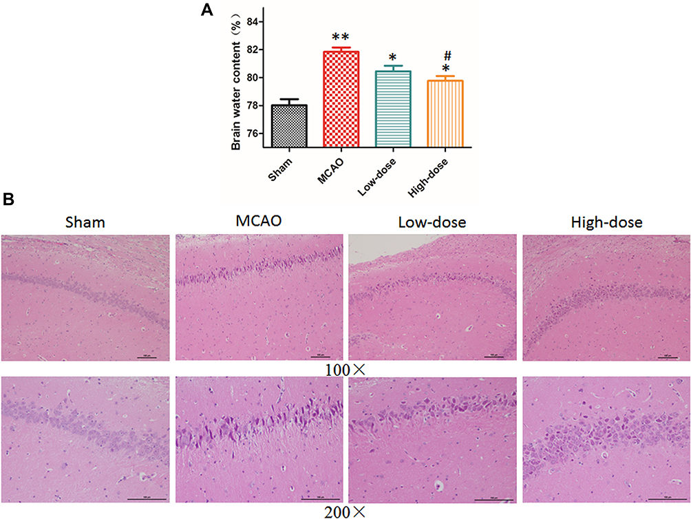 |
Figure 3 Effect of black bamboo rhizome extract on brain oedema in the MCAO rat models. (A) Brain tissue water content in each group. *P<0.05, **P<0.01, vs sham group; #P<0.05, vs MCAO group. (B) Haematoxylin and eosin staining showed that neuronal degeneration and necrosis were significantly decreased in the high-dose drug group. Abbreviation: MCAO, middle cerebral artery occlusion. |
Treatment with BBRE Improved Learning and Memory Abilities
A longer escape latency in the positioning navigation experiment and fewer instances of crossing the platforms in the space exploration experiment indicate deficits in learning and memory abilities. The escape latencies of rats in the sham, low-dose drug, and high-dose drug groups at each detection time point were gradually reduced from the 1st day to the 5th day of training, indicating that the rats had normal learning and memory abilities. The time to find the platform was significantly decreased in the drug groups, especially in the high-dose drug group, compared with that of the MACO group. On the 6th day of the experiment, after the platform in the target quadrant was removed, the time spent in the target quadrant was the longest in the sham group and the shortest in the MCAO group. The time spent in the target quadrant was significantly higher in the drug groups than in the MCAO group. In addition, the number of times the rats crossed the hidden platform was the highest in the sham group, while that in the MCAO group was the lowest. The number of platform crossings in the drug groups was significantly higher than that in the MCAO group, and the effect was the most obvious in the high-dose group (P<0.05, Figure 4).
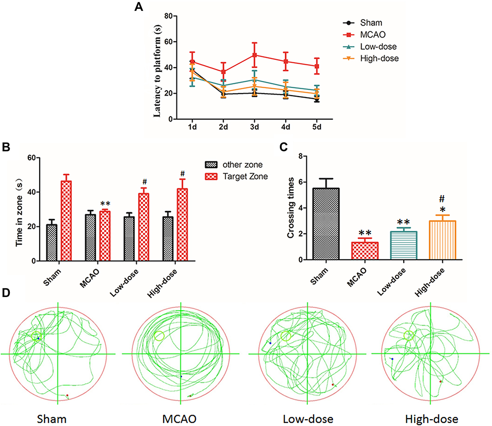 |
Figure 4 Effect of black bamboo rhizome extract on learning and memory abilities in MCAO rat models. (A) escape latency; (B) time spent in the target zone; (C) platform crossing (times); (D) movement pattern of each group. *P<0.05, **P<0.01, vs sham group. # P<0.05 vs MCAO group. Abbreviation: MCAO, middle cerebral artery occlusion. |
Treatment with BBRE Upregulated BDNF and CREB Expression in the Hippocampus
Compared with the sham group, the model group, low-dose drug group, and high-dose drug group showed significantly decreased expression of CREB in the hippocampus (P<0.05, P<0.01). Compared with the MCAO model group, the low-dose and high-dose drug groups showed significantly increased expression of CREB, and the effect was more obvious in the high-dose drug group than in the low-dose group (P<0.01). The model and low-dose drug groups showed lower expression of BDNF in the hippocampus than did the sham group (P<0.05). Moreover, compared with the MCAO model group, the drug treatment groups showed increased BDNF expression, and a significant difference was found between the high-dose drug group and the model group (# P<0.05). There was no significant difference between the low-dose group and the model group (Figure 5).
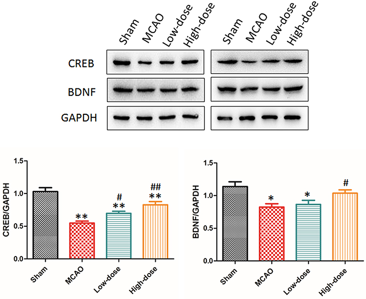 |
Figure 5 Effect of black bamboo rhizome extract on the expression of CREB and BDNF in the hippocampus in MCAO rat models.*P<0.05, **P<0.01, vs sham group; #P<0.05, vs MCAO group; ##P<0.05, vs low-dose group. Abbreviations: BDNF, brain-derived neurotrophic factor; CREB, cyclic adenosine phosphate response element-binding protein; MCAO, middle cerebral artery occlusion. |
Treatment with BBRE Increased Bcl-2 Expression and Decreased Caspase 3 and Bax Expression
Expression levels of caspase 3 and Bax were higher in the model group than in the sham group, and those of bcl-2 were lower in the model group than in the sham operation group (P<0.01). Notably, expression levels of caspase 3 and Bax were lower in the drug treatment groups than in the model group, but those of bcl-2 were higher in the drug treatment groups than in the model group (P < 0.01, Figure 6).
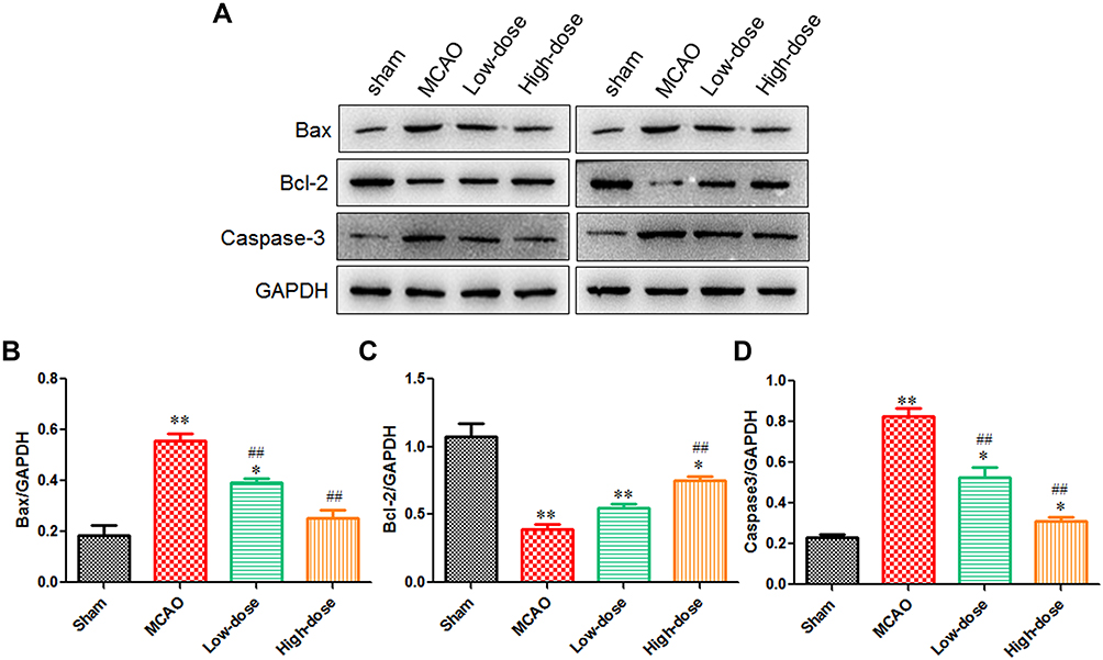 |
Figure 6 Effect of black bamboo rhizome extract on expression of Bax, Bcl-2, and Caspase 3 in MCAO rat models. (A) Western blot of Bax, Bcl-2, and Caspase 3 in rats of difference groups; (B) Expression level of Bax in rats of different groups; (C) Expression level of Bcl-2 in rats of different groups; (D) Expression level of Caspase 3 in rats of different groups. *P<0.05, **P<0.01, vs sham group; ## P<0.01, vs MCAO group. Abbreviation: MCAO, middle cerebral artery occlusion. |
Discussion
The rhizome of black bamboo is a type of Dong medicine; black bamboo is widely distributed in the south of the Yangtze River in China. Previous investigations have found that the rhizome of black bamboo mainly contains triterpenoids, saponins, alkaloids, phenols, amino acids, sugars and their glycosides, and volatile oils. Research has shown that panaxnotoginsenosides, ginsenosides, Rgl, and Rbl play important roles in the treatment of ischaemic cerebral infarction.8 Poria triterpenoids can significantly inhibit HL-60 cell viability and effectively remove superoxide anion, hydrogen peroxide, and hydroxyl free radicals.9–11 In addition, triterpenoids have been shown to have different degrees of inhibitory effects on various bacilli, cocci, and viruses.12 Notably, accumulating studies have shown that the rhizome of black bamboo protects optic ganglion cells and lowers blood sugar.13,14 In this study, we found that treatment with BBRE improved neurological impairment scores, reduced the cerebral infarction area, decreased cerebral oedema, improved learning and memory, and increased expression of CREB and BDNF proteins in rats with cerebral ischaemia-reperfusion injury. Our findings suggest that treatment with BBRE protects the nerve cells and improves cognitive dysfunction caused by cerebral ischaemic disease.
The CREB transcription factor is an important downstream factor in the extracellular regulated protein kinases pathway, which is involved in the generation, proliferation, survival, and neuroprotection of neurons, as well as brain development. In addition, CREB reduces brain injury and improves learning and memory.15–17 For example, Li et al showed that Curdione improved the cognitive function of rats by upregulating the expression of CREB/BDNF in cerebral ischaemia-reperfusion injury, and Chava18,19 found that CREB stimulated angiogenesis and reduced vascular endothelial leakage by upregulating the expression of Ang-1. These findings are in line with our results showing that the effects of BBRE on brain oedema caused by cerebral ischaemia-reperfusion injury might be modulated by the CREB signalling pathway.
BDNF is a neurotrophic factor known to promote nerve growth. It is widely distributed in the nervous system, especially in the hippocampus and cortex, and plays an important role in maintaining neuronal function and repairing neurons by participating in the growth of axons and dendrites and the formation of synapses.20 Therefore, decreased levels of BDNF expression in the brain tissue likely reflect the degree of damage to cognitive function. In addition, the effect of BDNF on neurons is regulated by CREB in the brain, and CREB activity is modulated by phosphorylation of CREB. In fact, phosphorylated CREB is known to be a regulatory factor that induces the expression of BDNF, and BDNF expression promotes phosphorylation of CREB.21,22 This study found that the drug treatment groups showed significantly increased expression of BDNF and CREB and improved cognition function (P<0.01 or P<0.05). BDNF is highly expressed in normal brain tissues. We found that the expression of BDNF was decreased in the MCAO group, but it was significantly increased in the high-dose group compared with that in the MCAO group (P<0.05). These findings suggest the neuroprotective effect of BBRE.
Nerve cell apoptosis is closely related to Bcl-2 and caspase-3. The Bcl-2 protein family is mainly involved in the endogenous apoptosis pathway of cells, as well as the regulation of cell apoptosis, and has been proven to inhibit apoptosis.23,24 Caspase-3 is a key protease in the caspase family and is an important protein for the initiation of apoptosis. After acute cerebral infarction, the expression level of Bcl-2 decreases, the expression level of caspase-3 increases, and the overall anti-apoptotic ability decreases. Our study suggests that BBRE improves cognitive function of rats by regulating the expression of Bcl-2, caspase 3, and Bax in the brain in rats with cerebral ischaemia-reperfusion injury.
To the best of our knowledge, this study is the first to assess the neuroprotective effect of BBRE against cerebral ischaemia-reperfusion injury. Our findings provide a basis for future research. Further studies are needed to identify the active ingredients. The present findings suggest that the rhizome of black bamboo roots, a Dong medicine, can significantly improve the learning and memory ability in cerebral ischaemia/reperfusion injury model rats, probably due to increased expression of CREB and BDNF.
Funding
This study was supported by a project supported by Scientific Research Fund of Hunan Provincial Education Department (No. 19A356) and the Science and Technology Plan of Huaihua City (No. 2015S2202).
Disclosure
Chuan-An Yi and Yu-Hong Jiang should be considered the co-first authors. Chuan-An Y and Xing-Guang Wan are co-corresponding authors. The authors have no conflicts of interest to declare.
References
1. Zheng HJ, Zhan YH. Modern Chinese Medicinal Materials Identification Manual. Beijing: China Medical Science and Technology Press; 2001.
2. Nanjing University of Chinese Medicine. Dictionary of Chinese Medicine (2nd). Version 2. Shanghai: Shanghai Science and Technology Press; 2006.
3. Wang QQ, Fan YH, Wang Y, Xiao QP, He CH. Antioxidant activity of aqueous extract of rhizome of phyllostachys nigra (Lodd. ex Lindl.) Munro in vitro and in vivo. Xumu Yu Shouyi. 2017;49(6):163–169.
4. Fan YH, He CH, Wang QQ, Li YH, Xiao QP, Nie LF A new application of water extract of rhizome of black bamboo root, China, ZL107041930A; 2017.
5. Bederson JB, Pitts LH, Tsuji M, Nishimura MC, Davis RL, Bartkowski H. Rat middle cerebral artery occlusion: evaluation of the model and development of a neurologic examination. Stroke. 1986;17(3):472–476. doi:10.1161/01.str.17.3.472
6. Swanson RA, Morton MT, Tsao-Wu G, Savalos RA, Davidson C, Sharp FR. A semiautomated method for measuring brain infarct volume. J Cereb Blood Flow Metab. 1990;10(2):293–295. doi:10.1038/jcbfm.1990.47
7. Xie J, Hong E, Ding B, et al. Inhibition of NOX4/ROS suppresses neuronal and blood-brain barrier injury by attenuating oxidative stress after intracerebral hemorrhage. Front Cell Neurosci. 2020;14:1–16. doi:10.3389/fncel.2020.578060
8. Huang XP, Deng CQ, Qiu YY, Wang B, Tang YH, Zeng R. Effects of composition of astragaloside IV and three active ingredients of Panaxnotoginseng on oxidative stress and Nrf2/HO-1 pathway after cerebral ischemia/reperfusion in mice. Zhong Guo Yao Li Xue Tong Bao. 2013;29(11):1596–1601.
9. Huang SQ, Li YZ, Fan H, et al. Research progress on chemical constituents and pharmacological activities of Caulophyllum robustom Chinese. Zhongguo Yesheng Zhiwu Ziyuan. 2020;39(3):48–62.
10. Cui XH, Zhang P, Zhu D. The current research progress on pharmacological activity of triterpenoids in Poria cocos. Zhongguo Yaowu Jiangjixue. 2019;14(12):123–125.
11. Cui HR, Wang RL, Guo WB, et al. Research advances in chemical components, pharmacological activities and clinical application of poria cocos. Xibei Yaoxue Zazhi. 2019;5:694–700.
12. Yang L, Jiang H, Wang XY, Liu C. Research progress on chemical constituents and pharmacological effects of Trinia scabiosaefolia Fisch. Zhongyiyao Xinxi. 2012;29(4):169–172.
13. Jung SH, Lee JM, Lee HJ, Kim CY, Lee EH, Um BH. Aldose reductase and advanced glycation endproducts inhibitory effect of Phyllostachys nigra. Biol Pharm Bull. 2007;30(8):1569–1572. doi:10.1248/bpb.30.1569
14. Lee HJ, Kim KA, Kang KD, et al. The compound isolated from the leaves of Phyllostachys nigra protects oxidative stress-induced retinal ganglion cells death. Food Chem Toxicol. 2010;48(6):1721–1727. doi:10.1016/j.fct.2010.03.052
15. Yang J, Jiang H, Chen SS, et al. Lentivirus-mediated RNAi targeting CREB binding protein attenuates neointimal formation and promotes re-endothelialization in balloon injured rat carotid artery. Cell Physiol Biochem. 2010;26(3):441–448. doi:10.1159/000320567
16. Bourtchuladze R, Frenguelli B, Blendy J, Cioffi D, Schutz G, Silva AJ. Deficient long-term memory in mice with a targeted mutation of the cAMP-responsive element-binding protein. Cell. 1994;79(1):59–68. doi:10.1016/0092-8674(94)90400-6
17. Josselyn SA, Shi C, Carlezon WA
18. Chava KR, Tauseef M, Sharma T, Mehta D. Cyclic AMP response element-binding protein prevents endothelial permeability increase through transcriptional controlling p190RhoGAP expression. Blood. 2012;119(1):308–319. doi:10.1182/blood-2011-02-339473
19. Chen J, Jiang H, Yang J, Chen SS, Xu L. Down-regulation of CREB-binding protein expression blocks thrombin-mediated endothelial activation by inhibiting acetylation of NF-κB. Int J Cardiol. 2012;154(2):147–152. doi:10.1016/j.ijcard.2010.09.003
20. Kirkland RA, Saavedra GM, Franklin JL. Rapid activation of antioxidant defenses by nerve growth factor suppresses reactive oxygen species during neuronal apoptosis: evidence for a role in cytochrome c redistribution. J Neurosci. 2007;27(42):11315–11326. doi:10.1523/JNEUROSCI.3590-07.2007
21. Autry AE, Monteggia LM. Brain-derived neurotrophic factor and neuropsychiatric disorders. Pharmacol Rev. 2012;64(2):238–258. doi:10.1124/pr.111.005108
22. Zhang LL, Ying J, Hua FZ, et al. Saikosaponin A attenuates cognitive function via cAMP/CREB signaling pathway in mice after traumatic brain injury. Linchuang Mazui Xue Zazhi. 2016;32(5):484–487.
23. Ma J, Bao L, Xia X, et al. miR-128b promotes cerebral infarction by regulating the expressions of BCL-2 and CAPASE3. World Neurosurg. 2019;123:e245–e251. doi:10.1016/j.wneu.2018.11.144
24. Che QQ, Huang T, Zhang YD, Qian XJ. Effect of miR-124 on neuronal apoptosis in rats with cerebral infarction through Wnt/ β-catenin signaling pathway. Eur Rev Med Pharmacol Sci. 2019;23(15):6657–6664. doi:10.26355/eurrev_201908_18556
 © 2021 The Author(s). This work is published and licensed by Dove Medical Press Limited. The full terms of this license are available at https://www.dovepress.com/terms.php and incorporate the Creative Commons Attribution - Non Commercial (unported, v3.0) License.
By accessing the work you hereby accept the Terms. Non-commercial uses of the work are permitted without any further permission from Dove Medical Press Limited, provided the work is properly attributed. For permission for commercial use of this work, please see paragraphs 4.2 and 5 of our Terms.
© 2021 The Author(s). This work is published and licensed by Dove Medical Press Limited. The full terms of this license are available at https://www.dovepress.com/terms.php and incorporate the Creative Commons Attribution - Non Commercial (unported, v3.0) License.
By accessing the work you hereby accept the Terms. Non-commercial uses of the work are permitted without any further permission from Dove Medical Press Limited, provided the work is properly attributed. For permission for commercial use of this work, please see paragraphs 4.2 and 5 of our Terms.