
Back to Journals » International Journal of Chronic Obstructive Pulmonary Disease » Volume 20
Biomarkers (NLR, PLR, SII) for Frequent COPD Exacerbations: Diagnostic and Clinical Management Implications in a Retrospective Study
Authors Fu Y, Wang Y, Wang Y, Mou T, He X, Wang J, Xiong A, Qing B, Wu D, Li G
Received 9 December 2024
Accepted for publication 21 March 2025
Published 5 April 2025 Volume 2025:20 Pages 987—998
DOI https://doi.org/10.2147/COPD.S510118
Checked for plagiarism Yes
Review by Single anonymous peer review
Peer reviewer comments 4
Editor who approved publication: Prof. Dr. Richard Russell
Yufen Fu,1– 3 Yuxin Wang,1,4 Yujiao Wang,1,4 Ting Mou,1,4 Xiang He,2,4 Junyi Wang,2,4 Anying Xiong,2,4 Bomiao Qing,2,4 Dehong Wu,2,4 Guoping Li1,2,4
1Department of Pulmonary and Critical Care Medicine, Affiliated Hospital of Southwest Medical University, Luzhou, 646000, People’s Republic of China; 2Laboratory of Allergy and Precision Medicine, Chengdu Institute of Respiratory Health, the Third People’s Hospital of Chengdu, Affiliated Hospital of Southwest Jiaotong University, Chengdu, 610031, People’s Republic of China; 3Department of Pulmonary and Critical Care Medicine, Longchang People’s Hospital, Neijiang, 642150, People’s Republic of China; 4Department of Pulmonary Medicine, Chengdu Third People’s Hospital branch of National Clinical Research Center for Respiratory Disease, Affiliated Hospital of Chongqing Medical University, Chengdu, 610031, People’s Republic of China
Correspondence: Guoping Li, Email [email protected]
Objective: To evaluate the diagnostic and predictive value of the neutrophil-to-lymphocyte ratio (NLR), platelet-to-lymphocyte ratio (PLR), and systemic immune-inflammation index (SII) for frequent exacerbations of chronic obstructive pulmonary disease (AECOPD), and to develop a risk stratification scoring system to optimize clinical management in resource-limited healthcare settings.
Patients and Methods: This retrospective observational study enrolled 16,849 AECOPD patients, categorized into frequent exacerbators (≥ 2 exacerbations/year, n=3,488) and non-frequent exacerbators (< 2 exacerbations/year, n=13,361). Comparative analyses of clinical characteristics and inflammatory biomarkers (NLR, PLR, SII, CRP, PCT) were conducted. Spearman correlation, receiver operating characteristic (ROC) curve analysis, and binary logistic regression were employed to assess biomarker performance. A risk scoring system was developed using odds ratios (OR) and regression coefficients (β) of NLR and PLR.
Results: The frequent exacerbators group exhibited significantly higher median NLR (6.71 vs 5.10, P < 0.001), mean PLR (239 ± 204 vs 218 ± 195, P < 0.001), and median SII (1,137.48 vs 847.54, P < 0.001). NLR, PLR and SII showed strong positive correlations with CRP and PCT (P < 0.001). ROC analysis identified NLR (specificity = 84.1%) and PLR (sensitivity = 55%) as optimal diagnostic indicators. Regression analysis confirmed NLR and PLR as independent risk factors for frequent exacerbations. The risk stratification system categorized patients into low-risk (< 290 points; annual exacerbation rate 17%), intermediate-risk (290– 768 points; 19.1%), and high-risk (> 768 points; 23.4%) groups.
Conclusion: NLR and PLR serve as cost-effective biomarkers for identifying high-risk frequent exacerbators patients with COPD in primary care settings. The percentile-based scoring system enables management strategies to address clinical needs in resource-constrained healthcare environments.
Keywords: COPD, exacerbation, NLR, PLR, biomarkers
Graphical Abstract:

Introduction
Chronic obstructive pulmonary disease (COPD) is a prevalent disease that markedly diminishes the quality of life for those affected, imposing a significant economic strain on families and society. This chronic airway disease is characterized by various pulmonary manifestations and progressive airway obstruction, which arise from conditions such as bronchitis and bronchiolitis, as well as alveolar damage associated with emphysema, Patients commonly present with persistent respiratory symptoms, including dyspnea, cough, sputum production, and episodes of acute exacerbation.1
COPD is characterized by a high prevalence and mortality rate, as well as a significant socio-economic impact. The 2018”the China Pulmonary Health [CPH]study”, which included 50,991 adults across 10 provinces and cities, reported a COPD prevalence of 8.6% in individuals over 20 years of age, increasing to 13.7% in those over 40. This corresponds to an estimated 100 million individuals who are affected by this condition in China.2 The prevalence of COPD is influenced by several factors, including an aging population, smoking and secondhand smoke exposure, ambient and household air pollution, genetic susceptibility, aberrant inflammatory responses, abnormal lung development, and tuberculosis. These factors contribute to the financial burden of COPD, with acute exacerbations representing the primary source of medical costs associated with the disease. Therefore, the development of straightforward and expeditious diagnostic methods is essential for the timely identification and diagnosis of COPD, potentially enhancing its evaluation and management.
AECOPD is clinically defined as an acute deterioration of respiratory symptoms—dyspnea, cough, and sputum production—within 14 days. These symptoms may be accompanied by tachypnoea and tachycardia. These exacerbations are frequently triggered by respiratory infections or air pollution, which lead to an increase in local or systemic inflammation, they may also arise from other etiologies that compromise airway integrity.1 Patients experiencing two or more exacerbations in the past year are classified as having frequent exacerbations.3,4 The impact of these exacerbations on health status, quality of life, and pulmonary function is considerable, while the economic burden they impose is also substantial.5,6 Nevertheless, the precise inflammatory profiles associated with frequent exacerbations in COPD patients remain poorly understood. Furthermore, the current absence of reliable biomarkers for AECOPD has resulted in a notable deficit in research focused on the identification of predictive biomarkers for frequent exacerbations.
C-reactive protein (CRP), a well-established acute-phase inflammatory biomarker, has limited sensitivity and specificity7–9and performs inconsistently across different patient populations. CRP levels can also be influenced by non-infectious factors such as patient age, cardiovascular disease, obesity, dyslipidemia, smoking, and alcohol consumption.10–15 Procalcitonin (PCT), commonly used to differentiate bacterial from non-bacterial infections,16–18 can be confounded by underlying chronic inflammatory states and airway colonization with Bacteria.19,20 PCT detection is costly, may yield false-negative results, and PCT-guided therapy has not been shown to reduce antibiotic use.21 Metagenomic next-generation sequencing (mNGS) offers a highly sensitive and pragmatic approach, complementing conventional microbiological tests (CMT) by identifying a broad spectrum of pathogens with high sensitivity and specificity.22–26 However, mNGS is expensive and requires interdisciplinary collaboration involving bioinformatics, microbiology, and clinical medicine. Additionally, miR-155 expression in smokers with COPD correlates positively with disease severity,27 and miR-21 is positively associated with disease severity and pulmonary function impairment in COPD patients.28 The expression levels of epithelial–mesenchymal transition markers were detected by Western blot and immunofluorescence.29 Metabolomic and genomic analyses of plasma have identified sphingomyelins as strongly associated with emphysema and glycosphingolipids as linked to COPD exacerbations.30,31 Given the heterogeneity of COPD, a single inflammatory biomarker cannot fully capture the complexity of AECOPD, including differentiating between infectious and non-infectious causes, or bacterial versus viral etiologies. Dynamic monitoring of inflammatory biomarker levels before, during, and after acute exacerbations is essential for assessing therapeutic efficacy. However, most novel inflammatory biomarkers are costly to detect and remain largely in the research phase, requiring validation through multicenter cohort studies to establish their clinical value.
In the United States, primary care providers are responsible for the majority of antibiotic prescriptions, with family physicians issuing the highest number of such prescriptions.32 Recent studies have shown that combining multiple biomarkers enhances diagnostic and predictive accuracy. Previous research has demonstrated that NLR, PLR, and SII are recombinant inflammatory markers that provide greater diagnostic and predictive value than single biomarkers.33–35 However, the effectiveness of NLR, PLR, and SII in diagnosing patients with frequent COPD exacerbations remains uncertain. Therefore, this study aims to comprehensively assess the diagnostic potential of NLR, PLR, and SII in identifying patients at risk for frequent AECOPD.
Subject Profile
In this retrospective analysis, we enrolled 16,849 subjects and categorized them into two cohorts: 13,361 subjects with Non-Frequent AECOPD and 3,488 subjects with Frequent AECOPD. The Non-Frequent AECOPD cohort comprised patients who experienced fewer than two exacerbations over the preceding year. In contrast, the Frequent AECOPD cohort included those who had experienced two or more exacerbations during the same period. Complete blood count data for all participants were extracted from the Third People’s Hospital of Chengdu, Sichuan Province, China, and covered the period from January 1, 2013, to May 1, 2023.
This study was conducted by the Declaration of Helsinki, and the protocol was reviewed and approved by the institutional review board of the Third People’s Hospital of Chengdu (Project No. Ethics Committee of Chengdu Third People’s Hospital [2024]-S-388).
Data and Sample Collection
According to the Global Initiative for Chronic Obstructive Lung Disease (GOLD) 2017, patients with two or more exacerbations per year are considered to have frequent exacerbations,36 patients were classified into two groups: frequent exacerbators, defined as those who had experienced two or more exacerbations in the previous year, and non-frequent exacerbators (Figure 1). Demographic data, clinical features, treatment strategies, blood cell counts, inflammatory markers, and sputum pathogen profiles were obtained from electronic medical records. Blood cell counts and inflammatory markers were assessed upon admission, before the initiation of antibiotic and steroid therapy. We calculated and analyzed the NLR, PLR, and SII using the following formulas: NLR = neutrophil count/lymphocyte count, PLR = platelet count/lymphocyte count, and SII = platelet count × neutrophil count/lymphocyte count.
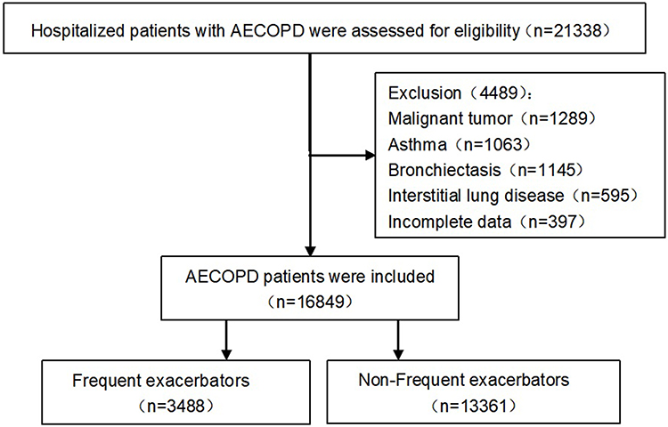 |
Figure 1 Flowchart of the study population. |
Statistical Analysis
All statistical analyses were conducted using R version 4.3.2. Continuous variables were expressed as mean with standard deviation (SD) for normally distributed variables or median with interquartile range (IQR) for skewed variables. Quantity and percentage were employed for categorical variables. Differences in clinical characters between the two groups were evaluated using the T-test and Mann–Whitney U-test for continuous variables, while the Chi-squared test and Fisher exact probability method were used for categorical variables, as appropriate. Spearman correlation coefficient (r) was calculated to identify the existence of correlation. Receiver Operating Characteristic (ROC) curve analysis was applied to assess the diagnostic sensitivity and specificity of NLR, PLR, and SII. Additionally, we examined the sensitivity and specificity of combinations of two or three of these markers. Diagnostic accuracy was determined by the area under the curve (AUC), with higher values indicating superior discriminatory power. Binary logistic regression was utilized to identify individual risk factors. The power calculation using the R pwr package resulted in a power of 1 (exceeding the 0.8 threshold), confirming sufficient sample size to detect the hypothesized effect. A risk scoring system was constructed using odds ratios (OR) and regression coefficients (β) of NLR and PLR. Statistical significance was established for group differences at P<0.05.
Results
Clinical Characteristics of Frequent and Non-Frequent Exacerbators Patients
From January 2013 to May 2023, our retrospective analysis enrolled 16,849 individuals with AECOPD. The cohort included 4,723 females and 12,126 males, with a mean age of 75.8 ± 10.2 years. Participants were classified into two groups: 3,488 patients with frequent exacerbators (mean age 77.3 ± 9.67 years; 75.9% male) and 13,361 patients with non-frequent exacerbators (mean age 75.4 ± 10.3 years; 70.9% male). Table 1 outlines the demographic and clinical profiles. Table 2 demonstrates that patients experiencing frequent exacerbations had significantly higher levels of leukocytes, neutrophils, NLR, PLR, SII, and CRP, compared to those with non-frequent exacerbators (P<0.001). However, platelet, lymphocyte, and PCT levels did not differ significantly between the two groups (P>0.05).
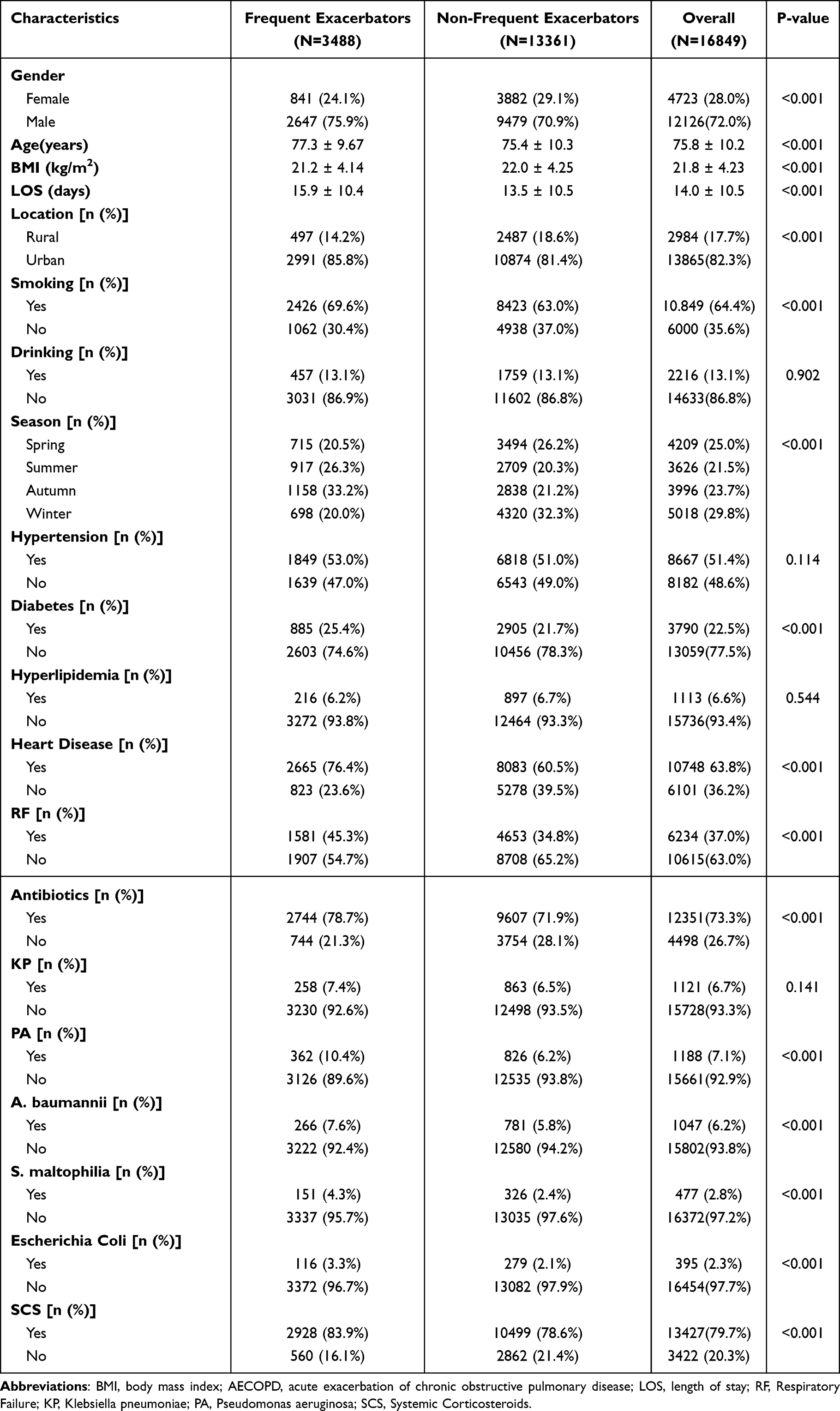 |
Table 1 The Demographic Characteristics of Frequent and Non-Frequent Exacerbators |
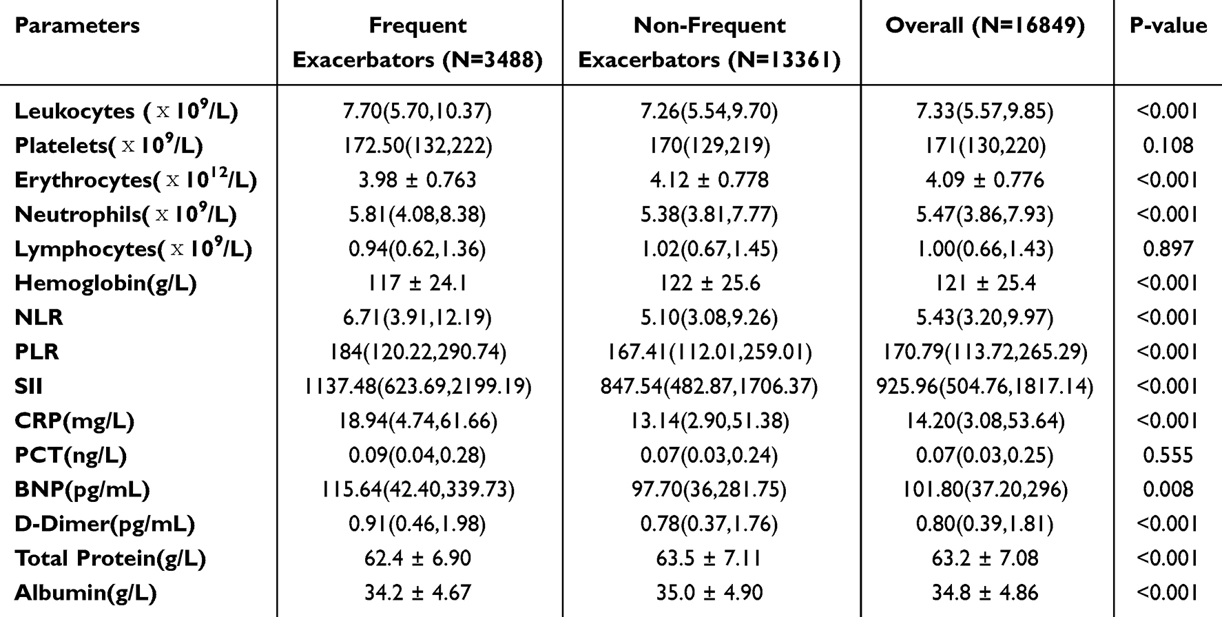 |
Table 2 Clinical Complete Blood Count Parameters of the Study Patients |
The Level of CRP, PCT, NLR, PLR and SII in Two Groups
In our study, median PCT levels were 0.09 ng/mL (IQR 0.04–0.28) in frequent exacerbators and 0.07 ng/mL (IQR 0.03–0.24) in non-frequent exacerbators (P=0.555). Median CRP values were 18.94 mg/L (IQR 4.74–61.66) in frequent exacerbators and 13.14 mg/L (IQR 2.90–51.38) in non-frequent exacerbators (P<0.001). Median NLR values were 6.71 (IQR 3.91–12.19) in frequent exacerbators and 5.10 (IQR 3.08–9.26) in non-frequent exacerbators (P<0.001). PLR medians were 184 (IQR 120.22–290.74) in frequent exacerbators and 167.41 (IQR 112.01–259.01) in non-frequent exacerbators (P<0.001). SII medians were 1137.48 (IQR 623.69–2199.19) in frequent exacerbators and 847.54 (IQR 482.87–1706.37) in non-frequent exacerbators (P<0.001) (Table 2).
The Correlation of PCT, CRP and NLR, PLR, and SII
Spearman correlation analysis revealed significant associations between PCT and the inflammatory markers: NLR (rs = 0.4105, P < 0.001), PLR (rs = 0.2047, P < 0.001), and SII (rs = 0.3184, P < 0.001). Additionally, CRP showed significant correlations with NLR (rs = 0.4239, P < 0.001), PLR (rs = 0.3015, P < 0.001), and SII (rs = 0.4127, P < 0.001) (Figure 2). All participants were included in the Spearman correlation analysis (Table 3).
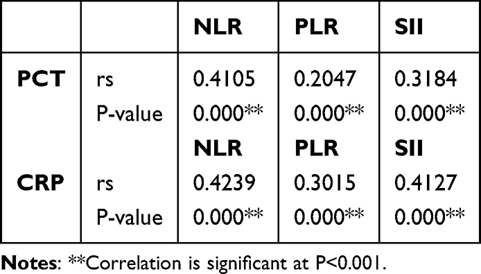 |
Table 3 Correlations Between All Inflammatory Markers |
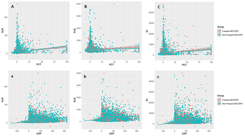 |
Figure 2 Spearman correlations between PCT and inflammatory biomarkers in all participants: (A) NLR, rs = 0.4105, P < 0.001; (B) PLR, rs = 0.2047, P < 0.001; (C) SII, rs = 0.3184, P < 0.001. Correlations between CRP and inflammatory biomarkers: (a) NLR, rs = 0.4239, P < 0.001; (b) PLR, rs = 0.3015, P < 0.001; (c) SII, rs = 0.4127, P < 0.001. |
Diagnostic Evaluation of NLR, PLR and SII
The receiver operating characteristic (ROC) curve analysis for NLR, PLR, and SII is presented in Figure 3. NLR demonstrated a sensitivity of 46.0% and a specificity of 84.1% at a cut-off value of 8.958, with an area under the curve (AUC) of 0.6965. For PLR, the optimal cut-off was 187.981, yielding an AUC of 0.6283, a sensitivity of 55.0%, and a specificity of 64.1%. SII had a sensitivity of 52.7% and a specificity of 72.4% at a cut-off value of 1221.867, with an AUC of 0.6713. NLR+PLR had a sensitivity of 50.4% and a specificity of 77.8% at a cut-off value of 0.425, with an AUC of 0.6902. NLR+SII had a sensitivity of 47.1% and a specificity of 82.1% at a cut-off value of 0.433, with an AUC of 0.6955. PLR+SII had a sensitivity of 54.8% and a specificity of 69.7% at a cut-off value of 0.411, with an AUC of 0.6650. NLR+PLR+SII had a sensitivity of 49.5% and a specificity of 78.8% at a cut-off value of 0.428, with an AUC of 0.6901 (Figure 3). This study identified NLR as having the highest diagnostic accuracy and specificity for frequent AECOPD. Although we assessed combinations of two or three markers, no improvement in diagnostic accuracy or sensitivity was observed. Given the suboptimal sensitivity of NLR, its results should be interpreted with caution. The diagnostic evaluations for individual markers and their combinations are detailed in Table 4.
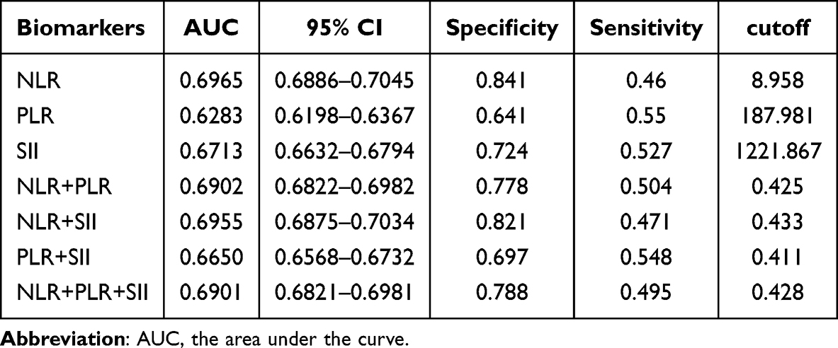 |
Table 4 Diagnostic Accuracy of the NLR, PLR, and SII |
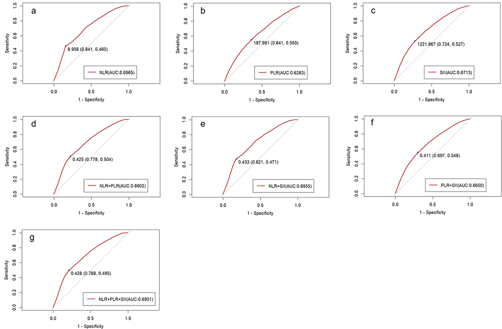 |
Figure 3 Receiver Operating Characteristic (ROC) curves for single and combined markers to evaluate diagnostic accuracy: (a) NLR, AUC = 0.6965; (b) PLR, AUC = 0.6283; (c) SII, AUC = 0.6713; (d) NLR + PLR, AUC = 0.6902; (e) NLR + SII, AUC = 0.6955; (f) PLR + SII, AUC = 0.6650; (g) NLR + PLR + SII, AUC = 0.6901. |
Risk Factors for the Development of Frequent AECOPD
In a binary logistic regression model, frequent exacerbators were designated as the dependent variable, with NLR, PLR, and SII included as covariates. The analysis revealed that NLR (OR=1.052, P<0.001) and PLR (OR=1.001, P=0.001) were significant risk factors for patients with frequent exacerbators, while SII (OR=1, P=0.14) was not significant (Table 5).
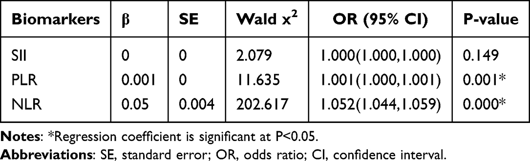 |
Table 5 Binary Logistic Regression Analysis of NLR, PLR, and SII |
Scoring System-Guided Clinical Stratification
Based on logistic regression beta coefficients and odds ratios (OR), SII was excluded (P > 0.05) with subsequent scoring assigned to NLR and PLR: 1 point per NLR unit increase and 50 points per PLR unit increase (Table 6). Using R software, total scores were calculated for each patient. Risk stratification was performed using percentile thresholds: low-risk (<290 points; annual exacerbation rate 17%), intermediate-risk (290–768 points; 19.1%), and high-risk (>768 points; 23.4%) groups (Table 7).
 |
Table 6 Scoring Criteria for NLR and PLR |
 |
Table 7 Risk Stratification and Incidence of Frequent Exacerbations |
Discussion
The primary findings of this study reveal that COPD patients with frequent acute exacerbations exhibit more pronounced inflammatory responses, with significant elevations in inflammatory markers such as the NLR, PLR, and SII. These markers not only identify patients prone to frequent exacerbations but also serve as independent risk factors for predicting such events. A risk scoring system based on these markers facilitates stratified management of COPD patients.
COPD is associated with both enhanced airway and systemic inflammation and, during states of exacerbation, the severity of inflammation is significantly increased.37 A study has indicated that NLR and PLR were higher in acute exacerbation of COPD and used for predicting hospitalization.36 NLR is a simple and cost-effective marker that does not require additional laboratory workup.38 In a study, NLR was identified as a highly-sensitive marker predicting and identifying mortality in patients with community-acquired pneumonia.39 In another study, NLR was found to be superior to CRP and neutrophil and to leukocyte counts alone, in identifying bacteremia in an emergency setting.40 Similarly, NLR was seen to increase in patients with advanced lung cancer and was found useful for the evaluation of disease severity and prognosis.41 A study has identified the optimal NLR cutoff value for acute exacerbations of COPD as 2.84 and the optimal PLR cutoff value as 112.23.42 In a study, NLR ≥2.91 and PLR ≥156.53 have been identified as independent risk factors for lung cancer in COPD patients.43 Our study found significant correlations between NLR, PLR, and SII with CRP and PCT, with notable elevations in these markers among COPD patients experiencing frequent acute exacerbations. The optimal cutoff values were determined to be 8.958 for NLR and 187.981 for PLR, indicating a more pronounced inflammatory response in patients with frequent exacerbations. We speculate that patients with frequent acute exacerbations of COPD may also have a higher likelihood of developing lung cancer.
Studies have shown that the combination of NLR and PLR can enhance diagnostic value by distinguishing lung cancer patients from healthy individuals.44 In our study, we evaluated the diagnostic value of NLR, PLR, and SII individually and in combination for frequent acute exacerbations. NLR demonstrated the highest AUC value of 0.6955, with a specificity of 84.1% and sensitivity of 46%. PLR had an AUC value of 0.6283, with the highest sensitivity among all markers at 55%, albeit with lower specificity. The combined use of these markers did not enhance diagnostic value, possibly due to differences in region, sample size, and outcome measures. These results suggest that NLR may hold some diagnostic value for frequent acute exacerbations, but its predictive accuracy should be interpreted with caution due to its suboptimal sensitivity. We suggest that NLR and PLR results be interpreted alongside traditional inflammatory markers such as CRP or PCT to ensure better prognostic accuracy. Future integration with genomics and metabolomics data holds promise for a more comprehensive elucidation of the inflammatory mechanisms underlying AECOPD.
Chronic inflammation and oxidative stress lead to structural and functional damage of lung tissue, rendering the airways more susceptible to invasion by external pathogens. This results in the recruitment and activation of inflammatory cells, exacerbating lung tissue damage and the inflammatory response.45–47 Our study revealed that patients with frequent acute exacerbations exhibited significantly higher levels of white blood cells, CRP, and neutrophils compared to those with infrequent exacerbations, NLR, PLR and SII reflect the dynamic changes in systemic inflammation. The significant elevation of these indices in COPD patients with frequent acute exacerbations indicates an imbalance in the body’s inflammatory levels and immune-inflammatory equilibrium.48,49 One study demonstrated that NLR and PLR levels are significantly elevated in COPD patients with lung cancer, serving as independent predictors.43 Additionally, compared to stable patients, NLR, MLR, and PLR levels are markedly increased in AECOPD patients, with elevated NLR and MLR identified as risk factors for AECOPD, whereas PLR is not.50 Recent research indicates that NLR, PLR, and SII may be potential biomarkers for early gastric cancer.51 Our study found that, according to binary logistic regression analysis, NLR and PLR are independent predictors of frequent acute exacerbations in COPD, whereas SII is not. Although SII has been identified as a significant predictor of patient outcomes, including prognosis and mortality, in various cardiovascular diseases (CVD) and cancer patient populations.52,53 This discrepancy may be related to population characteristics and disease heterogeneity, necessitating further validation through multicenter, prospective studies. These indices reflect systemic inflammation, offering a safe, noninvasive, economical, and simple biological indicator that guides clinical assessment of inflammatory status.
Based on the β values and OR values derived from regression analysis, this study has developed a scoring system to stratify the risk of AECOPD patients and thereby estimate the probability of frequent acute exacerbations. The scoring system indicates that the higher the risk stratification, the greater the probability of frequent acute exacerbations. The application of this scoring system will aid in optimizing the clinical management of AECOPD patients and the rational allocation of limited medical resources.
However, our study had several limitations. First, the single-center retrospective design limits the generalizability of the findings to more diverse populations. Future research should include prospective, multicenter studies across different ethnicities with larger sample sizes to confirm the diagnostic accuracy and predictive value of NLR, PLR, and SII in AE-COPD patients. Second, future studies should conduct regular CBC tests throughout the treatment process to explore the dynamic relationship between NLR/PLR/SII levels and frequent AE-COPD. Third, further investigation into the role of NLR, PLR, SII, and other inflammatory markers in COPD management is warranted. Additionally, research on the performance of these markers across different COPD phenotypes is needed to optimize personalized treatment strategies. Lastly, future work should assess the ability of NLR, PLR, and SII to differentiate between bacterial and non-bacterial infections in patients with frequent acute exacerbations, which could guide antibiotic therapy.
Conclusion
Our study shows that NLR, PLR, and SII levels are elevated in frequent AECOPD patients, serving as cost-effective markers for exacerbation frequency and severity. A scoring system based on these markers aids COPD management in primary care.
Ethics Approval and Consent to Participate
This study was approved by the by the Ethics Committee of Chengdu Third People’s Hospital (Project No. Ethics Committee of Chengdu Third People’s Hospital [2024]-S-388). The Chengdu Third People’s Hospital waived the need for informed consent as the study was retrospective and This study collected data anonymously.
Funding
This work was supported by the Chengdu Science and Technology Bureau Major Science and Technology Application Demonstration Project (2021-YF09-00102-SN), Chengdu Medical Research Project (2021011), Chengdu High-Level Clinical Key Specialty Construction Project, The Third People’s Hospital of Chengdu Clinical Research Program: Mechanistic Study on Exhausted Neutrophil Subsets Induced by Fine Particulate Matter in Bronchial Asthma(CSY-YN-03-2024-027), The Impact of Air Pollutants on the Immune Microenvironment and Pro-Cancer Mechanisms in the Development of Lung Cancer and Molecular Drug Screening(2023PI01), Mechanism Study on the Regulation of TH17 Cell Differentiation by Fungal Components in Fine Particulate Matter through Exhausting Neutrophil PD-L1/CD80 cis-Action to Exacerbate Asthma(CSY-YN-01-2023-002).
Disclosure
The authors report no conflicts of interest in this work.
References
1. Agustí A, Celli BR, Criner GJ, et al. Global initiative for chronic obstructive lung disease 2023 report: GOLD executive summary. Am J Respir Crit Care Med. 2023;207(7):819–837. doi:10.1164/rccm.202301-0106PP
2. Wang C, Xu J, Yang L, et al. Prevalence and risk factors of chronic obstructive pulmonary disease in China (the China Pulmonary Health [CPH] study): a national cross-sectional study. Lancet. 2018;391(10131):1706–1717. doi:10.1016/s0140-6736(18)30841-9
3. Hurst JR, Vestbo J, Anzueto A, et al. Susceptibility to exacerbation in chronic obstructive pulmonary disease. N Engl J Med. 2010;363(12):1128–1138. doi:10.1056/NEJMoa0909883
4. Le Rouzic O, Roche N, Cortot AB, et al. Defining the “frequent exacerbator” phenotype in COPD: a hypothesis-free approach. Chest. 2018;153(5):1106–1115. doi:10.1016/j.chest.2017.10.009
5. JAEC-C W, Miravitlles M, Hurst JR, et al. Management of COPD exacerbations: a European Respiratory Society/American thoracic society guideline. Eur Respir J. 2017;49(3). doi:10.1183/13993003.00791-2016
6. Yin P, Wang H, Vos T, et al. A subnational analysis of mortality and prevalence of COPD in China From 1990 to 2013: findings from the global burden of disease study 2013. Chest. 2016;150(6):1269–1280. doi:10.1016/j.chest.2016.08.1474
7. Hurst JR, Donaldson GC, Perera WR, et al. Use of plasma biomarkers at exacerbation of chronic obstructive pulmonary disease. Am J Respir Crit Care Med. 2006;174(8):867–874. doi:10.1164/rccm.200604-506OC
8. Bolatkale M, Duger M, Ülfer G, et al. A novel biochemical marker for community-acquired pneumonia: ischemia-modified albumin. Am J Emerg Med. 2017;35(8):1121–1125. doi:10.1016/j.ajem.2017.03.018
9. Viasus D, Del Rio-Pertuz G, Simonetti AF, et al. Biomarkers for predicting short-term mortality in community-acquired pneumonia: a systematic review and meta-analysis. J Infect. 2016;72(3):273–282. doi:10.1016/j.jinf.2016.01.002
10. Hausfater P. Biomarkers and infection in the emergency unit. Med Mal Infect. 2014;44(4):139–145. doi:10.1016/j.medmal.2014.01.002
11. Mitsuma SF, Mansour MK, Dekker JP, et al. Promising new assays and technologies for the diagnosis and management of infectious diseases. Clin Infect Dis Apr. 2013;56(7):996–1002. doi:10.1093/cid/cis1014
12. Pearson TA, Mensah GA, Alexander RW, et al. Markers of inflammation and cardiovascular disease: application to clinical and public health practice: a statement for healthcare professionals from the centers for disease control and prevention and the American Heart Association. Circulation. 2003;107(3):499–511. doi:10.1161/01.cir.0000052939.59093.45
13. Craig WY, Palomaki GE, Haddow JE. Cigarette smoking and serum lipid and lipoprotein concentrations: an analysis of published data. BMJ. 1989;298(6676):784–788. doi:10.1136/bmj.298.6676.784
14. Albert MA, Glynn RJ, Ridker PM. Alcohol consumption and plasma concentration of C-reactive protein. Circulation. 2003;107(3):443–447. doi:10.1161/01.cir.0000045669.16499.ec
15. Aronson D, Bartha P, Zinder O, et al. Obesity is the major determinant of elevated C-reactive protein in subjects with the metabolic syndrome. Int J Obes Relat Metab Disord. 2004;28(5):674–679. doi:10.1038/sj.ijo.0802609
16. Wacker C, Prkno A, Brunkhorst FM, Schlattmann P. Procalcitonin as a diagnostic marker for sepsis: a systematic review and meta-analysis. Lancet Infect Dis. 2013;13(5):426–435. doi:10.1016/s1473-3099(12)70323-7
17. Karzai W, Oberhoffer M, Meier-Hellmann A, Reinhart K. Procalcitonin--a new indicator of the systemic response to severe infections. Infection. 1997;25(6):329–334. doi:10.1007/bf01740811
18. Ergan B, Şahin AA, Topeli A. Serum Procalcitonin as a biomarker for the prediction of bacterial exacerbation and mortality in severe COPD exacerbations requiring mechanical ventilation. Respiration. 2016;91(4):316–324. doi:10.1159/000445440
19. Pantzaris ND, Spilioti DX, Psaromyalou A, Koniari I, Velissaris D. The use of serum procalcitonin as a diagnostic and prognostic biomarker in chronic obstructive pulmonary disease exacerbations: a literature review update. J Clin Med Res. 2018;10(7):545–551. doi:10.14740/jocmr3458w
20. Daubin C, Fournel F, Thiollière F, et al. Ability of procalcitonin to distinguish between bacterial and nonbacterial infection in severe acute exacerbation of chronic obstructive pulmonary syndrome in the ICU. Ann Intensive Care. 2021;11(1):39. doi:10.1186/s13613-021-00816-6
21. Lindenauer PK, Shieh MS, Stefan MS, et al. Hospital procalcitonin testing and antibiotic treatment of patients admitted for chronic obstructive pulmonary disease exacerbation. Ann Am Thorac Soc. 2017;14(12):1779–1785. doi:10.1513/AnnalsATS.201702-133OC
22. Niu S, Liu D, Yang Y, Zhao L. Clinical utility of metagenomic next-generation sequencing in the diagnosis of invasive pulmonary aspergillosis in acute exacerbation of chronic obstructive pulmonary disease patients in the intensive care unit. Front Cell Infect Microbiol. 2024;14:1397733. doi:10.3389/fcimb.2024.1397733
23. Shi Y, Peng JM, Hu XY, Yang QW, Wang Y. Metagenomic next-generation sequencing for detecting Aspergillosis pneumonia in immunocompromised patients: a retrospective study. Front Cell Infect Microbiol. 2023;13:1209724. doi:10.3389/fcimb.2023.1209724
24. Qin W, Guo T, You T, Tian R, Cui X, Wang P. Metagenomic next generation sequencing of bronchoalveolar lavage fluids for the identification of pathogens in patients with pulmonary infection: a retrospective study. Diagn Microbiol Infect Dis. 2024;110(1):116402. doi:10.1016/j.diagmicrobio.2024.116402
25. Gao Q, Li L, Su T, et al. A single-center, retrospective study of hospitalized patients with lower respiratory tract infections: clinical assessment of metagenomic next-generation sequencing and identification of risk factors in patients. Respir Res. 2024;25(1):250. doi:10.1186/s12931-024-02887-y
26. Zheng Y, Liu W, Xiao T, Chen H, Liu Y. Clinical utility of metagenomic next-generation sequencing on bronchoalveolar lavage fluid in diagnosis of lower respiratory tract infections. BMC Pulm Med. 2024;24(1):422. doi:10.1186/s12890-024-03237-w
27. De Smet EG, Van Eeckhoutte HP, Avila cobos F, et al. The role of miR-155 in cigarette smoke-induced pulmonary inflammation and COPD. Mucosal Immunology. 2020;13(3):423–436. doi:10.1038/s41385-019-0241-6
28. Crunkhorn S. miR-21 antagomir reverses COPD pathology. Nat Rev Drug Discov. 2022;21(1):20. doi:10.1038/d41573-021-00199-0
29. Liang X, He X, Li Y, et al. Lyn regulates epithelial-mesenchymal transition in CS-exposed model through Smad2/3 signaling. Respir Res. 2019;20(1):201. doi:10.1186/s12931-019-1166-z
30. Bowler RP, Jacobson S, Cruickshank C, et al. Plasma sphingolipids associated with chronic obstructive pulmonary disease phenotypes. Am J Respir Crit Care Med. 2015;191(3):275–284. doi:10.1164/rccm.201410-1771OC
31. Berdyshev EV, Serban KA, Schweitzer KS, Bronova IA, Mikosz A, Petrache I. Ceramide and sphingosine-1 phosphate in COPD lungs. Thorax. 2021;76:821–825. doi:10.1136/thoraxjnl-2020-215892
32. Hicks LA, Bartoces MG, Roberts RM, et al. US outpatient antibiotic prescribing variation according to geography, patient population, and provider specialty in 2011. Clin Infect Dis. 2015;60(9):1308–1316. doi:10.1093/cid/civ076
33. Faria SS, Fernandes PC Jr, Silva MJ, et al. The neutrophil-to-lymphocyte ratio: a narrative review. Ecancermedicalscience. 2016;10:702. doi:10.3332/ecancer.2016.702
34. Yang W, Wang X, Zhang W, et al. Neutrophil-lymphocyte ratio and platelet-lymphocyte ratio are 2 new inflammatory markers associated with pulmonary involvement and disease activity in patients with dermatomyositis. Clin Chim Acta. 2017;465:11–16. doi:10.1016/j.cca.2016.12.007
35. Liu X, Ge H, Feng X, et al. The combination of hemogram indexes to predict exacerbation in stable chronic obstructive pulmonary disease. Front Med. 2020;7:572435. doi:10.3389/fmed.2020.572435
36. Vogelmeier CF, Criner GJ, Martinez FJ, et al. Global strategy for the diagnosis, management, and prevention of chronic obstructive lung disease 2017 report. GOLD executive summary. Am J Respir Crit Care Med. 2017;195(5):557–582. doi:10.1164/rccm.201701-0218PP
37. Gan WQ, Man SF, Senthilselvan A, Sin DD. Association between chronic obstructive pulmonary disease and systemic inflammation: a systematic review and a meta-analysis. Thorax. 2004;59(7):574–580. doi:10.1136/thx.2003.019588
38. Kurtipek E, Bekci TT, Kesli R, Sami SS, Terzi Y. The role of neutrophil-lymphocyte ratio and platelet-lymphocyte ratio in exacerbation of chronic obstructive pulmonary disease. J Pak Med Assoc. 2015;65(12):1283–1287.
39. de Jager CP, Wever PC, Gemen EF, et al. The neutrophil-lymphocyte count ratio in patients with community-acquired pneumonia. PLoS One. 2012;7(10):e46561. doi:10.1371/journal.pone.0046561
40. de Jager CP, van Wijk PT, Mathoera RB, de Jongh-Leuvenink J, van der Poll T, Wever PC. Lymphocytopenia and neutrophil-lymphocyte count ratio predict bacteremia better than conventional infection markers in an emergency care unit. Crit Care. 2010;14(5):R192. doi:10.1186/cc9309
41. Unal D, Eroglu C, Kurtul N, Oguz A, Tasdemir A. Are neutrophil/lymphocyte and platelet/lymphocyte rates in patients with non-small cell lung cancer associated with treatment response and prognosis? Asian Pac J Cancer Prev. 2013;14(9):5237–5242. doi:10.7314/apjcp.2013.14.9.5237
42. Şahin F, Koşar AF, Aslan AF, Yiğitbaş B, Uslu B. Serum biomarkers in patients with stable and acute exacerbation of chronic obstructive pulmonary disease: a comparative study. J Med Biochem. 2019;38(4):503–511. doi:10.2478/jomb-2018-0050
43. Ma A, Wang G, Du Y, et al. The clinical relevance of neutrophil-to-lymphocyte ratio and platelet-to-lymphocyte ratio in chronic obstructive pulmonary disease with lung cancer. Front Oncol. 2022;12:902955. doi:10.3389/fonc.2022.902955
44. Zhu X, Song H, Chen Y, Han F, Wang Q, Cui Y. Neutrophil-to-lymphocyte ratio and platelet-to-lymphocyte ratio in blood to distinguish lung cancer patients from healthy subjects. Dis Markers. 2020;2020:8844698. doi:10.1155/2020/8844698
45. Greene CJ, Nguyen JA, Cheung SM, et al. Macrophages disseminate pathogen associated molecular patterns through the direct extracellular release of the soluble content of their phagolysosomes. Nat Commun. 2022;13(1):3072. doi:10.1038/s41467-022-30654-4
46. Ciaston I, Dobosz E, Potempa J, Koziel J. The subversion of toll-like receptor signaling by bacterial and viral proteases during the development of infectious diseases. mol Aspects Med. 2022;88:101143. doi:10.1016/j.mam.2022.101143
47. Thoma-Uszynski S, Stenger S, Takeuchi O, et al. Induction of direct antimicrobial activity through mammalian toll-like receptors. Science. 1544-1547;291(5508). doi:10.1126/science.291.5508.1544
48. Hu B, Yang XR, Xu Y, et al. Systemic immune-inflammation index predicts prognosis of patients after curative resection for hepatocellular carcinoma. Clin Cancer Res. 2014;20(23):6212–6222. doi:10.1158/1078-0432.Ccr-14-0442
49. Wu Y, Jiang M, Qin Y, Lin F, Lai M. Single and combined use of neutrophil-lymphocyte ratio, platelet-lymphocyte ratio and carcinoembryonic antigen in diagnosing gastric cancer. Clin Chim Acta. 2018;481:20–24. doi:10.1016/j.cca.2018.02.027
50. Cai C, Zeng W, Wang H, Ren S. Neutrophil-to-Lymphocyte Ratio (NLR), Platelet-to-Lymphocyte Ratio (PLR) and Monocyte-to-Lymphocyte Ratio (MLR) as Biomarkers in Diagnosis Evaluation of Acute Exacerbation of Chronic Obstructive Pulmonary Disease: a Retrospective, Observational Study. Int J Chron Obstruct Pulmon Dis. 2024;19:933–943. doi:10.2147/copd.S452444
51. Zhang J, Zhang L, Duan S, Li Z, Li G, Yu H. Single and combined use of the platelet-lymphocyte ratio, neutrophil-lymphocyte ratio, and systemic immune-inflammation index in gastric cancer diagnosis. Front Oncol. 2023;13:1143154. doi:10.3389/fonc.2023.1143154
52. Fest J, Ruiter R, Mulder M, et al. The systemic immune-inflammation index is associated with an increased risk of incident cancer-A population-based cohort study. Int, J, Cancer. 2020;146(3):692–698. doi:10.1002/ijc.32303
53. Fani L, Van der willik KD, Bos D, et al. The association of innate and adaptive immunity, subclinical atherosclerosis, and cardiovascular disease in the Rotterdam study: a prospective cohort study. PLoS Med. 2020;17(5):e1003115. doi:10.1371/journal.pmed.1003115
 © 2025 The Author(s). This work is published and licensed by Dove Medical Press Limited. The
full terms of this license are available at https://www.dovepress.com/terms
and incorporate the Creative Commons Attribution
- Non Commercial (unported, 4.0) License.
By accessing the work you hereby accept the Terms. Non-commercial uses of the work are permitted
without any further permission from Dove Medical Press Limited, provided the work is properly
attributed. For permission for commercial use of this work, please see paragraphs 4.2 and 5 of our Terms.
© 2025 The Author(s). This work is published and licensed by Dove Medical Press Limited. The
full terms of this license are available at https://www.dovepress.com/terms
and incorporate the Creative Commons Attribution
- Non Commercial (unported, 4.0) License.
By accessing the work you hereby accept the Terms. Non-commercial uses of the work are permitted
without any further permission from Dove Medical Press Limited, provided the work is properly
attributed. For permission for commercial use of this work, please see paragraphs 4.2 and 5 of our Terms.