Back to Journals » Clinical, Cosmetic and Investigational Dentistry » Volume 18
Nanoparticle-Assisted Endodontic Irrigation: A Systematic Review of Preclinical Studies
Authors Hussein HM ![]() , Ali AH
, Ali AH
Received 20 March 2026
Accepted for publication 6 May 2026
Published 13 May 2026 Volume 2026:18 610759
DOI https://doi.org/10.2147/CCIDE.S610759
Checked for plagiarism Yes
Review by Single anonymous peer review
Peer reviewer comments 2
Editor who approved publication: Professor Christopher E. Okunseri
Hashim Mueen Hussein,1,2 Ahmed Hamid Ali1
1Aesthetic and Restorative Dentistry Department, College of Dentistry, University of Baghdad, Baghdad, Iraq; 2Department of Conservative Dentistry, College of Dentistry, Mustansiriyah University, Baghdad, Iraq
Correspondence: Ahmed Hamid Ali, Aesthetic and Restorative Dentistry Department, College of Dentistry, University of Baghdad, Baghdad, Iraq, Email [email protected]
Background: Conventional root canal irrigants such as sodium hypochlorite (NaOCl) and ethylenediaminetetraacetic acid (EDTA) are widely used because of their antimicrobial and chelating properties; however, their effectiveness may be limited by biofilm resistance and complex root canal anatomy, and they may adversely affect dentin microstructure and regenerative potential.
Objective: This systematic review evaluated preclinical evidence on nanoparticle-based endodontic irrigation with respect to antimicrobial and antibiofilm activity, dentin-related outcomes, and biocompatibility or regeneration-related effects.
Methods: PubMed/MEDLINE and ScienceDirect were systematically searched for English-language studies published from January 2015 to December 2025 using the following Boolean strategy: (“Nanoparticles” OR “Nano-based”) AND (“Root canal irrigant” OR “Antibiofilm”) AND (“Dentin surface” OR “Biocompatibility”). Eligible studies were original preclinical investigations evaluating nanoparticle-assisted irrigation or disinfection in extracted human-tooth models. After screening 695 records against predefined eligibility criteria, 12 studies were included.
Results: Compared with conventional irrigants, nanoparticle-based systems showed promising antibacterial and antibiofilm effects, particularly against Enterococcus faecalis. Activation approaches, including photodynamic, sonic, and ultrasonic methods, were frequently associated with enhanced antimicrobial performance. Some studies also reported improved smear-layer removal, preservation of dentin ultrastructure, and favorable mechanical outcomes. In addition, several biology-related studies suggested acceptable cytocompatibility, growth-factor release, and improved healing-related responses under experimental conditions.
Conclusion: Nanoparticle-assisted irrigation shows promising preclinical potential as an adjunct to conventional root canal disinfection. However, heterogeneity in study design and the absence of clinical evidence limit definitive conclusions and preclude routine clinical recommendations at present.
Keywords: biocompatibility, biofilms, dental disinfectants, dentin, nanoparticles, root canal irrigation
Introduction
The root canal system’s complicated and challenging structure1 complicates the chemomechanical debridement and elimination of microbes. Biofilms are structured colonies of bacteria that adhere firmly to surfaces and are very hard to remove. Their presence is a major reason why root canal treatments fail. The main goal of endodontic therapy is to eliminate this biofilm.2,3 Although a range of intracanal medicaments and irrigating solutions are employed routinely, many microorganisms exhibit persistence or resistance to conventional irrigants, thereby limiting the efficacy of the current protocols.4 Ethylene diamine tetraacetic acid (EDTA) has chelation ability, removing the smear layer, while sodium hypochlorite (NaOCl) is a common irrigant that kills germs, but both have the drawback that they are unable to preserve dentin structure, and may weaken it.5 As a result, researchers are investigating new approaches, such as the use of nanotechnology to overcome these issues in conventional irrigants.6
Nanotechnology is a new and promising field of research in endodontics that could help solve these problems.6 One way to create new interactions with biological systems is to make engineered materials at the nanoscale, which is defined here as 1–100 nm.7
The main reason researchers study nanoparticles (NPs) is that they have a complex and tunable structure, which often includes a functionalized core-shell-surface design. Antimicrobials can be carefully designed to target specific areas, control how quickly they are released, and be moved around in the right way.8 These structural features may enable deeper penetration into intricate biofilms and dentinal tubules, more controlled and sustained antimicrobial release, and more localized treatment with potentially fewer adverse effects than conventional irrigants. However, some NPs may have the opposite effect, making the dentin more stable or even promoting it remineralize.9 Researchers are investigating numerous different NPs to utilize these properties. Some examples are metallic NPs (such as silver) that kill bacteria, polymeric carriers (such as chitosan) that might carry drugs, and ceramic particles (such as nanodiamonds) that can eliminate smear layers.6,10
The oligodynamic effect is what makes many NPs efficient in eliminating bacteria. NPs may reduce infections by breaking down cell membranes, making reactive oxygen species (ROS) that eradicate bacteria, breaking down microbial deoxyribonucleic acid (DNA), and blocking important metabolic pathways. Unlike traditional antibiotics, which often target a single species, nanoparticles may affect multiple microbial targets simultaneously. NPs’ ability to affect many species at once makes them a good choice for existing irrigation procedures in root canal treatment, which is especially beneficial in the elimination of bacteria that do not respond to antibiotics. Also, in inhibiting biofilms that last a long time in the mouth.9,11
In vitro evidence backs up this possibility. For example, a 0.1% nanosilver gel worked better against Enterococcus faecalis (E. faecalis) biofilm than chlorhexidine gluconate (CHX) and camphorated phenol.12 Chitosan nanoparticles (CNPs) and CHX exhibit a synergistic antibacterial effect,13 whereas the suppression of E. faecalis biofilm was markedly improved when silver NPs were combined with calcium hydroxide (Ca(OH)2).14
The NPs might be helpful for physical debridement as well as killing bacteria. Silver nanoparticles (AgNPs) added to irrigants not only have strong antibacterial properties, but they also facilitate breaking down the biofilm matrix and make it easier to remove debris.15,16 In the same way, irrigation fluids made with newer materials such as nanodiamonds work as bioactive abrasives. Their main job is to make the surface cleaner for obturation by promoting the removal of the stubborn smear layer from the walls of the canal. This process may not work with standard chelators such as EDTA.10,11
One potentially important advantage of nanoparticle-based strategies is their possible interaction with dentin itself. Unlike NaOCl, which may weaken dentin structure, some final-rinse formulations containing nanoparticles such as silver, titanium dioxide, or zinc oxide have been associated with increased fracture resistance of root-filled teeth.17 One proposed explanation is that nanoparticles may penetrate dentinal tubules and interact with the dentin matrix, which could contribute to structural reinforcement and improved sealing with filling materials.
Collectively, owing to their high surface area and unique physicochemical properties, NPs represent a promising alternative capable of serving a dual therapeutic goal: enhancing biofilm eradication while simultaneously supporting dentin remineralization and structural stability.18
Over the past decade, interest in nanoparticle-assisted strategies in endodontics has increased substantially, and several reviews have discussed nanomaterials in broad endodontic applications. However, a focused synthesis of recent evidence on nanoparticle-based irrigation strategies in extracted human-tooth models remains limited, particularly across the major outcome domains relevant to endodontic irrigation. These domains include antimicrobial and antibiofilm performance, dentin-related effects such as smear-layer removal and mechanical behavior, and biocompatibility or regeneration-related findings. In addition, important translational issues, including formulation variability, lack of protocol standardization, and absence of clinical evidence, remain relevant when interpreting the current literature. Unlike broader reviews of nanomaterials in endodontics, the present review was designed to provide a focused, systematic synthesis of preclinical evidence specifically related to nanoparticle-assisted irrigation strategies in extracted human-tooth models across antimicrobial, dentin-related, and biologic outcome domains.
Accordingly, this systematic review was undertaken to evaluate preclinical studies on nanoparticle-assisted endodontic irrigation and disinfection strategies performed in extracted human-tooth models. The objective was to synthesize evidence regarding antimicrobial and antibiofilm activity, effects on dentin microstructure and mechanical properties, and biocompatibility or regeneration-related outcomes.
Methods
Search Strategy
Two electronic databases, PubMed/MEDLINE and ScienceDirect, were systematically searched for eligible studies. Screenshots of the original database searches corresponding to the initial record counts are provided in Supplementary Figures S1 and S2. The search strategy included the terms “Nanoparticles” OR “Nano-based” AND (“Root canal irrigant” OR “Antibiofilm”) AND (“Dentin surface” OR “Biocompatibility”). The search was restricted to English-language studies published between 1 January 2015 and 31 December 2025. Both databases were last searched on 23 January 2026. The full search strategies and database-specific filters used for each source are provided in Supplementary Table S1.
Eligibility Criteria
The included studies were original research articles evaluating nanoparticle (NP)-based endodontic irrigants or disinfection strategies used as root canal irrigants or final rinse solutions. Eligible studies were required to use extracted human teeth with natural root canal anatomy (ex vivo/in vitro tooth models) and report outcomes related to antimicrobial/antibiofilm efficacy, dentin hard-tissue properties (eg., smear-layer removal, surface morphology, microhardness, remineralization, mechanical performance), and/or cytotoxicity/biocompatibility.
Exclusion Criteria
Scoping reviews, systematic reviews, meta-analyses, and case reports were excluded, as were conference abstracts, editorials, letters, books, guidelines, indexes, and other non–peer-reviewed publications. Studies focusing solely on NP applications in sealers, obturation materials, restorative procedures, or other non-irrigation uses were excluded. Studies not using extracted human teeth were excluded, including those based on bovine/porcine teeth, dentin blocks, synthetic or standardized artificial canal models (eg., resin blocks), hydroxyapatite discs, membrane/plate models, or purely planktonic assays without an extracted-tooth root canal system. Duplicate records and studies lacking sufficient methodological detail were excluded. Retracted articles were excluded. These model-based exclusions were applied to maintain methodological comparability with extracted human teeth and natural root canal anatomy for outcomes related to irrigation, dentin substrate, and canal-wall assessment.
Study Selection
All retrieved records were imported into EndNote (Clarivate, USA) for reference management and removal of duplicate entries; a representative screenshot of duplicate management is provided in Supplementary Figure S3. The study selections were conducted in two sequential stages. Initially, titles and abstracts were screened against the predefined eligibility criteria. Subsequently, full texts of potentially relevant studies were retrieved and assessed for final inclusion. Two reviewers (H.M.H. and A.H.A.) independently screened titles/abstracts and full-text records, and any disagreements were resolved through discussion.
Data Extraction
Data from all included studies were extracted independently by the two reviewers using a standardized and pre-piloted data extraction form in Microsoft Excel (Microsoft, USA). Extracted data included study identification (author and year), NP characteristics, study design, comparator or control protocols, and reported outcomes related to antimicrobial/antibiofilm efficacy, dentin properties, and biological or biocompatibility effects. Outcome data sought from each study included antimicrobial and antibiofilm results (eg., bacterial reduction, colony-forming unit (CFU) counts, biofilm disruption, anti-adhesion effects), dentin-related or mechanical outcomes (eg., smear-layer removal, surface morphology, microhardness, fracture resistance, push-out bond strength, and dentin ultrastructural changes), and biologic or biocompatibility outcomes (eg., cytotoxicity, cell viability, genotoxicity, growth-factor release, and healing-related responses). All reported results that were relevant to these predefined outcome domains were extracted. Other extracted data included the study model, nanoparticle type, activation or delivery method, comparator groups, and study design. Any missing or unclear information was not estimated and was recorded only as reported in the original studies.
The initial database search yielded 695 records, including 213 from PubMed/MEDLINE and 482 from ScienceDirect. After duplicate removal, 677 records remained for screening. Following application of the predefined eligibility criteria, 12 studies were included in the final review, as shown in Figure 1, which summarizes the study selection and exclusion process used in this review. A detailed breakdown of the full-text articles excluded after eligibility assessment and their primary reasons for exclusion is provided in Supplementary Table S2.19–50 For synthesis, included studies were grouped into three predefined outcome domains: antimicrobial/antibiofilm performance, dentin-related/mechanical outcomes, and biologic/biocompatibility outcomes. Studies were assigned to one or more synthesis domains according to the outcomes they reported, and some studies contributed to more than one domain. The review authors did not contact the authors of the included studies to obtain or confirm additional information.
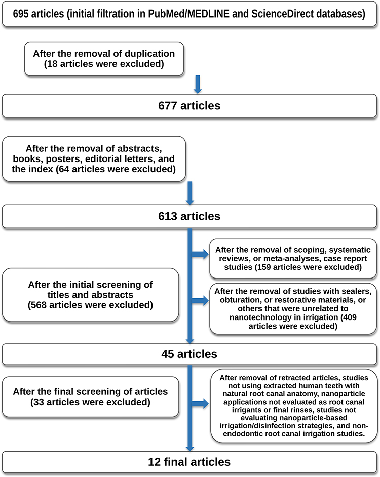 |
Figure 1 PRISMA flow diagram of study selection. |
Methodological Quality Appraisal
The quality of the included studies was assessed independently by two reviewers using a structured approach suitable for preclinical in vitro and ex vivo dental studies. The assessment considered sample selection, use of control groups, reporting of randomization or allocation, blinding where applicable, adequacy of nanoparticle characterization, completeness of outcome reporting, and appropriateness of statistical analysis. Any disagreements were resolved through discussion. Because of substantial methodological heterogeneity across nanoparticle platforms, activation protocols, study models, and outcome measures, no pooled quantitative analysis was performed. Findings were therefore summarized narratively as reported in the original studies. No data conversion or imputation was performed. Study selection was presented in a flow diagram, included study characteristics and findings were summarized in an integrated summary table, and reasons for exclusion of full-text studies were summarized in a supplementary table. This systematic review was not registered. No review protocol was prepared.
Results
A total of twelve original studies met the eligibility criteria and were included in this systematic review. Evidence was synthesized across three outcome domains: (i) antimicrobial and antibiofilm performance, (ii) dentin cleaning and hard-tissue/mechanical effects, and (iii) biocompatibility and regeneration-related biological outcomes. In most studies, extracted human teeth served as models for intracanal infection or biofilm, predominantly using Enterococcus faecalis as the test microorganism. The included studies evaluated nanoparticle-based strategies both as standalone irrigants/final rinses and as activation-assisted protocols, including diode-laser/photodynamic and sonic/ultrasonic approaches.
Overall, the included studies showed heterogeneous methodological quality. Common limitations included incomplete reporting of randomization and blinding, variability in nanoparticle characterization, and substantial diversity in activation protocols and outcome measures. The study-level assessment of methodological quality domains in the included studies is summarized in Supplementary Table S3.51–62
The included studies are summarized in a single integrated table (Table 1), which presents the study groups, model, and key findings across antimicrobial/antibiofilm, dentin-related/mechanical, and biologic/biocompatibility outcome domains. Across the included studies, nanoparticle-assisted protocols produced measurable antimicrobial and antibiofilm effects in extracted-tooth canal infection models, while dentin-related/mechanical and biologic outcomes were more variably reported.
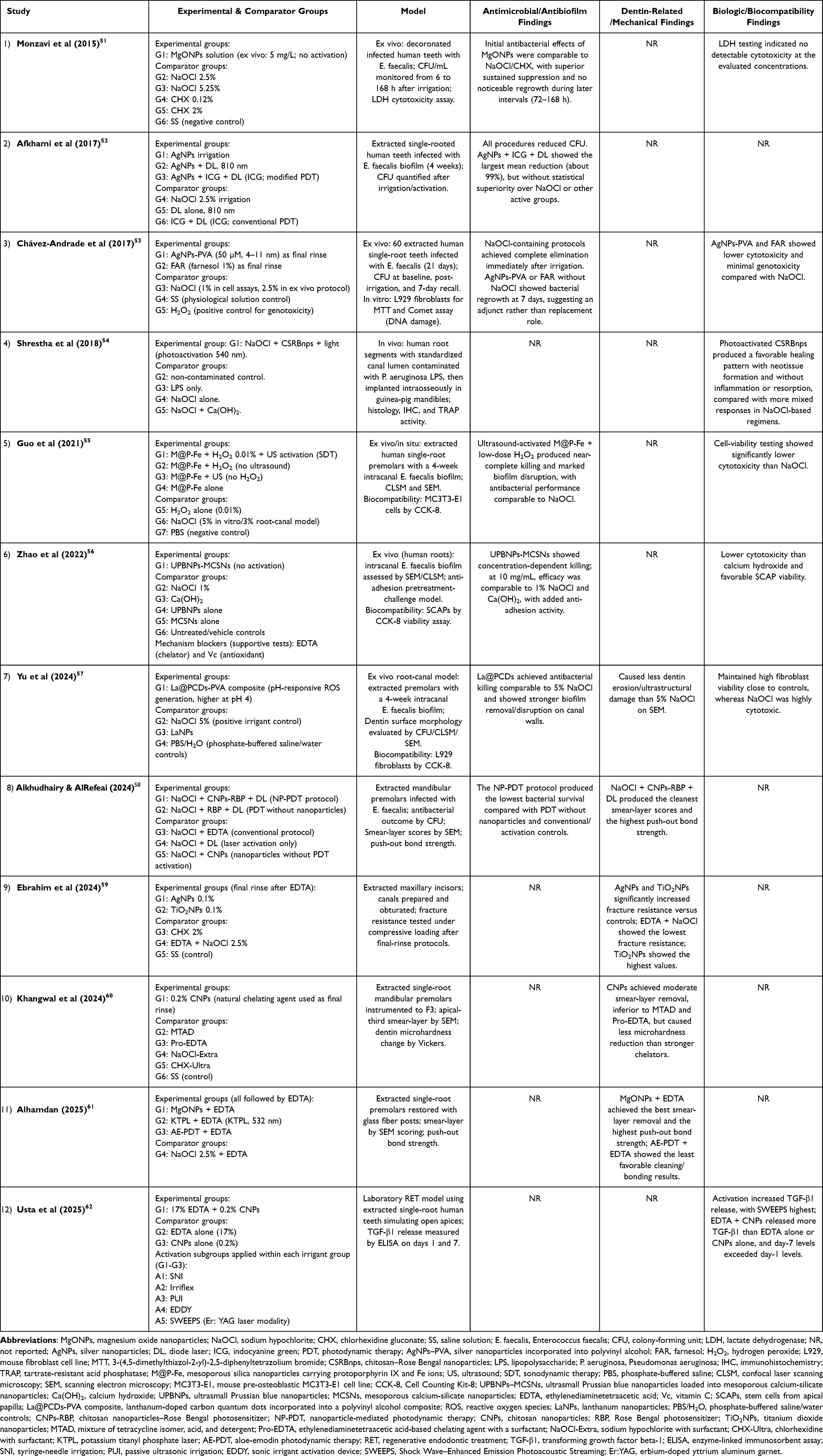 |
Table 1 Summary of Included Studies, Experimental and Comparator Groups, Models, and Key Findings Across Antimicrobial/Antibiofilm, Dentin-Related/Mechanical, and Biologic/Biocompatibility Outcomes |
Across the antimicrobial/antibiofilm studies (Table 1), all NP approaches produced measurable reductions in E. faecalis in extracted-tooth canal infection models, but the magnitude and consistency differed by platform and by whether an adjunct activation step was used. Light-mediated protocols were assessed using diode-laser–based strategies and PDT. Afkhami et al in 2017,52 reported that all tested groups reduced CFU, with the combined silver nanoparticles (AgNPs)–indocyanine green (ICG)–diode-laser protocol showing the highest mean reduction (≈99%), although it was not statistically superior to NaOCl or other activated groups. Alkhudhairy and AlRefeai in 2024,58 similarly reported that adding Chitosan nanoparticles-Rose Bengal photosensitizer (CNPs-RBP) to a NaOCl and diode-laser PDT protocol resulted in the lowest bacterial survival among their treatment arms.
A second group of studies evaluated catalytic or ROS-amplifying platforms in which antibiofilm effects were assessed with a trigger (eg., ultrasound/sonodynamic protocols or pH-responsive ROS generation). Guo et al55 reported near-complete killing and marked biofilm disruption when mesoporous silica nanoparticles carrying protoporphyrin IX (PpIX) and Fe ions (M@P-Fe) were combined with low-dose hydrogen peroxide (H2O2) and ultrasound (sonodynamic therapy), with antibacterial performance comparable to NaOCl in their tooth model. Yu et al57 reported that lanthanum-doped carbon quantum dots incorporated into a polyvinyl alcohol composite (La@PCDs-PVA) achieved antibacterial activity comparable to 5% NaOCl and demonstrated stronger biofilm removal/disruption on canal walls. Zhao et al in 2022,56 showed concentration-dependent antibiofilm activity for ultrasmall Prussian blue nanoparticles loaded into mesoporous calcium-silicate nanoparticles (UPBNPs-MCSNs), with performance at higher concentrations comparable to 1% NaOCl/Ca(OH)2 and an additional anti-adhesion effect in pretreatment-challenge experiments. Overall, Table 1 indicates consistent antibiofilm activity across NP systems, with activation/delivery steps frequently associated with greater reductions under the reported experimental conditions.
Within Table 1, dentin-related and mechanical outcomes included smear-layer removal, dentin microhardness changes, fracture resistance, push-out bond strength, and dentin surface preservation. The mechanical factors, such as fracture resistance and/or push-out bond strength, and cleaning efficacy, were investigated in multiple studies, so there was a clinical focus on preserving dentin and canal walls clean.
Two push-out tests analyzed the removal of smear layers and bonding to root dentin. Alhamdan61 reported that aloe-emodin-photodynamic therapy (AE-PDT) and EDTA had the worst cleaning and bonding findings, while magnesium oxide nanoparticles (MgONPs) and EDTA had the best smear-layer scores and the strongest push-out bond strength, especially in the cervix area. In a related study, Alkhudhairy and AlRefeai in 2024,58 found that NaOCl, CNPs-RBP, and diode laser (DL) produced the strongest push-out bond strength and the cleanest smear-layer scores when compared to NaOCl, EDTA, and other activation approaches in the same experimental setup.
Other studies emphasized the balance between cleaning and dentin preservation. Khangwal et al60 reported that 0.2% CNPs achieved moderate smear-layer removal while producing less reduction in microhardness compared with stronger chelators such as a mixture of tetracycline isomer, acid, and detergent (MTAD) and Pro-EDTA. Mechanical reinforcement was evaluated by Ebrahim et al,59 who reported that AgNPs or TiO2NPs used as final rinses significantly increased fracture resistance compared with control protocols, whereas the EDTA followed by NaOCl sequence showed the lowest fracture resistance. Finally, Yu et al57 reported dentin surface morphology outcomes on scanning electron microscopy (SEM), where La@PCDs caused less erosive/ultrastructural change than 5% NaOCl. Overall, Table 1 shows that dentin-related outcomes ranged from enhanced smear-layer removal and higher push-out bond strength to more conservative dentin effects, including lower microhardness reduction, reduced erosive surface changes, and improved fracture resistance in some models.
Within Table 1, biologic and biocompatibility evidence clustered into three main outcome types: in vivo healing responses after endotoxin contamination and disinfection, growth-factor release relevant to regenerative endodontics, and cytotoxicity/viability outcomes in mammalian cells.
Shrestha et al54 reported an in vivo model using human root segments contaminated with Pseudomonas aeruginosa lipopolysaccharide (P. aeruginosa LPS), where photoactivated chitosan–rose bengal nanoparticles (CRBnps) produced a favorable healing pattern (neotissue formation without inflammation or resorption) comparable to non-contaminated controls, while NaOCl-based regimens (± Ca(OH)2) showed mixed inflammatory/resorptive responses. Usta et al62 used a laboratory regenerative endodontic treatment (RET) model and reported that both irrigant composition and activation method influenced transforming growth factor beta-1 (TGF-β1) release: Shock Wave–Enhanced Emission Photoacoustic Streaming (SWEEPS) yielded the highest levels, followed by the EDDY system, a sonic irrigant activation device, and passive ultrasonic irrigation, and EDTA with CNPs consistently produced higher TGF-β1 than EDTA alone or CNPs alone, with higher release at day 7 than day 1.
Cell-based biocompatibility endpoints were reported for multiple NP strategies, also represented in Table 1. Chávez-Andrade et al reported that silver nanoparticles incorporated into polyvinyl alcohol (AgNPs-PVA) and farnesol (FAR) 1% showed lower cytotoxicity than NaOCl and minimal genotoxicity in the Comet assay after short exposure. Guo et al55 reported higher viability (lower cytotoxicity) for their M@P-Fe-based sonodynamic strategy compared with NaOCl in the mouse pre-osteoblastic MC3T3-E1 cell line (MC3T3-E1) cells. Monzavi et al in 201551 reported no detectable cytotoxicity for MgONPs at tested concentrations using a lactate dehydrogenase (LDH) assay. Yu et al57 reported that La@PCDs maintained high L929 fibroblast viability close to controls, whereas NaOCl was highly cytotoxic under their test conditions. Zhao et al56 reported favorable stem cells from the apical papilla (SCAP) viability for ultrasmall Prussian blue nanoparticles loaded into mesoporous calcium-silicate nanoparticles; no activation (UPBNPs–MCSNs) relative to Ca(OH)2. Collectively, Table 1 indicates that several NP platforms showing antimicrobial activity were also associated with more favorable cytocompatibility outcomes than NaOCl in corresponding cell assays, alongside regeneration-related findings such as dentin growth-factor release under activated irrigation conditions.
Figure 2 provides a conceptual overview of the representative nanoparticle platforms, activation approaches, mechanisms, and principal outcome domains discussed in this review. It links key mechanisms (membrane disruption, ROS/ion release, and biofilm/extracellular polymeric substances (EPS) destabilization) to outcomes across three domains: antimicrobial/antibiofilm efficacy, dentin cleaning/preservation, mechanical performance, and biocompatibility/regeneration-relevant effects. Overall, it illustrates NP irrigation as a multifunctional strategy where material design and activation jointly shape clinically relevant outcomes.
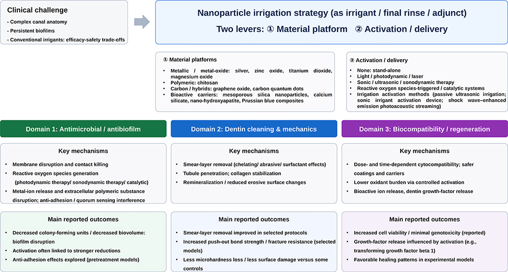 |
Figure 2 Conceptual summary of nanoparticle-assisted endodontic irrigation. |
Overview of Nanoparticles in Endodontic Irrigation
To understand why nanoparticles are increasingly being investigated as adjuncts in endodontic irrigation, it is essential first to examine their fundamental physicochemical properties and how these differ from conventional irrigation agents.
General Properties of Nanoparticles
Nanomaterials are defined as materials possessing at least one dimension within the 1–100 nm range.9,63 Their ultrasmall size, high surface area-to-mass ratio, and enhanced chemical reactivity enable interactions with biological systems at the subcellular level.64,65 The antimicrobial activity of NPs is typically explained by three principal mechanisms: oxidative stress induction, metal ion release, and non-oxidative interactions.9 Positively charged NPs can accumulate on negatively charged bacterial cell surfaces, destabilising the cell wall, increasing membrane permeability, and promoting leakage of intracellular contents.63,66
The ROS generation is a central pathway; ROS (including superoxide and hydroxyl radicals) can induce oxidative damage to lipids, proteins, and nucleic acids.9,11 This multi-targeted mode of action-affecting membranes, proteins, nucleic acids, and metabolism-may reduce the likelihood of resistance development relative to single-target antimicrobials.67
Why Nanoparticles are Promising for Endodontic Irrigation
In endodontics, the ongoing difficulty of eliminating intracanal biofilms using only traditional chemo-mechanical methods is what led to the study of NPs.3,65 Endodontic biofilms consist of approximately 10–15% bacterial cells embedded within an EPS matrix, which constitutes about 85–90% of the total composition. This matrix makes things more stable and helps fight off germs at the same time.64 E. faecalis is one of the most important organisms that has been linked to chronic illness.3
There is a chance that common irrigants such as NaOCl, CHX, and EDTA will break down quickly or only release active chemicals for a short time, which will limit the antibacterial effects that are kept.11 The NPs might be able to get around these problems by going deeper into dentinal tubules and biofilms. Additionally, they might have antibacterial effects that last longer because they are released in a controlled way.11 Their large surface area makes it easier for them to interact with microbial targets, but their very small size makes it easier to reach the complex canal structure.67
Evidence supporting specific systems includes reports that AgNPs show efficacy comparable to 5.25% NaOCl against E. faecalis.68 ZnO NPs have been reported to increase antibacterial activity and reduce bacterial adhesion to dentin walls by up to 95%.68 TiO2 NPs in irrigation solutions have been associated with increased fracture resistance of treated roots.68 MgONP solutions (5–10 mg/L) demonstrate antibacterial efficacy comparable to 5.25% NaOCl and 2% CHX against common endodontic pathogens.68 In addition, chitosan nanoparticles (CNPs) exhibit chelating capacity and can dissolve inorganic smear layer components.69,70 As final irrigants, 0.2% CNPs have shown smear layer removal comparable to 17% EDTA, while producing higher dentin microhardness and lower surface roughness.70 CNPs may also penetrate dentinal tubules and maintain antimicrobial effects for up to three months.71 Graphene oxide (GO) nanosheets have been reported to disrupt established biofilms by penetrating the EPS matrix.66,71
Nanoparticles in Endodontic Irrigation: Overview of Main Classes
Figure 3 illustrates the representative nanoparticle classes discussed in relation to endodontic irrigation.
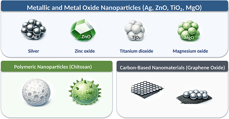 |
Figure 3 Representative nanoparticle classes in endodontic irrigation. |
Metallic and Metal Oxide Nanoparticles (Ag, ZnO, TiO2, MgO)
- Silver nanoparticles (AgNPs): The AgNPs, which are 1 to 100 nm in size, may get into the small spaces of root canals and dentinal tubules, which makes disinfection better.65,67 They kill bacteria by making ROS, breaking down DNA, and stopping adenosine triphosphate (ATP) synthesis.71 AgNPs work as well as 5.25% NaOCl against E. faecalis when used as irrigants.68 Long-term exposure may cause bacteria to adapt or become resistant, and AgNPs may change the color of dentin and be cytotoxic in a way that depends on their concentration.68,71
- Zinc oxide nanoparticles (ZnONPs): ZnONPs stop E. faecalis from forming biofilms and make it much harder for bacteria to stick to surfaces.66 As irrigants, they might cut down on how much bacteria stick to dentin walls by as much as 95%.68
- Titanium dioxide nanoparticles (TiO2NPs) are generally biocompatible and possess photocatalytic antimicrobial properties. Studies have shown that they make endodontically treated roots more resistant to breaking in irrigation solutions.68
- Magnesium oxide nanoparticles (MgONPs): Aqueous MgONP solutions (5–10 mg/L) demonstrate antibacterial efficacy comparable to 5.25% NaOCl and 2% CHX against Candida albicans (C. albicans), Staphylococcus aureus (S. aureus), and E. faecalis.68
Polymeric Nanoparticles (Chitosan)
Chitosan is a natural polysaccharide that kills bacteria by interacting with their membranes through electrostatic forces. This makes the membranes more permeable, which kills the cells.11,71 The CNPs have chelating properties that facilitate breaking down the inorganic smear layer components.69,70 As a final irrigant, 0.2% CNPs removed smear layers better than NaOCl and as well as 17% EDTA. CNPs had a higher microhardness (49.88 ± 2.34 Vickers hardness number (VHN)) than EDTA (45.04 ± 4.02 VHN) and a lower surface roughness (0.84 ± 0.23 μm) than EDTA (2.41 ± 0.09 μm). This means that they could be used instead of EDTA for final irrigation.70
Carbon-Based Nanomaterials (Graphene Oxide)
The GO nanosheets exhibit antibacterial properties through physical membrane disruption, commonly known as the “nano-knife” effect, and the generation of ROS.71,72 GO might disrupt established biofilms by penetrating the EPS matrix and hindering quorum sensing (QS).66,71 Some researchers think that silver nanoparticle-graphene oxide composites (AgNPs-GO composites) could be good alternatives to NaOCl for irrigation.68
Mechanisms of Antimicrobial Action of Nanoparticles
A significant feature of nanoparticle-assisted irrigation strategies is their ability to operate through multiple antimicrobial pathways rather than depending on a single chemical mechanism.
Physicochemical Basis of Activity
The physicochemical properties of NPs have a big effect on how well they kill bacteria or other microorganisms. Because they have a larger surface area, smaller particles are often more active because they can more easily come into contact with microbial targets. The 5 nm AgNPs, for instance, have been shown to have stronger effects.71 A positive surface charge increases the electrostatic attraction to bacterial cell envelopes that have a negative charge. This helps the bacteria stick to the surface, which can then damage and break down the cell membrane. The shape of the particles, the chemistry of their surfaces, and their composition all have a big effect on how well NPs kill bacteria and how they work in the body.6,71
Contact-Mediated Killing and Membrane Disruption
Direct contact is one of the main ways that NPs kill microbes. Positively charged NPs stick to the walls of bacterial cells, which can change the shape of the cell, make the membrane more permeable, and sometimes even break the membrane, letting the cell’s contents leak out.71 This mechanism is particularly relevant for cationic polymeric systems such as chitosan, where membrane interaction constitutes a primary route of bactericidal action.
Reactive Oxygen Species Generation and Oxidative Stress
The H2O2 and hydroxyl radicals (OH) are two examples of the ROS (ROS) that many NPs produce; these radicals cause oxidative stress and harm to the lipids, proteins, and nucleic acids of bacteria.71 The PDT accelerates the generation of ROS following irradiation, and other activation-dependent techniques are predicated on this mechanism, which is especially crucial for metallic and metal oxide NPs.6
Metal Ion Release
The antibacterial effect of metallic and metal oxide NPs may be amplified by the release of ions, such as silver (Ag⁺) and zinc (Zn2⁺), which disrupt metabolic processes, bind to proteins, and impede enzyme activity.71 This is why ion release is a good addition to contact-mediated and oxidative processes; it often makes these systems work on more than one target at a time.
Biofilm Penetration and Extracellular Polymeric Substance Disruption
Because they are so small, NPs can get into dentinal tubules and the extracellular polymeric substance (EPS) matrix. This could let them get to microbial niches that regular irrigants cannot reach.71,73 Additional evidence indicates that AgNPs may inhibit biofilm development by reducing EPS production, thereby compromising biofilm integrity.73
Inhibition of Quorum Sensing (QS)
The QS controls biofilm growth, pathogenicity, and coordinated behavior in a community. Some NPs, such as AgNPs and chitosan-based systems, can mess with this.71,73 So, antibiofilm effectiveness may come from both disrupting QS and killing bacteria directly.
Interaction of Nanoparticles with Dentin
Nanoparticle (NP)-based systems differ from traditional irrigants because they aim to preserve dentin structure, which is becoming a key goal in modern endodontics, not just eliminating bacteria.
Smear Layer Removal and Surface Morphology
It is very important to eliminate the smear layer so that disinfectants can work well and irrigants can get into dentinal tubules. The CNPs are chelating agents that are effective at getting rid of the smear layer while causing less erosion of the dentin than EDTA.70,74 This means that CNPs have been linked to less roughness on the surface after treatment than EDTA, which suggests that they protect the dentin microstructure better.70
Impact on the Exposed Collagen Matrix
Traditional irrigants can harm the organic parts of dentin. For example, NaOCl breaks down collagen, and EDTA can cause collagen to change shape in some situations. On the other hand, CNPs may stick to dentin collagen, which would make the collagen matrix stronger and less likely to break down, which could make it more resistant to fractures.71 In studies that compared the two, biosynthesized AgNPs were found to keep the structure of collagen better than NaOCl.75
Mechanical Properties and Microhardness
Irrigants may change the way dentin works mechanically, especially its microhardness. EDTA and NaOCl are often linked to lower microhardness because they cause demineralization and/or degradation of the organic matrix. The CNPs, on the other hand, are linked to higher microhardness values than EDTA, which means that less mineral is lost and demineralization is less violent.70,74
Dentin Discoloration
One clinically important drawback of AgNPs is that they can cause dentin to change color or turn black when silver sulfide precipitates are made.67,71 On the other hand, other nanoparticle systems, such as chitosan (CS), titanium dioxide (TiO2), and zinc oxide (ZnO), have not been linked to clinically significant discoloration very often.63,71
Chemical, Remineralisation, and Regenerative Effects
Some NP systems may offer extra benefits for endodontic results in addition to disinfection. Chitosan has been shown to help remineralization by making crystals form on dentin collagen.70 Nano-hydroxyapatite (nHAp) may get into dentinal tubules and block them, making it easier to seal them and add minerals back to them.11 In addition, bioactive glass (BG) and mesoporous calcium silicate nanoparticles (MCSNs) can release ions that create an antibacterial environment and promote apatite formation, aligning with regenerative and reparative objectives.71
How Nanoparticles Work with Tissues Around the Teeth
For clinical translation, antimicrobial performance must be considered alongside biologic compatibility and host-tissue responses.
Cytotoxicity to Fibroblasts and Oral Cells
Biocompatibility is critical for clinical translation, particularly for irrigants that may contact periapical tissues. The cytotoxicity of nanoparticle systems is formulation-dependent and may be influenced by particle size, surface charge, coating, dissolution behavior, concentration, exposure duration, and the target cell type.6,66,67,76,77 Accordingly, safety cannot be generalized across nanoparticle classes or even across preparations within the same class, and biologic interpretation should consider the physicochemical characteristics of each formulation.6,66,67
- Silver nanoparticles (AgNPs): Toxicity is frequently linked to the release of Ag⁺ ions and the generation of ROS, typically escalating with increased concentrations. Numerous studies demonstrate that optimizing formulations—by modifying particle size and surface characteristics—can enhance cytocompatibility at reduced doses, indicating that safety cannot be universally applied to all AgNP preparations.66,67
- Zinc oxide nanoparticles (ZnO NPs): Zinc oxide nanoparticles (ZnO NPs) are frequently regarded as less toxic to fibroblasts compared to AgNPs; however, outcomes may differ based on particle size, shape, and the specific cell types examined.76
- Iron oxide nanoparticles (IONPs): Numerous studies indicate that iron oxide nanoparticles (IONPs) exhibit low cytotoxicity and favorable biocompatibility, thereby endorsing their prospective applications—particularly in contexts that leverage magnetic activation or directed distribution.77
- Polymeric nanoparticles: They are often seen as safer options because they are generally more biocompatible and may reduce the harmful effects that can happen with metal-based materials, especially when they interact with ions. Still, you should not assume that something is biologically compatible just because of the base polymer composition. Realistic biological evaluation is crucial, as formulation-specific factors—such as crosslinking components, residual solvent traces, surface functionalization, and particle charge characteristics—can significantly influence cellular behavior and host tissue responses.6
Effects on Dental Pulp Stem Cells and Regenerative Potential
In regenerative endodontics, it is very important for disinfection methods to keep dental pulp stem cells (DPSCs) alive and help tissue heal.
- Nano-hydroxyapatite (nHAp): Because it is similar to natural apatite found in teeth and bone, nHAp is thought to be very biocompatible. It has also been linked to the production of reparative dentin.11,78
- Chitosan nanoparticles (CNPs): Chitosan-based systems are well known for being biocompatible and have been proposed as parts of disinfection methods that are compatible with regeneration. However, the results may vary depending on the molecular properties and formulation parameters.63,64
- Bioactive glass nanoparticles (BGNPs): BGNPs systems may help the development of dentin bridges with little inflammation and have been shown not to slow down DPSCs growth, which is in line with their high levels of bioactivity and ability to regenerate.11
Inflammatory Response and Host-Tissue Compatibility
In addition to direct cytotoxicity, nanoparticle formulations may influence inflammatory signaling and host-tissue responses that are relevant to periapical healing.
- Silver nanoparticles: Some models show that AgNPs may have anti-inflammatory effects by stopping the production of pro-inflammatory cytokines. However, these effects depend on the formulation and dosage.67
- CNPs: The antibacterial effects of chitosan may be lessened by clinically important “tissue inhibitors,” such as pulp-derived components and serum albumin. This shows how important it is to test settings that are very similar to those in body.66 Functionalized systems, exemplified by rose bengal–chitosan nanoparticles, are proposed to amalgamate antibacterial and anti-inflammatory attributes, encompassing lipopolysaccharide (LPS) inactivation and diminished inflammatory markers upon photoactivation.66
Discussion
Interpretation of the Findings
Overall, the included preclinical studies suggest that nanoparticle-assisted irrigation strategies may enhance antimicrobial and antibiofilm activity and, in some settings, support more favorable dentin-related or biologic outcomes than conventional approaches. However, the evidence remains heterogeneous across nanoparticle platforms, activation methods, study models, and reported endpoints, which limits direct comparison and precludes quantitative synthesis. Accordingly, the findings should be interpreted as promising but preliminary preclinical evidence rather than definitive support for routine clinical use.
Limitations and Safety Considerations
Although nanoparticle-based irrigation offers several potential advantages, its safety, possible toxicity, and clinical reliability must be carefully evaluated before clinical use. This review was also limited by restriction to English-language studies, use of two electronic databases, and substantial methodological heterogeneity across the included preclinical studies, which precluded meta-analysis and formal certainty assessment.
Nanotoxicology and Risk Assessment
Small size, high surface area, and greater reactivity are all things that make antimicrobial drugs work better. However, these same things can also make them more toxic, so each NP formulation needs to be carefully evaluated for risk.66 The endodontic failure rate, which is always between 18% and 26%, shows that we need better ways to disinfect. However, candidate nanomaterials need to be tested in conditions that really show how biofilms behave and how the root canal environment limits them.64,65 In addition, tissue-derived inhibitors such as serum albumin may alter nanoparticle behavior and antimicrobial performance, further emphasizing the need for ex vivo and in vivo models that better reflect clinical exposure conditions and biological complexity.66
Local and Systemic Adverse Effects
- Silver nanoparticles: Dentin discoloration and dose-dependent cytotoxicity to mammalian cells are the main problems. These are often linked to free Ag⁺ release and oxidative stress caused by ROS.66,67
- Systemic exposure considerations: NP biodistribution is size-dependent; very small particles may undergo rapid clearance, whereas larger particles may be sequestered by the reticuloendothelial system. Accordingly, design must balance canal penetration and antimicrobial efficacy against the need to minimise systemic exposure risk, particularly in the context of potential apical extrusion.64
Approaches to Toxicity Assessment
Although many studies report promising in vitro findings, robust translation requires clinically relevant models and in vivo confirmation, particularly where irrigants may contact periapical tissues.66 The current evidence base is limited by inconsistencies in experimental design, variable reporting of NP synthesis/characterisation, and a relative lack of clinical data, contributing to reduced certainty of conclusions.6 Comprehensive physicochemical characterization, particularly particle size distribution, surface charge, morphology, coating, and dissolution kinetics, is essential for interpreting both efficacy and biocompatibility outcomes and for enabling meaningful comparison across studies.3,64
Design Strategies to Reduce Nanotoxicity
Multiple “safer-by-design” strategies have been proposed to optimise the balance between antimicrobial efficacy and host compatibility.
- Formulation engineering: For example, positively charged ~9 nm AgNPs have shown strong antibacterial activity while remaining cytocompatible to fibroblasts, illustrating how tuning particle size and surface charge can improve the benefit–risk profile.66
- Encapsulation and controlled release: Mesoporous or inorganic carriers (eg., silica coatings) can encapsulate AgNPs to support sustained ion release while reducing acute cytotoxicity compared with free AgNPs.66,67
- Polymeric nanoparticle platforms: Polymeric systems may offer a relative safety advantage by enabling controlled, sustained delivery and reducing reliance on high-concentration bolus exposure, although they still require rigorous evaluation due to formulation-dependent effects.6
- Activation-dependent systems: The PDT is one method that uses light to stimulate NPs to make more ROS. This allows for spatial and temporal control of antimicrobial effects and may reduce host-tissue exposure when done correctly.11,79
Clinical Implications and Future Directions
Although several included studies reported favorable antimicrobial, dentin-related, or biologic findings under controlled experimental conditions, the current evidence base remains limited to preclinical in vitro, ex vivo, and laboratory/in vivo models.51–62 Broader literature also highlights formulation variability, incomplete protocol standardization, and the absence of clinical trials as major barriers to clinical translation.3,6,8,66 Accordingly, nanoparticle-assisted irrigation is not yet established in routine clinical practice. At present, these approaches are more reasonably viewed as adjunctive strategies, such as final-rinse or activation-assisted protocols, rather than direct replacements for conventional irrigants.51,53,54,57–62 This broader concept is also supported by non-nanoparticle activation-assisted irrigation studies, such as the SMART approach, which have reported enhanced microbial reduction and root canal cleaning with synergistic irrigant–activation protocols.80 Further standardized preclinical investigations and well-designed animal and clinical studies are required to define optimal formulation, dosage, delivery, safety, and true clinical benefit.3,6,8,66
Conclusions
Across the included preclinical studies, nanoparticle-assisted irrigation strategies showed promising antimicrobial and antibiofilm effects, particularly in Enterococcus faecalis root canal models. Findings related to smear-layer removal, dentin preservation, and mechanical outcomes were more variable across studies. In addition, biologic and regeneration-related observations, including growth-factor release, cytocompatibility, and healing-related responses, remain preliminary and are based on limited experimental evidence.
Because the current evidence is derived mainly from heterogeneous in vitro, ex vivo, and laboratory/in vivo models, nanoparticle-assisted systems should be regarded as promising adjunctive strategies rather than established clinical alternatives to conventional irrigants. At present, the evidence is strongest for antimicrobial and antibiofilm potential in controlled models, more mixed for dentin-related outcomes, and still preliminary for regenerative relevance, long-term substantivity, safety, and clinical superiority over established irrigants. Further standardized preclinical investigations and well-designed in vivo and clinical studies are required before routine clinical implementation can be recommended.
Data Sharing Statement
The data supporting the findings of this review are available from the corresponding author upon reasonable request. No analytic code was generated for this systematic review.
Funding
There is no funding to report.
Disclosure
The authors report no conflicts of interest in this work.
References
1. Mamat R, Nik Abdul Ghani NR. The complexity of the root canal anatomy and its influence on root canal debridement in the apical region: a review. Cureus. 2023;15(11):e49024. doi:10.7759/cureus.49024
2. Neelakantan P, Romero M, Vera J, et al. Biofilms in endodontics-current status and future directions. Int J Mol Sci. 2017;18(8). doi:10.3390/ijms18081748.
3. Oncu A, Huang Y, Amasya G, Sevimay FS, Orhan K, Celikten B. Silver nanoparticles in endodontics: recent developments and applications. Restor Dent Endod. 2021;46(3):e38. doi:10.5395/rde.2021.46.e38
4. Zou X, Zheng X, Liang Y, et al. Expert consensus on irrigation and intracanal medication in root canal therapy. Int J Oral Sci. 2024;16(1):23. doi:10.1038/s41368-024-00280-5
5. Gomes B, Aveiro E, Kishen A. Irrigants and irrigation activation systems in Endodontics. Brazilian Dental J. 2023;34(4):1–19. doi:10.1590/0103-6440202305577
6. Orozco MFS, IdA M, Gonzalez OL, et al. Antimicrobial polymeric nanoparticles in endodontics: a systematic review. J Nanotechnol. 2025;2025(1):3896901. doi:10.1155/jnt/3896901
7. Jeevanandam J, Barhoum A, Chan YS, Dufresne A, Danquah MK. Review on nanoparticles and nanostructured materials: history, sources, toxicity and regulations. Beilstein J. Nanotechnol. 2018;9:1050–1074. doi:10.3762/bjnano.9.98
8. Wong J, Zou T, Lee AHC, Zhang C. The potential translational applications of nanoparticles in endodontics. Int J Nanomed. 2021;16:2087–2106. doi:10.2147/IJN.S293518
9. Wang L, Hu C, Shao L. The antimicrobial activity of nanoparticles: present situation and prospects for the future. Int J Nanomed. 2017;12:1227–1249. doi:10.2147/IJN.S121956
10. Huang CS, Hsiao CH, Chang YC, et al. A novel endodontic approach in removing smear layer using nano and submicron diamonds with intracanal oscillation irrigation. Nanomaterials. 13(10).
11. Mierzejewska ŻA, Rusztyn B, Łukaszuk K, Borys J, Borowska M, Antonowicz B. The latest advances in the use of nanoparticles in endodontics. Appl Sci. 2024;14(17):7912. doi:10.3390/app14177912
12. Bo D, Kayombo CM. Effect of nanosilver gel, chlorhexidine gluconate, and camphorated phenol on enterococcus faecalis biofilm. Int Scholarly Res Notices. 2014;2014:380278. doi:10.1155/2014/380278
13. Barreras US, Méndez FT, Martínez RE, Valencia CS, Rodríguez PR, Rodríguez JP. Chitosan nanoparticles enhance the antibacterial activity of chlorhexidine in collagen membranes used for periapical guided tissue regeneration. Mater Sci Eng C Mater Biol Appl. 2016;58:1182–1187. doi:10.1016/j.msec.2015.09.085
14. Zhang FH, Li M, Wei ZJ, Zhao B. The effect of a combined nanoparticulate/calcium hydroxide medication on the biofilm of Enterococcus faecalis in starvation phase. Shanghai Kou Qiang Yi Xue. 2016;25(1):11–15.
15. Wu D, Fan W, Kishen A, Gutmann JL, Fan B. Evaluation of the antibacterial efficacy of silver nanoparticles against Enterococcus faecalis biofilm. J Endodontics. 2014;40(2):285–290. doi:10.1016/j.joen.2013.08.022
16. Chávez-Andrade GM, Tanomaru-Filho M, Basso Bernardi MI, de Toledo Leonardo R, Faria G, Guerreiro-Tanomaru JM. Antimicrobial and biofilm anti-adhesion activities of silver nanoparticles and farnesol against endodontic microorganisms for possible application in root canal treatment. Arch Oral Biol. 2019;107:104481. doi:10.1016/j.archoralbio.2019.104481
17. Jowkar Z, Hamidi SA, Shafiei F, Ghahramani Y. The effect of silver, zinc oxide, and titanium dioxide nanoparticles used as final irrigation solutions on the fracture resistance of root-filled teeth. Clin Cosmet Investig Dent. 2020;12:141–148. doi:10.2147/CCIDE.S253251
18. Yin U IX, Xu A, Chen VW, Zhang KJ, Chu MY, Chu CH. Use of antimicrobial nanoparticles for the management of dental diseases. Nanomaterials. 2025;15(3). doi:10.3390/nano15030209
19. Shrestha S, Torneck CD, Kishen A. Dentin conditioning with bioactive molecule releasing nanoparticle system enhances adherence,viability,and differentiation of stem cells from apical papilla. J Endodontics. 2016;42(5):717–723. doi:10.1016/j.joen.2016.01.026
20. Akbari T, Pourhajibagher M, Hosseini F, et al. The effect of indocyanine green loaded on a novel nano-graphene oxide for high performance of photodynamic therapy against Enterococcus faecalis. Photodiagn Photodyn Ther. 2017;20:148–153. doi:10.1016/j.pdpdt.2017.08.017
21. Rosa RAD, Santini MF, Figueiredo JAP, et al. Effectiveness of photodynamic therapy associated with irrigants over two biofilm models. Photodiagn Photodyn Ther. 2017;20:169–174. doi:10.1016/j.pdpdt.2017.10.003
22. Souza MA, Lima G, Pazinatto B, Bischoff KF, Palhano HS, Cecchin D. Evaluation of antimicrobial activity of association of chlorhexidine to photosensitizer used in photodynamic therapy in root canals infected by Enterococcus faecalis. Photodiagn Photodyn Ther. 2017;19:170–174. doi:10.1016/j.pdpdt.2017.06.007
23. Batinić M, Ročan M, Budimir A, Anić I, Bago I. Comparison of final disinfection protocols using antimicrobial photodynamic therapy and different irrigants after single-file reciprocating instrumentation against intracanal bacterial biofilm - An in vitro study. Photodiagn Photodyn Ther. 2018;24:153–157. doi:10.1016/j.pdpdt.2018.10.006
24. Bukhari S, Kim D, Liu Y, Karabucak B, Koo H. Novel endodontic disinfection approach using catalytic nanoparticles. J Endodontics. 2018;44(5):806–812. doi:10.1016/j.joen.2017.12.003
25. Daood U, Parolia A, Elkezza A, et al. An in vitro study of a novel quaternary ammonium silane endodontic irrigant. Dental Materials. 2019;35(9):1264–1278. doi:10.1016/j.dental.2019.05.020
26. Ioannidis K, Niazi S, Mylonas P, Mannocci F, Deb S. The synthesis of nano silver-graphene oxide system and its efficacy against endodontic biofilms using a novel tooth model. Dent. Mater. 2019;35(11):1614–1629. doi:10.1016/j.dental.2019.08.105
27. Li FC, Borkar S, Ramachandran A, Kishen A. Novel activated microbubbles-based strategy to coat nanoparticles on root canal dentin: fluid dynamical characterization. J Endodontics. 2019;45(6):797–802. doi:10.1016/j.joen.2019.02.011
28. Naseri M, Eftekhar L, Gholami F, Atai M, Dianat O. The effect of calcium hydroxide and nano-calcium hydroxide on microhardness and superficial chemical structure of root canal dentin: an ex vivo study. J Endodontics. 2019;45(9):1148–1154. doi:10.1016/j.joen.2019.06.002
29. Savitha A, SriRekha A, Vijay R, Ashwija C, Jaykumar C, Jaykumar T. An in vivo comparative evaluation of antimicrobial efficacy of chitosan, chlorhexidine gluconate gel and their combination as an intracanal medicament against Enterococcus faecalis in failed endodontic cases using real time polymerase chain reaction (qPCR). Saudi Dent. J. 2019;31(3):360–366. doi:10.1016/j.sdentj.2019.03.003
30. Daood U, Parolia A, Matinlinna J, Yiu C, Ahmed HMA, Fawzy A. Properties of a modified quaternary ammonium silane formulation as a potential root canal irrigant in endodontics. Dental Materials. 2020;36(12):e386–e402. doi:10.1016/j.dental.2020.09.008
31. Marinković J, Ćulafić DM, Nikolić B, et al. Antimicrobial potential of irrigants based on essential oils of Cymbopogon martinii and Thymus zygis towards in vitro multispecies biofilm cultured in ex vivo root canals. Arch Oral Biol. 2020;117:104842. doi:10.1016/j.archoralbio.2020.104842
32. Daood U, Bapat RA, Sidhu P, et al. Antibacterial and antibiofilm efficacy of k21-E in root canal disinfection. Dent. Mater. 2021;37(10):1511–1528. doi:10.1016/j.dental.2021.08.001
33. Hussein H, Kishen A. Engineered Chitosan-based Nanoparticles Modulate Macrophage-Periodontal Ligament Fibroblast Interactions in Biofilm-mediated Inflammation. J Endodontics. 2021;47(9):1435–1444. doi:10.1016/j.joen.2021.06.017
34. Singh K, Ali A, Shrestha A, Magalhaes M, Kishen A. Assessing macrophage polarization in nanoparticle-guided wound repair using a lipopolysaccharide contaminated intraosseous model. J Endodontics. 2022;48(1):109–116. doi:10.1016/j.joen.2021.09.011
35. Minhaco V, Maquera Huacho PM, Mancim Imbriani MJ, et al. Improving antimicrobial activity against endodontic biofilm after exposure to blue light-activated novel curcumin nanoparticle. Photodiagn Photodyn Ther. 2023;42:103322. doi:10.1016/j.pdpdt.2023.103322
36. Teimoory N, Yegane-Sefidan F, Nouri R, Erfanparast L, Rezai Y, Vatandoost M. Antibacterial behavior of silver diamine fluoride, sodium hypochlorite and ozone gel on enterococcus faecalis in root canal of deciduous teeth. Open Dent J. 2023;10:17.
37. Zhang J, Neupane N, Dahal PR, et al. Antibiotic-loaded boron nitride nanoconjugate with strong performance against planktonic bacteria and biofilms. ACS Appl. Bio Mater. 2023;6(8):3131–3142. doi:10.1021/acsabm.3c00247
38. Jafari Semnani S, Moghadam KN, Jafari Z, Chiniforush N. Comparative effects of the conventional, ultrasonic, and laser-activated irrigation on penetration depth of three photosensitizers in the root canal system. Photodiagn Photodyn Ther. 2024;49:104286. doi:10.1016/j.pdpdt.2024.104286
39. Selvaraj K, Venkatesan LS, Ganapathy D, Sathishkumar P. Treatment of dental biofilm-forming bacterium Streptococcus mutans using tannic acid-mediated gold nanoparticles. Microb Pathogenesis. 2024;189:106568. doi:10.1016/j.micpath.2024.106568
40. Ci H, Xin B, Hubbard AW, et al. Investigation of endodontic disinfection by combination of laser and nanobubble technology. J Endodontics. 2025;51(12):1803–1813. doi:10.1016/j.joen.2025.08.016
41. Hu P, Chen H, Qian C, et al. Antimicrobial and anti-biofilm activity of dichlorophen-functionalized gold nanoparticles against carbapenem-resistant enterobacteriaceae. Int J Nanomed. 2025;20:10255–10277. doi:10.2147/IJN.S532807
42. Hwang J, Askar M, Smoczer C, Young L, Ferracciolo J, Paurazas S. Cytotoxicity and genotoxicity of triton all-in-one irrigation solution: an invitro study. J Endodontics. 2026;52(4):632–637. doi:10.1016/j.joen.2025.11.002
43. Ismael M, Edwin M, Juliah K. Biogenic synthesis of silver nanoparticles using Sida cuneifolia leaf extract for enhanced antibacterial, cytotoxic, and anti-biofilm activities. Biotechnology Notes. 2025;6:196–208. doi:10.1016/j.biotno.2025.07.003
44. Jouhar R, Halim MS, Quadri SA, Ahmed MA, Shah F. Antimicrobial efficacy of methylene blue-functionalized graphene oxide-mediated photodynamic therapy against Enterococcus faecalis in root canal disinfection. J Taibah Univ Sci. 2025;20(4):558–567. doi:10.1016/j.jtumed.2025.07.012
45. Kaukab A, Nekkanti S. Antimicrobial efficacy of intracanal medicaments incorporated with nanoparticles in primary teeth: an in vitro study. The Scientific World J. 2025;2025:5182716. doi:10.1155/tswj/5182716
46. Pradeep M, Cecil A, Nidhita S, Rajakumar G, Thirumalaivasan N. Exploring plant-derived carbon dots: dual-functional nanomaterials for endodontic infections and enhanced bio-imaging. J. Oral Biol. Craniofac. Res. 2025;15(2025):1408–1414. doi:10.1016/j.jobcr.2025.08.030
47. Rajan B, Abdelmoneim D, Salem AS, et al. Nanoencapsulation, biocompatibility and antibiofilm properties of chitosan/nisin Z spheres. Arch Oral Biol. 2025;173:106193. doi:10.1016/j.archoralbio.2025.106193
48. Rao MH, Rajkumar K, Pavithra G, et al. Antibiofilm efficiency of silver and copper nanoparticle incorporated calcium hydroxide as an intracanal medicament: an in vitro study. J. Oral Biol. Craniofac. Res. 2025;15(2):319–324. doi:10.1016/j.jobcr.2025.01.020
49. Silva E, da Silva DVF, Massa GDS, et al. Improved ROOT CANAL DISINFECTION THROUGH EXTENDED SODIUM HYPOCHLORITE EXPOSURE AND RENEWAL AFTER PREPARATION PROCEdures. J Endodontics. 2026;52(3):451–457. doi:10.1016/j.joen.2025.11.023
50. Xu X, Wang P, Tong F, et al. Polydopamine-coated magnetic nanoplatform for magnetically guided penetration and enhanced antibacterial efficacy in root canal biofilm elimination. Polymers. 17(10).
51. Monzavi A, Eshraghi S, Hashemian R, Momen-Heravi F. In vitro and ex vivo antimicrobial efficacy of nano-MgO in the elimination of endodontic pathogens. Clin. Oral Investig. 2015;19(2):349–356. doi:10.1007/s00784-014-1253-y
52. Afkhami F, Akbari S, Chiniforush N. Entrococcus faecalis elimination in root canals using silver nanoparticles,photodynamic therapy,diode laser, or laser-activated nanoparticles: an in vitro study. J Endodontics. 2017;43(2):279–282. doi:10.1016/j.joen.2016.08.029
53. Chávez-Andrade GM, Tanomaru-Filho M, Rodrigues EM, et al. Cytotoxicity, genotoxicity and antibacterial activity of poly(vinyl alcohol)-coated silver nanoparticles and farnesol as irrigating solutions. Arch Oral Biol. 2017;84:89–93. doi:10.1016/j.archoralbio.2017.09.028
54. Shrestha A, Friedman S, Torneck CD, Kishen A. Bioactivity of photoactivated functionalized nanoparticles assessed in lipopolysaccharide-contaminated root canals in vivo. J Endodontics. 2018;44(1):104–110. doi:10.1016/j.joen.2017.08.021
55. Guo J, Xu Y, Liu M, et al. An MSN-based synergistic nanoplatform for root canal biofilm eradication via Fenton-enhanced sonodynamic therapy. J Mat Chem B. 2021;9(37):7686–7697. doi:10.1039/D1TB01031J
56. Zhao X, Wang Y, Zhu T, et al. Mesoporous calcium-silicate nanoparticles loaded with prussian blue promotes enterococcus faecalis ferroptosis-like death by regulating bacterial redox pathway ROS/GSH. Int J Nanomed. 2022;17:5187–5205. doi:10.2147/IJN.S382928
57. Yu L, Zhang C, Yang J, Li L. A novel ph-responsive nano-sized lanthanum-doped polyvinyl alcohol-carbon quantum dot composite for root canal irrigation. Int J Nanomed. 2024;19:11343–11356. doi:10.2147/IJN.S475872
58. Alkhudhairy F, AlRefeai MH. Chitosan nanoparticles, Rose Bengal-chitosan activated photodynamic therapy as final irrigant on pushout bond strength, smear layer removal efficacy and antibacterial effectiveness against E.faecalis. Photodiagn Photodyn Ther. 2024;49:104316. doi:10.1016/j.pdpdt.2024.104316
59. Ebrahim MI, Hadhoud FM, Alqarni AA, Al Harthi SM, Shawli HT. Influence of final irrigation with titanium dioxide and silver nanoparticles on the fracture resistance of endodontically treated roots. Saudi Dent. J. 2024;36(12):1549–1552. doi:10.1016/j.sdentj.2024.10.004
60. Khangwal M, Solanki R, Rahman H, Vinay S, Bagde N, Bagde N. Effectiveness of chitosan nanoparticles, and novel chemical irrigants with surfactant on smear layer removal and microhardness alteration. J. Oral Biol. Craniofac. Res. 2024;14(5):578–584. doi:10.1016/j.jobcr.2024.06.005
61. Alhamdan MM. Canal disinfectants: potassium titanyl phosphate laser, magnesium oxide nanoparticles, and aloe-emodin PDT on smear layer removal and bond strength of glass fiber post to root dentin. Photodiagn Photodyn Ther. 2025;53:104615. doi:10.1016/j.pdpdt.2025.104615
62. Usta SN, Avcı E, Oktay AN, Keskin C. Combination of chitosan nanoparticles, edta, and irrigation activation enhances tgf-β1 release from dentin: a laboratory study. J Endodontics. 2025;51(8):1081–1087. doi:10.1016/j.joen.2025.04.011
63. Raura N, Garg A, Arora A, Roma M. Nanoparticle technology and its implications in endodontics: a review. Biomater Res. 2020;24(1):21. doi:10.1186/s40824-020-00198-z
64. Manojkanna K, Chandana CS. Nanoparticles in endodontics - a review. J Adv. Pharm. Educ. Res. 2017;7(2):58–60.
65. Yahya EM, Jamel RS. Nanoparticles as endodontic irrigation: an update overview. Al-Rafidain Dent J. 2023;23:37–52. doi:10.33899/rdenj.2023.133586.01161
66. Shrestha A, Kishen A. Antibacterial Nanoparticles in Endodontics: a Review. J Endodontics. 2016;42(10):1417–1426. doi:10.1016/j.joen.2016.05.021
67. Afkhami F, Forghan P, Gutmann JL, Kishen A. Silver nanoparticles and their therapeutic applications in endodontics: a narrative review. Pharmaceutics. 15(3). doi:10.3390/pharmaceutics15030715
68. Sharifi R, Vatani A, Sabzi A, Safaei M. A narrative review on application of metal and metal oxide nanoparticles in endodontics. Heliyon. 2024;10(15):e34673. doi:10.1016/j.heliyon.2024.e34673
69. A DC-P, Bramante CM, Duarte MA, et al. Chelating and antibacterial properties of chitosan nanoparticles on dentin. Restor Dent Endod. 2015;40(3):195–201. doi:10.5395/rde.2015.40.3.195
70. Ratih Diatri N, Enggardipta Raras A, Kartikaningtyas Aqilla T. The effect of chitosan nanoparticle as a final irrigation solution on the smear layer removal, micro-hardness and surface roughness of root canal dentin. Open Dent J. 2020;14.
71. Capuano N, Amato A, Dell’Annunziata F, et al. Nanoparticles and their antibacterial application in endodontics. Antibiotics. 2023;12(12):1690. doi:10.3390/antibiotics12121690
72. Yin Z IX, Zhao J, Mei IS, Li ML, Chu Q, Chu CH. The antibacterial mechanism of silver nanoparticles and its application in dentistry. Int J Nanomed. 2020;15:2555–2562. doi:10.2147/IJN.S246764
73. Pérez-Sáenz MG, Martínez-Martínez RE, Zaragoza-Contreras EA, et al. Antibacterial and anti-adherence efficacy of silver nanoparticles against endodontic biofilms: an in vitro and ex vivo study. Pharmaceutics. 2025;17(7). doi:10.3390/pharmaceutics17070831.
74. Wakas H, Al-Zaka IM. Effectiveness of continuous irrigating solution on the microhardness of root dentin. Mustansiria Dentl J. 2025;21(2):12–24.
75. Chandran N, Ramesh S, Haridas R, Kamath A, Kader A, Maheesan K. Pre- and post-irrigation with biosynthesized and chemically synthesized silver nano-particles: a comparative analysis of dentin micro-hardness, surface roughness, and chemical changes. J. Stomatol. 2025;78:21–31. doi:10.5114/jos.2025.148391
76. Afkhami F, Chen Y, Walsh LJ, Peters OA, Xu C. Application of Nanomaterials in Endodontics. BME Frontiers. 2024;5:0043. doi:10.34133/bmef.0043
77. Al-Mustwfi ES, Al-Huwaizi HF. Evaluation of microhardness in conservative root dentin treatment techniques after irrigation with iron oxide nanoparticles delivered with an external magnetic field. 2025;15(14):7728.
78. Zakrzewski W, Dobrzyński M, Zawadzka-Knefel A, et al. Nanomaterials Application in Endodontics. Materials. 2021;14(18). doi:10.3390/ma14185296.
79. Kannan KP, Gunasekaran V, Sreenivasan P, Sathishkumar P. Recent updates and feasibility of nanodrugs in the prevention and eradication of dental biofilm and its associated pathogens-A review. Journal of Dentistry Apr. 2024;143:104888. doi:10.1016/j.jdent.2024.104888
80. Foroughi M, Abolmaali S, Abedi H, Ravenel T. Enhancing endodontic outcomes with the synergistic microbicidal and activated root-cleansing technique (smart): a novel approach to root canal irrigation. Medicina. 2025;61(5). doi:10.3390/medicina61050874
 © 2026 The Author(s). This work is published and licensed by Dove Medical Press Limited. The
full terms of this license are available at https://www.dovepress.com/terms
and incorporate the Creative Commons Attribution
- Non Commercial (unported, 4.0) License.
By accessing the work you hereby accept the Terms. Non-commercial uses of the work are permitted
without any further permission from Dove Medical Press Limited, provided the work is properly
attributed. For permission for commercial use of this work, please see paragraphs 4.2 and 5 of our Terms.
© 2026 The Author(s). This work is published and licensed by Dove Medical Press Limited. The
full terms of this license are available at https://www.dovepress.com/terms
and incorporate the Creative Commons Attribution
- Non Commercial (unported, 4.0) License.
By accessing the work you hereby accept the Terms. Non-commercial uses of the work are permitted
without any further permission from Dove Medical Press Limited, provided the work is properly
attributed. For permission for commercial use of this work, please see paragraphs 4.2 and 5 of our Terms.