Back to Journals » International Journal of Nanomedicine » Volume 19
Advancing Tissue Damage Repair in Geriatric Diseases: Prospects of Combining Stem Cell-Derived Exosomes with Hydrogels
Authors Shi L, Zhou Y, Yin Y, Zhang J, Chen K, Liu S, Chen P ![]() , Jiang H, Liu J, Wu Y
, Jiang H, Liu J, Wu Y ![]()
Received 22 December 2023
Accepted for publication 19 April 2024
Published 29 April 2024 Volume 2024:19 Pages 3773—3804
DOI https://doi.org/10.2147/IJN.S456268
Checked for plagiarism Yes
Review by Single anonymous peer review
Peer reviewer comments 2
Editor who approved publication: Professor Jie Huang
Ling Shi,1 Yunjun Zhou,2 Yongkui Yin,1 Jin Zhang,3 Kaiyuan Chen,1 Sen Liu,1 Peijian Chen,1 Hua Jiang,2 Jieting Liu,1 Yan Wu1
1College of Life Science, Mudanjiang Medical University, Mudanjiang, 157000, People’s Republic of China; 2The Affiliated Hongqi Hospital, Mudanjiang Medical University, Mudanjiang, 157000, People’s Republic of China; 3Clinical Laboratory, Zhejiang Medical & Health Group Quzhou Hospital, Quzhou, 324004, People’s Republic of China
Correspondence: Yan Wu; Jieting Liu, College of Life Science, Mudanjiang Medical University, 3 Tong Xiang Street, Ai Min District, Mudanjiang, 157000, People’s Republic of China, Tel +86-0453-6984647, Email [email protected]; [email protected]
Abstract: Geriatric diseases are a group of diseases with unique characteristics related to senility. With the rising trend of global aging, senile diseases now mainly include endocrine, cardiovascular, neurodegenerative, skeletal, and muscular diseases and cancer. Compared with younger populations, the structure and function of various cells, tissues and organs in the body of the elderly undergo a decline as they age, rendering them more susceptible to external factors and diseases, leading to serious tissue damage. Tissue damage presents a significant obstacle to the overall health and well-being of older adults, exerting a profound impact on their quality of life. Moreover, this phenomenon places an immense burden on families, society, and the healthcare system.In recent years, stem cell-derived exosomes have become a hot topic in tissue repair research. The combination of these exosomes with biomaterials allows for the preservation of their biological activity, leading to a significant improvement in their therapeutic efficacy. Among the numerous biomaterial options available, hydrogels stand out as promising candidates for loading exosomes, owing to their exceptional properties. Due to the lack of a comprehensive review on the subject matter, this review comprehensively summarizes the application and progress of combining stem cell-derived exosomes and hydrogels in promoting tissue damage repair in geriatric diseases. In addition, the challenges encountered in the field and potential prospects are presented for future advancements.
Keywords: exosomes, biomaterials, hydrogels, geriatric diseases, injury repair
Introduction
According to the recently released UN report, World Population Prospects 2022, the total global population has reached 8 billion. Increasing life expectancy and declining fertility rates are expected to intensify the aging problem in the global population. As age increases, a number of factors such as inadequate secretion of hormone levels and organ perfusion in the body lead to a gradual decline in physiological functions in the elderly. These include vascular sclerosis, neuronal degeneration, decreased skin elasticity, and reduced bone density. These physiological changes affect the overall health status and the ability to repair tissue damage in the elderly.1 According to the immunological theory of aging, there is a decline observed in the immune response against exogenous antigens in individuals as they age,2 while the immune response against autoantigens increases. Thus, the elderly population is susceptible to diseases with unique pathological characteristics such as diverse pathogenesis, severe pathological features, and greater variability.3 The inflammation, reduced ability of angiogenesis and matrix remodeling lead to prolonged tissue regeneration and poor repair.4 Tissue regeneration and repair in geriatric diseases are still predominantly reliant on conventional approaches worldwide, with limited remarkable advancements achieved to date. As a result, various geriatric diseases continue to pose a great threat to the life and health of the elderly population (Figure 1).
 |
Figure 1 Geriatric diseases are mainly associated with five major factors: inadequate hormone levels in the organism, inadequate organ perfusion, reduced immune response against exogenous antigens, lack of exercise, and Emotional factor, characterized by diverse causes, severe pathological features, and large disease variability. Therefore, it becomes a great threat to the life and health of the elderly population. (Image created with BioRender.com). |
With the rapid development of regenerative medicine, exosomes derived from stem cells have gradually become important biological materials to promote tissue repair and regeneration. Exosomes are membranous vesicles with a 30–100 nm diameter containing bioactive molecules including nucleic acids, lipids, and proteins.Notably, exosomes are readily available and considered to be safe for use in various applications. Furthermore, exosomes have demonstrated their potential to play a multifaceted role in tissue repair processes by exerting anti-inflammatory, antioxidant, angiogenic, and apoptosis-inhibiting effects. These unique characteristics have led to their extensive utilization in diverse fields such as disease treatment, regenerative medicine, and tissue engineering.5 In particular, stem cell-derived exosomes have good biocompatibility, low immune rejection, easy engineering, and functional similarity to stem cells. These qualities offer a certain degree of advantage by mitigating the limitations associated with teratogenicity and abnormal accumulation that may arise in traditional stem cell therapies. Therefore, stem cell-derived exosomes are the most promising nanomedicines for disease treatment.6 For example, Luisa Pascucci et al added high concentrations of paclitaxel to the medium of bone marrow-derived MSCs based on the carrier advantage of exosomes, which caused MSCs to take up paclitaxel and release exosomes containing the drug. Subsequently, these exosomes were translocated into pancreatic cancer cells to exert targeted antitumor effects.7 According to the experiments of Cui et al human adipose-derived exosomes can significantly improve learning and memory deficits by reducing the levels of pro-inflammatory cytokines (IL-1β and TNF-α).8 The enkephalinase contained in exosomes can cross the blood-brain barrier and effectively cleave Aβ peptides in the brain. Therefore, exosomes can also be used in the treatment of Alzheimer’s disease.9 Nonetheless, the application of exosomes does come with certain limitations. The relatively short half-life of exosomes and challenges related to large-scale preparation can impact their therapeutic efficacy and hinder their widespread application in practical settings. These limitations need attention to fully harness the potential of exosomes as therapeutic agents.
Hydrogels are distinguished by their water retention, flexibility, biodegradability, and biocompatibility, all of which result from their stable, three-dimensional hydrophilic networks.10 The integration of exosomes with biomaterials such as hydrogels, aiming to extend the retention time of exosomes at tissue injury sites without affecting their bioactivity, has emerged as a prominent area of research in recent years. In this context, an earlier study by Xu et al, demonstrated the successful encapsulation of exosomes derived from bone marrow mesenchymal stem cells (BMSCs) in a thermosensitive hydrogel by modifying poly(N-isopropylacrylamide) (PNIPAAm) with the photothermal material black phosphorus (BP).11 Furthermore, the in vitro experiments substantiated that near-infrared (NIR) laser irradiation of the BP-based nanomaterial induces a reversible cascade reaction in the hydrogel at localized high temperatures, resulting in the mechanical contraction of the hydrogel, thus enabling the controlled release of a large quantity of exosomes along with the water molecules. In addition, recent studies have revealed that BP hydrogels loaded with BMSC-derived exosomes exhibit good biocompatibility and can significantly promote bone regeneration by enhancing the proliferative potential and osteogenic differentiation of mesenchymal stem cells (MSCs).11 Thus, the synergistic application of exosomes and hydrogels can complement each other’s strengths. This approach also prevents the rapid clearance of exosomes, while facilitating the concurrent use of the tissue repair capabilities of exosomes and hydrogels, thereby optimizing therapeutic outcomes. Thus, this synergy may have potential applications in the treatment of age-related diseases by fostering tissue repair and regeneration.
According to statistical data published in 2018 by the Centers for Medicare & Medicaid Services, arthritis, coronary heart disease, and diabetes are among the ten most prevalent chronic diseases among individuals aged 65 and older, affecting 35%, 29%, and 27% of the older population, respectively. These findings underscore the importance of diabetes and its associated complications, cardiovascular diseases, and osteoarthritis as prevalent health concerns among the aging population. This review synthesizes the research progress on the combination of stem cell-derived exosomes and hydrogels in promoting tissue injury repair in age-related diseases. This study highlights the mechanisms and therapeutic potential of this novel treatment approach across various tissue injuries and seeks to contribute theoretical insights into innovative therapies designed to facilitate tissue repair in age-related conditions.
Exosomes
Stem cells are a group of undifferentiated totipotent cells characterized by their remarkable self-renewal capacity and rapid proliferation rate. Due to their diverse sources, widespread availability, and remarkable potential for differentiation, stem cells have found extensive applications in the field of regenerative medicine.12 However, stem cells also have limitations such as tumorigenic risk, organismal rejection, and ethical restrictions.13 Among the primary mechanisms of stem cell therapy for disease treatment is the paracrine mechanism, in which the secreted exosomes can reflect the biological functions of stem cells with higher safety and therapeutic efficacy.14 In addition, exosomes have smaller size, higher structural stability, and lower storage cost compared to stem cells. Moreover, exosomes have a high safety profile and low immunogenicity because they are low in membrane-bound proteins and cannot proliferate and divide. Therefore, the possibility of immune rejection and tumor formation in the organism is low.15 At present, exosomes are increasingly used in medicine and contribute to the prevention and treatment of diseases. In addition, they have promising applications in tissue repair.
Sources, Biosynthetic Processes, and Common Preparation Schemes
Exosomes, characterized by their membranous structure that encapsulates a wide variety of RNAs, proteins, and lipids, typically range from 30 to 100 nm in diameter.16 They primarily originate from the intraluminal vesicles of multivesicular bodies created by the inward formation of lysosomal microvesicles. These vesicles are then released into the extracellular matrix (ECM) after the outer membrane of the multivesicular body fuses with the plasma membrane. It can be released by most types of cells, such as mesenchymal stem cells,17 T cells,18 tumor cells,19 and epithelial cells.20 Exosomes are also distributed in blood, serum,21 breast milk,22 amniotic fluid,23 cerebrospinal fluid,24 saliva,25 and bile.26 Their biosynthetic pathway involves the cellular uptake of extracellular proteins, lipids, metabolites, and other substances through a process known as endocytosis, resulting in the production of early endosomes within the cell. While the membrane buds inward, these substances and cytoplasm are co-wrapped in intraluminal vesicles (ILVs) within the multivesicular body (MVB). In addition, early endosomes gradually transform into late endosomes, which can form additional multivesicular bodies. Two metabolic pathways exist for the final formed multivesicular bodies: (1) multivesicular bodies bind to lysosomes or autophagic lysosomes and undergo material degradation; (2) multivesicular bodies fuse with the cell membrane through microtubules and cytoskeleton and are secreted extracellularly by cytosolic exocytosis to form the final exosomes (Figure 2A).
Exosomes interact with recipient cells in two ways: (1) the proteins or lipids on the surface of exosomes bind to receptors on the surface of recipient cells, forming signal complexes and activating signaling pathways; (2) exosomes can directly fuse with target cell membranes through endocytosis and engulfment.
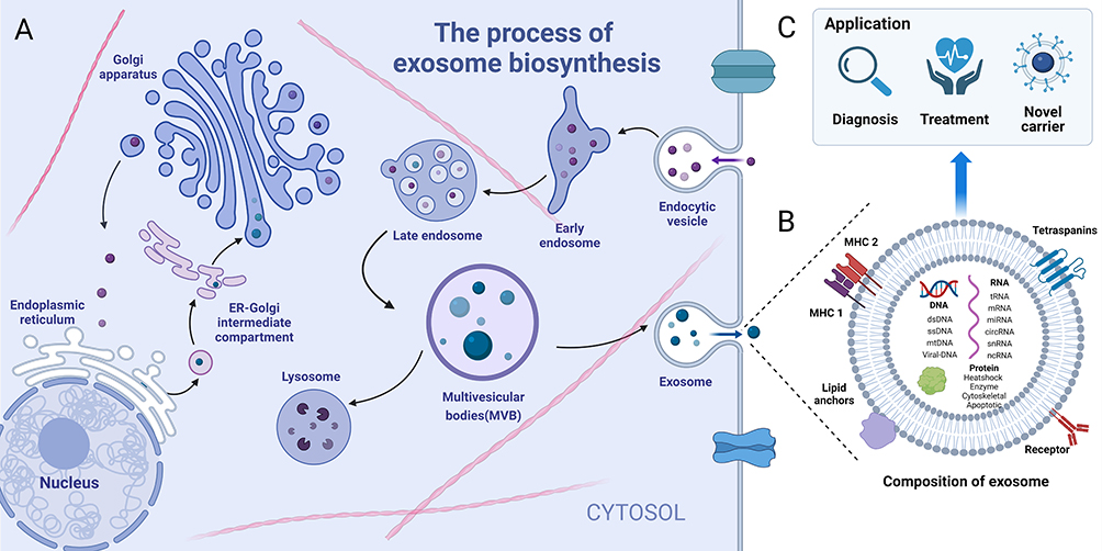 |
Figure 2 Biogenesis, composition, and application of exosomes. (A) The biosynthetic pathway of exosomes; (B) Composition of exosomes: proteins, nucleic acids, lipids, receptors, etc; (C) The primary applications of exosomes: diagnosis and treatment of diseases, novel carriers for drug transport, etc. (Image created with BioRender.com). |
Diverse methods are currently available to prepare exosomes including traditional methods such as ultracentrifugation, precipitation,27 density gradient centrifugation,28 and immunomagnetic bead method.29 However, these conventional methods often encounter issues such as time-consuming procedures, low yield, and low purity of the derived exosomes. With technological advancements, a range of emerging separation and purification methods have been introduced, including volume exclusion chromatography (SEC),30 nano-definite lateral displacement (nano-DLD),31 and microfluidic separation technology.32 However, these novel approaches still face challenges in practical applications, such as high costs and complex operations. As a result, an ideal separation solution that can effectively address these limitations while ensuring high efficiency, affordability, and simplicity remains elusive.
Primary Components and Applications
The proteins of exosomes (eg, tetra-transmembrane proteins, membrane-linked proteins, heat shock proteins, etc.) have multiple functions such as targeting/adhesion, anti-apoptosis, membrane fusion, signal transduction, and metabolism.33,34 Exosomes are rich in lipids, including glycosphingolipids, sphingomyelin (SM), cholesterol (CHOL), and phosphatidylserine (PS). These lipids contribute to the formation of exosomes and facilitate their release into the extracellular compartment, stimulating biological processes such as calcium inward flow.35 In addition, exosomes contain a large number of nucleic acids (DNA, mRNA, miRNA, tRNA, ncRNA, etc). Therefore, exosomes can be used as markers for early tumor diagnosis.36 Lin et al found that the microRNA expression profile of tumor-derived exosomes could identify exosome hsa-miR-483-5p as a potential biomarker for hepatocellular carcinoma.37 In addition, Guo et al demonstrated that long-stranded non-coding RNA (lncRNA), UCA1, from hypoxic tumor-derived exosomes promote angiogenesis in pancreatic cancer through the miR-96-5p / AMOTL2 axis.38 Therefore, the contents (nucleic acids, proteins, etc) contained in stem cell-derived exosomes and their membrane are of great significance in clinical studies (Figure 2B and C).
As a “cell-free” therapeutic approach, exosomes have been widely implemented in the management of various diseases. Jiang et al, for instance, utilized a combination of ultrafiltration and purification methods to isolate exosomes from the culture medium of urine-derived stem cells (USCs), which were then applied to investigate renal injuries associated with diabetic nephropathy.39 The resultant findings demonstrated that the USC-derived exosomes had the capacity to mitigate the high glucose-induced apoptotic potential of podocytes, significantly reduce urinary volume and albumin excretion in diabetic rats, and promote the proliferation of glomerular endothelial cells.39 However, the impact of different cellular treatment modalities on exosome functions warrants further attention. In this context, an earlier study by Li et al highlighted the importance of preconditioning strategies on exosomal function; herein, preconditioning BMSCs under simulated hypoxia/reoxygenation conditions increased the levels of miR-21-5p in exosomes.40 On the other hand, MSC-secreted exosomes were found to specifically act on the PTEN enzyme and the PDCD4 protein to inhibit both the intrinsic and extrinsic apoptotic pathways, resulting in a reduction of pulmonary edema, lung dysfunction, and inflammation caused by ischemia-reperfusion injury.40
Hydrogel
Hydrogels, characterized by their stable, three-dimensional hydrophilic networks, are typically synthesized using a multifaceted approach involving physical, chemical, and enzymatic cross-linking techniques.41 Their unique structural properties have led to their widespread applications across the fields of drug delivery, tissue engineering, and regenerative medicine.42 The polymers used for hydrogel fabrication are diverse and can be broadly categorized into three groups according to their source: natural polymers and synthetic polymers.43 Natural hydrogels, derived from biological sources, can be categorized into various polysaccharides and proteins, such as hyaluronic acid, alginate, chitosan, agarose, collagen, gelatin, silk fibroin, and fibrin.44 In contrast, synthetic polymers often employ polyethylene glycol (PEG), polyvinyl alcohol (PVA), poly(acrylic acid) (PAA), poly(lactic-co-glycolic acid) (PLGA), poly(acrylamide) PAAm, and poly(2-hydroxyethyl methacrylate) (PHEMA) to improve the physical and chemical properties of natural hydrogels, resulting in materials with enhanced mechanical strength, spatial precision, and the ability to control the internal dynamic microenvironment.45 In addition to these polymers, the fabrication of hydrogel also involves the utilization of self-assembling peptides, DNA molecules, polylactic acid, and ECM.46
Preparation Method
Diverse methods for hydrogel preparation are currently available. Free radical polymerization is a traditional method for developing polymer-based hydrogels and includes four approaches: bulk polymerization, solution polymerization, suspension polymerization, and emulsion polymerization. Hydrogel polymer precursors are synthesized into hydrogels by cross-linking, including physical, chemical, and irradiation cross-linking, with chemical cross-linking being an important and commonly used method for preparing hydrogels today (Figure 3A). In addition, extensive research efforts have contributed to the progressive advancement of key technologies utilized in hydrogel scaffolds for tissue engineering. These include emulsification, lyophilization (Figure 3B), gas foaming, 3D printing, and porogenic leaching techniques (Figure 3C). The development of these technological tools has made hydrogel materials easily accessible and affordable for large-scale use.47
 |
Figure 3 Common preparation (synthesis) methods of hydrogels. (A) free radical polymerization, physical, irradiation, and chemical cross-linking methods; (B) lyophilization technique; (C) leaching technique. Reproduced from Mantha S, Pillai S, Khayambashi P, et al. Smart Hydrogels in Tissue Engineering and Regenerative Medicine. Materials. 2019;12(20):3323. © 2019 by the authors. Creative Commons Attribution (CC BY) license (http://creativecommons.org/licenses/by/4.0/).47 |
The introduction of 3D printing technology has completely transformed the production of hydrogel scaffolds. 3D printing allows for the precise fabrication of three-dimensional scaffold structures tailored to specific tissue architectures and functional requirements. It is particularly crucial in mimicking the microenvironment of natural tissues and facilitating cellular growth.48 Thus, the versatility of 3D printing expands the applicability of hydrogel scaffolds in a wider array of tissue engineering and regenerative medicine applications and markedly improves their efficacy. Furthermore, the development of hydrogel microspheres via specific fabrication techniques, such as emulsification, introduces novel hydrogel forms with specific sizes and release properties. These microspheres allow for the encapsulation and precise release of drug molecules, thereby offering great potential for advancing drug delivery systems and cellular therapies.49 This diversity in the fabrication of hydrogels effectively yields different types of hydrogels, thereby offering a wide range of materials and application possibilities in the biomedical field.
Material Properties Required for Loaded Exosomes
Biomaterials supporting exosomes derived from stem cells are generally required to have high water content, good water retention, excellent biocompatibility and a microstructure similar to that of the extracellular matrix. Hydrogels are considered to be the biological functional materials closest to living tissues, so hydrogels combine with exosomes to promote the exosomes to better play their functions.
Biocompatibility
The good biocompatibility of hydrogels can provide suitable microenvironmental conditions for cell growth and act as an effective delivery matrix for bioactive molecules.50 Biocompatibility can be described as “the benign expression of the relationship between a material and its biological environment”, ie, it is less likely to trigger hemolysis, inflammatory reactions, immune reactions, etc., in the body.51 Pasquale Picone et al investigated various properties of xylene glycol hydrogel membrane (XG-PVA) for skin wound healing, in which biocompatibility was assessed by evaluating the damage mechanisms that may be activated by the biomaterial when in contact with the biological environment, such as oxidative stress, mitochondrial dysfunction, and apoptosis. XG-PVA membrane presented good biocompatibility without activating any immunogenic response, confirming the safety of this material for application in skin wound repair.52
High Water Content
Hydrogels are a class of hydrated polymeric materials with ≥90% water content. With the help of a hydrophilic 3D cross-linked network structure, hydrogels can hold large amounts of water or other water-based fluids (eg, cell nutrient solution, tissue culture fluid) without disintegrating. Liu et al observed that injectable hydrogels have shown great potential in scaffolds applied to tissue engineering due to their high water content, similarity to natural extracellular matrix (ECM), a porous framework for cell transplantation and proliferation, minimally invasive properties, and ability to match irregular defects, showing great potential for use as 3D cell culture scaffolds in tissue engineering.53
Capable of Protecting the Loaded Substance
As loading systems, hydrogels protect the loaded substances (eg, cells, bioactive molecules) and maintain their structure and function for longer periods. Tyson Smyth et al found that unmodified tumor exosomes were rapidly absorbed by the reticuloendothelial system (RES) in the liver and spleen when injected intravenously. This mechanism reduced the accumulation of their loaded drug in the tumor tissue, thus limiting the use of exosomes as a drug delivery vehicle.54 After being injected inside the organism, exosomes are susceptible to rapid clearance by the immune system, which reduces the efficiency of drug delivery and diminishes the therapeutic effect.55 Based on animal experiments, however, a very different effect was observed when hydrogels were used as exosome-carrying scaffolds. Wang et al prepared an injectable adhesive thermosensitive multifunctional polysaccharide dressing (FEP). The dressing had multifunctional properties such as efficient antimicrobial activity, rapid hemostatic ability, and good UV shielding properties. Compared to the exosome group, the exosome-loaded hydrogel group had a higher number of endothelial cells and greater angiogenic capacity.56
Degradability
The hydrogels combined with exosomes requires degradability, which promotes the sustained release of exosomes.Since exosomes are located in the three-dimensional meshwork of the hydrogel, their release rate is related to the structure, swelling rate, and degradation rate of the hydrogel. According to Shilan Shafei et al, alginate hydrogels loaded with adipose-derived stem cell exosomes promote tissue regeneration in whole skin wounds. The hydrogel was gradually degraded during immersion in the culture medium for 240 h, resulting in a slow release of exosomes. This release pattern is more conducive to skin repair and accelerates skin repair processes such as re-epithelialization, collagen deposition, and angiogenesis.57
Preparation Methods and Advantages of Hydrogel-Loaded Exosomes
The fabrication of exosomes loaded with hydrogels encompasses three primary methodologies: in situ gel formation, the “Breathing method”, mixing and crosslinking, and in situ gel formation. Each method has its advantages and disadvantages, necessitating a careful evaluation based on specific requirements and experimental conditions.
Firstly, the “Breathing method” utilizes the hydrophilic and adsorptive properties of hydrogels.58 This method entails the initial extraction of excess water from the swollen hydrogel by employing a specific solvent in order to completely expose its internal voids. Subsequently, exosomes are introduced directly into these voids, leading to the incorporation of exosomes into the hydrogel. This approach is noted for its simplicity, as it directly utilizes hydrogel voids for loading without the need for complex equipment or techniques, resulting in enhanced loading efficiency. However, the compatibility between exosome size and hydrogel pore dimensions is constrained by the precision in void size of the hydrogel required to prevent exosome leakage (caused by large void size) or inadequate entry (caused by small void size). This condition highlights a significant limitation in terms of the compatibility between exosome size and hydrogel pore dimensions.
Secondly, the mixing and crosslinking method entails combining exosomes with a hydrogel precursor solution, followed by the addition of a crosslinking agent or utilizing a physical method to induce gelation.59 This facilitates greater control over the structure and properties of the resulting hydrogel. Nonetheless, the process of crosslinking may potentially compromise the integrity and bioactivity of the exosomes. Furthermore, the compatibility between the crosslinking agent and the exosomes warrants careful consideration.
Thirdly, the in situ gel formation method, involves the use of a dual-chamber syringe to simultaneously inject exosomes and the hydrogel precursor into the target site.60 Once injected, they react with each other to form a hydrogel that encapsulates the exosomes. This method enables the direct formation of the hydrogel at the target location, thereby facilitating localized drug delivery for tissue engineering applications, with the capability to adjust the hydrogel formation rate and properties as needed. However, it is important to consider the potential toxicity of the adhesive in the body when using this method.
The integration of exosomes within hydrogels provides a multifaceted approach that is crucial and advantageous in enhancing therapeutic outcomes and tissue regeneration ability. Hydrogels enhance the stability and bioactivity of exosomes by protecting them from external factors, such as enzymatic degradation and oxidation,61 and evading rapid clearance by the immune system.55 This property ensures their sustained presence and functioning within the body. Furthermore, the ability of hydrogels to facilitate the precise release of exosomes at specific times and rates is crucial in maintaining therapeutic efficacy and decreasing the need for frequent administration.62 In addition, hydrogels can modify exosomes chemically or physically to endow them with specific properties, allowing for more precise delivery to the pathological sites and thereby augmenting therapeutic outcomes.63 Moreover, the ability of hydrogels to be administered via injections also simplifies the process of delivering exosomes, rendering them highly suitable for clinical applications.64 Finally, the conducive microenvironment provided by hydrogels promotes cell growth and tissue repair and synergistically fosters the tissue repair and regeneration processes carried out via exosomes.65 In summary, the incorporation of exosomes into hydrogels is not only necessary but also advantageous, holding significant importance in enhancing therapeutic outcomes and facilitating tissue regeneration.
Diabetes Mellitus and Its Complications
Disease Overview and Traditional Therapies
Diabetes mellitus is a metabolic disorder that is experiencing a significant global rise in prevalence. The decline in organism function, pharmacological factors, psychological factors, and lifestyle changes collectively contribute to the increased susceptibility of the elderly population to diabetes. According to statistics,66 approximately 463 million adults aged 20–79 years (9.3% of the world population) currently have diabetes. However, most patients have chronically high levels of blood glucose, leading to chronic complications of the kidneys, eyes, nerves, heart, and skin, such as diabetic nephropathy, diabetic retinopathy, diabetic neuropathy, diabetic cardiomyopathy, and diabetic foot.67
In recent years, the application of stem cell-derived exosomes to the treatment of the above-mentioned diabetic complications has yielded good results. Bone marrow MSC-derived exosomes promote the recovery of renal function in diabetic mice by inhibiting the expression of pro-inflammatory cytokines in the kidney and epithelial damage in the proximal tubules of the kidney induced by high-fat diet and STZ.68 For diabetic retinopathy, exosomes derived from human umbilical cord mesenchymal stem cells (hUCMSCs) can regulate miR-126 by targeting HMGB1 to ameliorate hyperglycemia-induced retinal inflammation (Zhang et al, 2019).69 The combination of exosomes and hydrogels for the treatment of diabetes and its complications has focused on diabetic wound healing. This study, focusing on this disease, analyzes the potential mechanisms and efficacy of this novel treatment approach. The interaction of multiple factors such as diabetic neuropathy, peripheral vascular disease, infection, and trauma causes poor wound healing and eventually the formation of severe foot ulcers, known as diabetic foot (Figure 4).70 This complication imposes huge financial stress and psychological burden on both the patient and their family. Hospital admissions for foot ulcers in the diabetic population are increasing significantly. The cost of care is more than 10 times higher in patients with diabetic foot compared to individuals without diabetes.71 Consequently, it is urgent to develop treatments for diabetic wound healing.
 |
Figure 4 The formation of diabetic foot. Normal wounds in diabetic patients gradually develop into chronic hard-to-heal wounds due to hypoxia, increased ROS (reactive oxygen species) and inflammatory factors, and impaired angiogenesis. (Image created with BioRender.com). |
The current conventional treatments for diabetic wounds include the use of growth factors, wound dressings, and skin substitutes. Each of these treatments has distinct advantages and disadvantages.72 When released, growth factors affect the wound microenvironment, promote intercellular communication, and increase differentiation, proliferation, and migration of endothelial cells and fibroblasts, facilitating wound healing.73 However, this therapeutic approach has its limitations. Specifically, growth factors are rapidly degraded by the action of protein hydrolases, making the treatment less effective.74 While wound dressings have the advantage of creating a moist and oxygenated environment for skin tissue, they may not effectively prevent bacterial invasion from the external environment, leading to the potential risk of secondary infections. On the other hand, skin substitutes can offer both a moist environment and a barrier against external bacteria.75 However, as allografts, they may trigger immune rejection in some cases.76
Molecular Mechanisms of Wound Repair by Exosomes
Wound healing is divided into four stages: hemostatic, inflammatory, proliferative, and remodeling stages (Figure 5).
 |
Figure 5 Involvement of exosomes in various stages of wound healing; (A) Promote the conversion of M1 type macrophages to M2 type; (B) Reduces ROS; (C) stimulate proliferation and migration of vascular endothelial cells; (D) promote proliferation of fibroblasts; (E) prevent fibroblasts from differentiating into myofibroblasts. (Image created with BioRender.com). |
Hemostatic
The hemostatic phase is achieved mainly through vasoconstriction, platelet aggregation, and activation of coagulation factors to initiate endogenous and exogenous coagulation pathways. Human embryonic stem cell-derived exosomes (HESC-Exos), induce angiogenesis and improve wound healing through the Nrf2 (a major regulator of oxidative stress, ie, nuclear factor erythroid 2-related factor 2) signaling pathway.
Furthermore, exosomes exert beneficial effects on vascular endothelial cells by reversing senescence and enhancing their proliferation, migration, and angiogenesis capabilities.77 Additionally, a prior study has demonstrated the significant procoagulant potential of extracellular vesicles derived from umbilical cord MSCs, indicating that exosomes may enhance tissue coagulation capacity and promote wound healing.78
Inflammatory
The inflammatory response, which has an important role in regulating wound healing and regeneration, is characterized by the activation of immune cells such as mast cells, neutrophils, and macrophages to proliferate and secrete inflammatory cytokines and chemokines to clear pathogens and debris.79 Although a moderate inflammatory response is effective in preventing infection, elevated levels of reactive oxygen species (ROS) in a high-glucose environment lead to increased production of IL-8 by keratinocytes. The corresponding increase in neutrophil infiltration causes an excessive inflammatory response and hinders wound healing.80 However, exosomes secreted by MSCs have anti-inflammatory and anti-apoptotic functions and can promote the regression of inflammation in several ways. First, miRNAs contained in exosomes can regulate the inflammatory response. For example, human umbilical cord MSC-derived exosomes expressing miR-150-5p promote skin wound healing by activating the PI3K/AKT pathway of PTEN to reduce the inflammatory response.81 Second, these exosomes inhibit the secretion of pro-inflammatory factors. According to Li et al, macrophage-derived exosomes significantly reduced the expression of inflammatory factors such as tumor necrosis factor (TNF-α) and interleukin (IL-6) compared to the blank group. These exosomes exert anti-inflammatory effects by reducing the secretion of proinflammatory enzymes and cytokines.82 Finally, these exosomes induce M1 to M2 type macrophage polarization to elevate the M2/M1 polarization ratio, and Liu et al found that melatonin-stimulated MSC-derived exosomes regulate macrophage M1 and M2 polarization by targeting the PTEN/AKT pathway, increasing the M2/M1 ratio, alleviating the inflammatory response and improving diabetic wound healing.83 In addition, exosomes can also reduce oxidative stress-related protein levels to promote wound healing in a diabetic rat model.84
Proliferative
The proliferative process involves granulation tissue generation, re-epithelialization, and angiogenesis. Exosomes can use their powerful messaging capabilities to promote fibroblast and endothelial cell proliferation, form granulation tissue, and regulate the migration of keratin-forming cells to the wound center.85 In the high glucose state, the production of advanced glycosylation end products (AGEs) is accelerated. This substance can directly induce endothelial cell apoptosis, causing structural damage to the vascular endothelium and increasing vascular permeability. In addition, endothelial cell damage leads to subendothelial collagen exposure. Subsequently, activation of platelets, adhesion, release, aggregation, and coagulation at the damaged site form thrombi, leading to narrowed lumen and increased resistance to blood flow. Exosomes promote angiogenesis, provide oxygen, nutrients, and metabolic pathways to the wound, and synergistically promote wound healing. Key angiogenesis-promoting miRNAs contained in exosomes can activate endogenous repair mechanisms in target cells. For example, EPC-Exos enhances endothelial cell proliferation, migration, and angiogenesis in a miR-126-dependent manner and downregulates SPRED1 to activate Raf/ERK signaling.86 In addition, exosomes can deliver angiogenesis-promoting related proteins to stimulate the rapid proliferation and migration of vascular endothelial cells and promote wound healing. According to Ting Deng et al, gastric cancer (GC)cell-derived exosomes can act as vectors to deliver miR-155, which promotes angiogenesis by inhibiting c-MYB and increasing VEGF (vascular endothelial growth factor) expression.87
Remodeling
The remodeling process is a critical stage of wound healing that determines the extent of scar formation. In this phase, exosomes play a vital role in improving wound healing outcomes and aesthetics by preventing fibroblasts from differentiating into myofibroblasts, which reduces scar formation.88 During the pre-healing phase, exosomes promote the formation of type I and type III collagens, while in the late healing phase, they increase the ratio of type III to type I collagen and the ratio of TGF-β3 to TGF-β1, contributing to proper extracellular matrix remodeling.89 Additionally, exosomes can increase matrix metalloproteinase-3 (MMP3) expression in skin dermal fibroblasts through activation of the ERK MAPK pathway and elevate the ratio of MMP3 to tissue inhibitor of matrix metalloproteinase-1 (TIMP1), thereby promoting extracellular matrix reconstitution.90
Combination Therapy
Stem cell exosomes have been found to promote skin wound repair through various mechanisms (Table 1). Therefore, the application of stem cell exosomes for diabetic wound healing appears to be a feasible approach. Nonetheless, certain limitations still need to be addressed. One notable limitation in the application of exosomes is their tendency to undergo rapid clearance in the body, resulting in low effective utilization and the potential for secondary damage to the skin when multiple doses are administered. To mitigate these challenges to a certain extent, the utilization of exosomes in combination with biomaterials offers a potential solution. In terms of promoting angiogenesis, a high glucose environment can cause dysfunction of blood vessels and smooth muscle cells, leading to impaired vascular networks and impeding wound healing. Zhang et al loaded the human umbilical cord MSC-derived exosomes into poly (vinyl alcohol) (PVA)-alginate (ALG) nanohydrogels. The exosomes could activate ERK1/2 signaling by promoting ERK1/2 phosphorylation, enhance the expression of molecules such as SMA/SR-B1/CD31, and promote HUVEC proliferation, migration, and angiogenesis.26 In terms of regulating the wound microenvironment, anti-inflammation is a key component in promoting wound healing since a high-glucose environment leads to an excessive inflammatory response and damages the cells surrounding the wound site. Geng et al loaded bone marrow MSC-derived exosomes into carboxyethyl chitosan (CEC)-dialkylated methylcellulose (DCMC) hydrogels. This strategy not only promoted angiogenesis but also induced M1 to M2 macrophage polarization to reduce inflammatory effects.91 Furthermore, the regulation of oxidative stress levels within the microenvironment of the diabetic wound is also a crucial factor in promoting effective tissue repair.92 Immune and other cells produce more ROS under hyperglycemic conditions,93 which trigger multiple cellular mechanisms such as activation of protein kinase C, NF-κB pathway-mediated inflammation, and altered vascular permeability.94 Zhang et al developed a novel PEG/Ag/CNT- M+E hydrogel and assayed ROS content in mouse skin tissues. According to their experimental data, the fluorescence intensity of the PEG/Ag/CNT-M+E hydrogel group was significantly lower than that of the untreated diabetic group, demonstrating significant ROS scavenging properties and excellent antioxidant capacity of the hydrogel.95
 |
Table 1 Combined Administration of Stem Cell Derived Exosomes and Hydrogels in the Treatment of Diabetic Wound |
In terms of promoting neurogenesis, diabetic neuropathy is also an important hindrance to wound healing in diabetic patients. According to a previous study, 50% of diabetic foot patients developed diabetic neuropathy.105 Therefore, it is extremely important to promote neuronal growth. Shi et al loaded gingival mesenchymal stem cell-derived exosomes (GMSC-Exos) into chitosan-silk hydrogels and evaluated nerve regeneration in a diabetic rat model at 2 weeks postoperatively by NEFH immunofluorescence staining. GMSC-exos significantly increases nerve density, providing novel evidence that sheds light on their effect on neuronal growth in a diabetic rat skin defect model (Figure 6D–J).102 In addition to the direct loading of exosomes into hydrogels to promote diabetic wound healing, exosomes were also modified to further promote their therapeutic effects. Tao et al used gene overexpression techniques to modify synovial mesenchymal stem cells (SMSCs). Although SMSCs can significantly promote fibroblast proliferation, they can barely promote angiogenesis. Therefore, the angiogenic capacity of endothelial progenitor cells was transferred to SMSCs by overexpression of miRNA-126-3p. Ultimately, miRNA-126-3p overexpression of synovial MSC-derived exosomes was loaded into chitosan hydrogels to achieve the synergistic promotion of wound healing.106
 |
Figure 6 Stem cell-derived exosome-bound hydrogel promotes chronic diabetic wound healing; (A) Electron microscopy images of hUCMSCs-exos; (B) Detection of wound healing rate. (C) VEGF and TGFβ-1 expression levels. Reproduced with permission from Dove Medical Press. Yang J, Chen Z, Pan D, Li H, Shen J. Umbilical Cord-Derived Mesenchymal Stem Cell-Derived Exosomes Combined Pluronic F127 Hydrogel Promote Chronic Diabetic Wound Healing and Complete Skin Regeneration. IJN. 2020;15:5911–5926.101 (D)Schematic illustration of the isolation of GMSC-derived exosomes and preparation of chitosan/silk hydrogel; (E) Quantitative analysis of the wound closure rates; (F) Quantitative analysis of the total length in the three groups at each time point.; (G) Quantitative analysis of the percentage of collagen in each group at each time point; (H) Quantitative analysis of the number of microvessels per field; (I) Representative immunofluorescent images of neurofilament heavy chain (red fluorescence) were detected at 2 weeks post-surgery; (J) Quantitative analysis of the nerve fiber density in each group at 2 weeks post-surgery. a, P < 0.05 compared to the control group; b, P < 0.05 compared to the hydrogel group. Reproduced from Shi Q, Qian Z, Liu D, et al. GMSC-Derived Exosomes Combined with a Chitosan/Silk Hydrogel Sponge Accelerates Wound Healing in a Diabetic Rat Skin Defect Model. Front Physiol. 2017;8:904. © 2017 Shi, Qian, Liu, Sun, Wang, Liu, Xu and Guo. Creative Commons Attribution License (CC BY).102 |
Good hydrogel properties will also further promote the effect of the combination of the two. Wang et al developed an injectable peptide hydrogel (FHE hydrogel) with self-healing properties and antimicrobial activity to promote chronic diabetic wound healing from a biomaterial perspective. The self-healing property of this hydrogel is reflected in the rapid and autonomous self-healing ability of the biomaterial after external damage. This feature allows the structural stability of the material to be maintained during the wound-healing process. Experimental data demonstrate the powerful antibacterial activity of FHE hydrogel. The hydrogel effectively killed Escherichia coli (gram-negative bacteria) and Staphylococcus aureus (gram-positive bacteria), indicating the good antibacterial ability of FHE hydrogel. Combining adipose mesenchymal stem cell-derived exosomes with this hydrogel promotes HUVEC proliferation, migration, and angiogenesis, accelerates granulation tissue formation, re-epithelialization, and collagen remodeling, and ultimately synergistically accelerates wound healing.63 The ideal hydrogel scaffold for wound healing should be suitable for complex and irregular wound trauma spaces in addition to having good antimicrobial capacity, injectability, and biocompatibility, allowing bioactive substances to adhere to the target site and exert their biological effects. Thus, Yang et al innovatively developed a thermosensitive hydrogel (Pluronic F-127). This hydrogel exists as a liquid at low temperatures and as a semi-solid gel at high temperatures. This reversible thermal response behavior gives Pluronic F-127 the aforementioned properties. In addition, PluronicF-127 was loaded with human umbilical cord mesenchymal stem cell-derived exosomes (hUCMSCs-Exos), which increased CD31 and Ki67 expression, enhanced granulation tissue regeneration, upregulated VEGF/TGF-β1 expression, and jointly promote diabetic wound healing (Figure 6A–C).101
As mentioned above, the integration of stem cell-derived exosomes into hydrogels presents a novel therapeutic approach for chronic diabetic wounds, offering significant advantages over conventional therapies. Exosome-loading hydrogels not only promote the proliferation and differentiation of cells that are essential for the wound healing process, but also prevent the rapid clearance of exosomes, ensuring their sustained and stable functioning within the body due to the protective function of hydrogels. Furthermore, hydrogels provide exposed skin tissues with a moist and oxygen-rich healing environment, while also exerting a profound antimicrobial effect to ensure the safety and efficacy of the treatment process. Thus, it is rational to hypothesize that the widespread application of this therapeutic approach may offer substantial relief to elderly diabetic patients suffering from the physical and psychological challenges associated with their condition.
Cardiovascular Disease
Disease Overview and Traditional Therapies
Cardiovascular disease (CVD) poses the greatest risk to human health and represents a major cause of mortality and disability among the elderly population.107 It encompasses various conditions such as hypertension, coronary atherosclerotic heart disease, myocardial infarction, and heart failure. In Europe alone, CVD remains the leading cause of death, resulting in the loss of more than 60 million potential lives each year.108 The main cause of high mortality from cardiovascular disease is ischemic heart disease. Coronary atherosclerotic heart disease is the classic form of ischemic heart disease.109 Its pathogenesis involves atherosclerosis of the coronary arteries, which causes narrowing of the lumen and inadequate blood supply, leading to myocardial ischemia and hypoxia, which in turn leads to myocardial infarction (Figure 7A). After the occurrence of myocardial infarction, the ischemic heart cannot maintain its structural integrity as the endocardial ATP content decreases, in which irreversible damage occurs to cardiomyocytes, eventually leading to apoptosis and necrosis (Figure 7B). Necrotic myocardial tissue undergoes three phases: inflammation, proliferation, and long-term remodeling. During this process, infarct wall thinning and left ventricular dilatation eventually lead to the onset and progression of heart failure (Figure 7C).110
 |
Figure 7 Causes of myocardial infarction and repair of myocardial tissue. (A) The “fragile plaque” model. Myocardial infarction is usually caused by the formation of occlusive thrombi in the coronary arteries due to rupture of fragile plaques; (B) early evidence of irreversible myocardial cell damage in ischemic myocardium: as ATP levels decrease, myocardial cells can no longer maintain their structural integrity and exhibit ultrastructural changes of damage, such as disruption of myocardial cell membranes and the presence of amorphous density of mitochondria; (C) The repair process after myocardial infarction. The repair process after myocardial infarction. Alarmins released by necrotic cardiomyocytes (alarm hormones, that is, endogenous biological mediators released by white blood cells and epithelial cells to the outside of the cell when the body is in the state of tissue injury and inflammatory response or physiological stress, which can enhance the immune response) trigger a strong inflammatory response to clear the dead cells and stromal debris in the infarction area. Removal of dead cells inhibits pro-inflammatory signaling and the transition to the proliferative phase. Conversion of fibroblasts into myofibroblasts and deposition of extracellular matrix proteins form an abundant neovascular network. Finally, during maturation, the extracellular matrix is hooked up, while the infarcted fibroblasts become quiescent and may undergo apoptosis. Reproduced from Frangogiannis NG. Pathophysiology of Myocardial Infarction. In: Terjung R, ed. Comprehensive Physiology. 1st ed. Wiley; 2015:1841–1875. © 2015 American Physiological Society.110 |
Current protocols for the treatment of cardiovascular disease involve mainly preventive interventions and post-onset treatment. Three treatment modalities are indicated for end-stage myocardial infarction and heart failure: drug therapy, heart transplantation, and cardiac assist device implantation. However, there are certain limitations associated with these treatment strategies.111 Heart transplantation is considered as the only treatment for end-stage heart failure. The shortage of donors, immune rejection after transplantation, and the high cost of treatment have limited the clinical use of this treatment. Despite the certain success of cardiac assist devices, the highly invasive nature of the implantation procedure is not the best treatment modality for elderly patients with an underlying disease. Considering the significant impact of cardiovascular disease on patient well-being, there is an urgent demand for innovative treatments that can effectively repair the damaged myocardium. These advancements aim to improve their quality of life and provide them with better therapeutic outcomes.112
Molecular Mechanisms of Exosome Repair of Injured Myocardium
Stem cell repair of myocardium to treat cardiovascular diseases has certain drawbacks and uncertainties, such as immune rejection, tumorigenicity, and ethical safety.13 The successful application of exosomes in tissue repair provides a novel idea to improve myocardial injury and treat end-stage cardiovascular diseases. Thus, the potential molecular mechanisms of exosome therapy for cardiovascular diseases were analyzed (Figure 8).
 |
Figure 8 Exosomes derive from MSCs of different origins can be combined with hydrogels by tissue engineering to repair injured myocardium through myocardial injection. The molecular mechanisms involved primarily include inhibition of apoptosis, regulation of cellular autophagy, anti-oxidative stress, controlling inflammation, promotion of angiogenesis, and anti-myocardial fibrosis. (Image created with BioRender.com). |
Inhibition of Apoptosis
Myocardium is susceptible to apoptosis or necrosis in an ischemic or hypoxic environment, or under the influence of oxidative stress. Stem cell-derived exosomes protect myocardial tissue by resisting apoptosis. miR-210 in MSC exosomes improves cardiomyocyte survival rate and in vivo cardiac function under hypoxic conditions in vitro by regulating the expression of PI3K/Akt and p53.113 In addition, a study transfected mouse BMSC with a lentivirus carrying doxycycline (DOX)-induced GATA-4 and collected exosomes released by BMSC. These exosomes induced BMSC differentiation into cardiomyocyte-like cells and reduced hypoxia-induced apoptosis in cardiomyocytes. Recovery of cardiac function, increased cardiac vascular density, an elevated number of cardiac endothelial cells, and decreased apoptotic cardiomyocytes were observed when GATA-4-BMSC exosomes were injected into mice 48 hours after the onset of myocardial infarction (MI).114
Regulation of Cellular Autophagy
Under mild stress conditions (eg, transient hypoxia and low-level oxidative stress), cardiomyocytes can preserve cell viability by transporting some damaged proteins or organelles to lysosomes for degradation and ATP production after being wrapped by autophagic vacuoles. However, severe ischemic conditions can lead to excessive autophagy, resulting in excessive self-digestion of essential proteins and organelles, which in turn accelerates cell death and attenuates cardiac function. Therefore, exosome regulation of excessive cellular autophagy facilitates cardiomyocyte survival. During cardiac hypoxia/reoxygenation (H/R)-induced injury, miR-21 in MSC-derived exosomes can inhibit H/R stimulation-induced apoptosis and autophagy via the PTEN/Akt/mTOR signaling pathway.115 Exosomes can also inhibit ischemia/reperfusion (I/R)-induced cellular autophagy by increasing the expression of the apoptotic protein p62, decreasing the ratio of LC3-II to LC3-I, upregulating Bcl-2, activating the mTORC1 / p-4eBP1 pathway, and downregulating the ubiquitin ligase Traf6.116
Anti-Oxidative Stress Effects
Oxidative stress damage produced by ischemia-reperfusion (I/R) causes lipid peroxidation reactions and reduces cell membrane fluidity, which affects cell membrane ion channel function and damages cell membranes.117 This process also causes severe damage to genetic material DNA,118 and organelles (eg, mitochondria) and is a major cause of decreased cardiomyocyte survival. Oxidized low-density lipoproteins (ox-LDL) from lipid peroxidation reactions are recognized and phagocytosed by macrophages, which in turn form foam cells and induce coronary atherosclerotic heart disease. Exosomes secreted by MSCs exhibit strong resistance to oxidative stress. Fatih Arslan et al found that exosomes can activate multiple pathways to protect the heart, such as increasing the levels of ATP and NADH, elevating phosphorylated Akt and GSK-3β content, and decreasing phosphorylated-c-JNK content in ischemic/reperfused hearts, and attenuating oxidative stress.119 In addition, miR-214 derived from BMSC-Exos effectively inhibits oxidative stress by altering the CaMKII pathway and decreasing the level of reactive oxygen species production in cardiomyocytes.120
Anti-Inflammatory Effects
This effect acts through two pathways: (1) regulation of T cells; Wei et al observed that miRNA-181a in exosomes can upregulate Foxp3 levels in regulatory T cells (Tregs), which, by means of Foxp3 binding to T cell nuclear factors, allows Tregs activation and ultimately suppresses the immune response.121 (2) Promotion of M1-type macrophage to M2-type macrophage polarization. Zhao et al observed that miRNA-182 in mouse bone marrow-derived MSC-derived exosomes promoted M2-type macrophage polarization via TLR4/Akt signaling pathway. Furthermore, miRNA-182 could synergistically control inflammation by activating the Pl3k/Akt signaling pathway to reduce M1-type pro-inflammatory macrophages and increase the number of M2-type anti-inflammatory macrophages.122
Promotion of Angiogenesis
During myocardial ischemia, blood vessels narrow or become blocked for a short period, thereby preventing the supply of oxygen-rich blood to the heart. Therefore, promotion of angiogenesis is essential for the treatment of ischemic heart disease. Exosomes contain pro-angiogenic miRNAs (eg, miRNA-21, miRNA-132, miRNA-210), which act as carriers to load proteins such as pro-angiogenic factors and platelet-derived growth factor D to regulate the gene expression of downstream vascular pathways. This mechanism enhances the proliferation, migration, and angiogenesis capacity of endothelial cells, accelerates angiogenesis in the infarcted region, increases the supply of nutrients and oxygen to it, and thus improves the function of ischemic myocardium.123 Ma et al used Akt-overexpressed human umbilical cord MSC-derived exosomes (Akt-Exo) to treat acute myocardial infarction. Their experimental data showed that Akt-Exo accelerated the proliferation and migration of endothelial cells and upregulated the expression of platelet-derived growth factor D (PDGF-D), demonstrating a significant ameliorative effect of Akt-Exo on cardiac function in the test animals.124
Anti-Myocardial Fibrosis
Myocardial fibrosis is an important process in the repair of ischemic cardiomyocytes. Primary fibrosis can prevent the rupture of the ventricular wall. However, persistent excessive fibrosis can lead to chronic ischemic heart disease.125 Stem cell-derived exosomes have been shown to prevent excessive myocardial tissue fibrosis to some extent. Ischemic preconditioned MSC-derived exosomes ameliorate cardiac fibrosis by secreting miR-22 that targets Mecp2, a methylated CpG-binding protein in fibroblasts.126 In addition, these exosomes inhibit myocardial fibrosis by suppressing transforming growth factor β-mediated conversion of fibroblasts to myofibroblasts.127
Combination Therapy
The combination therapy involving hydrogel and exosome has demonstrated significant advancements in the treatment of cardiovascular disorders (Table 2). Lee et al investigated the application effectiveness of an implantable hydrogel known as Algisyl-LVR™ in the treatment of heart failure. According to their experimental data, end-diastolic volume (EDV) and end-systolic volume (ESV) decreased significantly 3 months after treatment, while ejection fraction and volume mean wall thickness increased significantly. In addition, myofiber stress became more uniform after treatment. Therefore, the material has shown efficacy in the treatment of myocardial infarction and heart failure.128 In addition, hydrogel injection alone can reduce oxidative stress, increase myocardial conductivity, and inhibit metalloproteinase-induced malignant remodeling, thereby treating cardiovascular disease.129 Exosomes or hydrogels alone have been administered to treat cardiovascular diseases with good therapeutic effects. In practical application, exosome is primarily administered intravenously. However, after injection, exosomes accumulate in organs such as the liver and kidney or are phagocytosed by immune cells,130 resulting in the very low levels of themin heart tissue. Therefore, an innovative combination of the two was developed for the synergistic treatment of cardiovascular diseases. Han et al injected functional peptide hydrogels (PGN hydrogels, obtained by mixing PA-GHRPS peptide with NapFF peptide) loaded with human umbilical cord MSC-derived exosomes (UMSC-Exo) into a rat myocardial infarction model. Their experimental outcomes showed that this combination therapy reduced inflammation, cardiac fibrosis, and apoptosis, promoted angiogenesis, and improved cardiac function.131
The combination of natural and synthetic materials gave the composite good biocompatibility, degradability, and some mechanical strength and elasticity to obtain the biological properties and physicochemical properties required for cardiac repair. nSi-Gel, a nanocomposite hydrogel, was prepared by Waters et al by mixing synthetic silicate nanosheets with the gelatin phase. Compared to controls, nSi-Gel loaded with exosomes derived from human umbilical cord vein endothelial cells resulted in a considerable enhancement in capillary density, reduction in the scar area, and improvement in cardiac function in treating myocardial infarction.132
 |
Table 2 Combined Administration of Stem Cell Derived Exosomes and Hydrogels for the Treatment of Cardiac Repair |
Smart hydrogels are currently attracting significant research attention, serving as a focal point of investigation. Notably, pH-sensitive and temperature-sensitive hydrogels have emerged as promising areas of study. These hydrogels exhibit a remarkable ability to respond to external stimuli, leading to changes in their mechanical or physical properties. This unique characteristic allows the hydrogel to be administered in a liquid state, providing matrix support and mechanical reinforcement to the infarcted heart tissue. Moreover, this advantageous feature enhances the drug-loading capacity and injectability of the hydrogel.142 Liu et al prepared chitosan/β-glycerophosphate sodium temperature-sensitive hydrogels and encapsulated exosomes secreted by human umbilical cord mesenchymal stem cells in them. The thermosensitive nature of the biomaterial allowed it to maintain a liquid state for an extended period at or below room temperature while transitioning into a gel-like state at body temperature. This thermosensitivity contributes to the smart hydrogel’s favorable histocompatibility, prolonged retention of exosomes within the body, a notable enhancement of blood perfusion, and improved recovery of tissue function following ischemia. Consequently, smart hydrogel exhibits substantial therapeutic effects in several fields such as tissue repair and regeneration.
To summarize, the use of this novel combination therapy can effectively deliver exosomes and other bioactive substances directly to the damaged myocardial tissues, thus circumventing the challenges associated with the uneven distribution of pharmaceuticals in the body and enhancing therapeutic outcomes. In addition, hydrogels are renowned for their exceptional biocompatibility, facilitating a gradual process of degradation and absorption within the body. This characteristic significantly reduces the practical strain placed on patients. Furthermore, given that exosomes are typically derived from the patient’s own cells, the risk of immune rejection and allergic reactions is markedly reduced, contributing to the enhanced safety profile of the therapy. Importantly, hydrogels, as drug carriers, can continuously release exosomes at the damaged site to facilitate the long-term efficacy of therapeutic molecules. In addition to increasing convenience, this property reduces the frequency and dosage of treatments. Therefore, this combination therapy holds significant potential for use in the treatment of cardiovascular diseases, such as myocardial infarction, while also improving treatment outcomes and quality of life for a wide patient population.
Osteoarthritis
Disease Overview and Traditional Therapies
Osteoarthritis, a degenerative joint disease caused by many factors such as age, inflammation, trauma, and strain,143 is characterized by damage to articular cartilage, hyperostosis at the joint edges, and osteophyte formation.144 Currently, OA is the fourth most disabling disease worldwide and is predicted to be the primary cause of disability by 2030.145 The incidence of osteoarthritis is surging with the aging of the population, lifestyle changes, and the gradual increase in the number of middle-aged, elderly, and obese people. The prevalence of OA in the middle-aged and elderly population is as high as 40%-80%, and the ultimate disability rate increases to 50%. Consequently, OA severely compromises the quality of life for affected individuals and imposes a substantial economic burden on both their families and society at large.146
The progression of osteoarthritis is characterized by an increase in the secretion of pro-inflammatory factors by hypertrophic chondrocytes and proliferating synovial cells. Infiltration of inflammatory cells results in abnormal metabolic and biological behavior of chondrocytes in articular cartilage, which in turn leads to an imbalance in cartilage tissue remodeling. The articular cartilage structure is degraded by extracellular matrix-degrading enzymes (a disintegrin and metalloproteinase with thrombospondin motifs [ADAMTs] and matrix metalloproteinases [MMPs]) secreted by the damaged chondrocytes, followed by apoptosis of the chondrocytes, deposition of large amounts of calcium salts in the matrix, progressive ossification of the cartilage, and ultimately, bone redundancy and loss of joint function.147 In clinical practice, osteoarthritis is often treated with medication or intra-articular injections of glucocorticoids and sodium Hyaluronate to achieve short-term analgesia and anti-inflammatory effects. However, the degeneration of joint cartilage cannot be avoided in cases of progressive deterioration, surgical interventions such as joint fusion or artificial joint replacement may be necessary. However, these procedures are subjected to several drawbacks, including postoperative mobility limitations and the risk of infection associated with metal prostheses.148 Hence, there is a pressing need to explore the underlying mechanisms of osteoarthritis and develop targeted therapeutic approaches to prevent its onset and slow down disease progression. In this context, the utilization of exosomes as a potential treatment modality for osteoarthritis has garnered significant attention in recent years.
Molecular Mechanisms of Cartilage Tissue Repair by Exosomes
The potential mechanisms by which exosomes contribute to cartilage tissue repair partially overlap with those described earlier for skin repair in diabetes and heart tissue repair. These mechanisms involve controlling inflammation by inhibiting chondrocyte apoptosis through pathways such as regulation of the adenosine-mediated AKT/ERK signaling pathway,149 promoting cell proliferation, migration, differentiation, and inducing macrophage conversion to M2 type.150 Furthermore, other molecular mechanisms contributing to the beneficial effects of exosomes in cartilage tissue repair also exist (Figure 9).
 |
Figure 9 Molecular mechanisms via which exosomes promote cartilage repair: repair of mitochondrial functional damage, reduction of oxidative stress damage, protection of extracellular matrix and chondrocytes, re-induction of type II collagen (type II collagen) and aggregated proteoglycan (aggrecan) expression in mature articular chondrocytes, etc. (Image created with BioRender.com). |
Decrease in the Content of MMP and ADAMTs
In the process of cartilage tissue degradation, substances such as MMPs and ADAMTs play a significant role.151 Exosomes have the potential to protect chondrocytes by reducing the levels of MMPs and ADAMTs. In a study by He et al, the effect of BMSC-derived exosomes on cartilage matrix function was investigated, particularly in chondrocytes treated with IL-1β, a key mediator of chondrocyte destruction in osteoarthritis. The outcomes showed that IL-1β treatment led to a considerable increase in the mRNA expression of MMP-13 and ADAMTS-5. However, when treated with exosomes, the expression of these substances was significantly downregulated.152 This suggests that exosomes have the potential to modulate the expression of MMPs and ADAMTs, thereby protecting cartilage from degradation.
Re-Induction of Type II Collagen and Aggrecan Expression in Mature Articular Chondrocytes
In a report by Zhang et al, MSC-derived exosomes were added to cartilage defects in rats, leading to a significant increase in newly formed cartilage tissue. This was accompanied by a notable increase in the expression of sulphated glycosaminoglycan (s-GAG) and type II collagen. Furthermore, there was a decrease in the expression of type I collagen. These combined effects contribute to the synergistic repair of cartilage tissue.153
Repair of Mitochondrial Functional Damage
Mitochondrial dysfunction and the associated oxidative stress may induce chondrocyte senescence. In addition, chondrocytes with osteoarthritis have damaged mitochondria,154 and reduced electron transport chain (ETC) compared to normal chondrocytes.155 The resulting disruption of the energy balance within the chondrocytes induces oxidative stress and eventually a vicious cycle leading to the deterioration of the disease.156 Therefore, restoration of mitochondrial function is essential for chondrocyte repair. Exosomes containing glycolytic enzymes and ATP-rich binding proteins are involved in regulating ATP synthesis.157,158 This enables them to repair mitochondrial functional damage, maintain energy homeostasis, and support cellular functions. Qi et al conducted experiments and observed that the addition of IL-1β to chondrocyte culture led to reduced cell viability, increased apoptosis, and significant changes in mitochondrial membrane potential. However, when BMSC-derived exosomes (BMSC-Exos) were added, these changes were largely eliminated. This suggests that BMSC-Exos can promote ATP synthesis and assist chondrocytes in maintaining energy homeostasis, thereby preserving cellular function.159
Reduction of Oxidative Stress Injury
Jin et al highlighted the modulatory effect of microRNA-9-5p in exosomes derived from bone marrow-derived mesenchymal stem cells (BM-MSCs) on rat OA model and measured the indexes related to oxidative stress injury (NO, MDA, iNOS, COX2, SOD). After treatment, the expression levels of the above indicators were significantly reduced. Therefore, BM-MSCs can reduce oxidative stress injury.160
Combination Therapy
In the treatment of osteoarthritis with exosomes, dispersion can occur when the exosome suspension is injected into the site of cartilage defects. In situ hydrogels, however, retain the biological properties of exosomes and adhere closely to natural cartilage tissue. Sang et al synthesized a thermosensitive injectable hydrogel for the durable retention of primary chondrocyte-derived exosomes by in situ cross-linking of Pluronic F-127 and hyaluronic acid. The hydrogel sustained the release of exosomes, positively regulated chondrocyte proliferation, migration, and differentiation, and effectively induced polarization of M1 to M2 macrophages to control inflammation.161 Nikhil et al combined exosomes with extracts from chitosan-gelatin-chondroitin sulfate (CGC) cryogels. This combination therapy exhibited an improved therapeutic outcome than the above two substances alone for the treatment of osteoarthritis.162 Shen et al highlighted the effect of exosomes derived from bone marrow mesenchymal stem cells after hypoxic pretreatment (H-Exos) and exosomes derived from stem cells without hypoxic treatment on articular chondrocytes (ACs). Their findings indicated that H-Exos significantly enhanced the proliferation, migration, and anabolic and anti-inflammatory effects of articular chondrocytes. Subsequently, they utilized an injectable filamentous protein (SF) hydrogel (SF/ACs/H-Exos) containing ACs and H-Exos for the repair of cartilage defects. The experimental results demonstrated that this material effectively promoted cartilage regeneration in vivo.163 Furthermore, experiments conducted by Chen et al also supported the mechanism of exosomes facilitating chondrocyte repair, They fabricated an ECM/GelMA/exosome scaffold using desktop-stereolithography (SLA) technology and implanted it subcutaneously in mice. The scaffold effectively restored chondrocyte mitochondrial dysfunction, enhanced chondrocyte migration, and had significant therapeutic effects on cartilage tissue repair (Figure 10).164
 |
Figure 10 Application of exosome-loaded hydrogel system for repairing cartilage damage and bone defects. (A) ECM/GelMA/exosome bioprinting and osteochondral defect implantation based on stereolithography; (B) exosomes promote cell migration, shown for quantitative analysis; (C) exosome (rotenone or rotenone + exosome) promote restoration of mitochondrial function in chondrocytes. The mtDNA content and intracellular ATP levels were measured in chondrocytes treated with rotenone or rotenone + exosome; IL-1β or IL-1β + exosome. n = 5, *p< 0.05, **p< 0.01; (D) Efficacy of ECM/GelMA/exosome scaffold on cartilage defect repair: typical photographs of cartilage defect area repair at 6 and 12 weeks. Reproduced from Chen P, Zheng L, Wang Y, et al. Desktop-stereolithography 3D printing of a radially oriented extracellular matrix/mesenchymal stem cell exosome bioink for osteochondral defect regeneration. Theranostics. 2019;9(9):2439–2459. © Ivyspring International Publisher. Creative Commons Attribution (CC BY-NC) license (https://creativecommons.org/licenses/by-nc/4.0/).164 |
The combination therapy of exosomes and hydrogels can also be applied to other orthopedic diseases, such as growth plate injury and fractures, as shown in Table 3. In the case of growth plate injury, the inflammation-induced inhibition of cartilage matrix proteins greatly hinders chondrocyte regeneration. To address this, Guan et al developed a combination therapy where exosomes derived from bone marrow mesenchymal stem cells (BMSC) were loaded into GM-OCS (gelatin methacryloyl-conjugated aldehyde-functionalized chondroitin sulfate) hydrogels. The GM-OCS-Exos hydrogels significantly promoted extracellular matrix (ECM) synthesis and further enhanced chondrocyte anabolism by suppressing inflammation. Ultimately, this approach facilitated growth plate injury repair through ECM remodeling.165 The regeneration of bone cells is extremely important for fracture repair. A novel coral hydroxyapatite (CHA)/silk fibroin (SF)/glycolic chitosan (GCS)/difunctionalized polyethylene glycol (DF-PEG) self-healing hydrogel was prepared by Wang et al. Histological analysis showed that the hydrogel promoted bone tissue and microangiogenesis and significantly elevated the expression of bone morphogenetic protein 2 (BMP-2), indicating the role of this hydrogel in promoting bone repair. The hydrogel was even more effective when loaded with human umbilical cord mesenchymal stem cell (hucMSC)-derived exosomes.166 In addition, Yang et al combined human umbilical cord MSC-derived exosomes with a hyaluronic acid-alginate (HA-ALG) hydrogel system containing hydroxyapatite (HAP) and applied it in a cranially defective SD mouse model. They observed a significant increase in CD31 expression, an increase in the number of mouse embryonic osteoblasts (MC3T3-E1), and a significant upregulation of the expression levels of osteogenic genes and proteins (ALP, OCN, COL1A1). Thus, the combination therapy significantly promoted bone regeneration.167
 |
Table 3 Combined Administration of Exosomes and Hydrogels in the Treatment of Orthopedic Diseases |
Comparatively, conventional treatments for osteoarthritis may offer some symptomatic relief but fall short in enhancing patient quality of life, minimizing adverse effects, and lowering recurrence rates. In contrast, the combination therapy of hydrogels and exosomes allows exosomes to precisely target the lesion site to foster the repair and regeneration of joint cartilage and synovial tissue. Furthermore, localized administration effectively minimizes the risk of long-term oral medication-induced damage to liver and kidney functions, thereby substantially reducing adverse effects. Unlike joint replacement surgery, this therapeutic approach does not disrupt daily activities, enjoys greater patient acceptance, and aids in the mitigation of psychological distress.
Conclusion
The combination of stem cell-derived exosomes and hydrogels has emerged as a promising therapeutic method in the treatment of various geriatric diseases. Stem cell-derived exosomes offer advantages such as wide availability, ease of collection, and high safety profile, while hydrogels provide desirable properties like biocompatibility, biodegradability, and water retention. This combination therapy has shown great potential in tissue regeneration therapy and holds promise for addressing the challenges associated with geriatric diseases.
Furthermore, the utilization of hydrogel-loaded exosomes not only presents a novel approach for repairing tissue damage but also expands the potential applications for other advanced biotechnologies. For instance, exosomes, due to their specific biomarkers, can be used for disease diagnosis and monitoring. The synergy between exosomes and hydrogels enables the development of highly sensitive biosensors capable of precise biomarker detection and disease progression tracking. Moreover, exosomes contribute to the activation of the immune system and the suppression of tumor growth in tumor immunotherapy. Hydrogel-mediated exosome delivery to tumor sites precisely enhances the effectiveness of immunotherapy.
However, several challenges need to be addressed for the widespread application of this therapy. Large-scale extraction of exosomes, storage limitations, and the selection of hydrogel materials that can meet diverse tissue repair conditions are important areas of focus. While animal experiments have provided valuable insights, there is still a gap between animal models and the physiological environment in humans. Therefore, further clinical trials are needed to explore optimal dosage forms, doses, administration frequencies, and long-term therapeutic effects.In the near future, as the understanding of the pathogenesis of geriatric diseases advances, more ideal hydrogel-loaded exosomes may be developed for the treatment of these conditions, benefiting a larger number of elderly patients. Recent years have witnessed some clinical trials exploring the application of exosomes and hydrogels in geriatric diseases, as summarized in Table 4. However, it must be noted that combination therapy is still in the early stages of development, with a relatively limited scope of application. Further research on this method for tissue repair treatment of geriatric diseases will contribute to enhancing the quality of life for the elderly population.
 |
Table 4 Clinical Trials Related to Exosome and Hydrogel Applications in Geriatric Diseases (Source: https://clinicaltrials.gov/) |
Acknowledgments
We thank Bullet Edits Limited for the linguistic editing and proofreading of the manuscript.
Funding
This work was supported by the Central Finance Supports Local Colleges and Universities Talent Development Funding from Heilongjiang Provincial Department of Finance [2020GSP09]; 2021 Heilongjiang Province key research and development plan project[GZ20210121].
Disclosure
The authors report no conflicts of interest in this work.
References
1. Marti GP, Liu L, Zhang X, et al. Wound Healing in the Elderly. In: Rosenthal RA, Zenilman ME, Katlic MR, editors. Principles and Practice of Geriatric Surgery. Springer New York; 2011:107–127. doi:10.1007/978-1-4419-6999-6_8
2. Suárez-Formigo GM, Saavedra-Hernández D. Biomodulina T may restore immunity in older adults. MEDICC Rev. 2020;22(3). doi:10.37757/MR2020.V22.N3.11
3. Forman DE, Maurer MS, Boyd C, et al. Multimorbidity in older adults with cardiovascular disease. J Am Coll Cardiol. 2018;71(19):2149–2161. doi:10.1016/j.jacc.2018.03.022
4. Gosain A, DiPietro LA. Aging and wound healing. World j Surg. 2004;28(3):321–326. doi:10.1007/s00268-003-7397-6
5. Liu J, Ren L, Li S, et al. The biology, function, and applications of exosomes in cancer. Acta Pharmaceutica Sinica B. 2021;11(9):2783–2797. doi:10.1016/j.apsb.2021.01.001
6. Mi P, Liu JL, Qi BP, Wei BM, Xu CZ, Zhu L. Stem cell-derived exosomes for chronic wound repair. Cell Tissue Res. 2023;391:419–423. doi:10.1007/s00441-023-03742-0
7. Pascucci L, Coccè V, Bonomi A, et al. Paclitaxel is incorporated by mesenchymal stromal cells and released in exosomes that inhibit in vitro tumor growth: a new approach for drug delivery. J Control Release. 2014;192:262–270. doi:10.1016/j.jconrel.2014.07.042
8. Cui G, Wu J, Mou F, et al. Exosomes derived from hypoxia‐preconditioned mesenchymal stromal cells ameliorate cognitive decline by rescuing synaptic dysfunction and regulating inflammatory responses in APP/PS1 mice. FASEB j. 2018;32(2):654–668. doi:10.1096/fj.201700600R
9. Katsuda T, Oki K, Ochiya T. Potential Application of Extracellular Vesicles of Human Adipose Tissue-Derived Mesenchymal Stem Cells in Alzheimer’s Disease Therapeutics. In: Turksen K editor. Stem Cell Renewal and Cell-Cell Communication. Methods in Molecular Biology. New York: Springer; 2014:171–181. doi:10.1007/7651_2014_98
10. Spang MT, Christman KL. Extracellular matrix hydrogel therapies: in vivo applications and development. Acta Biomater. 2018;68:1–14. doi:10.1016/j.actbio.2017.12.019
11. Xu T, Hua Y, Mei P, Zeng D, Jiang S, Liao C. Black phosphorus thermosensitive hydrogels loaded with bone marrow mesenchymal stem cell-derived exosomes synergistically promote bone tissue defect repair. J Mat Chem B. 2023;11:4396–4407. doi:10.1039/D3TB00341H
12. Naji A, Eitoku M, Favier B, Deschaseaux F, Rouas-Freiss N, Suganuma N. Biological functions of mesenchymal stem cells and clinical implications. Cell Mol Life Sci. 2019;76(17):3323–3348. doi:10.1007/s00018-019-03125-1
13. Song CG, Zhang YZ, Wu HN, et al. Stem cells: a promising candidate to treat neurological disorders. Neural Regen Res. 2018;13(7):1294. doi:10.4103/1673-5374.235085
14. Chang C, Yan J, Yao Z, Zhang C, Li X, Mao H. Effects of mesenchymal stem cell‐derived paracrine signals and their delivery strategies. Adv Healthcare Mater. 2021;10(7):2001689. doi:10.1002/adhm.202001689
15. Marote A, Teixeira FG, Mendes-Pinheiro B, Salgado AJ. MSCs-derived exosomes: cell-secreted nanovesicles with regenerative potential. Front Pharmacol. 2016;7. doi:10.3389/fphar.2016.00231
16. Phan J, Kumar P, Hao D, Gao K, Farmer D, Wang A. Engineering mesenchymal stem cells to improve their exosome efficacy and yield for cell‐free therapy. J Extracellular Vesicle. 2018;7(1):1522236. doi:10.1080/20013078.2018.1522236
17. Xunian Z, Kalluri R. Biology and therapeutic potential of mesenchymal stem cell‐derived exosomes. Cancer Sci. 2020;111(9):3100–3110. doi:10.1111/cas.14563
18. Azoulay-Alfaguter I, Mor A. Proteomic analysis of human T cell-derived exosomes reveals differential RAS/MAPK signaling. Eur J Immunol. 2018;48(11):1915–1917. doi:10.1002/eji.201847655
19. Lin LY, Du LM, Cao K, et al. Tumour cell-derived exosomes endow mesenchymal stromal cells with tumour-promotion capabilities. Oncogene. 2016;35(46):6038–6042. doi:10.1038/onc.2016.131
20. Royce SG, Patel KP, Mao W, Zhu D, Lim R, Samuel CS. Serelaxin enhances the therapeutic effects of human amnion epithelial cell‐derived exosomes in experimental models of lung disease. Br J Pharmacol. 2019;
21. Yu G, Jung H, Kang YY, Mok H. Comparative evaluation of cell- and serum-derived exosomes to deliver immune stimulators to lymph nodes. Biomaterials. 2018;162:71–81. doi:10.1016/j.biomaterials.2018.02.003
22. Hock A, Miyake H, Li B, et al. Breast milk-derived exosomes promote intestinal epithelial cell growth. J Pediat Surg. 2017;52(5):755–759. doi:10.1016/j.jpedsurg.2017.01.032
23. Sheller-Miller S, Menon R. Isolation and characterization of human amniotic fluid-derived exosomes. In: Methods in Enzymology. Elsevier; 2020:181–194. doi:10.1016/bs.mie.2020.07.006
24. Anderson MR, Pleet ML, Enose‐Akahata Y, et al. Viral antigens detectable in CSF exosomes from patients with retrovirus associated neurologic disease: functional role of exosomes. Clin Translat Med. 2018;7(1). doi:10.1186/s40169-018-0204-7
25. Palanisamy V, Sharma S, Deshpande A, Zhou H, Gimzewski J, Wong DT. Nanostructural and transcriptomic analyses of human saliva derived exosomes. PLoS One. 2010;5(1):e8577. doi:10.1371/journal.pone.0008577
26. Yao Y, Jiao D, Li Z, et al. Roles of bile-derived exosomes in hepatobiliary disease. BioMed Res Internat 2021;2021:1–14. doi:10.1155/2021/8743409
27. Coughlan C, Bruce KD, Burgy O, et al. Exosome isolation by ultracentrifugation and precipitation and techniques for downstream analyses. Curr Protoc Cell Bio. 2020;88(1). doi:10.1002/cpcb.110
28. Zhang Z, Wang C, Li T, Liu Z, Li L. Comparison of ultracentrifugation and density gradient separation methods for isolating Tca8113 human tongue cancer cell line-derived exosomes. Oncol Lett. 2014;8(4):1701–1706. doi:10.3892/ol.2014.2373
29. Jiawei S, Zhi C, Kewei T, Xiaoping L. Magnetic bead-based adsorption strategy for exosome isolation. Front Bioeng Biotechnol. 2022;10:942077. doi:10.3389/fbioe.2022.942077
30. An M, Wu J, Zhu J, Lubman DM. Comparison of an optimized ultracentrifugation method versus size-exclusion chromatography for isolation of exosomes from human serum. J Proteome Res. 2018;17(10):3599–3605. doi:10.1021/acs.jproteome.8b00479
31. Wunsch BH, Smith JT, Gifford SM, et al. Nanoscale lateral displacement arrays for the separation of exosomes and colloids down to 20 nm. Nature Nanotech. 2016;11(11):936–940. doi:10.1038/nnano.2016.134
32. Su W, Li H, Chen W, Qin J. Microfluidic strategies for label-free exosomes isolation and analysis. TrAC Trend Analy Chem. 2019;118:686–698. doi:10.1016/j.trac.2019.06.037
33. Théry C, Zitvogel L, Amigorena S. Exosomes: composition, biogenesis and function. Nat Rev Immunol. 2002;2(8):569–579. doi:10.1038/nri855
34. Simpson RJ, Lim JW, Moritz RL, Mathivanan S. Exosomes: proteomic insights and diagnostic potential. Expert Review of Proteomics. 2009;6(3):267–283. doi:10.1586/epr.09.17
35. Hade MD, Suire CN, Suo Z. Mesenchymal stem cell-derived exosomes: applications in regenerative medicine. Cells. 2021;10(8):1959. doi:10.3390/cells10081959
36. Salehi M, Sharifi M. Exosomal miRNAs as novel cancer biomarkers: challenges and opportunities. J Cell Physiol. 2018;233(9):6370–6380. doi:10.1002/jcp.26481
37. Lin J, Lin W, Bai Y, et al. Identification of exosomal hsa-miR-483-5p as a potential biomarker for hepatocellular carcinoma via microRNA expression profiling of tumor-derived exosomes. Exp. Cell. Res. 2022;417(2):113232. doi:10.1016/j.yexcr.2022.113232
38. Guo Z, Wang X, Yang Y, et al. Hypoxic tumor-derived exosomal long noncoding RNA UCA1 promotes angiogenesis via miR-96-5p/AMOTL2 in pancreatic cancer. Mol Ther Nucleic Acids. 2020;22:179–195. doi:10.1016/j.omtn.2020.08.021
39. zhen JZ, mei LY, Niu X, et al. Exosomes secreted by human urine-derived stem cells could prevent kidney complications from type I diabetes in rats. Stem Cell Res Ther. 2016;7(1):24. doi:10.1186/s13287-016-0287-2
40. wei LJ, Wei L, Han Z, Chen Z. Mesenchymal stromal cells-derived exosomes alleviate ischemia/reperfusion injury in mouse lung by transporting anti-apoptotic miR-21-5p. Eur. J. Pharmacol. 2019;852:68–76. doi:10.1016/j.ejphar.2019.01.022
41. Chander S, Kulkarni GT, Dhiman N, Kharkwal H. Protein-Based Nanohydrogels for Bioactive Delivery. Front Chem. 2021;9:573748. doi:10.3389/fchem.2021.573748
42. Kasai RD, Radhika D, Archana S, et al. A review on hydrogels classification and recent developments in biomedical applications. Int J Polym Mater Polym Biomater. 2023;72(13):1059–1069. doi:10.1080/00914037.2022.2075872
43. Mehta P, Sharma M, Devi M. Hydrogels: an overview of its classifications, properties, and applications. J Mech Behav Biomed Mater. 2023;147:106145. doi:10.1016/j.jmbbm.2023.106145
44. Catoira MC, Fusaro L, Di Francesco D, Ramella M, Boccafoschi F. Overview of natural hydrogels for regenerative medicine applications. J Mater Sci Mater Med. 2019;30(10):115. doi:10.1007/s10856-019-6318-7
45. Madduma‐Bandarage USK, Madihally SV. Synthetic hydrogels: synthesis, novel trends, and applications. J of Applied Polymer Sci. 2021;138(19):50376. doi:10.1002/app.50376
46. Ju Y, Hu Y, Yang P, Xie X, Fang B. Extracellular vesicle-loaded hydrogels for tissue repair and regeneration. Mater Today Bio. 2023;18:100522. doi:10.1016/j.mtbio.2022.100522
47. Mantha S, Pillai S, Khayambashi P, et al. Smart hydrogels in tissue engineering and regenerative medicine. Materials. 2019;12(20):3323. doi:10.3390/ma12203323
48. Yang L, Fan L, Lin X, Yu Y, Zhao Y. Pearl powder hybrid bioactive scaffolds from microfluidic 3D printing for bone regeneration. Adv Sci. 2023;10(34):2304190. doi:10.1002/advs.202304190
49. Chi H, Qiu Y, Ye X, Shi J, Li Z. Preparation strategy of hydrogel microsphere and its application in skin repair. Front Bioeng Biotechnol. 2023;11:1239183. doi:10.3389/fbioe.2023.1239183
50. Hong Y, Lin Z, Yang Y, Jiang T, Shang J, Luo Z. Biocompatible conductive hydrogels: applications in the field of biomedicine. IJMS. 2022;23(9):4578. doi:10.3390/ijms23094578
51. Naahidi S, Jafari M, Logan M, et al. Biocompatibility of hydrogel-based scaffolds for tissue engineering applications. Biotechnol Adv. 2017;35(5):530–544. doi:10.1016/j.biotechadv.2017.05.006
52. Picone P, Sabatino MA, Ajovalasit A, Giacomazza D, Dispenza C, Di Carlo M. Biocompatibility, hemocompatibility and antimicrobial properties of xyloglucan-based hydrogel film for wound healing application. Int J Biol Macromol. 2019;121:784–795. doi:10.1016/j.ijbiomac.2018.10.078
53. Liu M, Zeng X, Ma C, et al. Injectable hydrogels for cartilage and bone tissue engineering. Bone Res. 2017;5(1):17014. doi:10.1038/boneres.2017.14
54. Smyth T, Kullberg M, Malik N, Smith-Jones P, Graner MW, Anchordoquy TJ. Biodistribution and delivery efficiency of unmodified tumor-derived exosomes. J Control Release. 2015;199:145–155. doi:10.1016/j.jconrel.2014.12.013
55. Yamashita T, Takahashi Y, Nishikawa M, Takakura Y. Effect of exosome isolation methods on physicochemical properties of exosomes and clearance of exosomes from the blood circulation. Eur J Pharm Biopharm. 2016;98:1–8. doi:10.1016/j.ejpb.2015.10.017
56. Wang M, Wang C, Chen M, et al. Efficient angiogenesis-based diabetic wound healing/skin reconstruction through bioactive antibacterial adhesive ultraviolet shielding nanodressing with exosome release. ACS Nano. 2019;13(9):10279–10293. doi:10.1021/acsnano.9b03656
57. Shafei S, Khanmohammadi M, Heidari R, et al. Exosome loaded alginate hydrogel promotes tissue regeneration in full‐thickness skin wounds: an in vivo study. J Biomed Mater Res. 2020;108(3):545–556. doi:10.1002/jbm.a.36835
58. Thomas V, Yallapu MM, Sreedhar B, Bajpai SK. Breathing‐in/breathing‐out approach to preparing nanosilver‐loaded hydrogels: highly efficient antibacterial nanocomposites. J of Applied Polymer Sci. 2009;111(2):934–944. doi:10.1002/app.29018
59. Qin Y, Wang L, Gao Z, Chen G, Zhang C. Bone marrow stromal/stem cell-derived extracellular vesicles regulate osteoblast activity and differentiation in vitro and promote bone regeneration in vivo. Sci Rep. 2016;6(1):21961. doi:10.1038/srep21961
60. Deepthi S, Jayakumar R. Alginate nanobeads interspersed fibrin network as in situ forming hydrogel for soft tissue engineering. Bioact. Mater. 2018;3(2):194–200. doi:10.1016/j.bioactmat.2017.09.005
61. Bakadia BM, Qaed Ahmed AA, Lamboni L, et al. Engineering homologous platelet-rich plasma, platelet-rich plasma-derived exosomes, and mesenchymal stem cell-derived exosomes-based dual-crosslinked hydrogels as bioactive diabetic wound dressings. Bioact. Mater. 2023;28:74–94. doi:10.1016/j.bioactmat.2023.05.002
62. Rice JJ, Martino MM, De Laporte L, Tortelli F, Briquez PS, Hubbell JA. Engineering the regenerative microenvironment with biomaterials. Adv Healthc Mater. 2013;2(1):57–71. doi:10.1002/adhm.201200197
63. Wang C, Wang M, Xu T, et al. Engineering bioactive self-healing antibacterial exosomes hydrogel for promoting chronic diabetic wound healing and complete skin regeneration. Theranostics. 2019;9(1):65–76. doi:10.7150/thno.29766
64. Zhang M, Zhang R, Chen H, et al. Injectable supramolecular hybrid hydrogel delivers IL-1β-stimulated exosomes to target neuroinflammation. ACS Appl Mater Interfaces. 2023;15(5):6486–6498. doi:10.1021/acsami.2c19997
65. Liu Z, Tong H, Li J, et al. Low-stiffness hydrogels promote peripheral nerve regeneration through the rapid release of exosomes. Front Bioeng Biotechnol. 2022;10:922570. doi:10.3389/fbioe.2022.922570
66. Russo MP, Grande-Ratti MF, Burgos MA, Molaro AA, Bonella MB. Prevalencia de diabetes, características epidemiológicas y complicaciones vasculares. ACM. 2023;93(1):8494. doi:10.24875/ACM.21000410
67. Forbes JM, Cooper ME. Mechanisms of Diabetic Complications. Physiol Rev. 2013;93(1):137–188. doi:10.1152/physrev.00045.2011
68. Nagaishi K, Mizue Y, Chikenji T, et al. Mesenchymal stem cell therapy ameliorates diabetic nephropathy via the paracrine effect of renal trophic factors including exosomes. Sci Rep. 2016;6(1):34842. doi:10.1038/srep34842
69. Zhang W, Wang Y, Kong Y. Exosomes derived from mesenchymal stem cells modulate miR-126 to ameliorate hyperglycemia-induced retinal inflammation via targeting HMGB1. Invest Ophthalmol Vis Sci. 2019;60(1):294. doi:10.1167/iovs.18-25617
70. Mishra SC, Chhatbar KC, Kashikar A, Mehndiratta A. Diabetic foot. BMJ. 2017;j5064. doi:10.1136/bmj.j5064
71. Hicks CW, Selvarajah S, Mathioudakis N, et al. Burden of infected diabetic foot ulcers on hospital admissions and costs. Ann Vasc Surg. 2016;33:149–158. doi:10.1016/j.avsg.2015.11.025
72. Lv H, Liu H, Sun T, Wang H, Zhang X, Xu W. Exosome derived from stem cell: a promising therapeutics for wound healing. Front Pharmacol. 2022;13:957771. doi:10.3389/fphar.2022.957771
73. Dolati S, Yousefi M, Pishgahi A, Nourbakhsh S, Pourabbas B, Shakouri SK. Prospects for the application of growth factors in wound healing. Growth Factors. 2020;38(1):25–34. doi:10.1080/08977194.2020.1820499
74. Golchin A, Hosseinzadeh S, Roshangar L. The role of nanomaterials in cell delivery systems. Med Mol Morphol. 2018;51(1):1–12. doi:10.1007/s00795-017-0173-8
75. Dai C, Shih S, Khachemoune A. Skin substitutes for acute and chronic wound healing: an updated review. J Dermatological Treat. 2020;31(6):639–648. doi:10.1080/09546634.2018.1530443
76. Alrubaiy L. Skin substitutes: a brief review of types and clinical applications. OMJ. 2009. doi:10.5001/omj.2009.2
77. Kahroba H, Davatgaran-Taghipour Y. Exosomal Nrf2: from anti-oxidant and anti-inflammation response to wound healing and tissue regeneration in aged-related diseases. Biochimie. 2020;171–172:103–109. doi:10.1016/j.biochi.2020.02.011
78. Silachev D, Goryunov K, Shpilyuk M, et al. Effect of MSCs and MSC-derived extracellular vesicles on human blood coagulation. Cells. 2019;8(3):258. doi:10.3390/cells8030258
79. Medhat D, Rodríguez CI, Infante A. Immunomodulatory Effects of MSCs in Bone Healing. IJMS. 2019;20(21):5467. doi:10.3390/ijms20215467
80. Lan CCE, Wu CS, Huang SM, Wu IH, Chen GS. High-Glucose Environment Enhanced Oxidative Stress and Increased Interleukin-8 Secretion From Keratinocytes. Diabetes. 2013;62(7):2530–2538. doi:10.2337/db12-1714
81. Xiu C, Zheng H, Jiang M, et al. MSCs-Derived miR-150-5p-expressing exosomes promote skin wound healing by Activating PI3K/AKT pathway through PTEN. Int J Stem Cells. 2022;15(4):359–371. doi:10.15283/ijsc21135
82. Li M, Wang T, Tian H, Wei G, Zhao L, Shi Y. Macrophage-derived exosomes accelerate wound healing through their anti-inflammation effects in a diabetic rat model. Artif Cells Nanomed Biotechnol. 2019;47(1):3793–3803. doi:10.1080/21691401.2019.1669617
83. Liu W, Yu M, Xie D, et al. Melatonin-stimulated MSC-derived exosomes improve diabetic wound healing through regulating macrophage M1 and M2 polarization by targeting the PTEN/AKT pathway. Stem Cell Res Ther. 2020;11(1):259. doi:10.1186/s13287-020-01756-x
84. Li X, Xie X, Lian W, et al. Exosomes from adipose-derived stem cells overexpressing Nrf2 accelerate cutaneous wound healing by promoting vascularization in a diabetic foot ulcer rat model. Exp Mol Med. 2018;50(4):1–14. doi:10.1038/s12276-018-0058-5
85. Choi EW, Seo MK, Woo EY, Kim SH, Park EJ, Kim S. Exosomes from human adipose-derived stem cells promote proliferation and migration of skin fibroblasts. Exp Dermatol. 2018;27(10):1170–1172. doi:10.1111/exd.13451
86. Jia Y, Zhu Y, Qiu S, Xu J, Chai Y. Exosomes secreted by endothelial progenitor cells accelerate bone regeneration during distraction osteogenesis by stimulating angiogenesis. Stem Cell Res Ther. 2019;10(1):12. doi:10.1186/s13287-018-1115-7
87. Deng T, Zhang H, Yang H, et al. RETRACTED: exosome miR-155 Derived from Gastric Carcinoma Promotes Angiogenesis by Targeting the c-MYB/VEGF Axis of Endothelial Cells. Mol Ther Nucleic Acids. 2020;19:1449–1459. doi:10.1016/j.omtn.2020.01.024
88. Fang S, Xu C, Zhang Y, et al. Umbilical cord-derived mesenchymal stem cell-derived exosomal MicroRNAs Suppress myofibroblast differentiation by inhibiting the transforming growth Factor-β/SMAD2 pathway during wound healing. Stem Cells Translat Med. 2016;5(10):1425–1439. doi:10.5966/sctm.2015-0367
89. Hu L, Wang J, Zhou X, et al. Exosomes derived from human adipose mensenchymal stem cells accelerates cutaneous wound healing via optimizing the characteristics of fibroblasts. Sci Rep. 2016;6(1):32993. doi:10.1038/srep32993
90. Wang L, Hu L, Zhou X, et al. Exosomes secreted by human adipose mesenchymal stem cells promote scarless cutaneous repair by regulating extracellular matrix remodelling. Sci Rep. 2017;7(1):13321. doi:10.1038/s41598-017-12919-x
91. Geng X, Qi Y, Liu X, Shi Y, Li H, Zhao L. A multifunctional antibacterial and self-healing hydrogel laden with bone marrow mesenchymal stem cell-derived exosomes for accelerating diabetic wound healing. Biomat Advan. 2022;133:112613. doi:10.1016/j.msec.2021.112613
92. Wang S, Zheng H, Zhou L, et al. Nanoenzyme-reinforced injectable hydrogel for healing diabetic wounds infected with multidrug resistant bacteria. Nano Lett. 2020;20(7):5149–5158. doi:10.1021/acs.nanolett.0c01371
93. Hu H, Jiang H, Ren H, Hu X, Wang X, Han C. AGEs and chronic subclinical inflammation in diabetes: disorders of immune system: aGEs and Diabetic Chronic Inflammation. Diabetes Metab Res Rev. 2015;31(2):127–137. doi:10.1002/dmrr.2560
94. Paneni F, Beckman JA, Creager MA, Cosentino F. Diabetes and vascular disease: pathophysiology, clinical consequences, and medical therapy: part I. Eur Heart J. 2013;34(31):2436–2443. doi:10.1093/eurheartj/eht149
95. Zhang Y, Li M, Wang Y, et al. Exosome/metformin-loaded self-healing conductive hydrogel rescues microvascular dysfunction and promotes chronic diabetic wound healing by inhibiting mitochondrial fission. Bioact. Mater. 2023;26:323–336. doi:10.1016/j.bioactmat.2023.01.020
96. Hu N, Cai Z, Jiang X, et al. Hypoxia-pretreated ADSC-derived exosome-embedded hydrogels promote angiogenesis and accelerate diabetic wound healing. Acta Biomater. 2023;157:175–186. doi:10.1016/j.actbio.2022.11.057
97. Wang K, Dong R, Tang J, et al. Exosomes laden self-healing injectable hydrogel enhances diabetic wound healing via regulating macrophage polarization to accelerate angiogenesis. Chem Eng J. 2022;430:132664. doi:10.1016/j.cej.2021.132664
98. Huldani H, Kozlitina IA, Alshahrani M, et al. Exosomes derived from adipose stem cells in combination with hyaluronic acid promote diabetic wound healing. Tissue Cell. 2023;85:102252. doi:10.1016/j.tice.2023.102252
99. Yuan M, Liu K, Jiang T, et al. GelMA/PEGDA microneedles patch loaded with HUVECs-derived exosomes and Tazarotene promote diabetic wound healing. J Nanobiotechnol. 2022;20(1):147. doi:10.1186/s12951-022-01354-4
100. Hu Y, Wu B, Xiong Y, et al. Cryogenic 3D printed hydrogel scaffolds loading exosomes accelerate diabetic wound healing. Chem Eng J. 2021;426:130634. doi:10.1016/j.cej.2021.130634
101. Yang J, Chen Z, Pan D, Li H, Shen J. Umbilical cord-derived mesenchymal stem cell-derived exosomes combined pluronic F127 hydrogel promote chronic diabetic wound healing and complete skin regeneration. IJN. 2020;15:5911–5926. doi:10.2147/IJN.S249129
102. Shi Q, Qian Z, Liu D, et al. GMSC-derived exosomes combined with a chitosan/silk hydrogel sponge accelerates wound healing in a diabetic rat skin defect model. Front Physiol. 2017;8:904. doi:10.3389/fphys.2017.00904
103. Song Y, You Y, Xu X, et al. Adipose‐derived mesenchymal stem cell‐derived exosomes biopotentiated extracellular matrix hydrogels accelerate diabetic wound healing and skin regeneration. Adv Sci. 2023;10(30):2304023. doi:10.1002/advs.202304023
104. Shi Y, Wang S, Wang K, et al. Relieving macrophage dysfunction by inhibiting SREBP2 activity: a hypoxic mesenchymal stem cells‐derived exosomes loaded multifunctional hydrogel for accelerated diabetic wound healing. Small. 2024:2309276. doi:10.1002/smll.202309276
105. Pop-Busui R, Ang L, Holmes C, Gallagher K, Feldman EL. Inflammation as a Therapeutic Target for Diabetic Neuropathies. Curr Diab Rep. 2016;16(3):29. doi:10.1007/s11892-016-0727-5
106. Tao SC, Guo SC, Li M, Ke QF, Guo YP, Zhang CQ. Chitosan wound dressings incorporating exosomes derived from MicroRNA-126-overexpressing synovium mesenchymal stem cells provide sustained release of exosomes and heal full-thickness skin defects in a diabetic rat model. Stem Cells Translat Med. 2017;6(3):736–747. doi:10.5966/sctm.2016-0275
107. Coll PP, Roche V, Olsen JS, Voit JH, Bowen E, Kumar M. The prevention of cardiovascular disease in older adults. J Am Geriatr Soc. 2020;68(5):1098–1106. doi:10.1111/jgs.16353
108. Townsend N, Kazakiewicz D, Lucy Wright F, et al. Epidemiology of cardiovascular disease in Europe. Nat Rev Cardiol. 2022;19(2):133–143. doi:10.1038/s41569-021-00607-3
109. Virani SS, Alonso A, Benjamin EJ, et al. Heart disease and stroke statistics—2020 update: a report from the American Heart Association. Circulation. 2020;141(9). doi:10.1161/CIR.0000000000000757
110. Frangogiannis NG. Pathophysiology of Myocardial Infarction. In: Terjung R, editor. Comprehensive Physiology.
111. Brower RW, Fioretti P, Simoons M, Haalebos M, Rulf EN, Hugenholtz PG. Surgical versus non-surgical management of patients soon after acute myocardial infarction. Heart. 1985;54(5):460–465. doi:10.1136/hrt.54.5.460
112. Reis LA, Chiu LLY, Feric N, Fu L, Radisic M. Biomaterials in myocardial tissue. J Tissue Eng Regen Med. 2016;10(1):11–28. doi:10.1002/term.1944
113. Cheng H, Chang S, Xu R, et al. Hypoxia-challenged MSC-derived exosomes deliver miR-210 to attenuate post-infarction cardiac apoptosis. Stem Cell Res Ther. 2020;11(1):224. doi:10.1186/s13287-020-01737-0
114. He JG, Li HR, Han JX, et al. GATA-4-expressing mouse bone marrow mesenchymal stem cells improve cardiac function after myocardial infarction via secreted exosomes. Sci Rep. 2018;8(1):9047. doi:10.1038/s41598-018-27435-9
115. Huang Z, Wu S, Kong F, et al. MicroRNA-21 protects against cardiac hypoxia/reoxygenation injury by inhibiting excessive autophagy in H9c2 cells via the Akt/mTOR pathway. J Cell Mol Med. 2017;21(3):467–474. doi:10.1111/jcmm.12990
116. Liu L, Jin X, Hu CF, Li R, Zhou Z, Shen CX. Exosomes derived from mesenchymal stem cells rescue myocardial ischaemia/reperfusion injury by inducing cardiomyocyte autophagy via AMPK and akt pathways. Cell Physiol Biochem. 2017;43(1):52–68. doi:10.1159/000480317
117. Ferguson SW, Wang J, Lee CJ, et al. The microRNA regulatory landscape of MSC-derived exosomes: a systems view. Sci Rep. 2018;8(1):1419. doi:10.1038/s41598-018-19581-x
118. Martins SG, Zilhão R, Thorsteinsdóttir S, Carlos AR. Linking oxidative stress and DNA damage to changes in the expression of extracellular matrix components. Front Genet. 2021;12:673002. doi:10.3389/fgene.2021.673002
119. Arslan F, Lai RC, Smeets MB, et al. Mesenchymal stem cell-derived exosomes increase ATP levels, decrease oxidative stress and activate PI3K/Akt pathway to enhance myocardial viability and prevent adverse remodeling after myocardial ischemia/reperfusion injury. Stem Cell Res. 2013;10(3):301–312. doi:10.1016/j.scr.2013.01.002
120. Wang Y, Zhao R, Liu D, et al. Exosomes derived from miR-214-enriched bone marrow-derived mesenchymal stem cells regulate oxidative damage in cardiac stem cells by targeting CaMKII. Oxid Med Cell Longev. 2018;2018:1–21. doi:10.1155/2018/4971261
121. Wei Z, Qiao S, Zhao J, et al. miRNA-181a over-expression in mesenchymal stem cell-derived exosomes influenced inflammatory response after myocardial ischemia-reperfusion injury. Life Sci. 2019;232:116632. doi:10.1016/j.lfs.2019.116632
122. Zhao J, Li X, Hu J, et al. Mesenchymal stromal cell-derived exosomes attenuate myocardial ischaemia-reperfusion injury through miR-182-regulated macrophage polarization. Cardiovas Res. 2019;115(7):1205–1216. doi:10.1093/cvr/cvz040
123. Wang K, Jiang Z, Webster KA, et al. Enhanced cardioprotection by human endometrium mesenchymal stem cells driven by exosomal MicroRNA-21. Stem Cells Translat Med. 2017;6(1):209–222. doi:10.5966/sctm.2015-0386
124. Ma J, Zhao Y, Sun L, et al. Exosomes derived from akt -modified human umbilical cord mesenchymal stem cells improve cardiac regeneration and promote angiogenesis via activating platelet-derived growth factor D. Stem Cells Translat Med. 2017;6(1):51–59. doi:10.5966/sctm.2016-0038
125. Talman V, Ruskoaho H. Cardiac fibrosis in myocardial infarction—from repair and remodeling to regeneration. Cell Tissue Res. 2016;365(3):563–581. doi:10.1007/s00441-016-2431-9
126. Feng Y, Huang W, Wani M, Yu X, Ashraf M. Ischemic preconditioning potentiates the protective effect of stem cells through secretion of exosomes by targeting Mecp2 via miR-22. PLoS ONE. 2014;9(2):e88685. doi:10.1371/journal.pone.0088685
127. Shao L, Zhang Y, Lan B, et al. MiRNA-sequence indicates that mesenchymal stem cells and exosomes have similar mechanism to enhance cardiac repair. Biomed Res Int. 2017;2017:1–9. doi:10.1155/2017/4150705
128. Lee LC, Wall ST, Klepach D, et al. Algisyl-LVRTM with coronary artery bypass grafting reduces left ventricular wall stress and improves function in the failing human heart. Int J Cardiol. 2013;168(3):2022–2028. doi:10.1016/j.ijcard.2013.01.003
129. Hu W, Yang C, Guo X, et al. Research advances of injectable functional hydrogel materials in the treatment of myocardial infarction. Gels. 2022;8(7):423. doi:10.3390/gels8070423
130. Assis ACM, Carvalho JL, Jacoby BA, et al. Time-dependent migration of systemically delivered bone marrow mesenchymal stem cells to the infarcted heart. Cell Transplant. 2010;19(2):219–230. doi:10.3727/096368909X479677
131. Han C, Zhou J, Liang C, et al. Human umbilical cord mesenchymal stem cell derived exosomes encapsulated in functional peptide hydrogels promote cardiac repair. Biomater Sci. 2019;7(7):2920–2933. doi:10.1039/C9BM00101H
132. Waters R, Alam P, Pacelli S, Chakravarti AR, Ahmed RPH, Paul A. Stem cell-inspired secretome-rich injectable hydrogel to repair injured cardiac tissue. Acta Biomater. 2018;69:95–106. doi:10.1016/j.actbio.2017.12.025
133. Lin S, Zhu Y, Hu T, Wang K, Chen X. Novel design of nano-selenium loaded injectable hydrogel combined with mesenchymal stem cells-derived exosomes improving cardiac repair and nursing care after acute myocardial infarction. J Drug Delivery Sci Technol. 2023;87:104711. doi:10.1016/j.jddst.2023.104711
134. Wang Y, Wang J, Liu C, et al. Injectable decellularized extracellular matrix hydrogel loaded with exosomes encapsulating curcumin for prevention of cardiac fibrosis after myocardial infarction. J Mater Sci Technol. 2023;167:50–58. doi:10.1016/j.jmst.2023.06.005
135. Wang Q, Zhang L, Sun Z, et al. HIF-1α overexpression in mesenchymal stem cell-derived exosome-encapsulated arginine-glycine-aspartate (RGD) hydrogels boost therapeutic efficacy of cardiac repair after myocardial infarction. Mater Today Bio. 2021;12:100171. doi:10.1016/j.mtbio.2021.100171
136. Zou Y, Li L, Li Y, et al. Restoring cardiac functions after myocardial infarction–ischemia/reperfusion via an exosome anchoring conductive hydrogel. ACS Appl Mater Interfaces. 2021;13(48):56892–56908. doi:10.1021/acsami.1c16481
137. Hu X, Ning X, Zhao Q, et al. Islet-1 mesenchymal stem cells-derived exosome-incorporated angiogenin-1 hydrogel for enhanced acute myocardial infarction therapy. ACS Appl Mater Interfaces. 2022;14(32):36289–36303. doi:10.1021/acsami.2c04686
138. Yan C, Wang X, Wang Q, et al. A novel conductive polypyrrole‐chitosan hydrogel containing human endometrial mesenchymal stem cell‐derived exosomes facilitated sustained release for cardiac repair. Adv Healthc Mater. 2024;13:2304207. doi:10.1002/adhm.202304207
139. Das A, Nikhil A, Shiekh PA, Yadav B, Jagavelu K, Kumar A. Ameliorating impaired cardiac function in myocardial infarction using exosome-loaded gallic-acid-containing polyurethane scaffolds. Bioact. Mater. 2024;33:324–340. doi:10.1016/j.bioactmat.2023.11.009
140. Monguió-Tortajada M, Prat-Vidal C, Martínez-Falguera D, et al. Acellular cardiac scaffolds enriched with MSC-derived extracellular vesicles limit ventricular remodelling and exert local and systemic immunomodulation in a myocardial infarction porcine model. Theranostics. 2022;12(10):4656–4670. doi:10.7150/thno.72289
141. Chen P, Ning X, Li W, et al. Fabrication of Tβ4-Exosome-releasing artificial stem cells for myocardial infarction therapy by improving coronary collateralization. Bioact. Mater. 2022;14:416–429. doi:10.1016/j.bioactmat.2022.01.029
142. Constantin M, Bucatariu SM, Doroftei F, Fundueanu G. Smart composite materials based on chitosan microspheres embedded in thermosensitive hydrogel for controlled delivery of drugs. Carbohydr Polym. 2017;157:493–502. doi:10.1016/j.carbpol.2016.10.022
143. Woolf AD, Pfleger B. Burden of major musculoskeletal conditions. Bulletin of the World Health Organization. 2000;2000:1.
144. Cosenza S, Ruiz M, Toupet K, Jorgensen C, Noël D. Mesenchymal stem cells derived exosomes and microparticles protect cartilage and bone from degradation in osteoarthritis. Sci Rep. 2017;7(1):16214. doi:10.1038/s41598-017-15376-8
145. Rahimi M, Charmi G, Matyjaszewski K, Banquy X, Pietrasik J. Recent developments in natural and synthetic polymeric drug delivery systems used for the treatment of osteoarthritis. Acta Biomater. 2021;123:31–50. doi:10.1016/j.actbio.2021.01.003
146. Hootman JM, Helmick CG, Barbour KE, Theis KA, Boring MA. Updated projected prevalence of self-reported doctor-diagnosed arthritis and arthritis-attributable activity limitation among US Adults, 201–-2040: projected prevalence of arthritis in the US, 2015–2040. Arthritis Rheumatol. 2016;68(7):1582–1587. doi:10.1002/art.39692
147. Abramoff B, Caldera FE. Osteoarthritis. Med Clin North Am. 2020;104(2):293–311. doi:10.1016/j.mcna.2019.10.007
148. Lützner J, Kasten P, Günther KP, Kirschner S. Surgical options for patients with osteoarthritis of the knee. Nat Rev Rheumatol. 2009;5(6):309–316. doi:10.1038/nrrheum.2009.88
149. Ma D, Kou X, Jin J, et al. Hydrostatic compress force enhances the viability and decreases the apoptosis of condylar chondrocytes through integrin-FAK-ERK/PI3K pathway. IJMS. 2016;17(11):1847. doi:10.3390/ijms17111847
150. Wang R, Xu B. TGF-β1-modified MSC-derived exosomal miR-135b attenuates cartilage injury via promoting M2 synovial macrophage polarization by targeting MAPK6. Cell Tissue Res. 2021;384(1):113–127. doi:10.1007/s00441-020-03319-1
151. Bao C, He C. The role and therapeutic potential of MSC-derived exosomes in osteoarthritis. Arch Biochem Biophys. 2021;710:109002. doi:10.1016/j.abb.2021.109002
152. He L, He T, Xing J, et al. Bone marrow mesenchymal stem cell-derived exosomes protect cartilage damage and relieve knee osteoarthritis pain in a rat model of osteoarthritis. Stem Cell Res Ther. 2020;11(1):276. doi:10.1186/s13287-020-01781-w
153. Zhang S, Chuah SJ, Lai RC, Hui JHP, Lim SK, Toh WS. MSC exosomes mediate cartilage repair by enhancing proliferation, attenuating apoptosis and modulating immune reactivity. Biomaterials. 2018;156:16–27. doi:10.1016/j.biomaterials.2017.11.028
154. Wang Y, Zhao X, Lotz M, Terkeltaub R, Liu-Bryan R. Mitochondrial biogenesis is impaired in osteoarthritis chondrocytes but reversible via peroxisome proliferator-activated receptor γ coactivator 1α: ACTIVATION OF AMPK/SIRT-1/PGC-1α pathway reverses impaired mitochondrial biogenesis. Arthritis Rheumatol. 2015;67(8):2141–2153. doi:10.1002/art.39182
155. Ruiz-Romero C, Calamia V, Mateos J, et al. Mitochondrial dysregulation of osteoarthritic human articular chondrocytes analyzed by proteomics. Mol Cell Proteomics. 2009;8(1):172–189. doi:10.1074/mcp.M800292-MCP200
156. Liu H, Li Z, Cao Y, et al. Effect of chondrocyte mitochondrial dysfunction on cartilage degeneration: a possible pathway for osteoarthritis pathology at the subcellular level. Mol Med Rep. 2019. doi:10.3892/mmr.2019.10559
157. Kan S, Duan M, Liu Y, Wang C, Xie J. Role of mitochondria in physiology of chondrocytes and diseases of osteoarthritis and rheumatoid arthritis. Cartilage. 2021;13(2_suppl):1102S–1121S. doi:10.1177/19476035211063858
158. Lai RC, Yeo RWY, Tan KH, Lim SK. Mesenchymal stem cell exosome ameliorates reperfusion injury through proteomic complementation. Regen Med. 2013;8(2):197–209. doi:10.2217/rme.13.4
159. Qi H, Liu DP, Xiao DW, Tian DC, Su YW, Jin SF. Exosomes derived from mesenchymal stem cells inhibit mitochondrial dysfunction-induced apoptosis of chondrocytes via p38, ERK, and Akt pathways. Vitro Celldevbiol-Animal. 2019;55(3):203–210. doi:10.1007/s11626-019-00330-x
160. Jin Z, Ren J, Qi S. Exosomal miR-9-5p secreted by bone marrow–derived mesenchymal stem cells alleviates osteoarthritis by inhibiting syndecan-1. Cell Tissue Res. 2020;381(1):99–114. doi:10.1007/s00441-020-03193-x
161. Sang X, Zhao X, Yan L, et al. Thermosensitive Hydrogel loaded with primary chondrocyte-derived exosomes promotes cartilage repair by regulating macrophage polarization in osteoarthritis. Tissue Eng Regen Med. 2022;19(3):629–642. doi:10.1007/s13770-022-00437-5
162. Nikhil A, Kumar A. Evaluating potential of tissue‐engineered cryogels and chondrocyte derived exosomes in articular cartilage repair. Biotech & Bioengin. 2022;119(2):605–625. doi:10.1002/bit.27982
163. Shen K, Duan A, Cheng J, et al. Exosomes derived from hypoxia preconditioned mesenchymal stem cells laden in a silk hydrogel promote cartilage regeneration via the miR-205–5p/PTEN/AKT pathway. Acta Biomater. 2022;143:173–188. doi:10.1016/j.actbio.2022.02.026
164. Chen P, Zheng L, Wang Y, et al. Desktop-stereolithography 3D printing of a radially oriented extracellular matrix/mesenchymal stem cell exosome bioink for osteochondral defect regeneration. Theranostics. 2019;9(9):2439–2459. doi:10.7150/thno.31017
165. Guan P, Liu C, Xie D, et al. Exosome-loaded extracellular matrix-mimic hydrogel with anti-inflammatory property Facilitates/promotes growth plate injury repair. Bioact. Mater. 2022;10:145–158. doi:10.1016/j.bioactmat.2021.09.010
166. Wang L, Wang J, Zhou X, et al. A new self-healing Hydrogel Containing hucMSC-derived exosomes promotes bone regeneration. Front Bioeng Biotechnol. 2020;8:564731. doi:10.3389/fbioe.2020.564731
167. Yang S, Zhu B, Yin P, et al. Integration of human umbilical cord mesenchymal stem cells-derived exosomes with hydroxyapatite-embedded hyaluronic acid-alginate hydrogel for bone regeneration. ACS Biomater Sci Eng. 2020;6(3):1590–1602. doi:10.1021/acsbiomaterials.9b01363
168. Zhang FX, Liu P, Ding W, et al. Injectable Mussel‐Inspired highly adhesive hydrogel with exosomes for endogenous cell recruitment and cartilage defect regeneration. Biomaterials. 2021;278:121169. doi:10.1016/j.biomaterials.2021.121169
169. Zhang Y, Xie Y, Hao Z, et al. Umbilical mesenchymal stem cell-derived exosome-encapsulated hydrogels accelerate bone repair by enhancing angiogenesis. ACS Appl Mater Interfaces. 2021;13(16):18472–18487. doi:10.1021/acsami.0c22671
170. Jing X, Wang S, Tang H, et al. Dynamically bioresponsive DNA hydrogel incorporated with dual-functional stem cells from apical papilla-derived exosomes promotes diabetic bone regeneration. ACS Appl Mater Interfaces. 2022;14(14):16082–16099. doi:10.1021/acsami.2c02278
 © 2024 The Author(s). This work is published and licensed by Dove Medical Press Limited. The
full terms of this license are available at https://www.dovepress.com/terms
and incorporate the Creative Commons Attribution
- Non Commercial (unported, 3.0) License.
By accessing the work you hereby accept the Terms. Non-commercial uses of the work are permitted
without any further permission from Dove Medical Press Limited, provided the work is properly
attributed. For permission for commercial use of this work, please see paragraphs 4.2 and 5 of our Terms.
© 2024 The Author(s). This work is published and licensed by Dove Medical Press Limited. The
full terms of this license are available at https://www.dovepress.com/terms
and incorporate the Creative Commons Attribution
- Non Commercial (unported, 3.0) License.
By accessing the work you hereby accept the Terms. Non-commercial uses of the work are permitted
without any further permission from Dove Medical Press Limited, provided the work is properly
attributed. For permission for commercial use of this work, please see paragraphs 4.2 and 5 of our Terms.