
Back to Journals » International Journal of Nanomedicine » Volume 20
Advancing Nanomedicine: For Bone Defect Repair and Regeneration
Authors Niu Z, Fan Y, Yin L, Tong Y, Yao L, Ding S, Chen J, Bi Q, Xia C 
Received 7 June 2025
Accepted for publication 2 December 2025
Published 16 December 2025 Volume 2025:20 Pages 15043—15062
DOI https://doi.org/10.2147/IJN.S545353
Checked for plagiarism Yes
Review by Single anonymous peer review
Peer reviewer comments 4
Editor who approved publication: Professor Farooq A. Shiekh
Zexuan Niu,1– 4,* Yong Fan,1– 4,* Li Yin,1– 4 Yu Tong,1,3,4 Longtao Yao,1– 4 Sheyuan Ding,1,3,4 Jiaxin Chen,5 Qing Bi,1– 4 Chen Xia1,3,4
1Department of Sports Medicine, Zhejiang Provincial People’s Hospital (Affiliated People’s Hospital), Hangzhou Medical College, Hangzhou, Zhejiang, 310000, People’s Republic of China; 2Postgraduate Training Base Alliance of Wenzhou Medical University, Wenzhou, Zhejiang Province, 325000, People’s Republic of China; 3Center for Rehabilitation Medicine, Department of Orthopedics, Zhejiang Provincial People’s Hospital (Affiliated People’s Hospital), Hangzhou Medical College, Hangzhou, Zhejiang, 310000, People’s Republic of China; 4Institute of Sports Medicine and Osteoarthropathy of Hangzhou Medical College, Hangzhou, Zhejiang, 310000, People’s Republic of China; 5Center for Plastic & Reconstructive Surgery, Department of Plastic & Reconstructive Surgery, Zhejiang Provincial People’s Hospital, Affiliated People’s Hospital, Hangzhou Medical College, Hangzhou, 310014, People’s Republic of China
*These authors contributed equally to this work
Correspondence: Chen Xia, Email [email protected] Qing Bi, Email [email protected]
Abstract: The clinical significance of bone defects is not well understood; these pathological conditions not only compromise patients’ quality of life but may also result in permanent functional impairment if inadequately addressed. Consequently, the development of effective therapeutic interventions for bone defect repair and regeneration is a critical medical challenge. Recent advancements in nanotechnology, particularly engineered nanoparticle systems, have introduced promising new strategies for bone tissue regeneration. However, it is important to note that most nanoparticle-based approaches remain at the preclinical or experimental stage, and their clinical translation is still limited. These sophisticated nanomaterials enhance critical biological processes including osteoconduction, osteoinduction, and osteogenesis which collectively facilitate optimal bone healing. Notably, certain nanoparticles possess intrinsic properties that enable modulation of the inflammatory microenvironment and immunological responses during bone repair. Furthermore, the integration of nanoparticles with complementary biomaterials yielded composite systems with superior therapeutic efficacy in addressing complex bone defects. This comprehensive review summarizes the pathophysiological mechanisms underlying bone repair, systematically examines the preclinical and experimental therapeutic applications of various nanoparticle formulations across different phases of the bone-healing cascade, highlights recent technological innovations in nanoparticle engineering for enhanced bone regeneration, and critically discusses the existing limitations and challenges of clinical translation as well as promising future research directions in this rapidly evolving field.
Keywords: bone regeneration, bone defects, nanoparticles, osteogenesis
Introduction
Bones constitute an essential component of the human skeletal system, providing critical mechanical support and vital protection to internal organs.1 However, bone integrity can be significantly compromised by various pathological conditions, including severe infections,2 neoplastic diseases,3 traumatic injuries,4 and metabolic bone disorders.5 These conditions can result in substantial bone loss, triggering a complex pathophysiological cascade of healing and repair processes. This intrinsic healing process typically involves sequential and overlapping phases: an initial inflammatory phase to clear debris, followed by a reparative phase characterized by the formation of a soft callus and its subsequent replacement by a hard callus through the recruitment and differentiation of mesenchymal stem cells (MSCs) and osteoblasts. Finally, a prolonged remodeling phase occurs in which the immature woven bone is gradually resorbed by osteoclasts and replaced by organized lamellar bone, restoring the original structure and mechanical strength of the bone.6 The efficacy of this natural healing response diminishes with increasing trauma severity, advanced age, and unfavorable metabolic status.7 Critical-sized bone defects, defined as osseous voids where spontaneous healing is impaired and regeneration capabilities are limited to less than 10% of the original structure, manifest significant limitations in self-healing capacity and ultimately develop into persistent bone defects.8 Current therapeutic approaches for addressing bone defects include various modalities such as bone grafting procedures, gene-based interventions, and growth factor applications.9 Bone grafting is a fundamental treatment strategy characterized by diverse graft sources with excellent osteoconductive and osteoinductive properties.10 Furthermore, appropriate grafts demonstrate favorable tissue compatibility and minimize the risk of immune rejection and disease transmission concerns.11 Nevertheless, this approach has significant limitations: the procurement of suitable bone grafts remains restricted in quantity and surgical complexity,12 whereas some grafts exhibit suboptimal bioactivity, potentially resulting in postoperative complications, including infection,13 thrombosis,14 and avascular necrosis.15 Additionally, their effectiveness in restoring structural integrity in extensive bone defects requires further enhancement. Although gene therapy and growth factor-based treatments have demonstrated promising outcomes in experimental settings, concerns regarding biosafety profiles, prohibitive treatment costs, and adverse effects such as ectopic ossification have limited their clinical translation.16 Although most established clinical strategies for bone regeneration have achieved relatively satisfactory results, they have inherent flaws that require careful consideration in clinical practice. With the continuous development of nanomaterials in the medical field, a more comprehensive overview of the novel approaches using nanoparticles, along with the rationale for their development, is essential for effectively utilizing nanomaterials in new therapies to improve clinical outcomes in bone defect management.
What Do We Know About Bone Healing?
Bone healing is a complex17 dynamic process involving coordinated cellular activities and molecular signaling pathways. This section describes the normal pathophysiological process of bone healing, which consists of three overlapping phases: inflammation, repair, and remodeling.18
Despite the well-documented mechanisms of bone healing, current approaches often fail to address complex bone defects or accelerate bone regeneration. Next, we outline and enumerate the key factors involved in each stage and illustrate how pathological conditions at different stages influence the bone healing process (Figure 1).
 |
Figure 1 Brief pathophysiological process of bone healing and listing of some nanoparticles. |
Inflammatory Initiation and Resolution
The extent of bone injury healing is influenced by the initial inflammatory stage,19 and local and systemic responses regulate the reaction to injury through a cascade of cellular and molecular events.20 An inflammatory response occurs immediately after bone injury,21 with acute inflammation typically peaking within 24–48 h.
Within the first 24 h, a substantial influx of neutrophils to the site of bone injury initiates downstream healing responses.22 These neutrophils induce macrophage recruitment by producing factors such as CCL2 and interleukin (IL)-6.23 Both neutrophils and M1-polarized macrophages clear apoptotic cells and debris, creating an environment conducive to repair.
As inflammation is triggered and proinflammatory mediator synthesis decreases, immune cells gradually become cleared from the tissue.24 This process is crucial for maintaining a normal healing environment, preventing excessive inflammation, and continuously synthesizing proinflammatory mediators, thus helping to balance the repair cascade and promote the effective healing of bone injury.
However, vascular and neural damage due to bone defects can impair blood supply and reduce available nutrients and oxygen,25 which limits the transport of cytokines and growth factors necessary for inflammation resolution and progression to healing stages. These limitations highlight the need for nanomedicine interventions, such as nanoparticles, that can deliver anti-inflammatory agents directly to the site of injury, thereby improving healing outcomes. Alternatively, excessive release of inflammatory factors can lead to an exaggerated inflammatory response, further exacerbating tissue damage and delaying healing.22
During the mid-to late inflammatory phase, as acute stimuli diminish, the inflammatory response begins to wane. Neutrophils at the injury site undergo apoptosis and are phagocytosed by macrophages.26 Under the influence of anti-inflammatory cytokines such as IL-4, IL-10, and transforming growth factor-β (TGF-β), macrophages polarize from the M1 phenotype to the M2 phenotype.27 M2 macrophages play a significant role at this stage, secreting a large amount of anti-inflammatory factors such as IL-10, TGF-β, and IL-1 receptor antagonist (IL-1Ra), thereby suppressing the inflammatory response and reducing tissue damage.23 They also secrete a range of important growth factors, such as vascular endothelial growth factor (VEGF) and fibroblast growth factor (FGF), which directly promote the formation and maturation of blood vessels, as well as stimulate the proliferation and migration of fibroblasts, leading to better synthesis and deposition of collagen.28 When there is an imbalance of cytokines or changes in the inflammatory microenvironment, leading to a disruption in the polarization balance between M1/M2 macrophages, overexpression of M1 macrophages can prolong the inflammatory response, leading to prolonged or chronic nonhealing of bone tissue;29,30 Persistent M1 predominance may prolong inflammation, whereas excessive M2 activity might trigger fibrous tissue formation at the defect site, resulting in nonunion of bone.31 However, achieving this balance remains challenging in clinical settings, where nanomedicine approaches, such as exosome-based therapies, offer precision in modulating macrophage polarization and delivering therapeutic cargo, thus addressing unmet needs in bone healing.
Repair Phase Dynamics
Once the factors causing inflammation are controlled, the repair phase begins. During this phase, the body initiates reconstruction of damaged tissues, thereby promoting healing.32 The onset of the repair phase typically indicates that the inflammatory process is near its end, although these phases typically overlap, rather than occur sequentially. During this transition, inflammatory cell activity decreases while osteoblast proliferation accelerates, facilitating tissue repair.33
The early inflammatory response creates a complex network of cellular interactions among immune, endothelial, and skeletal stem cells, promoting angiogenesis, neurogenesis, and skeletal stem cell migration, differentiation, and proliferation.34 This process promotes the formation of blood vessels and nerves and the migration, differentiation, and proliferation of skeletal stem cells, thereby initiating bone injury repair.35 Subsequently, the repair phase, which partially overlaps with the inflammatory phase, is thought to involve the initial scab formation during repair, mainly by osteoblasts formed by the differentiation of skeletal stem cells from the periosteum through intramembranous osteogenesis. Sufficient oxygen and nutrients are delivered through circulation support neovascularization at the injury site.36
Bone regeneration occurs through intramembrane or endochondral ossification. Intramembranous ossification involves osteoblast proliferation in the periosteum to form a hard bone callus, whereas endochondral ossification involves MSC differentiation into chondrocytes followed by cartilage-to-bone conversion.37 However, the initial callus lacks the strength of the mature bone, and excessive mechanical stress can delay healing or cause secondary injuries. Nanomedicine strategies such as osteoinductive nanoparticles or stem cell–derived exosomes are being explored to enhance bone matrix formation and mechanical stability and address these limitations.38
An inappropriate microenvironment in the bone defect area may compromise the attraction of key cells, such as fibroblasts and bone progenitor cells, thereby limiting new bone formation. In the repair stage, although MSCs migrate to the fracture site and differentiate into osteoblasts to form woven bone, the initial callus lacks the strength and toughness of mature bone. Excessive physical stress during this period may lead to delayed healing or secondary injuries.39 Studies have demonstrated that tumor necrosis factor (TNF) plays a crucial role during the late healing phase, when cartilage transforms into woven bone. Both the level and timing of TNF expression are critical for successful bone regeneration,7 with TNF- deficiency delaying tissue formation within cartilage and impeding repair.40 These findings highlight the need for targeted therapies such as nanomedicine that can modulate cytokine expression or deliver osteogenic factors to overcome healing delays in complex bone defects.41
Remodeling Phase Regulation
The remodeling phase represents the final stage of bone injury repair, in which the original bone callus is remodeled to form a more robust and biomechanically compliant bone.42 When injured bone is filled with newly generated bone, absorption of the periosteal bone callus marks the beginning of the bone-remodeling stage.43
Bone remodeling involves spatiotemporally arranged cellular activities—activation, resorption, reversal, formation, and termination—coordinated by osteoclasts and osteoblasts.44,45 The balance between resorption and formation ensures continuous bone replacement and adaptation to mechanical load. However, disruptions in this balance due to nutrient deficiencies or chronic conditions such as diabetes can lead to nonunions or osteoporosis.46 Nanomedicine approaches, such as sustained-release bone morphogenetic protein (BMP)-loaded nanoparticles or exosomes, are being investigated to restore this balance and accelerate remodeling in compromised patients.47
At this stage, cytokines promote the proliferation and differentiation of osteoblasts through various mechanisms and regulate osteoclast activity. Balance and regulation of activity are crucial for bone healing. Cytokines such as BMPs48 and MSCs49 can directly stimulate the differentiation of stem cells into osteoblasts and enhance their proliferation and growth. A lack of negative regulatory signals or abnormal cytokine expression due to ischemia and hypoxia leads to excessive proliferation and differentiation of osteoblasts, which can result in the formation of abnormal bone tissues such as osteophytes or osteomas.50 Similarly, cytokines such as IL-1 and IL-6 activate osteoclast precursors and promote their differentiation into mature osteoclasts. Imbalances in signaling pathways, such as RANKL/RANK/OPG, can result in excessive osteoclast activity, affecting bone-healing outcomes.51,52 Research has also shown that osteoblast-derived VEGF regulates osteoclast maturation and differentiation during this stage, contributing to successful healing.53
Traditional treatment methods often fail to restore the complex balance between bone formation and resorption, particularly in patients with severe conditions. Nanomedicine offers new possibilities for the precise regulation of cellular activities and signaling pathways during the remodeling process. Integrating nanotechnology into bone healing models is expected to overcome the limitations of traditional methods and accelerate the recovery of bone function.
How Current Nanomedicines Treat Bone Healing and Regeneration ?
Nanoparticles are ultrafine particles with dimensions ranging from 1 to 100 nanometers.54 This nanoscale size confers unique physicochemical properties that are distinct from those of their bulk counterparts. Over recent decades, as the scientific exploration of nanotechnology has intensified,55 researchers have uncovered numerous advantageous characteristics of nanoparticles that make them particularly valuable for biomedical applications.
The primary advantage of nanoparticles is their cellular internalization capacity, which is attributable to their extremely small dimensions. This property enables nanoparticles to function effectively as drug delivery vehicles, shielding therapeutic agents from premature immune clearance and hepatic metabolism, and thereby enhancing drug efficacy and bioavailability.56,57 Furthermore, through strategic surface modifications, nanoparticles can achieve targeted delivery to specific tissues or cells, concentrating therapeutic agents at desired sites while minimizing systemic exposure and associated adverse effects.58
Nanotechnology has catalyzed significant breakthroughs in orthopaedics. Researchers have successfully employed nanoparticles as carriers of bioactive molecules such as growth factors to enhance bone regeneration.59 Additionally, nanoparticles have also been integrated into bone repair materials to improve their mechanical and biological properties. Certain nanoparticles with distinctive optical properties have found applications in bioimaging, offering novel approaches for disease diagnosis and therapeutic monitoring.60 Advanced nanomedicine approaches facilitate the development of biomimetic constructs that emulate natural osteochondral tissue, potentially restoring both structure and function in patients with bone defects.61 The application of nanotechnology in bone repair not only enhances tissue regeneration quality but also enables earlier detection of compromised bone tissue through biomimetic strategies that replicate the natural bone hierarchy and extracellular matrix composition.62
Despite these promising advances, challenges remain in translating nanomedicine approaches from the laboratory to clinical practice, including concerns regarding long-term biocompatibility, optimal dosing regimens, and standardization of manufacturing processes. Additionally, the field continues to debate the most effective nanoparticle composition and functionalization strategies for specific bone-repair applications.
This review aims to provide a comprehensive analysis of nanoparticle applications in bone repair, addressing the critical need for a systematic evaluation of current approaches. We examine the development trajectory of nanoparticles in orthopedics, critically assess the advantages and limitations of existing nanoparticle systems, highlight strategies for overcoming current challenges, and discuss emerging trends that will likely shape future research in this rapidly evolving field.
Which Nanoparticles Revolutionize Bone Repair?
The physiological process of bone healing has been intensively studied to improve therapeutic effects. In this process, nanoparticles, a new type of biomaterial, have a unique advantage because of their nanoscale properties, which can mimic natural bone tissue architecture and interact with cellular components at the molecular level.63 In the following sections, we categorize and introduce the applications of various nanoparticles in bone repair, including their physicochemical properties, biocompatibility, and potential toxicity.
Metal and Metal Oxide Nanoparticles
Owing to their unique physicochemical and mechanical properties, metal and metal oxide nanoparticles have a wide range of applications in bone repair. These nanoparticles can be incorporated into orthopedic implants and scaffolds via doping to enhance their antimicrobial properties, delivery of bioactive molecules, mechanical strength, osseointegration capabilities, cell labeling, and imaging capabilities.64 The physical properties of these nanoparticles, including size (typically 1–100 nm), shape (spherical, rod-like, or star-shaped), surface charge (positive, negative, or neutral), and surface modifications (polyethylene glycol (PEG), antibodies, or peptides), significantly influence their biological interactions and therapeutic efficacy.65
Gold Nanoparticles (AuNPs)
Gold nanoparticles (AuNPs) are notable for their straightforward fabrication, high stability, and precisely controllable size, typically ranging from 2 to 100 nm, with common morphologies including spherical, rod-, and star-shaped forms.66 Their surfaces can easily be functionalized with various groups to improve their biocompatibility and targeting abilities. AuNPs have been shown to promote bone formation by modulating key cellular signaling pathways, such as Wnt/β-catenin,67 and ERK/MAPK. Multiple studies have demonstrated that AuNPs significantly enhance the expression of essential osteogenic markers, including BMP-2, Runx-2, OCN, and Col-1, thereby supporting bone tissue regeneration.68
Yan et al69 elucidated the regulatory mechanism of noble metal nanoparticles in bone regeneration through a systematic investigation (Figure 2). Their findings demonstrated that gold nanostructures can modulate osteogenic differentiation via dual-directional regulation of autophagy-related proteins, specifically characterized by the upregulated expression of microtubule-associated protein light chain 3 coupled with the downregulation of chelator 1/p62. This molecular homeostatic mechanism provides novel theoretical insights into nanotechnology-mediated bone-tissue engineering. In addition, AuNPs modulated the immune response by inducing macrophage polarization from the proinflammatory M1 phenotype to the M2 phenotype. This shift promotes the release of tissue repair factors such as IL-10 and TGF- and enhances neovascularization through the upregulation of VEGF, collectively contributing to increased new bone formation.66
 |
Figure 2 Mechanistic illustration depicting the regulatory influence of gold nanoparticles on osteogenic differentiation pathways: (A) Scheme for the in vitro study. (B) Scheme for the in vivo study. (C) Fabrication of sheet-scaffold constructs. (D) The constructs were implanted into the dorsa of nude mice at 1week. Notes: Reproduced from Zhang Y, Wang P, Wang Y, et al. Gold nanoparticles promote the bone regeneration of periodontal ligament stem cell sheets through activation 630 of autophagy. Int J Nanomed. 2021;16:61–73.69 |
Although AuNPs are generally considered biocompatible, their safety profile is influenced by factors such as particle size, shape, and surface coating. Smaller particles may exhibit higher cytotoxicity because of increased cellular uptake.70 Surface modifications with biocompatible polymers such as PEG can help reduce potential toxicity and prolong circulation time in vivo.
Silver Nanoparticles
Silver nanoparticles (AgNPs), typically ranging from 1 to 100 nm with spherical or rod-like morphology and negative surface charge,71 can promote osteogenic differentiation of bone marrow MSCs (BMSCs) by inducing/activating TGFβ/BMP signaling.72 AgNPs can chemoattract MSCs from areas surrounding the injury site, thereby promoting the repair phase. Additionally, AgNPs reduce hypoxia-inducible factor (HIF)-1α, achieving anti-inflammatory effects.73
The antimicrobial activity of AgNPs is influenced by their size, shape, dosage, and stabilizer; it works by destroying bacterial cell walls, generating reactive oxygen species (ROS), and damaging DNA structures.74,75 However, the toxicity of AgNPs was dose-dependent, with potential cytotoxicity at higher concentrations due to silver ion release and oxidative stress induction. Surface coating with biocompatible polymers can mitigate these concerns, while maintaining therapeutic efficacy.
Zinc Oxide Nanoparticles
Zinc oxide nanoparticles (ZnONPs), which typically range from 5 to 50 nm in size and often exhibit a hexagonal or hemispherical shape with a positive surface charge,76 demonstrate favorable biocompatibility and cytocompatibility when used at appropriate concentrations.77 Studies have shown that ZnONPs exert anti-inflammatory effects by inhibiting LPS-induced NF-κB activation through the upregulation of A20.78 In addition, the gradual release of zinc ions from these nanoparticles effectively promotes the proliferation and osteogenic differentiation of BMSCs, while simultaneously reducing bacterial contamination.79
The biological effects of the ZnONPs were dose-dependent. Lower concentrations of ZnONPs are generally considered safe for bone cells; however, higher concentrations may induce oxidative stress and cytotoxicity.80
For instance, as illustrated in Figure 3, a research team developed a ZnO nanorod-drug-PLGA system that effectively utilized the antimicrobial properties of ZnO nanorods. This system not only demonstrates strong antimicrobial activity but also promotes bone defect regeneration and provides localized immunomodulatory effects, supporting both infection control and osteogenesis.81
 |
Figure 3 Schematic representation of the ZnO nanorod-drug-PLGA composite system demonstrating its multifunctional therapeutic mechanisms. Notes: Reprinted from Acta Biomaterialia, Volume 189, Hybrid zinc oxide nanocoating on titanium implants: controlled drug release for enhanced antibacterial andosteogenic performance in infectious conditions, Page no 589–604, Copyright 2024, with permission from Elsevier.81 |
Cerium Oxide Nanoparticles
Cerium oxide nanoparticles (CeO2-NPs), typically 10–200 nm in size with spherical or cubic shapes and a positive surface charge,82 are a class of inorganic nanoparticles known for their anti-inflammatory, anticancer, and pro-angiogenic properties.83 Recent studies have demonstrated that CeO2-NPs can promote the differentiation of BMSCs by activating the DHX15-p38 MAPK signaling pathway, thereby accelerating endochondral osteogenesis.84
In terms of biocompatibility, CeO2-NPs exhibit strong antioxidant capabilities, which help protect cells from oxidative stress. However, their safety profiles are concentration-dependent. Low to moderate concentrations are generally well tolerated; however, higher doses (greater than 100 µg/mL) may induce the generation of ROS and trigger inflammatory responses, potentially leading to cytotoxicity.85
Platinum Nanoparticles
Platinum nanoparticles (PtNPs), which typically range from 5 to 50 nm in size and most commonly exhibit spherical or cubic shapes with a negative surface charge, possess distinctive physical, chemical, and optical properties attributable to their high surface-to-volume ratios.86 PtNPs have been shown to inhibit osteoclast formation by interfering with the RANKL signaling pathway. Additionally, their ability to catalyze the breakdown of hydrogen peroxide and superoxide anions reduces the levels of ROS levels, thereby supporting bone healing.87
PtNPs are generally regarded as highly biocompatible, largely because of their antioxidant properties. However, it is important to note that even at low concentrations, PtNPs may trigger various cellular stress responses.88
Calcium Phosphate Nanoparticles
Calcium phosphate nanoparticles, which typically range from 20 to 200 nm in size and exhibit spherical or rod-like morphologies with negative surface charges,89 have been extensively utilized in bone tissue engineering.90 This widespread application is largely due to its compositional similarity to natural bone minerals and its excellent biocompatibility. Hydroxyapatite, the principal inorganic constituent of human hard tissue, is notable for its ability to integrate directly with native bone without the formation of fibrous tissue.
Research by Zhang et al91 demonstrated that alkaline phosphatase activity was significantly higher in groups treated with nanohydroxyapatite (nHAp) scaffolds than in control groups, highlighting the strong osteoinductive potential of nHAp. Moreover, RNA-based therapies have shown promise for promoting bone regeneration. Therefore, the development of delivery vectors that effectively protect, release, and facilitate the intracellular expression of genetic material is of great importance. Calcium phosphate nanoparticles have attracted attention because of their high affinity for nucleic acids, making them effective carriers for RNA interference (RNAi) delivery in bone tissue regeneration applications.73
Silicate-Based Nanoparticles
Mesoporous silica nanoparticles (MSNs), typically ranging from 50 to 200 nm in diameter and characterized by a spherical shape, tunable pore sizes,92 and negative surface charge, are highly valued owing to their adjustable mesoporous architecture and large specific surface area. These features allow efficient encapsulation and delivery of therapeutic agents.93,94 MSNs are known for their favorable biocompatibility and can be safely excreted from the body.95 Because of their excellent drug loading and controlled release capabilities, MSNs have been widely used in studies aimed at enhancing the delivery of osteogenic molecules and regulating the activity of cells involved in bone formation.96
The use of MSNs for intracellular delivery of small interfering RNAs (siRNAs) is particularly interesting. For example, MSNs have been used to deliver siRNAs targeting sclerostin to modulate the Wnt/β-catenin signaling pathway, thereby promoting bone regeneration.97
Although MSNs generally demonstrate good biocompatibility, their safety is influenced by variables, such as particle size, surface modifications, and degradation rate. Smaller MSNs are associated with hemolytic activity, but this risk can be reduced by surface functionalization with biocompatible polymers.98 Importantly, the primary degradation product of MSNs, silicic acid, is naturally excreted from the body, minimizing concerns regarding long-term toxicity.99
Polymer Nanoparticles
A wide range of synthetic and natural polymer nanoparticles, typically measuring between 1 and 1000 nm and exhibiting diverse shapes and surface charges,100 have been employed in antibiotic delivery systems. Early examples included nondegradable poly(methyl methacrylate) nanoparticles, which served as one of the first drug delivery carriers.101 In recent years, silk fibroin (SF) nanoparticles have gained increasing attention because of their favorable water solubility, biocompatibility, biodegradability, and nontoxicity.102 Incorporation of SF nanoparticles into polycaprolactone (PCL)/hyaluronic acid/minocycline electrospun composites has been shown to improve hydrophilicity, support osteoblast attachment, and accelerate mineralization. Furthermore, SF-based biomaterials loaded with growth factors have demonstrated enhanced interactions with chondrocytes and osteoblasts, thereby promoting bone tissue regeneration.103 Shi et al104 developed a composite incorporating polydopamine nanoparticles that exhibited synergistic effects within the bone microenvironment and contributed to improved bone healing.
Polymer nanoparticles exhibit varying biocompatibility profiles depending on their composition. Natural polymers (chitosan, alginate, and SF) generally exhibit excellent biocompatibility and biodegradability.105 Synthetic polymers may exhibit dose-dependent toxicity, with degradation products potentially causing inflammatory responses. Surface modification with hydrophilic polymers or bioactive molecules can enhance biocompatibility while maintaining the therapeutic functionality.106
Magnetic Nanoparticles
Magnetic nanoparticles (MNPs), typically measuring 1–100 nm in diameter and featuring a spherical iron oxide core with a biocompatible coating,107 can promote both osteogenesis and angiogenesis.108 Research led by Wu et al109 showed that combining MNPs with BMSCs can significantly enhance bone formation and vascularization, resulting in improved bone regeneration.
Iron oxide-based MNPs are generally considered biocompatible at therapeutic concentrations. However, at elevated doses, the release of iron ions may lead to oxidative stress.110 To address this issue, surface modification with biocompatible polymers, such as dextran, PEG, and chitosan, is commonly employed.
Carbon-Based Nanoparticles
Carbon-based nanomaterials such as carbon nanotubes (CNTs), graphene, graphene oxide (GO), and carbon dots (CDs) have garnered significant attention in bone-regeneration research owing to their outstanding mechanical strength and electrical conductivity.111
CNTs, which typically range from 1 to 100 nm in diameter and can extend up to several micrometers in length, possess a tubular structure and a negative surface charge.112 They enhance the mechanical properties of bone scaffolds and support cellular adhesion and proliferation. Owing to their high aspect ratio, CNTs structurally resemble the collagen fibrils in native bone, providing a favorable environment for osteoblast growth.113 The functionalization of CNT surfaces with carboxyl or hydroxyl groups can improve their dispersibility and biocompatibility.114
CDs, a class of zero-dimensional carbon nanomaterials that are typically <10 nm in diameter, exhibit unique photoluminescence, high water solubility, and excellent biocompatibility.115 Nitrogen-doped carbon quantum dots (N-CQDs), a subclass of CDs, further improve bioactivity through nitrogen incorporation, enhancing surface charge distribution, and facilitating interactions with biomolecules.116 Their small size enables efficient cellular uptake and interaction with intracellular targets.117 N-CQDs have been shown to promote osteogenic differentiation by modulating cellular signaling pathways and regulating oxidative stress.118
Graphene and GO, which are generally 5–20 nm in thickness and exhibit a sheet-like morphology with a negative surface charge,119 have also demonstrated the ability to promote osteogenic differentiation by facilitating cell adhesion and proliferation.120 Their capacity to adsorb and deliver osteogenic growth factors and therapeutic agents makes them promising candidates for bone tissue engineering applications. The abundant oxygen-containing functional groups on GO enable straightforward functionalization with a variety of bioactive molecules.
In terms of biocompatibility, carbon-based nanomaterials display dose-dependent effects, with their toxicity influenced by factors such as size, shape, surface chemistry, and aggregation state. Although CNTs are associated with pulmonary inflammation and fibrosis upon inhalation, graphene-based materials generally exhibit better biocompatibility.121
Nitrogen-Based Nanoparticles
Nitrogen-based nanoparticles, such as nitric oxide (NO)-releasing nanoparticles, represent a rapidly developing class for bone regeneration applications.
NO-releasing nanoparticles, which are available in various sizes and compositions with tunable surface properties, are designed to deliver controlled amounts of NO, a key signaling molecule in bone metabolism. These systems stimulate angiogenesis, modulate inflammatory responses, and enhance osteoblast differentiation and proliferation, thereby supporting bone healing.122
Nitrogen-based nanoparticles generally exhibit favorable biocompatibility because of their biodegradability and low inherent toxicity.123
Exosomes and Other Emerging Nanomaterials
Recent advances in nanotechnology have expanded the scope of bone-repair materials to include biologically derived nanovesicles and engineered hybrid systems. Among these, exosomes, which are natural extracellular vesicles (30–150 nm) secreted by cells, have emerged as revolutionary tools owing to their inherent biocompatibility, targeted delivery capabilities, and ability to regulate bone regeneration through multiple mechanisms.124 Exosomes carry bioactive cargo such as miRNAs, cytokines, and growth factors (BMP-2 and VEGF), which directly modulate bone healing processes.125 Owing to their small size and surface proteins such as CD9 and CD63, exosomes are readily taken up by cells and effectively reach bone injury sites, offering greater targeting precision than synthetic nanoparticles.126 In addition to exosomes, other innovative nanomaterials have also been developed. For example, nanoparticles coated with cell membranes, such as those wrapped in macrophage membranes, can inherit the targeting abilities of their source cells, thereby improving their homing efficiency to specific tissues.127 These advanced materials offer new possibilities for targeted and effective bone regeneration therapies.
Summary
Table 1 summarizes the characteristics, mechanisms of action, and biocompatibility profiles of various nanoparticles in bone healing.
 |
Table 1 Characteristics and Mechanisms of Nanoparticles in Bone Healing |
How to Engineer Next-Generation Nanomedicines?
The exploration of nanotechnology in bone repair has advanced significantly, with various nanoparticles used to facilitate bone tissue repair and reconstruction in different defect environments.148 A critical foundation for these applications lies in the synthesis techniques that govern the physicochemical properties and biocompatibility of nanoparticles. Despite their promising potential, these nanoparticles face challenges that researchers are addressing by combining them with complementary materials to enhance their functional properties, as illustrated in Figure 4.
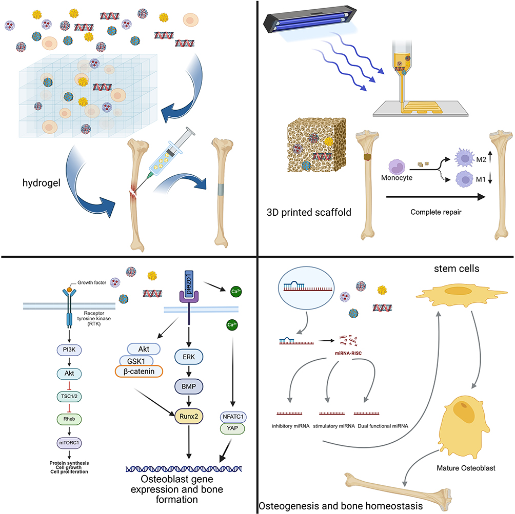 |
Figure 4 Nanoparticle-related derivative materials. |
Precision Nanoparticle Synthesis
The effectiveness of nanoparticles in bone regeneration is closely linked to the methods used for their synthesis, because these approaches determine their physicochemical characteristics and interactions within biological systems.149 Chemical synthesis methods, such as chemical reduction and sol-gel processes, allow for the fine-tuned control of particle size, shape, and crystallinity by manipulating nucleation and growth dynamics. Physical techniques, including laser ablation and ball milling, are valued for producing nanoparticles of high purity with minimal chemical residues, although additional processing may be required to achieve a uniform particle distribution. Recently, biological synthesis strategies have gained attention, utilizing plant extracts, fungi, or bacterial cultures to create nanoparticles with natural biocompatibility, aided by biomolecule-driven capping and stabilization mechanisms.150 Selecting an appropriate synthesis technique requires careful consideration of its compatibility with bone tissue environments and the intended functional outcomes. These advanced synthetic strategies provide a versatile platform for developing next-generation nanoparticle systems.
Smart Surface Engineering
In addition to precise synthesis, surface engineering and functionalization are essential for enhancing the performance of nanoparticles in bone regeneration. Techniques such as coating nanoparticles with biocompatible polymers, such as PEG or chitosan, can improve their stability, prolong circulation time, reduce immune responses, and promote cellular uptake.151 Functionalized nanoparticles with specific ligands, such as peptides or growth factors, enables targeted delivery to the bone tissue and supports cell adhesion and differentiation, further boosting osteogenic activity.152 Incorporating bioactive ions and adjusting the surface charge can stimulate bone formation and influence the interactions with the surrounding microenvironment. Moreover, designing nanoparticles to respond to local stimuli, such as pH or enzyme activity, allows for the controlled release of therapeutic agents at sites of bone injury or inflammation.153 Through these integrated surface modification strategies, the biocompatibility, bioactivity, and therapeutic precision of nanoparticles can be significantly improved, leading to more effective bone regeneration and improved tissue integration.
Hydrogel Nanocomposite Systems
Nanoparticles alone often exhibit limited biocompatibility, potentially triggering immune rejection and inflammatory responses.154 In addition, their inherent properties prevent stable drug release profiles. Researchers have addressed these limitations by incorporating nanoparticles into hydrogel matrices, which modulate release kinetics and provide structural stability for nanoparticle immobilization.155
Advanced Gel Formulations
Common gel matrices, including gelatin, sodium alginate, and polyacrylate, serve as effective carriers for enhancing nanoparticle stability.156 On this basis, nanogels with slow-release effects can be prepared and applied in the field of bone repair to improve the therapeutic effects.
Liu et al157 investigated osteogenesis mechanisms in diabetic microenvironments and developed a metformin-loaded zeolite imidazole framework (Met@ZIF-8)-modified hydrogel. This system exploited the acidic nature of diabetic inflammatory environments and the pH sensitivity of ZIF-8 to create targeted responses. By incorporating these nanoparticles into gelatin methacrylate, they produced a multifunctional composite hydrogel that was adaptable to irregular defects while mimicking the natural extracellular matrix. Further, they demonstrated a significantly increased number of CD206-positive macrophages, indicating excellent immunomodulatory properties along with robust ROS-scavenging capabilities, which significantly enhanced bone defect repair in ex vivo models.
Ji et al158 developed a biocompatible hydrogel with mechanical adaptability by loading adipose stem cell exosomes into gelatin nanoparticles. This system effectively modulates the macrophage phenotype through miR-451a regulation, promoting improved bone-healing outcomes.
Rinted Smart Scaffolds
Nanoparticles alone often fall short in effectively repairing bone defects of varying sizes and shapes, and conventional approaches such as autografts or metal implants also face limitations in achieving sustained therapeutic outcomes.159 3D printing technology emerges as a powerful solution, enabling the precise spatial distribution of nanoparticles within scaffolds to create hierarchical structures that mimic native bone. The integration of nanoparticles into 3D printed scaffolds is not merely additive; it is a strategic process to engineer composites that meet the multifaceted requirements of bone regeneration.160 For instance, the incorporation of nano-hydroxyapatite (nHA) or carbon nanotubes (CNTs) significantly enhances the compressive strength, modulus, and fracture toughness of the scaffold. nHA improves the biomineralization capacity, while CNTs, with their high aspect ratio and stiffness, efficiently transfer stress and inhibit crack propagation, leading to a scaffold with mechanical properties closer to those of natural bone.161 This addresses the critical need for a scaffold that is initially rigid enough to withstand physiological loads.
As demonstrated by Aisling Dunne et al, functionalized nHA within ECM scaffolds can directly influence cellular behavior, such as driving macrophage polarization from the pro-inflammatory M1 to the pro-regenerative M2 phenotype, which is crucial for modulating the inflammatory phase and transitioning to the repair phase.162 The high surface area of nanoparticles makes them ideal carriers for therapeutic agents (growth factors, antibiotics, miRNAs).163 When embedded in a 3D printed scaffold, they create a localized, sustained release system. This overcomes the limitations of systemic drug delivery and ensures a prolonged therapeutic effect at the defect site.
On the other hand, Binghong Luo’s group focused on replicating the unique three-dimensional microenvironment provided by the native bone ECM. They prepared chitosan whiskers via acid hydrolysis, which, after ultrasonic treatment in ionized water, exhibited a liquid crystal phase similar to that of collagen in bone ECM. These chitosan whiskers were then incorporated into hydrogels to mimic the viscoelastic properties and structural features of bone ECM. The integration of these hydrogels with 3D-printed poly(L-lactic acid) (PLLA) scaffolds, along with the addition of phytic acid to impart antimicrobial activity, resulted in a composite scaffold that supported cell proliferation, osteogenic differentiation, and angiogenesis. In vivo studies further demonstrated the scaffold’s ability to promote both vascularization and new bone formation, highlighting its potential for clinical bone regeneration applications.164
Signaling Pathway Nanodrugs
Nanotechnology offers unprecedented opportunities to modulate cellular behavior by precisely targeting the key signaling pathways involved in bone repair.165 Wang et al166 designed a 3D bioprinted scaffold incorporating BMSCs, RAW264.7, and MSNs loaded with BMP-4. This multifunctional scaffold exhibited excellent biocompatibility, and the sustained release of BMP-4 effectively triggered the osteogenic differentiation of BMSCs and osteoblasts through activation of the Smad signaling pathway, highlighting its therapeutic potential. Pan et al167 achieved controlled macrophage polarization by introducing IL-4-loaded calcium alginate-gelatin (Ca-GG) hydrogel microbeads. This strategy promoted the osteogenic differentiation of BMSCs via the TGF- 1/Smad pathway, further demonstrating the value of modulating the cellular microenvironment for bone regeneration.
With in-depth research in the field of genetic stem cells and exploration of nanoparticles, we have successfully realized the combination of nanoparticles with genetic stem cells, which has led to excellent results in the field of bone repair and is expected to bring new therapeutic approaches for bone healing.168 Addressing the challenge of hypoxia in bone defect sites, Sun et al62 developed an intelligent “bone microenvironment-regulating hydrogel” by encapsulating oxygen-carrying perfluorocarbons within oxygen-generating PLGA/PPS nanoparticles. This system reverses local hypoxic conditions, thereby upregulating BMAL1 expression in osteoblasts and enhancing osteogenic differentiation via the Nrf2-BMAL1 pathway. When combined with CPPL/GelMA hydrogels to further promote autophagy, these nanoparticles not only improved osteogenic outcomes, but also significantly accelerated bone tissue regeneration, offering a promising strategy for clinical bone repair.
At the level of genetic regulation, Cai et al169 introduced an innovative delivery platform for miR-29c using tetrahedral framework nucleic acids. This approach increases the bioavailability and efficacy of miR-29c, leading to enhanced activation of the Wnt signaling pathway and promotion of osteogenic activity in BMSCs. This has resulted in a marked improvement in bone tissue regeneration, opening new avenues for research and applications in bone tissue engineering.
Theranostic Nanoplatforms
To address the limited bioactivity of conventional bone-substitute materials, Pan et al170 developed copper- and manganese-doped borosilicate nanoparticles with a dual network structure. These nanoparticles not only supported the self-regulation of stem cell osteogenic differentiation but also promoted vascular regeneration by enhancing the proliferation of BMSCs. Notably, the controlled release of Cu2⁺ and Mn3⁺ ions from these materials led to the generation of hydroxyl radicals, which induce apoptosis in tumor cells. This innovative approach thus provides dual therapeutic benefits, facilitating bone regeneration while offering potential anticancer effects. El-Kamel et al171 employed a green synthesis method using cranberry extract to produce stabilized AgNPs with tunable size and morphology. This plant-based approach enables scalable production without the complexity of traditional cell cultures. The resulting AgNPs exhibited potent antimicrobial activity by disrupting cellular respiratory chains and division processes as well as by releasing bactericidal silver ions. In infected wound models, particularly those involving Staphylococcus aureus, these nanoparticles demonstrate significant antimicrobial efficacy and promote healing in a rat full-thickness skin excision model, underscoring their promise for future clinical applications.
Lee et al172 developed a novel complex by conjugating AuNPs with alendronate. This composite exhibited enhanced adhesion to bone surfaces and effectively inhibited osteoclast formation in a dose-dependent manner. These properties highlight its potential as a promising therapeutic agent for managing bone diseases.
Summary and Outlook
Rapid progress in nanotechnology and nanomedicine has positioned a wide array of nanomaterials, especially nanoparticles, as highly promising tools for biomedical applications.173 Metal-based nanoparticles offer several unique advantages: they can be manipulated using electromagnetic fields, possess strong antimicrobial properties, efficiently deliver bioactive molecules, and exhibit notable mechanical strength. These features render them particularly valuable for imaging, sensing, and drug delivery.64 In contrast, polymer-based nanoparticles provide superior thermal stability compared with traditional delivery systems and can be chemically tailored to carry various therapeutic agents. This versatility supports enhanced cell proliferation, new bone tissue formation, and controlled scaffold biodegradation.174 Although nanoparticles have shown great potential in several fields, their toxicity, stability issues, and production challenges limit their widespread use. Improvements are required to utilize this potential better. The following is an overview of the drawbacks, improvements, and future trends of existing nanomaterials.
Currently, the potential toxicity of nanoparticles or the adverse reactions they may cause limit their use in biomedical applications. In vivo or during storage, the stability of nanoparticles may suffer, which in turn may reduce their performance. The production of nanoparticles with consistent sizes and shapes is a challenging task essential for ensuring their functionality. In addition, the manufacture and application of nanoparticles may have a negative impact on the environment, especially in terms of waste disposal and recycling.
Future research should focus on the development of multifunctional composite nanoparticles that combine therapeutic and diagnostic functions on a single platform. These integrated systems hold great promise for advancements in cancer therapy, targeted drug delivery, and disease diagnostics. Furthermore, as our understanding of disease mechanisms and patient variability deepens, there is likely to be a shift towards designing personalized nanoparticles tailored to individual treatment needs. This personalized approach can significantly enhance therapeutic outcomes, while minimizing side effects.
The potential of nanoparticles for bone repair and regeneration is being increasingly recognized by the scientific community. By improving the functional performance and safety profiles of nanoparticles and adopting innovative manufacturing techniques and personalized therapeutic strategies, these materials are poised to have a transformative impact in bone repair, regenerative medicine, and related biomedical fields. Addressing fundamental issues such as toxicity, stability, and reproducibility is essential to facilitate the successful clinical translation of nanoparticle-based technologies and ultimately improve patient outcomes.
Ethical Approval and Consent to Participate
This study did not involve human participants, animals, or sensitive data requiring ethical approval. Therefore, no ethical approval or informed consent was necessary.
Author Contributions
All authors made a significant contribution to the work reported, whether that is in the conception, study design, execution, acquisition of data, analysis and interpretation, or in all these areas; took part in drafting, revising or critically reviewing the article; gave final approval of the version to be published; have agreed on the journal to which the article has been submitted; and agree to be accountable for all aspects of the work.
Funding
This research was funded by Basic Public Welfare Research Program of Zhejiang Province Award, Award Number: TGY24H060025. Recipient: Chen Xia.
Disclosure
The authors declare that they have no competing interests.
References
1. He T, Qin L, Chen S, et al. Bone-derived factors mediate crosstalk between skeletal and extra-skeletal organs. Bone Res. 2025;13(1).
2. Sheehy EJ, von Diemling C, Ryan E, et al. Antibiotic-eluting scaffolds with responsive dual-release kinetics facilitate bone healing and eliminate S. aureus infection. Biomaterials. 2025;313:122774.
3. Zhang Z, Li B, Zhang C, et al. Nanotechnology for understanding the tumor microenvironment heterogeneity in bone carcinoma. Adv Compos Hybrid Mater. 2025;8(2). doi:10.1007/s42114-025-01293-2
4. Jiang L, Zhang Y, Guo Z, Zhang Z, Wang C. Enhancing bone defect repair and acute wound healing: the synergistic effects of nanodiamond-enhanced gelatin methacryloyl hydrogels. ACS Appl Mater Interfaces. 2025;17(26):37421–37434. doi:10.1021/acsami.5c02573
5. Aleksova J, Ebeling P, Elder G. The effects of type 1 and type 2 diabetes mellitus on bone health in chronic kidney disease. Nat Rev Endocrinol. 2025;21(5):301–313.
6. Cheng J, Wang H, Li M, et al. Dual‐biomimetic bone adhesive with osteoimmunomodulatory capabilities for anatomical reconstruction of comminuted fractures. Adv Sci. 2025;12(32). doi:10.1002/advs.202501108
7. Duda GN, Geissler S, Checa S, Tsitsilonis S, Petersen A, Schmidt-Bleek K. The decisive early phase of bone regeneration. Nat Rev Rheumatol. 2023;19(2):78–95. doi:10.1038/s41584-022-00887-0
8. Tsang S-TJ, van Rensburg AJ, van Heerden J, Epstein GZ, Venter R, Ferreira N. The management of critical bone defects: outcomes of a systematic approach. Eurn J Orthop Surg Traumatol. 2024;34(6):3225–3231. doi:10.1007/s00590-024-04050-1
9. Wu Y, Song P, Wang M, Liu H, Jing Y, Su J. Extracellular derivatives for bone metabolism. J Adv Res. 2024;66:329–347. doi:10.1016/j.jare.2024.01.011
10. Robin M, Mouloungui E, Castillo Dali G, et al. Mineralized collagen plywood contributes to bone autograft performance. Nature. 2024;636(8041):100–107. doi:10.1038/s41586-024-08208-z
11. Zhang Q, Liu Z, He Y, et al. Osteoimmunity-Regulating biospun 3D silk scaffold for bone regeneration in critical-size defects. J Adv Res. 2025;2025.
12. Jiang X-R, Yang H-Y, Zhang -X-X, et al. Repair of bone defects with prefabricated vascularized bone grafts and double-labeled bone marrow-derived mesenchymal stem cells in a rat model. Sci Rep. 2017;7(1).
13. Wei H, Cui J, Lin K, Xie J, Wang X. Recent advances in smart stimuli-responsive biomaterials for bone therapeutics and regeneration. Bone Res. 2022;10(1). doi:10.1038/s41413-021-00180-y
14. Wu H, Huang J, Wu H, et al. Enhancement of in vitro and in vivo bone repair performance of decalcified bone/gelma by desferrioxamine. Sci Rep. 2025;15(1):14092.
15. Quan H, Ren C, He Y, Wang F, Dong S, Jiang H. Application of biomaterials in treating early osteonecrosis of the femoral head: research progress and future perspectives. Acta Biomater. 2023;164:15–73. doi:10.1016/j.actbio.2023.04.005
16. Grol MW. The evolving landscape of gene therapy strategies for the treatment of osteoarthritis. Osteoarthritis Cartilage. 2024;32(4):372–384. doi:10.1016/j.joca.2023.12.009
17. Jeffery EC. The role of hematopoiesis in bone repair: an update. Current Opinion Hematol. 2024;31(4):163–167. doi:10.1097/MOH.0000000000000821
18. Maruyama M, Rhee C, Utsunomiya T, et al. Modulation of the inflammatory response and bone healing. Front Endocrinol. 2020;11:11. doi:10.3389/fendo.2020.00011
19. Dang Y, Zhang Y, Luo G, et al. The decisive early phase of biomaterial-induced bone regeneration. Appl Mater Today. 2024;38:102236.
20. Grundnes O, Reikerås O. The importance of the hematoma for fracture healing in rats. Acta Orthop Scand. 2009;64(3):340–342. doi:10.3109/17453679308993640
21. Torres HM, Arnold KM, Oviedo M, Westendorf JJ, Weaver SR. Inflammatory processes affecting bone health and repair. Curr Osteoporosis Rep. 2023;21(6):842–853. doi:10.1007/s11914-023-00824-4
22. Shu L-Z, Zhang X-L, Ding Y-D, Lin H. From inflammation to bone formation: the intricate role of neutrophils in skeletal muscle injury and traumatic heterotopic ossification. Exp Mol Med. 2024;56(7):1523–1530. doi:10.1038/s12276-024-01270-7
23. Lynch CA, Acosta SA, Anderson DM, Rogers GE, Wilson-Rawls J, Rawls A. The transcription factor mohawk facilitates skeletal muscle repair via modulation of the inflammatory environment. Int J Mol Sci. 2024;25(9):5019. doi:10.3390/ijms25095019
24. Fullerton JN, Gilroy DW. Resolution of inflammation: a new therapeutic frontier. Nat Rev Drug Discov. 2016;15(8):551–567. doi:10.1038/nrd.2016.39
25. Megha KB, Joseph X, Akhil V, Mohanan PV. Cascade of immune mechanism and consequences of inflammatory disorders. Phytomedicine. 2021;91:153712.
26. Soares CLR, Wilairatana P, Silva LR, et al. Biochemical aspects of the inflammatory process: a narrative review. Biomed Pharmacother. 2023;168:115764.
27. Chen S, Saeed AFUH, Liu Q, et al. Macrophages in immunoregulation and therapeutics. Signal Transduct Target Ther. 2023;8(1):207.
28. McCauley J, Bitsaktsis C, Cottrell J. Macrophage subtype and cytokine expression characterization during the acute inflammatory phase of mouse bone fracture repair. J Orthop Res. 2020;38(8):1693–1702. doi:10.1002/jor.24603
29. Zhang C, Li D, Wang Z, et al. M1 type macrophage targeted anti-inflammatory exosomes derived from BMSC for the treatment of acute and chronic inflammatory diseases. Mater Des. 2024;240:112844.
30. Bender EC, Tareq HS, Suggs LJ. Inflammation: a matter of immune cell life and death. Npj Biomed Innov. 2025;2(1). doi:10.1038/s44385-025-00010-4
31. Hou J, Shi J, Chen L, et al. M2 macrophages promote myofibroblast differentiation of LR-MSCs and are associated with pulmonary fibrogenesis. Cell Commun Signal. 2018;16(1). doi:10.1186/s12964-018-0300-8
32. Mountziaris PM, Mikos AG. Modulation of the inflammatory response for enhanced bone tissue regeneration. Tissue Eng Part B Rev. 2008;14(2):179–186. doi:10.1089/ten.teb.2008.0038
33. Wynn TA, Vannella KM. Macrophages in tissue repair, regeneration, and fibrosis. Immunity. 2016;44(3):450–462. doi:10.1016/j.immuni.2016.02.015
34. Ren Q, Xing W, Jiang B, et al. Tenascin-C promotes bone regeneration via inflammatory macrophages. Cell Death Differ. 2025;32(4):763–775. doi:10.1038/s41418-024-01429-9
35. Koh TJ, DiPietro LA. Inflammation and wound healing: the role of the macrophage. Expert Rev Mol Med. 2011;13:e23.
36. Kenkre JS, Bassett JHD. The bone remodelling cycle. Ann Clin Biochem. 2018;55(3):308–327. doi:10.1177/0004563218759371
37. Coates BA, McKenzie JA, Buettmann EG, et al. Transcriptional profiling of intramembranous and endochondral ossification after fracture in mice. Bone. 2019;127:577–591. doi:10.1016/j.bone.2019.07.022
38. Farjaminejad S, Farjaminejad R, Garcia-Godoy F. Nanoparticles in bone regeneration: a narrative review of current advances and future directions in tissue engineering. J Funct Biomater. 2024;15(9):241.
39. Kanczler JM, Oreffo ROC. Osteogenesis and angiogenesis: the potential for engineering bone. Eur Cells Mater. 2008;15:100–114. doi:10.22203/eCM.v015a08
40. Gerstenfeld LC, Cho TJ, Kon T, et al. Impaired fracture healing in the absence of TNF-α signaling: the role of TNF-α in endochondral cartilage resorption. J Bone Miner Res. 2003;18(9):1584–1592. doi:10.1359/jbmr.2003.18.9.1584
41. Ju Y, Kent SJ. Balancing stealth and targeting to improve nanomedicine efficacy. Nature Nanotechnol. 2025;20(5):576–579. doi:10.1038/s41565-025-01926-z
42. Bolamperti S, Villa I, Rubinacci A. Bone remodeling: an operational process ensuring survival and bone mechanical competence. Bone Res. 2022;10(1). doi:10.1038/s41413-022-00219-8
43. Chutani K, Rai N, Sardar A, et al. Fortified Withaferin A accelerates the transition from fibrovascular to bone remodeling phase during endochondral bone formation to promote ossification. Front Endocrinol. 2025;16. doi:10.3389/fendo.2025.1540237
44. Schindeler A, McDonald MM, Bokko P, Little DG. Bone remodeling during fracture repair: the cellular picture. Semin Cell Dev Biol. 2008;19(5):459–466. doi:10.1016/j.semcdb.2008.07.004
45. Marsell R, Einhorn TA. The biology of fracture healing. Injury. 2011;42(6):551–555. doi:10.1016/j.injury.2011.03.031
46. Frost HM. Skeletal structural adaptations to mechanical usage (SATMU): 2. Redefining Wolff’s law: the remodeling problem. Anat Record. 2005;226(4):414–422. doi:10.1002/ar.1092260403
47. Huang X-Y, Zhou X-X, Yang H, et al. Directed osteogenic differentiation of human bone marrow mesenchymal stem cells via sustained release of BMP4 from PBVHx-based nanoparticles. Int J Biol Macromol. 2024;265:130649.
48. Rambhia KJ, Sun H, Feng K, et al. Nanofibrous 3D scaffolds capable of individually controlled BMP and FGF release for the regulation of bone regeneration. Acta Biomater. 2024;190:50–63. doi:10.1016/j.actbio.2024.10.044
49. Liu S, Liu D, Chen C, et al. MSC transplantation improves osteopenia via epigenetic regulation of notch signaling in lupus. Cell Metab. 2015;22(4):606–618. doi:10.1016/j.cmet.2015.08.018
50. Hankenson KD, Gagne K, Shaughnessy M. Extracellular signaling molecules to promote fracture healing and bone regeneration. Adv Drug Delivery Rev. 2015;94:3–12. doi:10.1016/j.addr.2015.09.008
51. Kim K, Lee S-H, Ha kim J, Choi Y, Kim N. NFATc1 Induces osteoclast fusion via up-regulation of Atp6v0d2 and the dendritic cell-specific transmembrane protein (DC-STAMP). Mol Endocrinol. 2008;22(1):176–185. doi:10.1210/me.2007-0237
52. Nakashima T, Kobayashi Y, Yamasaki S, et al. Protein expression and functional difference of membrane-bound and soluble receptor activator of NF-κB ligand: modulation of the expression by osteotropic factors and cytokines. Biochem Biophys Res Commun. 2000;275(3):768–775. doi:10.1006/bbrc.2000.3379
53. Hu K, Olsen BR. The roles of vascular endothelial growth factor in bone repair and regeneration. Bone. 2016;91:30–38.
54. Emerich DF, Thanos CG. Targeted nanoparticle-based drug delivery and diagnosis. J Drug Targeting. 2008;15(3):163–183. doi:10.1080/10611860701231810
55. Arcos D, Gómez-Cerezo N, Saiz-Pardo M, et al. Injectable mesoporous bioactive nanoparticles regenerate bone tissue under osteoporosis conditions. Acta Biomater. 2022;151:501–511. doi:10.1016/j.actbio.2022.07.067
56. Blanco E, Shen H, Ferrari M. Principles of nanoparticle design for overcoming biological barriers to drug delivery. Nature Biotechnol. 2015;33(9):941–951. doi:10.1038/nbt.3330
57. Jokerst JV, Gambhir SS. Molecular imaging with theranostic nanoparticles. Acc Chem Res. 2011;44(10):1050–1060. doi:10.1021/ar200106e
58. Mitchell MJ, Billingsley MM, Haley RM, Wechsler ME, Peppas NA, Langer R. Engineering precision nanoparticles for drug delivery. Nat Rev Drug Discov. 2020;20(2):101–124. doi:10.1038/s41573-020-0090-8
59. Chen L, Hong W, Ren W, Xu T, Qian Z, He Z. Recent progress in targeted delivery vectors based on biomimetic nanoparticles. Signal Transduct Target Ther. 2021;6(1):962.
60. Efthimiadou EK, Theodosiou M, Toniolo G, Abu-Thabit NY. Stimuli-responsive biopolymer nanocarriers for drug delivery applications. In: Stimuli Responsive Polymeric Nanocarriers for Drug Delivery Applications. Vol. 1. 2018:405–432.
61. Nauth A, Schemitsch E, Norris B, Nollin Z, Watson JT. Critical-size bone defects: is there a consensus for diagnosis and treatment? J Orthop Trauma. 2018;32(3):S7–S11. doi:10.1097/BOT.0000000000001115
62. Sun H, Xu J, Wang Y, et al. Bone microenvironment regulative hydrogels with ROS scavenging and prolonged oxygen-generating for enhancing bone repair. Bioact Mater. 2023;24:477–496. doi:10.1016/j.bioactmat.2022.12.021
63. Zhang YS, Khademhosseini A. Advances in engineering hydrogels. Science. 2017;356(6337). doi:10.1126/science.aaf3627
64. Wang N, Fuh JYH, Dheen ST, Senthil Kumar A. Functions and applications of metallic and metallic oxide nanoparticles in orthopedic implants and scaffolds. J Biomed Mater Res Part B. 2020;109(2):160–179. doi:10.1002/jbm.b.34688
65. Kiio TM, Park S. Physical properties of nanoparticles do matter. J Pharm Invest. 2020;51(1):35–51. doi:10.1007/s40005-020-00504-w
66. Gupta A, Singh S. Multimodal potentials of gold nanoparticles for bone tissue engineering and regenerative medicine: avenues and prospects. Small. 2022;18(29). doi:10.1002/smll.202201462
67. Qiao M, Tang W, Xu Z, et al. Gold nanoparticles: promising biomaterials for osteogenic/adipogenic regulation in bone repair. J Mat Chem B. 2023;11(11):2307–2333. doi:10.1039/D2TB02563A
68. Zhang D, Liu D, Zhang J, Fong C, Yang M. Gold nanoparticles stimulate differentiation and mineralization of primary osteoblasts through the ERK/MAPK signaling pathway. Mater Sci Eng C. 2014;42:70–77. doi:10.1016/j.msea.2013.12.066
69. Zhang Y, Wang P, Wang Y, et al. Gold nanoparticles promote the bone regeneration of periodontal ligament stem cell sheets through activation of autophagy. Int J Nanomed. 2021;16:61–73. doi:10.2147/IJN.S282246
70. Kus-Liśkiewicz M, Fickers P, Ben Tahar I. Biocompatibility and cytotoxicity of gold nanoparticles: recent advances in methodologies and regulations. Int J Mol Sci. 2021;22(20):10952.
71. Giri AK, Jena B, Biswal B, et al. Green synthesis and characterization of silver nanoparticles using Eugenia roxburghii DC. extract and activity against biofilm-producing bacteria. Sci Rep. 2022;12(1). doi:10.1038/s41598-022-12484-y
72. Zhang R, Lee P, Lui VCH, et al. Silver nanoparticles promote osteogenesis of mesenchymal stem cells and improve bone fracture healing in osteogenesis mechanism mouse model. Nanomed Nanotechnol Biol Med. 2015;11(8):1949–1959. doi:10.1016/j.nano.2015.07.016
73. Imtiyaz HZ, Simon MC. Hypoxia-Inducible Factors as Essential Regulators of Inflammation. In: Diverse Effects of Hypoxia on Tumor Progression. 2010:105–120.
74. Otari SV, Patil RM, Ghosh SJ, Thorat ND, Pawar SH. Intracellular synthesis of silver nanoparticle by actinobacteria and its antimicrobial activity. Spectrochimica Acta Part A. 2015;136:1175–1180. doi:10.1016/j.saa.2014.10.003
75. Xu L, Wang -Y-Y, Huang J, Chen C-Y, Wang Z-X, Xie H. Silver nanoparticles: synthesis, medical applications and biosafety. Theranostics. 2020;10(20):8996–9031. doi:10.7150/thno.45413
76. Singh K, Yadav S. Biosynthesis of a range of ZnO nanoparticles utilising Salvia hispanica L. seed extract and evaluation of their bioactivity. Sci Rep. 2025;15(1):4043.
77. Goodman SB, Pajarinen J, Yao Z, Lin T. Inflammation and bone repair: from particle disease to tissue regeneration. Front Bioeng Biotechnol. 2019;7:230.
78. Jarosz M, Olbert M, Wyszogrodzka G, Młyniec K, Librowski T. Antioxidant and anti-inflammatory effects of zinc. Zinc-dependent NF-κB signaling. Inflammopharmacology. 2017;25(1):11–24. doi:10.1007/s10787-017-0309-4
79. Chen X, Weng Z, Zhang H, et al. Nano-zinc oxide (nZnO) targets the AMPK-ULK1 pathway to promote bone regeneration. Stem Cell Res Ther. 2025;16(1):206.
80. Singh TA, Sharma A, Tejwan N, Ghosh N, Das J, Sil PC. A state of the art review on the synthesis, antibacterial, antioxidant, antidiabetic and tissue regeneration activities of zinc oxide nanoparticles. Adv Colloid Interface Sci. 2021;295:102495.
81. Zhou J, Wang H, Virtanen S, et al. Hybrid zinc oxide nanocoating on titanium implants: controlled drug release for enhanced antibacterial and osteogenic performance in infectious conditions. Acta Biomater. 2024;189:589–604. doi:10.1016/j.actbio.2024.09.039
82. Mohajeri M, Momenai R, Karami-Mohajeri S, Ohadi M, Raeisi Estabragh MA. Cerium oxide nanoparticles, physical and chemical properties, applications and toxicological implications: a review. Results Chem. 2025;15:102302.
83. Jiang L, Lin J, Zhao S, et al. ADAMTS5 in osteoarthritis: biological functions, regulatory network, and potential targeting therapies. Front Mol Biosci. 2021;8:703110.
84. Li J, Kang F, Gong X, et al. Ceria nanoparticles enhance endochondral ossification–based critical‐sized bone defect regeneration by promoting the hypertrophic differentiation of BMSCs via DHX15 activation. FASEB J. 2019;33(5):6378–6389. doi:10.1096/fj.201802187R
85. Wang R, He X, Bai J, et al. Cerium oxide nanoparticles-reinforced gelma hydrogel loading bone marrow stem cells with osteogenic and inflammatory regulatory capacity for bone defect repair. ACS Appl Mater Interfaces. 2024;16(49):67373–67384. doi:10.1021/acsami.4c15718
86. Jung Y-K, Park H-R, Cho H-J, et al. Degrading products of chondroitin sulfate can induce hypertrophy-like changes and MMP-13/ADAMTS5 production in chondrocytes. Sci Rep. 2019;9(1). doi:10.1038/s41598-019-52358-4
87. Tetsunaga T, Nishida K, Furumatsu T, et al. Regulation of mechanical stress-induced MMP-13 and ADAMTS-5 expression by RUNX-2 transcriptional factor in SW1353 chondrocyte-like cells. Osteoarthritis Cartilage. 2011;19(2):222–232. doi:10.1016/j.joca.2010.11.004
88. Jeyaraj M, Gurunathan S, Qasim M, Kang M-H, Kim J-H. A comprehensive review on the synthesis, characterization, and biomedical application of platinum nanoparticles. Nanomaterials. 2019;9(12):1719.
89. Xu W, Zhou H, Hu B, et al. Prussian blue‐derived nanocomposite synergized with calcium overload for three‐mode ROS outbreak generation to enhance oncotherapy. Adv Healthcare Mater. 2024;13(22). doi:10.1002/adhm.202400591
90. Richard MJ, Driban JB, McAlindon TE. Pharmaceutical treatment of osteoarthritis. Osteoarthritis Cartilage. 2023;31(4):458–466. doi:10.1016/j.joca.2022.11.005
91. Mueller M, Weinmann D, Toegel S, Holzer W, Unger FM, Viernstein H. Compounds from Caesalpinia sappan with anti-inflammatory properties in macrophages and chondrocytes. Food Funct. 2016;7(3):1671–1679. doi:10.1039/C5FO01256B
92. Rizzi F, Castaldo R, Latronico T, et al. High surface area mesoporous silica nanoparticles with tunable size in the sub-micrometer regime: insights on the size and porosity control mechanisms. Molecules. 2021;26(14):4247. doi:10.3390/molecules26144247
93. Lu J, Liong M, Li Z, Zink JI, Biocompatibility TF. Biodistribution, and drug‐delivery efficiency of mesoporous silica nanoparticles for cancer therapy in animals. Small. 2010;6(16):1794–1805. doi:10.1002/smll.201000538
94. Slowing I, Viveroescoto J, Wu C, Lin V. Mesoporous silica nanoparticles as controlled release drug delivery and gene transfection carriers☆. Adv Drug Delivery Rev. 2008;60(11):1278–1288. doi:10.1016/j.addr.2008.03.012
95. Wu J, Li H, Hu F, Luo P. Stevioside attenuates osteoarthritis via regulating Nrf2/HO-1/NF-κB pathway. J Orthop Transl. 2023;38:190–202. doi:10.1016/j.jot.2022.05.005
96. Kang W, Xu Q, Dong H, Wang W, Huang G, Zhang J. Eriodictyol attenuates osteoarthritis progression through inhibiting inflammation via the PI3K/AKT/NF-κB signaling pathway. Sci Rep. 2024;14(1):18853.
97. Czabotar PE, Garcia-Saez AJ. Mechanisms of BCL-2 family proteins in mitochondrial apoptosis. Nat Rev Mol Cell Biol. 2023;24(10):732–748. doi:10.1038/s41580-023-00629-4
98. Gu B, Zhao Q, Ao Y. Advances in immunomodulatory mesoporous silica nanoparticles for inflammatory and cancer therapies. Biomolecules. 2024;14(9):1057. doi:10.3390/biom14091057
99. Croissant JG, Butler KS, Zink JI, Brinker CJ. Synthetic amorphous silica nanoparticles: toxicity, biomedical and environmental implications. Nature Rev Mater. 2020;5(12):886–909.
100. Perumal S. Polymer nanoparticles: synthesis and applications. Polymers. 2022;14(24):5449. doi:10.3390/polym14245449
101. Avramović N, Mandić B, Savić-Radojević A, Simić T. Polymeric nanocarriers of drug delivery systems in cancer therapy. Pharmaceutics. 2020;12(4):298. doi:10.3390/pharmaceutics12040298
102. Zhou Z, Cui J, Wu S, Geng Z, Su J. Silk fibroin-based biomaterials for cartilage/osteochondral repair. Theranostics. 2022;12(11):5103–5124. doi:10.7150/thno.74548
103. Raja IS, Preeth DR, Vedhanayagam M, et al. Polyphenols-loaded electrospun nanofibers in bone tissue engineering and regeneration. Biomater Res. 2021;25(1). doi:10.1186/s40824-021-00229-3
104. Shi W, Gao Y, Wu Y, et al. A multifunctional polydopamine/genipin/alendronate nanoparticle licences fibrin hydrogels osteoinductive and immunomodulatory potencies for repairing bone defects. Int J Biol Macromol. 2023;249:126072.
105. Joyce K, Fabra GT, Bozkurt Y, Pandit A. Bioactive potential of natural biomaterials: identification, retention and assessment of biological properties. Signal Transduct Target Ther. 2021;6(1):175.
106. Elmowafy EM, Tiboni M, Soliman ME. Biocompatibility, biodegradation and biomedical applications of poly(lactic acid)/poly(lactic-co-glycolic acid) micro and nanoparticles. J Pharm Invest. 2019;49(4):347–380. doi:10.1007/s40005-019-00439-x
107. Stiufiuc GF, Stiufiuc RI. Magnetic nanoparticles: synthesis, characterization, and their use in biomedical field. Appl Sci. 2024;14(4):1623.
108. Xia Y, Sun J, Zhao L, et al. Magnetic field and nano-scaffolds with stem cells to enhance bone regeneration. Biomaterials. 2018;183:151–170. doi:10.1016/j.biomaterials.2018.08.040
109. Wu D, Chang X, Tian J, et al. Bone mesenchymal stem cells stimulation by magnetic nanoparticles and a static magnetic field: release of exosomal miR-1260a improves osteogenesis and angiogenesis. J Nanobiotechnol. 2021;19(1). doi:10.1186/s12951-021-00958-6
110. Nowak-Jary J, Machnicka B. In vivo biodistribution and clearance of magnetic iron oxide nanoparticles for medical applications. Int J Nanomed. 2023;18:4067–4100. doi:10.2147/IJN.S415063
111. Chen Y, Li X. The utilization of carbon-based nanomaterials in bone tissue regeneration and engineering: respective featured applications and future prospects. Med Novel Technol Dev. 2022;16:100168. doi:10.1016/j.medntd.2022.100168
112. Syduzzaman M, Islam Saad MS, Piam MF, Talukdar TA, Shobdo TT, Pritha NM. Carbon nanotubes: structure, properties and applications in the aerospace industry. Results Mater. 2025;25:100654.
113. Kim T, Sridharan I, Zhu B, Orgel J, Wang R. Effect of CNT on collagen fiber structure, stiffness assembly kinetics and stem cell differentiation. Mater Sci Eng. 2015;49:281–289. doi:10.1016/j.msec.2015.01.014
114. Assali M, Kittana N, Alhaj-Qasem S, et al. Noncovalent functionalization of carbon nanotubes as a scaffold for tissue engineering. Sci Rep. 2022;12(1). doi:10.1038/s41598-022-16247-7
115. Sen S, Bose A. Carbon dots: a review of innovations, applications, challenges, and future prospects. Inorg Chem Commun. 2025;173:113852.
116. Das R, Bandyopadhyay R, Pramanik P. Carbon quantum dots from natural resource: a review. Mater Today Chem. 2018;8:96–109. doi:10.1016/j.mtchem.2018.03.003
117. Elkun S, Ghali M, Sharshar T, Mosaad MM. Green synthesis of fluorescent N-doped carbon quantum dots from castor seeds and their applications in cell imaging and pH sensing. Sci Rep. 2024;14(1). doi:10.1038/s41598-024-78745-0
118. Majood M, Garg P, Chaurasia R, Agarwal A, Mohanty S, Mukherjee M. Carbon quantum dots for stem cell imaging and deciding the fate of stem cell differentiation. ACS Omega. 2022;7(33):28685–28693. doi:10.1021/acsomega.2c03285
119. Donato KZ, Tan HL, Marangoni VS, et al. Graphene oxide classification and standardization. Sci Rep. 2023;13(1). doi:10.1038/s41598-023-33350-5
120. Zare P, Aleemardani M, Seifalian A, Bagher Z, Seifalian AM. Graphene oxide: opportunities and challenges in biomedicine. Nanomaterials. 2021;11(5):1083. doi:10.3390/nano11051083
121. Luanpitpong S, Wang L, Rojanasakul Y. The effects of carbon nanotubes on lung and dermal cellular behaviors. Nanomedicine. 2014;9(6):895–912. doi:10.2217/nnm.14.42
122. Ma X, Luan Z, Zhao Q, Yang A, Li J. NIR-triggered release of nitric oxide by upconversion-based nanoplatforms to enhance osteogenic differentiation of mesenchymal stem cells for osteoporosis therapy. Biomater Res. 2024;28:0058.
123. Prakash A, Yadav S, Yadav U, Saxena PS, Srivastava A. Recent advances on nitrogen-doped carbon quantum dots and their applications in bioimaging: a review. Bull Mater Sci. 2023;46(1). doi:10.1007/s12034-022-02846-7
124. Chen Y-F, Luh F, Ho Y-S, Yen Y. Exosomes: a review of biologic function, diagnostic and targeted therapy applications, and clinical trials. J Biomed Sci. 2024;31(1). doi:10.1186/s12929-024-01055-0
125. Kim HI, Park J, Zhu Y, Wang X, Han Y, Zhang D. Recent advances in extracellular vesicles for therapeutic cargo delivery. Exp Mol Med. 2024;56(4):836–849. doi:10.1038/s12276-024-01201-6
126. Li L, Wang F, Zhu D, Hu S, Cheng K, Li Z. Engineering exosomes and exosome-like nanovesicles for improving tissue targeting and retention. Fundam Res. 2025;5(2):851–867. doi:10.1016/j.fmre.2024.03.025
127. Wu Y, Wan S, Yang S, et al. Macrophage cell membrane-based nanoparticles: a new promising biomimetic platform for targeted delivery and treatment. J Nanobiotechnol. 2022;20(1). doi:10.1186/s12951-022-01746-6
128. Noga M, Milan J, Frydrych A, Jurowski K. Toxicological aspects, safety assessment, and green toxicology of silver nanoparticles (AgNPs)—critical review: state of the art. Int J Mol Sci. 2023;24(6):5133.
129. Gulab H, Fatima N, Tariq U, et al. Advancements in zinc oxide nanomaterials: synthesis, properties, and diverse applications. Nano-Struct Nano-Objects. 2024;39:101271.
130. Agarwal H, Nakara A, Shanmugam VK. Anti-inflammatory mechanism of various metal and metal oxide nanoparticles synthesized using plant extracts: a review. Biomed Pharmacother. 2019;109:2561–2572. doi:10.1016/j.biopha.2018.11.116
131. Yang C, Liao X, Wang Y, et al. Biomimetic nanoplatform integrating CeO2 nanozymes and anti‐inflammatory peptides for osteoarthritis therapy. Small. 2025;21(32):2502677.
132. Jan H, Gul R, Andleeb A, et al. A detailed review on biosynthesis of platinum nanoparticles (PtNPs), their potential antimicrobial and biomedical applications. J Saudi Chem Soc. 2021;25(8):101297. doi:10.1016/j.jscs.2021.101297
133. Rai M, Ingle AP, Birla S, Yadav A, Santos CAD. Strategic role of selected noble metal nanoparticles in medicine. Crit Rev Microbiol. 2015;1–24. doi:10.3109/1040841X.2015.1018131
134. Sokolova V, Epple M. Biological and medical applications of calcium phosphate nanoparticles. Chem Eur J. 2021;27(27):7471–7488. doi:10.1002/chem.202005257
135. Liu Z, Yamada S, Otsuka Y, Peñaflor Galindo TG, Tagaya M. Surface modification of hydroxyapatite nanoparticles for bone regeneration by controlling their surface hydration and protein adsorption states. Dalton Trans. 2022;51(25):9572–9583. doi:10.1039/D2DT00969B
136. Dutta Gupta Y, Mackeyev Y, Krishnan S, Bhandary S. Mesoporous silica nanotechnology: promising advances in augmenting cancer theranostics. Cancer Nanotechnol. 2024;15(1):9.
137. Mora‐Raimundo P, Lozano D, Benito M, Mulero F, Manzano M, Vallet‐Regí M. Osteoporosis remission and new bone formation with mesoporous silica nanoparticles. Adv Sci. 2021;8(16). doi:10.1002/advs.202101107
138. Xu C, Xia X, Liu G, Wang Y, Xiao B. Zwitterionic polymers improve the therapeutic effect of silk fibroin-based nanomedicines against ulcerative colitis via the oral route. Precision Med Eng. 2025;2(2):100027. doi:10.1016/j.preme.2025.100027
139. Venkatesh Y, Naidu PVS, Rao PA, Kundrapu DB. Green synthesis of multifunctional nitrogen-doped carbon quantum dots (N-CQDs) derived from citrus lemon juice: applications on antioxidant and antimicrobial activities and highly selective detection for Fe3+ ion sensing. Res Chem Intermed. 2025;51(5):2621–2643. doi:10.1007/s11164-025-05567-8
140. Griffin KV, Saunders MN, Lyssiotis CA, Shea LD. Engineering immunity using metabolically active polymeric nanoparticles. Trends Biotechnol. 2025;43(6):1371–1384. doi:10.1016/j.tibtech.2024.11.016
141. Qiao R, Fu C, Forgham H, et al. Magnetic iron oxide nanoparticles for brain imaging and drug delivery. Adv Drug Delivery Rev. 2023;197. doi:10.1016/j.addr.2023.114822
142. Tang S-J, Rotherham M, El Haj AJ. Remote activation of the Wnt/β-Catenin signalling pathway using functionalised magnetic particles. PLoS One. 2015;10(3):e0121761.
143. Ijaz H, Mahmood A, Abdel-Daim MM, et al. Review on carbon nanotubes (CNTs) and their chemical and physical characteristics, with particular emphasis on potential applications in biomedicine. Inorg Chem Commun. 2023;155:111020.
144. Yang W, Li C, Han L. Mechanical properties of polycaprolactone bone scaffolds reinforced with carbon nanotube-modified tricalcium phosphate. Carbon Lett. 2024;35(1):267–276. doi:10.1007/s42823-024-00788-0
145. Fu C, Yang X, Tan S, Song L. Enhancing cell proliferation and osteogenic differentiation of MC3T3-E1 pre-osteoblasts by BMP-2 delivery in graphene oxide-incorporated PLGA/HA biodegradable microcarriers. Sci Rep. 2017;7(1). doi:10.1038/s41598-017-12935-x
146. Pei S, Cai S, Yan K, Zhou J, Luo K, Chen X. Synthesis of N-doped carbon quantum dots as an effective fluorescent sensor of Fe3+ Ions and a potent antibacterial agent. J Fluoresc. 2025;35(8):7339–7348. doi:10.1007/s10895-024-04112-x
147. Geng B, Li P, Fang F, et al. Antibacterial and osteogenic carbon quantum dots for regeneration of bone defects infected with multidrug-resistant bacteria. Carbon. 2021;184:375–385. doi:10.1016/j.carbon.2021.08.040
148. Sridhar S, Venugopal JR, Sridhar R, Ramakrishna S. Cardiogenic differentiation of mesenchymal stem cells with gold nanoparticle loaded functionalized nanofibers. Colloids Surf B. 2015;134:346–354. doi:10.1016/j.colsurfb.2015.07.019
149. Zhang G, Zhen C, Yang J, et al. Recent advances of nanoparticles on bone tissue engineering and bone cells. Nanoscale Adv. 2024;6(8):1957–1973. doi:10.1039/D3NA00851G
150. Khan H, Piccolella S, Pacifico S. Harnessing plant extracts for green nanoparticle synthesis: toward a sustainable future. Mater Today Sustainability. 2025;31.
151. Swarupa S, Thareja P. Techniques, applications and prospects of polysaccharide and protein based biopolymer coatings: a review. Int J Biol Macromol. 2024;266:131104.
152. Wietrzyk A, Hutny A, Jonderko L, et al. Protein-functionalized nanoparticles: emerging strategies in drug delivery. Biomed Pharmacother. 2025;189:118337.
153. Valentino A, Yazdanpanah S, Conte R, Calarco A, Peluso G. Smart nanocomposite hydrogels as next-generation therapeutic and diagnostic solutions. Gels. 2024;10(11):689. doi:10.3390/gels10110689
154. Salatin S, Maleki Dizaj S, Yari Khosroushahi A. Effect of the surface modification, size, and shape on cellular uptake of nanoparticles. Cell Biol Int. 2015;39(8):881–890. doi:10.1002/cbin.10459
155. Gao W, Zhang Y, Zhang Q, Zhang L. Nanoparticle-Hydrogel: a hybrid biomaterial system for localized drug delivery. Ann Biomed Eng. 2016;44(6):2049–2061. doi:10.1007/s10439-016-1583-9
156. Dash TK, Konkimalla VB. Poly-є-caprolactone based formulations for drug delivery and tissue engineering: a review. J Control Release. 2012;158(1):15–33. doi:10.1016/j.jconrel.2011.09.064
157. Lao A, Wu J, Li D, et al. Functionalized metal–organic framework‐modified hydrogel that breaks the vicious cycle of inflammation and ROS for repairing of diabetic bone defects. Small. 2023;19(36). doi:10.1002/smll.202206919
158. Li R, Li D, Wang H, et al. Exosomes from adipose-derived stem cells regulate M1/M2 macrophage phenotypic polarization to promote bone healing via miR-451a/MIF. Stem Cell Res Ther. 2022;13(1). doi:10.1186/s13287-022-02975-0
159. Kim Y, Kim J, Kim B, et al. Discovery and optimization of a series of vinyl sulfoximine-based analogues as potent Nrf2 activators for the treatment of multiple sclerosis. J Med Chem. 2024;67(19):17866–17892. doi:10.1021/acs.jmedchem.4c01907
160. Huang B, Li G, Cao L, et al. Nanoengineered 3D-printing scaffolds prepared by metal-coordination self-assembly for hyperthermia-catalytic osteosarcoma therapy and bone regeneration. J Colloid Interface Sci. 2024;672:724–735. doi:10.1016/j.jcis.2024.06.055
161. Elmofty AR, Abdel Aziz ME, Tash M, El-Hadad S. Development and characterization of hydroxyapatite and multiwall carbon nanotubes reinforced polypropylene biocomposites. Sci Rep. 2025;15(1). doi:10.1038/s41598-025-96082-8
162. Mahon OR, Browe DC, Gonzalez-Fernandez T, et al. Nano-particle mediated M2 macrophage polarization enhances bone formation and MSC osteogenesis in an IL-10 dependent manner. Biomaterials. 2020;239:119833.
163. Chenxi Z, Hemmat A, Thi NH, Afrand M. Nanoparticle-enhanced drug delivery systems: an up-to-date review. J Mol Liq. 2025;424:126999.
164. Li Y, Tang S, Luo Z, et al. Chitin whisker/chitosan liquid crystal hydrogel assisted scaffolds with bone-like ECM microenvironment for bone regeneration. Carbohydr Polym. 2024;332:121927.
165. Marino A, Battaglini M, Tapeinos C, Larrañaga A, Ciofani G. Innovative nanotechnology tools for the functional control and tracking of human stem cells. Mater Today Adv. 2022;16:100298. doi:10.1016/j.mtadv.2022.100298
166. Sun X, Ma Z, Zhao X, et al. Three-dimensional bioprinting of multicell-laden scaffolds containing bone morphogenic protein-4 for promoting M2 macrophage polarization and accelerating bone defect repair in diabetes mellitus. Bioact Mater. 2021;6(3):757–769. doi:10.1016/j.bioactmat.2020.08.030
167. Zhang J, Shi H, Zhang N, Hu L, Jing W, Pan J. Interleukin‐4‐loaded hydrogel scaffold regulates macrophages polarization to promote bone mesenchymal stem cells osteogenic differentiation via TGF‐β1/Smad pathway for repair of bone defect. Cell Proliferation. 2020;53(10). doi:10.1111/cpr.12907
168. Dong Y, Wu X, Chen X, Zhou P, Xu F, Liang W. Nanotechnology shaping stem cell therapy: recent advances, application, challenges, and future outlook. Biomed Pharmacother. 2021;137:111236.
169. Sun J, Chen X, Lin Y, Cai X. MicroRNA-29c -tetrahedral framework nucleic acids: towards osteogenic differentiation of mesenchymal stem cells and bone regeneration in critical-sized calvarial defects. Cell Proliferation. 2024;57. doi:10.1111/cpr.13624
170. Pang L, Zhao R, Chen J, et al. Osteogenic and anti-tumor Cu and Mn-doped borosilicate nanoparticles for syncretic bone repair and chemodynamic therapy in bone tumor treatment. Bioact Mater. 2022;12:1–15. doi:10.1016/j.bioactmat.2021.10.030
171. El-Kamel A, Ashour A, Raafat D, El-Goweily H. Green synthesis of silver nanoparticles using cranberry powder aqueous extract: characterization and antimicrobial properties. Int J Nanomed. 2015;7207. doi:10.2147/IJN.S87268
172. Bharadwaz A, Jayasuriya AC. Recent trends in the application of widely used natural and synthetic polymer nanocomposites in bone tissue regeneration. Mater Sci Eng C. 2020;110:110698.
173. Rafieian S, Mirzadeh H, Mahdavi H, Masoumi ME. A review on nanocomposite hydrogels and their biomedical applications. Sci Eng Composite Mater. 2019;26(1):154–174. doi:10.1515/secm-2017-0161
174. Bennet D, Kim S. Polymer nanoparticles for smart drug delivery. Appl Nanotechnol Drug Del. 2014;8.
 © 2025 The Author(s). This work is published and licensed by Dove Medical Press Limited. The
full terms of this license are available at https://www.dovepress.com/terms
and incorporate the Creative Commons Attribution
- Non Commercial (unported, 4.0) License.
By accessing the work you hereby accept the Terms. Non-commercial uses of the work are permitted
without any further permission from Dove Medical Press Limited, provided the work is properly
attributed. For permission for commercial use of this work, please see paragraphs 4.2 and 5 of our Terms.
© 2025 The Author(s). This work is published and licensed by Dove Medical Press Limited. The
full terms of this license are available at https://www.dovepress.com/terms
and incorporate the Creative Commons Attribution
- Non Commercial (unported, 4.0) License.
By accessing the work you hereby accept the Terms. Non-commercial uses of the work are permitted
without any further permission from Dove Medical Press Limited, provided the work is properly
attributed. For permission for commercial use of this work, please see paragraphs 4.2 and 5 of our Terms.
Recommended articles

bFGF-Loaded Mesoporous Silica Nanoparticles Promote Bone Regeneration Through the Wnt/β-Catenin Signalling Pathway
Shen M, Wang L, Feng L, Gao Y, Li S, Wu Y, Xu C, Pei G
International Journal of Nanomedicine 2022, 17:2593-2608
Published Date: 7 June 2022
Potential of Liposomal FTY720 for Bone Regeneration: Proliferative, Osteoinductive, Chemoattractive, and Angiogenic Properties Compared to Free Bioactive Lipid

Mouzoura P, Marazioti A, Gkartziou F, Metsiou DN, Antimisiaris SG
International Journal of Nanomedicine 2025, 20:239-265
Published Date: 7 January 2025
Zinc Doped Synthetic Polymer Composites for Bone Regeneration: A Promising Strategy to Repair Bone Defects

Chen L, Zhou C, Xie Q, Xia L, Liu L, Bao W, Lin H, Xiong X, Zhang H, Zheng Z, Zhao J, Liang W
International Journal of Nanomedicine 2025, 20:8567-8586
Published Date: 1 July 2025