")
Back to Journals » International Journal of General Medicine » Volume 16
Advances in the Mechanism of Action of Neutrophil Extracellular Traps in Gastrointestinal Tumors: A Review
Authors Zhang Z , Wang B, Tan B
Received 1 May 2023
Accepted for publication 23 June 2023
Published 30 June 2023 Volume 2023:16 Pages 2783—2789
DOI https://doi.org/10.2147/IJGM.S419542
Checked for plagiarism Yes
Review by Single anonymous peer review
Peer reviewer comments 2
Editor who approved publication: Dr Scott Fraser
Zaibo Zhang, Bingyu Wang, Bibo Tan
The Third Department of Surgery, The Fourth Hospital of Hebei Medical University, Shijiazhuang, People’s Republic of China
Correspondence: Bibo Tan, Email [email protected]
Introduction: Neutrophils are important immune cells in the body, extremely abundant, phagocytic and bactericidal, and usually involved in the defense against infectious diseases as immune become. However, a new reticulum structure has been discovered: neutrophil extracellular traps (NETs), which consists of various components such as DNA and proteins, etc. Current studies have found that NETs are closely associated with various diseases such as immune diseases, inflammation and tumors, and the study of the development and metastasis of gastrointestinal tumors has become a recent research hotspot. The clinical significance of NETs has been gradually highlighted, especially in the area of immunosuppression.
Methods: We reviewed a large amount of relevant literature, summarized the latest detection methods of NETs, explored the mechanism of NETs in gastrointestinal tumors and summarized the latest hotspot directions.
Results: NETs are involved in the development of gastrointestinal tumors, and are closely related to the proliferation and metastasis of gastrointestinal tumors. Higher levels of NETs are associated with poor prognosis of gastrointestinal tumors, promote local growth of tumors through various pathways, participate in tumor-related systemic injury, and promote tumor growth and metastasis by enhancing the mitochondrial function of tumor cells and awakening dormant tumor cells.
Discussion: NETs are highly expressed in tumors, and tumors and their microenvironment can promote the production of NETs, providing new ideas for the clinical diagnosis and treatment of gastrointestinal tumors. In this paper, we describe the basic information about NETs, explore the research mechanisms related to NETs in gastrointestinal tumors, and prospectively explore the clinical potential of hotspots and inhibitors related to NETs for gastrointestinal tumors, in order to provide new ideas and targets for the diagnosis and treatment of gastrointestinal tumors.
Keywords: neutrophil extracellular traps, gastrointestinal tumors, mechanism of action, research progress
Background
Gastrointestinal tumors are common clinical malignancies with extremely high morbidity and mortality rates, and recurrence and metastasis are the main causes of patient death.1 The effect of comprehensive management is unsatisfactory, and there is an urgent need to investigate new anti-cancer molecular targets.2–5 Neutrophils are the leukocytes with the largest proportion in peripheral blood and usually constitute the first line of defense,6–9 but excessive activation leads to the release of large amounts of cytotoxic mediators such as reactive oxygen species and cytokines, which damage endothelial cells and lead to cell death. Therefore, NETs are a double-edged sword, which play a role in removing toxic substances while also causing imbalance in regulatory mechanisms and causing pathological damage to the organism, and at the same time can secrete in vivo NETs are an edged sword, which can affect the microenvironment of gastrointestinal tumors by scavenging toxic substances and causing pathological damage to the organism.10–13 The presence of NETs in gastrointestinal tumor tissues has been found in a number of studies, and they are involved in tumor biology through various pathways, promoting tumor cell cycle progression and metastatic potential, which has become the focus of clinical research in gastrointestinal surgery. In this paper, we summarize the basic characteristics of NETs, related formation methods, detection methods and mechanisms of NETs in the development and progression of gastrointestinal tumors, and discuss the importance of NETs as a prognostic marker in the development of gastrointestinal tumors, which will further clarify the relationship between NETs and gastrointestinal tumors and will definitely provide new ideas for clinical prevention and treatment. This will provide new ideas for clinical prevention and treatment, and provide references for subsequent studies of related inhibitors.
NETs
Basic Characteristics of NETs
NETs are a special ultrastructure with a diameter of about 15–17 nm. It has two forms, the anchored net and the cell-free net, the cell-free net is considered to be divided; while the anchored net is described as linear, reticulated, without membrane structure envelope,12,14,15 when activated by tumor cells, it releases depolymerized DNA to the extracellular and uses this as a backbone to mosaic a variety of proteins, which together constitute NETs, which They usually include histones, elastase and peroxidase, etc.16 Participating in pathophysiological processes to achieve pathogen elimination, and mainly include two types, N1 and N2.17 The N1 phenotype is activated by reactive oxygen species or antibody-dependent cell-mediated cytotoxic effect on tumor cells and direct release of cytotoxicity to tumor cells. The N2 phenotype neutrophils promote tumor growth and disease progression through mechanisms such as reconstitution of the tumor extracellular matrix.
Mode of NETs Formation
The formation of NETs is mainly associated with the regulation of peptidy larginine deiminase and reactive oxygen species and usually involves nuclear division and disintegration of the nuclear envelope with loss of cell polarization. After appropriate stimulation, the nucleus of neutrophils is deformed, chromatin is sparse, separation of the nuclear and granular membranes occurs, and NETs are released from the cell into the extracellular compartment due to cell membrane cleavage.16,18 In addition,19 it has been suggested that NETs can occur independently of cell death, a unique, rapid and reductase-II-independent process involving the secretion of nuclear chromatin, accompanied by the release of granule proteins through degranulation,19 which accumulate outside the cell and leave active nucleated cells to continuously ingest microorganisms, expel nuclear DNA through vesicular transport mechanisms, and participate in the formation of NETs (Figure 1).18,20,21 Nucleated neutrophils have intact cell membranes The physiological properties, such as phagocytosis, are still retained. Among them, chromatin sparing is the most characteristic change in the composition of NETs.5,8,22 In addition, the release of large amounts of lipopolysaccharides in the presence of dysbiosis of the intestinal flora is an important prerequisite for stimulating formation.
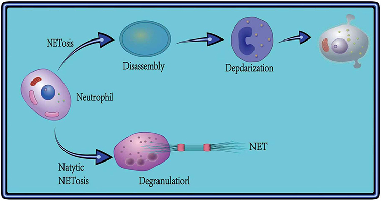 |
Figure 1 Formation of NETs. |
Detection Methods and Progress of NETs
There is increasing evidence that NETs can promote the dissemination and metastasis of tumor cells, therefore, the detection of NETs in clinical treatment is more meaningful.23–26 Effective detection methods for NETs are beneficial for early screening of tumors, monitoring of recurrence and prognosis.13,27 In recent years, the detection of NETs is mostly based on simple fluorescent labeling staining method, which has a wide range of measurement and simple operation, but there is a considerable error in marker screening.28 With the development of microscopy technology and digital analysis technology, the success rate of quantitative target NETs can be improved by the integrated use of sophisticated instruments (Table 1).
 |
Table 1 Advances in in vitro and in vivo Testing Technology digestive Tract Tumors |
Fluorescent staining is a very common in vitro assay, and NETs are a kind of reticulation structure released to the extracellular surface after neutrophil activation by spiking, which establishes the importance of staining in the detection of NETs. The same commonly used method also includes enzyme-linked immunosorbent assay, which is poorly reproducible, but the method is sensitive, rapid and can be automated at low cost, making it the preferred option for detecting NETs. With the advancement of detection technology, in recent years, a number of methods such as confocal microscopy, in vivo microscopy, and in vivo imaging have emerged to analyze and study the tissue morphology and structural characteristics of objects using high resolution and high magnification, thus improving the detection rate and accuracy. Specific in vivo and ex vivo detection technology advances are shown in Table 1.
NETs Regulatory Mechanism
NETs Degrade the Extracellular Matrix
The extracellular matrix reticulum acts as a barrier for tumor cells to invade and enter the circulatory system, therefore, the process of degrading the extracellular matrix reticulum can effectively promote the metastasis of GI tumors. It has been shown that protein-degrading enzymes present in NETs can enhance tumor invasion and metastasis by dissolving the extracellular matrix meshwork. Matrix metalloproteinase 9 (MMP-9) is an important component of NETs, and several studies29–31 have indicated that MMP-9 plays an important role in cell proliferation and apoptosis, and also degrades extracellular matrix components to prepare tumor cells for migration and accelerate GI tumor metastasis. Studies have shown5,22 that neutrophil elastase in NETs has synergistic effects with metalloproteinases and is able to degrade protein inhibitors. Most scholars believe that neutrophil elastase has a role in degrading extracellular matrix,23,24,32 but little has been reported on the specific mechanism of NETs neutrophil elastase in tumor metastasis, and more experimental studies are still needed to gain insight (Figure 2).
 |
Figure 2 NETs regulatory mechanism. |
NETs is Involved in Immunoregulation of Gastrointestinal Tumors
Immunomodulation is a fundamental step in the development of gastrointestinal tumors and an important step affecting the development of gastrointestinal tumors, which can be blocked or promoted by the immune system.17,33–36 NETs in gastrointestinal tumors tissues expresses programmed cell death protein 1 significantly higher than surrounding normal tissues,37 inhibiting the proliferation of CD8+ T cells and disrupting their anti-immune function, thus promoting gastrointestinal tumors progression.8,9,38–43 PD-1 overexpression in NETs has also been shown to be associated with tumor progression and a reduced natural killer cell response.5,9,44 Also, NETs can assist tumor cells in evading tumor cells by releasing the pro-inflammatory cytokine TNF-α activates nuclear factors that assist tumor cells to evade cytotoxic T cell killing.45–49 NETs inhibits CD4+T cell proliferation and prevents T cell attack on tumor cells, it’s also secretes growth factors to nourish gastrointestinal tumors cells. IL-10 is a potent immunosuppressive cytokine associated with cancer, and secretion of TGF-α and IL-10 by M2-type NETs induces expression of programmed death protein ligands as a way to inhibit the killing of gastrointestinal tumors by T cells.27,50,51 Immunomodulation plays a major role in the development of gastrointestinal tumors, activating activating factors, exerting programmed control, and influencing immune tolerance, and further understanding of immunomodulatory mechanisms, uncovering immune verification sites, and blocking immunosuppression are essential for the clinical treatment of gastrointestinal tumors.
NETs Promote Proliferation of Gastrointestinal Tumor Cells
Several studies have shown that NETs can promote tumor cell proliferation.8,52 It’s found that NETs-derived DNA in mouse pancreatic cancer cells promoted the growth of pancreatic cancer; deficiency of neutrophil elastase from NETs led to a decrease in tumor cell proliferation and an increase in apoptosis.32,53 In addition, the inflammatory microenvironment is one of the important features of tumor proliferation. NETs are closely associated with the inflammatory microenvironment of tumors, creating a microenvironment that promotes inflammation and thus further recruiting neutrophil aggregation.23,54 It’s found that blocking NETs improved the liver tumor microenvironment and slowed the progression to hepatocellular carcinoma by reducing mononuclear macrophage infiltration and their production of inflammatory cytokines. Lipopolysaccharide stimulates the upregulation of neutrophil complement receptor expression and activates the complement cascade reaction causing the formation of NETs, which promotes the conversion of coagulation and tumor-associated neutrophils to a pro-tumor phenotype (N2 type), leading to the development of small bowel cancer.
NETs Affect Tumor Vascular Stability
The maintenance and stability of vascular integrity mainly depends on the tight interconnection of endothelial cells of blood vessels, and an intact vascular wall plays a barrier role for tumor metastasis. NETs-related proteases promote tumor growth and metastasis by causing down-regulation of endothelial calcium mucin expression, which leads to disruption of the integrity of cell-cell junctions and vascular leakage. It has also been found that neutrophil elastase, a component of NETs, can stimulate the expression of vascular endothelial growth factor and promote tumor growth and invasion.
Hot Spot Exploration
Studies have shown that the formation and degradation of NETs is a complex process influenced by multiple factors. The current hot research on NETs in tumors is mainly in the NETs-related inhibitors.
The overexpression of protein arginine deiminase 4 (PAD4) in neutrophils has been found in many malignant tumors in humans9,45,55 and it has been found that overexpression of PAD4 in cancer cells can induce NETs reticulation46; and PAD4 inhibitors can promote gene expression, inhibit tumor cell proliferation, and thus induce apoptosis.47,56 However, whether PAD4 inhibitors affect tumor cell proliferation and distant metastasis by inhibiting the release of NETs from neutrophils still needs to be further confirmed. Whether NETs inhibitors have synergistic effects with chemotherapeutic agents, radiotherapy, targeted therapy and immunotherapy also needs to be further investigated. Suppression of T cell function is the focus of current research on tumor immunosuppressive mechanisms,52 however However, neutrophils can overexpress and secrete nitric oxide synthase, leading to T cell suppression.53,57–59 Anti-PD-L1 immunotherapy is a popular GI treatment, and studies have confirmed that anti-PD-1 immunotherapy can act on malignant tumor cells through mechanisms other than T lymphocyte involvement, while NETs can suppress T cells, further delaying the progression of malignant GI tumors and the disruption of the immune microenvironment. Therefore, using neutrophil mechanism as the target of anti-PD-L1 immunotherapy is a new breakthrough point for tumor immunotherapy targeting PD-L1/PD-1 signaling pathway54 and is expected to be applied in the clinical treatment process.
Conclusion
NETs, as an emerging research hotspot with wide clinical value, have opened up a whole new research field and provided new explanations for studying the biological behavior of gastrointestinal tumors. Not only can they kill pathogens, but also are closely related to tumor development and metastatic recurrence, and are important prognostic factors for tumor patients, participating in tumor progression through multiple pathways. Although the mechanism of NETs in gastrointestinal tumors has not been fully elucidated yet, and relevant experimental and research reports are very limited and only superficial mechanisms have been explored, this is an area worthy of further study and is expected to be further applied in the clinical setting, and assessing the effect of targeted NETs on tumor development will have great prospects for application. At present, NETs inhibitors have become a relatively new tumor treatment strategy in clinical research, which has attracted much attention. Due to the specificity of tumors and the lack of a large number of clinical studies and differences in animal models, the real application of NETs inhibitors in clinical treatment is still open to question, and a large number of experiments are still needed to study and explore, and further improvement of detection methods is needed to achieve early screening as much as possible. There is great potential for future development of related inhibitors, and targeted drugs for NETs formation mechanism can also provide more options to control the development of gastrointestinal tumors. Overall, these findings strongly suggest the use of NETs as a potential target.
Ethics Standards
This article does not contain or involve any human participants and/or animals.
Informed Consent
For this type of study, formal consent was not required.
Author Contributions
All authors made a significant contribution to the work reported, whether that is in the conception, study design, execution, acquisition of data, analysis and interpretation, or in all these areas; took part in drafting, revising or critically reviewing the article; gave final approval of the version to be published; have agreed on the journal to which the article has been submitted; and agree to be accountable for all aspects of the work.
Funding
The 14th Five-Year Plan of Hebei Medical University Clinical Medicine Innovation Research Team Support Program (Project No. 2022LCTD-A13); Medical Science Research Project of Hebei Provincial Health Care Commission (Project No. 20230123).
Disclosure
The authors declare no conflicts of interest in this work.
References
1. Hu W, Lee SML, Bazhin AV, et al. Neutrophil extracellular traps facilitate cancer metastasis: cellular mechanisms and therapeutic strategies. J Cancer Res Clin Oncol. 2022;149:1–20.
2. Zhao J, Jin J. Neutrophil extracellular traps: new players in cancer research. Front Immunol. 2022;13:937565. doi:10.3389/fimmu.2022.937565
3. Demkow U. Neutrophil Extracellular Traps (NETs) in cancer invasion, evasion and metastasis. Cancers. 2021;13(17):4495. doi:10.3390/cancers13174495
4. Colotta F, Allavena P, Sica A, et al. Cancer-related inf lammation, the seventh hallmark of cancer: links to genetic instability. Carcinogenesis. 2009;30(7):1073–1081. doi:10.1093/carcin/bgp127
5. Pieterse E, Rother N, Garsen M, et al. Neutrophil extracellular traps drive endothelial-to-mesenchymal transition. Arterioscler Thromb Vasc Biol. 2017;37(7):1371–1379. doi:10.1161/ATVBAHA.117.309002
6. Wu L, Saxena S, Awaji M, et al. Tumor-associated neutrophils in cancer: going pro. Cancers. 2019;11(4):E564. doi:10.3390/cancers11040564
7. Mizuno R, Kawada K, Itatani Y, et al. The role of tumor-associated neutrophils in colorectal cancer. Int J Mol Sci. 2019;20(3):E529. doi:10.3390/ijms20030529
8. Li H, Zhang K, Liu LH, et al. A systematic review of matrix metalloproteinase 9 as a biomarker of survival in patients with osteosarcoma. Tumour Biol. 2014;35(6):5487–5491. doi:10.1007/s13277-014-1717-3
9. Stadler SC, Vincent CT, Fedorov VD, et al. Dysregulation of PAD4-mediated citrullination of nuclear GSK3β activates TGF-β signaling and induces epithelial-to-mesenchymal transition in breast cancer cells. Proc Natl Acad Sci USA. 2013;110(29):11851–11856. doi:10.1073/pnas.1308362110
10. Chen Y, Han L, Qiu X, Wang G, Zheng J. Neutrophil extracellular traps in digestive cancers: warrior or accomplice. Front Oncol. 2021;11:766636. doi:10.3389/fonc.2021.766636
11. Szczerba BM, Castro-Giner F, Vetter M, et al. Neutrophils escort circulating tumour cells to enable cell cycle progression. Chin Pharmacol Bull. 2020;566:553–557.
12. Dabrowska D, Jablonska E, Garley M, et al. New aspects of the biology of neutrophil extracellular traps. Scand J Immunol. 2016;84(6):317–322. doi:10.1111/sji.12494
13. Papayannopoulos V, Metzler KD, Hakkim A, et al. Neutrophil elastase and myeloperoxidase regulate the formation of neutrophil extracellular traps. J Cell Biol. 2010;191(3):677–691. doi:10.1083/jcb.201006052
14. Vorobjeva NV. Neutrophil extracellular traps: new aspects. Moscow Univ Biol Sci Bull. 2020;75(4):173–188. doi:10.3103/S0096392520040112
15. Carmona-Rivera C, Kaplan MJ. Detection of sle antigens in neutrophil extracellular traps (nets). Methods Mol Biol. 2014;1134:151–161.
16. Fuchs TA, Abed U, Goosmann C, et al. Novel cell death program leads to neutrophil extracellular traps. J Cell Biol. 2007;176(2):231–241. doi:10.1083/jcb.200606027
17. Guglietta S, Chiavelli A, Zagato E, et al. Coagulation induced by C3aR-dependent NETosis drives protumorigenic neutrophils during small intestinal tumorigenesis. Nat Commun. 2016;7:11037. doi:10.1038/ncomms11037
18. Wang H, Zhang Y, Wang Q, et al. The regulatory mechanism of neutrophil extracellular traps in cancer biological behavior. Cell Biosci. 2021;11(1):193. doi:10.1186/s13578-021-00708-z
19. Pilsczek FH, Salina D, Poon KK, et al. A novel mechanism of rapid nuclea r neutrophil extracellular trap formation in response to Staphylococcus aureus. J Immunol. 2010;185(12):7413–7425. doi:10.4049/jimmunol.1000675
20. Boone BA, Orlichenko L, Schapiro NE, et al. The receptor for advanced glycation end products (RAGE) enhances autophagy and neutrophil extracellular traps in pancreatic cancer. Cancer Gene Ther. 2015;22(6):326–334. doi:10.1038/cgt.2015.21
21. Abdol Razak N, Elaskalani O, Metharom P. Pancreatic cancer-induced neutrophil extracellular traps: a potential contributor to cancer-associated thrombosis. Int J Mol Sci. 2017;18(3):487. doi:10.3390/ijms18030487
22. Kolaczkowska E, Jenne CN, Surewaard BG, et al. Molecular mechanisms of NET formation and degradation revealed by intravital imaging in the liver vasculature. Nat Commun. 2015;6:6673. doi:10.1038/ncomms7673
23. Li CH, Li KQ. Mechanism of tumor microangiogenesis and tumor invasion and metastasis. Zhonghua Zhong Liu Za Zhi. 2000;22(3):181.
24. Erpenbeck L, Schön MP. Neutrophil extracellular traps: protagonists of cancer progression. Oncogene. 2017;36(18):2483–2490. doi:10.1038/onc.2016.406
25. de Buhr N, Von Kockritz-Blickwede M. How neutro⁃ phil extracellular traps become visible. J Immunol Res. 2016;2016:4604713. doi:10.1155/2016/4604713
26. Wislez M, Antoine M, Rabbe N, et al. Neutrophils promote aerogenous spread of lung adenocarcinoma with bronchioloalveolar carcinoma features. Clin Cancer Res. 2007;13(12):3518–3527. doi:10.1158/1078-0432.CCR-06-2558
27. Park J, Wysocki RW, Amoozgar Z, et al. Cancer cells induce metastasis supporting neutrophil extracellular DNA traps. Sci Transl Med. 2016;8(361):361ra138. doi:10.1126/scitranslmed.aag1711
28. Wilson TJ, Nannuru KC, Futakuchi M, et al. Cathepsin G-mediated enhanced TGF-beta signaling promotes angiogenesis via upregulation of VEGF and MCP-1. Cancer Lett. 2010;288(2):162–169. doi:10.1016/j.canlet.2009.06.035
29. Yazdani HO, Roy EC, Omerci AJ, et al. Neutrophil extracellular traps drive mitochondrial homeostasis in tumors to augment growth. Cancer Res. 2019;79:5626–5639. doi:10.1158/0008-5472.CAN-19-0800
30. Qiao Y, Jiang J, Zhang Z, et al. Heparin reduces endothelial cell damage induced by neutrophil extracellular traps. Zhonghua Wei Zhong Bing JiJiu Yi Xue. 2017;29(4):342–346.
31. Miller-Ocuin JL, Liang X, Boone BA, et al. DNA released from neutrophil extracellular traps (NETs) activates pancreatic stellate cells and enhances pancreatic tumor growth. Oncoimmunology. 2019;8(9):e1605822. doi:10.1080/2162402X.2019.1605822
32. Houghton AM, Rzymkiewicz DM, Ji H. Neutrophil elastase-mediated degradation of IRS-1 accelerates lung tumor growth. Nat Med. 2010;16(2):219–223. doi:10.1038/nm.2084
33. Gong L, Cumpian AM, Caetano MS, et al. Promoting effect of neutrophils on lung tumorigenesis is mediated by CXCR2 and neutrophil elastase. Mol Cancer. 2013;12(1):154. doi:10.1186/1476-4598-12-154
34. Warnatsch A, Ioannou M, Wang Q, et al. Neutrophil extracellular traps license macrophages for cytokine production in atherosclerosis. Science. 2015;349(6245):316–320. doi:10.1126/science.aaa8064
35. van der Windt DJ, Sud V, Zhang H, et al. Neutrophil extracellular traps promote inflammation and development of hepatocellular carcinoma in nonalcoholic steatohepatitis. Hepatology. 2018;68(4):1347–1360. doi:10.1002/hep.29914
36. Najmeh S, Cools-Lartigue J, Rayes RF, et al. Neutrophil extracellular traps sequester circulating tumor cells via β1-integrin mediated interactions. Int J Cancer. 2017;140:2321–2330. doi:10.1002/ijc.30635
37. Rayes RF, Mouhanna JG, Nicolau I, et al. Primary tumors induce neutrophil extracellular traps with targetable metastasis promoting effects. JCI Insight. 2019;5(pii):128008. doi:10.1172/jci.insight.128008
38. Tohme S, Yazdani HO, Al-Khafaji AB, et al. Neutrophil extracellular traps promote the development and progression of liver metastases after surgical stress. Hpb. 2017;19:S26–S27.
39. Clark RA, Klebanoff SJ. Neutrophil-mediated tumor cell cytotoxicity: role of the peroxidase system. J Exp Med. 1975;141(6):1442–1447. doi:10.1084/jem.141.6.1442
40. Zivkovic M, Poljak-Blazi M, Egger G, et al. Oxidative burst and anticancer activities of rat neutrophils. BioFactors. 2005;24(1–4):305–312. doi:10.1002/biof.5520240136
41. Eruslanov EB, Bhojnagarwala PS, Quatromoni JG, et al. Tumor-associated neutrophils stimulate T cell responses in early-stage human lung cancer. J Clin Invest. 2014;124(12):5466–5480. doi:10.1172/JCI77053
42. Arelaki S, Arampatzioglou A, Kambas K, et al. Gradient infiltration of neutrophil extracellular traps in colon cancer and evidence for their involvement in tumour growth. PLoS One. 2016;11(5):e0154484. doi:10.1371/journal.pone.0154484
43. Yee PP, Wei Y, Kim S, et al. Neutrophil-induced ferroptosis promotes tumor necrosis in glioblastoma progression. Nat Commun. 2020;11(1):5424. doi:10.1038/s41467-020-19193-y
44. Zhou SL, Yin D, Hu ZQ, et al. A positive feedback loop be⁃ tween cancer stem-like cells and tumor ⁃ associated neutrophils controls hepatocellular carcinoma progression. Hepatology. 2019;70(4):1214–1230. doi:10.1002/hep.30630
45. Chang X, Han J, Pang L, et al. Increased PADI4 expression in blood and tissues of patients with malignant tumors. BMC Cancer. 2009;9:40. doi:10.1186/1471-2407-9-40
46. Leshner M, Wang S, Lewis C, et al. PAD4 mediated histone hypercitrullination induces heterochromatin decondensation and chromatin unfolding to form neutrophil extracellular trap-like structures. Front Immunol. 2012;3:307. doi:10.3389/fimmu.2012.00307
47. Li P, Yao H, Zhang Z, et al. Regulation of p53 target gene expression by peptidylarginine deiminase 4. Mol Cell Biol. 2008;28(15):4745–4758. doi:10.1128/MCB.01747-07
48. Li P, Wang D, Yao H, et al. Coordination of PAD4 and HDAC2 in the regulation of p53-target gene expression. Oncogene. 2010;29(21):3153–3162. doi:10.1038/onc.2010.51
49. Brinkmann V. Neutrophil extracellular traps in the second decade. J Innate Immun. 2018;10(5–6):414–421. doi:10.1159/000489829
50. Stravitz RT, Sanyal AJ, Reisch J, et al. Effects of Nacetylcysteine on cytokines in non-Acetaminophen acute liver failure: potential mechanism of improvement in transplant-free survival. Liver Int. 2013;33(9):1324–1331. doi:10.1111/liv.12214
51. D’Amico F, Vitale A, Piovan D, et al. Use of N-acetylcysteine during liver procurement: a prospective randomized controlled study. Liver Transpl. 2013;19(2):135–144. doi:10.1002/lt.23527
52. Newton JM, Hanoteau A, Liu H, et al. Immune micro⁃ environment modulation unmasks therapeutic benefit of radio-therapy and checkpoint inhibition. J Immuno Ther Cancer. 2019;7(1):216. doi:10.1186/s40425-019-0698-6
53. Li P, Lu M, Shi J, et al. Dual roles of neutrophils in metastatic colonization are governed by the host NK cell status. Nat Commun. 2020;11(1):4387. doi:10.1038/s41467-020-18125-0
54. Martín-Ruiz A, Fiuza-Luces C, Martínez-Martínez E, et al. Effects of anti-PD-1 immunotherapy on tumor regression: insights from a patient-derived xenograft model. Sci Rep. 2020;10(1):7078. doi:10.1038/s41598-020-63796-w
55. Jung HS, Gu J, Kim JE, et al. Cancer cell induced neutrophil extracellular traps promote both hypercoagulability and cancer progression. PLoS One. 2019;14(4):e0216055. doi:10.1371/journal.pone.0216055
56. Yang C, Sun W, Cui W, et al. Procoagulant role of neutrophil extracellular traps in patients with gastric cancer. Int J Clin Exp Pathol. 2015;8(11):14075–14086.
57. Massbe S, Grahl L, Von Ruehl ML, et al. Reciprocal coupling of coagulation and innate immunity via neutrophil serine proteases. Nat Med. 2010;16(8):887–896. doi:10.1038/nm.2184
58. Zhang Y, Wang C, Yu M, et al. Neutrophil extracellular traps induced by activated platelets contribute to procoagulant activity in patients with colorectal cancer. Thromb Res. 2019;180:87–97. doi:10.1016/j.thromres.2019.06.005
59. Nie M, Yang L, Bi X, et al. Neutrophil extracellular traps induced by IL8 promote diffuse large B-cell lymphoma progression via the TLR9 signaling. Clin Cancer Res. 2019;25(6):1867–1879. doi:10.1158/1078-0432.CCR-18-1226
 © 2023 The Author(s). This work is published and licensed by Dove Medical Press Limited. The full terms of this license are available at https://www.dovepress.com/terms.php and incorporate the Creative Commons Attribution - Non Commercial (unported, v3.0) License.
By accessing the work you hereby accept the Terms. Non-commercial uses of the work are permitted without any further permission from Dove Medical Press Limited, provided the work is properly attributed. For permission for commercial use of this work, please see paragraphs 4.2 and 5 of our Terms.
© 2023 The Author(s). This work is published and licensed by Dove Medical Press Limited. The full terms of this license are available at https://www.dovepress.com/terms.php and incorporate the Creative Commons Attribution - Non Commercial (unported, v3.0) License.
By accessing the work you hereby accept the Terms. Non-commercial uses of the work are permitted without any further permission from Dove Medical Press Limited, provided the work is properly attributed. For permission for commercial use of this work, please see paragraphs 4.2 and 5 of our Terms.