Back to Journals » Journal of Inflammation Research » Volume 14
Acute Phase Responses Vary Between Children of HbAS and HbAA Genotypes During Plasmodium falciparum Infection
Authors Tetteh M, Addai-Mensah O, Siedu Z , Kyei-Baafour E , Lamptey H , Williams J, Kupeh E, Egbi G, Kwayie AB, Abbam G, Afrifah DA, Debrah AY, Ofori MF
Received 20 January 2021
Accepted for publication 26 March 2021
Published 14 April 2021 Volume 2021:14 Pages 1415—1426
DOI https://doi.org/10.2147/JIR.S301465
Checked for plagiarism Yes
Review by Single anonymous peer review
Peer reviewer comments 3
Editor who approved publication: Professor Ning Quan
Mary Tetteh,1,2,* Otchere Addai-Mensah,1,* Zakaria Siedu,3,4 Eric Kyei-Baafour,3 Helena Lamptey,3 Jovis Williams,3 Edward Kupeh,5 Godfred Egbi,6 Anna Boadi Kwayie,2 Gabriel Abbam,1,7 David Amoah Afrifah,1 Alexander Yaw Debrah,1 Michael Fokuo Ofori3,4
1Department of Medical Diagnostics, Faculty of Allied Health Sciences, Kwame Nkrumah University of Science and Technology, Kumasi, Ghana; 2Laboratory Department, District Hospital, Begoro, Ghana; 3Immunology Department, Noguchi Memorial Institute for Medical Research, College of Health Sciences, University of Ghana, Accra, Ghana; 4West Africa Centre for Cell Biology of Infectious Pathogens, Department of Biochemistry, Cell and Molecular Biology, University of Ghana, Accra, Ghana; 5Laboratory Department, Tema Polyclinic, Tema, Ghana; 6Nutrition Department, Noguchi Memorial Institute for Medical Research, College of Health Sciences, University of Ghana, Accra, Ghana; 7University Clinic Laboratory, University of Education, Winneba, Ghana
*These authors contributed equally to this work
Correspondence: Michael Fokuo Ofori
Immunology Department, Noguchi Memorial Institute for Medical Research, College of Health Sciences, University of Ghana, Post Office Box LG581, Legon, Accra, Ghana
Tel +233 244 715975
Fax +233 302 502182
Email [email protected]
Purpose: Haemoglobin genotype S is known to offer protection against Plasmodium falciparum infections but the mechanism underlying this protection is not completely understood. Associated changes in acute phase proteins (APPs) during Plasmodium falciparum infections between Haemoglobin AA (HbAA) and Haemoglobin AS (HbAS) individuals also remain unclear. This study aimed to evaluate changes in three APPs and full blood count (FBC) indices of HbAA and HbAS children during Plasmodium falciparum infection.
Methods: Venous blood was collected from three hundred and twenty children (6 months to 15 years) in Begoro in Fanteakwa District of Ghana during a cross-sectional study. Full blood count (FBC) indices were measured and levels of previously investigated APPs in malaria patients; C-reactive protein (CRP), ferritin and transferrin measured using Enzyme-Linked Immunosorbent Assays.
Results: Among the HbAA and HbAS children, levels of CRP and ferritin were higher in malaria positive children as compared to those who did not have malaria. The mean CRP levels were significantly higher among HbAA children (p=0.2e-08) as compared to the HbAS children (p=0.43). Levels of transferrin reduced in both HbAA and HbAS children with malaria, but the difference was only significant among HbAA children (p=0.0038), as compared to the HbAS children. No significant differences were observed in ferritin levels between HbAA and HbAS children in both malaria negative (p=0.76) and positive (p=0.26) children. Of the full blood count indices measured, red blood cell count (p=0.044) and haemoglobin (Hb) levels (p=0.017) differed between HbAA and HbAS in those without malaria, with higher RBC counts and lower Hb levels found in HbAS children. In contrast, during malaria, lymphocyte and platelet counts were elevated, whilst granulocytes and Mean Cell Haematocrit counts were reduced among children of the HbAS genotypes.
Conclusion: Significant changes in APPs were found in HbAA children during malaria as compared to HbAS children, possibly due to differences in malaria-induced inflammation levels. This suggests that the HbAS genotype is associated with better control of P. falciparum infection-induced inflammatory response than HbAA genotype.
Keywords: acute phase response, APR, acute phase proteins, APP, Plasmodium falciparum, C-reactive protein, ferritin, transferrin
Introduction
The pathophysiological response to infection with Plasmodium falciparum (P. falciparum) depends on several different factors including previously acquired immunity and the production of cytokines such as interleukin-1 (IL-1) and TNF-α, IFN-γ, and interleukin-6 (IL-6).1 In malaria infections, initial inflammatory responses are noted to be beneficial in reducing parasite growth.2,3 On the other hand, when the proliferation of the parasites are not suppressed, the increased number of infected erythrocytes (IEs) will end up interfering with haemodynamics leading to anaemia, and stimulate sequestration of IEs in the microvasculature.4,5
Although the body releases anti-inflammatory cytokines such as IL-10, IL-4, IL-17, and IL-13 to balance the undesirable effects of pro-inflammatory cytokines,6–8 the acute phase response (APR) is the hallmark response, purposeful to prevent any inflammation-mediated damages to tissues, activate the repair process and remove harmful molecules.9 It is characterized by changes in the serum concentration of some proteins, known as acute phase proteins (APPs), increasing the levels of positive acute-phase proteins and decreasing the levels of negative acute-phase proteins.10,11 Secretion of the APPs occurs in the hepatocytes and their synthesis is controlled mainly by IL-6 and other pro-inflammatory cytokines, such as TNF-α and IFN-γ, released during P. falciparum infection.9,10,12
Since an inflammatory response is a hallmark of the early innate immune response in malaria, previous studies have established a positive correlation between expression of some APPs and severity of P. falciparum malaria, and proposed the possibility of using APR to predict the severity of malaria.1,10 However, in malaria-endemic areas, some hereditary factors, especially for the sickle-cell trait carriers (haemoglobin type AS),13 have been and are under natural selection because they confer relative protection against severe malaria disease.14–16 Despite this, the associated changes in APPs concentration between HbAA and HbAS individuals during malaria remain unclear.
Based on the above, we investigated the levels of three APPs: CRP, serum ferritin and serum transferrin in children with HbAS and HbAA. These proteins are crucial markers of inflammation, and have been investigated by previous studies in malaria patients.10,17–19 Also we compared changes in full blood count (FBC) indices in the children and how they associate with P. falciparum infection. The data from this study suggest that the HbAS genotype is associated with better control of P. falciparum malaria-induced inflammatory response than the HbAA genotype, with resultant milder APR during P. falciparum infection/malaria in children with the HbAS genotype compared to the HbAA genotype. This observation could potentially explain the relative protection against severe malaria in HbAS children.
Materials and Methods
Ethical Statement
The School of Medical Sciences, Kwame Nkrumah, University of Science and Technology (KNUST), Ghana, Committee on Human Ethics and Publication approved this study (CHRPE/AP/308/19), Permission was also sought from the Begoro District Hospital and approval was given for the facility to be used as the study site. The study complied with the Helsinki declaration for conducting research involving human subjects. Written informed consent was obtained from each parent/guardian of all study participants.
Study Design and Population
The study was cross-sectional and was conducted at the Begoro District Hospital in the Fanteakwa District in the Eastern Region of Ghana. It has an area of 1150 square kilometres and lies within longitudes “0º32.5” West and “0º10” East and Latitude 6º40ʹ, with an average temperature of 24°C. The study population comprised of individuals between the ages of 6 months to 15 years who came to the out-patient department (OPD) of the Begoro District Hospital with clinical signs of malaria and were referred to the laboratory by a Physician for a malaria test. A simple random sampling procedure where every other suspected child reporting to the laboratory for testing was recruited in the study between May to August 2019 (rainy season in Ghana), 320 children were recruited using a proportionate rate of malaria prevalence in the Fanteakwa district to be 28% at 95% confidence interval, and 5% margin of error.
The children were tested for malaria with a rapid diagnostic test (RDT), and microscope slides prepared for Giemsa staining for the malaria parasite density estimation. Participants haemoglobin were phenotyped and those with HbAA and HbAS phenotype (HbAA=258, HbAS=62) were prioritized for blood sampling (male=146, female=174). Venous blood samples (2–3 mL) were collected into ethylenediaminetetraacetic acid (EDTA) tubes from participants whose parent or guardian consented to donate blood and were clinically examined and those who were not severely ill were allowed to participate in the study. Blood samples were immediately separated by centrifugation and plasma stored frozen at −30°C until ready to be used. Concurrently, few drops of the remaining pelleted RBCs were spotted on filter papers (Whatman, USA) and preserved at −30°C for retrospective molecular analysis (PCR) to confirm malaria RDT and microscopy results. The weight (kg) and Height (m) of the children were measured and used in calculating the body mass index (BMI) in Kg/m2.
Malaria Diagnosis and Haemoglobin Phenotyping
The Carestart™ Ag P.f rapid diagnostic test kit (Access Bio Inc. New Jersey, USA) was used for primary diagnosis of malaria and confirmed it by microscopic examination of 10% Giemsa-stained thick blood films by two independent experienced microscopists, using finger-pricked capillary blood samples. Discordant microscopy results were re-examined by a third microscopist. Parasite densities were recorded as a ratio of parasites to white blood cells (WBCs) in thick blood films, considering an assumed WBC count of 8000/µL of blood.20 Also, retrospectively DNA samples were extracted from the dried blood spots (DBS), for molecular testing (PCR). Concurrently with the blood films, slides were prepared from each participant for a metabisulphite screening test and the Hb phenotype of children with positive sickling metabisulphite test were further determined using Hb electrophoresis.21
Quantification of Full Blood Count Indices and Acute-Phase Proteins
Full blood count (FBC) analysis was performed using the URIT 3000 Plus Hematology Analyser (URIT Medical Electronic Group, China) and plasma levels of human ferritin and transferrin were measured using commercially prepared ELISA kits from MyBioSource (Cat no: MBS771449 and MBS773765 respectively, USA), according to the manufacturer’s instructions. Also, the levels of the C- reactive protein (CRP) was determined with ELISA, by testing for the CRP antibodies in the plasma against 2µg/mL of a commercially prepared CRP capture antigen (R&D Systems). The detection ranges for the ferritin kit, transferrin kit and CRP kit were 20ng/mL −800ng/mL, 0.1nmol/L −8nmol/L, and 15.6 pg/mL – 1000pg/mL, respectively.
Data Analysis
Normality test was performed on the data and those not normally distributed were log transformed. The demographics and clinical parameters of the children are presented as frequency and subgroup proportions and compared them between malaria negative and positive groups using Chi-Square test. While continuous variables such as the FBC indices and the APPs were expressed as mean ± (SD) and compared between the HbAA and HbAS and between malaria negative and positive using t-test or Wilcoxon signed-rank test based on the normality of the data. APPs data were log-transformed before analysis. All analysis were performed with R version 3.6.3 and considered all p-values less than 0.05 at 95% CI to be statistically significant.
Results
Patient’s Characteristics During Plasmodium falciparum Infection
Comparison of the demographic and other related factors between individuals who tested positive for P. falciparum compared to the negative individuals revealed discrepancies in how some of these factors were associated with P. falciparum infection. Assessment of age group, gender, haemoglobin phenotype, Body Mass Index (BMI) status, Insecticide treated nets (ITNs) use, clinical signs/symptoms and the antimalarial history of the study participants were done to determine their association with P. falciparum infection. Gender, ITNs use and BMI did not significantly contribute to the P. falciparum infection of the children (p>0.05) (Table 1).
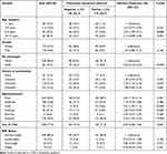 |
Table 1 Descriptive Summary of Demographic Information of the Study Participants |
However, after stratifying the data by age groups, children aged above one year had a general tendency to test positive for P. falciparum compared to children younger than one year. Children between the ages of 1–5 years (p=0.22) and 10–15 years (p=0.055) only exhibited a relatively higher risk of developing P. falciparum infection compared to those below one year, with corresponding odds of 1.53 and 2.10, respectively. While P. falciparum infection risk among children between the ages of 5–8 years was significantly higher (p=0.028) compared to the children below 1 year. The haemoglobin genotype of the children also showed a significant association with P. falciparum infection, with the infection detection risk being 80.0% less likely among children who are sickling positive (Hb AS) compared with the sickling negative group (HbAA) (p-value <0.0001).
When the antimalarial history of the children and P. falciparum infection was assessed, the children with antimalarial medication history <3 months were significantly likely to test positive for P. falciparum compare to those who did not present with antimalarial history (OR=2.34, P-value = 0.018). While those who presented with an unknown history (OR=1.39, P-value = 0.453) and antimalarial history beyond 3 months (OR=1.98, p-value = 0.065) did not significantly have a higher P. falciparum infection risk compared to those with no antimalarial history. Individuals who presented with clinical symptoms of fever, chills, anaemia and vomiting were more observed among the P. falciparum positive children compared to the negative children (p-value <0.05) but symptoms like headache and expectedly diarrhoea, did not significantly defer among the P. falciparum positive and negative individuals (p>0.05).
Changes in Full Blood Count Indices and Acute Phase Proteins During Plasmodium falciparum Infection
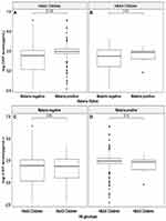
To investigate the effect of haemoglobin genotype on changes in the FBC indices and acute-phase proteins, the full blood count (FBC) indices and levels of the acute phase proteins during P. falciparum infection, the data was stratified into P. falciparum positive and negative. Within each group, levels of the FBC indices and the acute phase proteins between children with HbAA and HbAS genotype were compared (Table 2). Without P. falciparum infection, levels of most FBC indices did not significantly differ between the HbAA and HbAS children, except in their RBC count (p=0.044) and Hb levels (p=0.017). However, among those with P. falciparum infection, significant differences were observed in the levels of lymphocytes (LYM) (p=0.026), platelets (PLT) (p=0.014), Granulocytes (GRAN) (p=0.032) and MCH (p=0.026) counts between the HbAA and HbAS children. Mean CRP level was significantly elevated from 17.7±57.9 µg/mL in the P. falciparum negative population to 22.3±66.3 µg/mL in the malaria positive individuals (Table 2), and the CRP levels between the two groups was significantly different (p=7.2e-09) (Supplementary Figure S1). However, it was only the individuals with the HbAA genotype that exhibited significantly elevated CRP levels during P. falciparum infection (p=0.2e-08) (Figure 1A). A weak positive correlation between age and ferritin and transferrin, and negative correlation between age and c-reactive protein were observed. However, no correlation was observed between age and the distribution of HbAA and HbSS (Table 2).
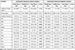 |
Table 2 Changes in FBC Indices and Acute Phase Proteins During Plasmodium falciparum Infection |
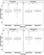
Similarly, the mean serum ferritin level also increased from 628.2±211.7 µg/L in the P. falciparum negative children to 654.2 ±209.6 µg/L in the positive children (Table 2), but neither the HbAA nor HbAS P. falciparum positive children had a significant elevated serum ferritin levels than the P. falciparum negative children (Figure 2). Overall, between the HbAA and HbAS individuals, in both the P. falciparum positive and negative children, the CRP and ferritin levels were relatively elevated among the HbAA children than in the HbAS individuals during P. falciparum infection (Table 2, Figures 1 and 2).
Unlike the C-reactive protein and ferritin, transferrin levels were rather negatively associated with malaria, generally decreasing from 6.4±1.5 nmol/L in the P. falciparum negative individuals to 5.8±1.3 nmol/L in the P. falciparum positive individuals (Table 2). In the P. falciparum negative group, no significant differences were observed in the transferrin levels between the P. falciparum negative HbAA (6.4±1.4 nmol/L) and HbAS (6.4±1.5 nmol/L) individuals (p= 0.974).
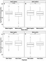
However, in the malaria positive group, there was an appreciable lower but not statistically significant mean transferrin levels (5.8 ±1.2 nmol/L) in the HbAA individuals as compared to the HbAS individuals (6.2 ± 1.5 nmol/L) (p=0.136). Overall, the serum transferrin levels significantly plummet in the HbAA individuals during malaria (p=0.004) but not in the HbAS individuals (p=0.6) (Figure 3).
Transferrin Significantly Decreased in HbAA Individuals During Plasmodium Infection/Malaria
Unlike the CRP and ferritin, levels of plasma transferrin plummet upon Plasmodium infection, decreasing from 6.4±1.5 nmol/L in the P. falciparum negative individuals to 5.8±1.3 nmol/L in the P. falciparum positive individuals (Table 2). When the data were stratified into malaria positive and negative cases, in the P. falciparum negative group, the HbAS individuals had similar levels of plasma transferrin as the HbAA individuals (p=0.974). However, in the P. falciparum positive group, a lower mean transferrin level (5.8 ±1.2 nmol/L) in the HbAA compared to the HbAS individuals (6.2 ± 1.5 nmol/L) was observed but the difference was not statistically significant (p=0.136) (Table 2).
Also, upon P. falciparum infection, there was a significant decrease in the plasma transferrin levels between the P. falciparum negative and P. falciparum positive HbAA individuals (p=0.004) (Figure 3A), compared to in the HbAS individuals (p=0.6) (Figure 3B). However, between the HbAA and HbAS individuals, in both the P. falciparum negative and positive individuals, there was no significant change in the transferrin levels (Figure 3B and C).
Discussion
In malaria-endemic areas, some hereditary factors, especially sickle-cell trait carriers (haemoglobin type AS),13 have been and are under natural selection because they seem to confer relative protection against severe malaria disease.14–16 Hence, this study examined the changes in concentration of C-reactive protein, plasma ferritin, plasma transferrin and full blood count indices of HbAA and HbAS Ghanaian children during malaria infection to determine changes in APPs and acute phase response levels in HbAA and HbAS individuals during P. falciparum malaria.
Previous studies have established a link between changes in APPs concentrations with Plasmodium falciparum malaria and the ability to use the APR to predict the severity of malaria.1,10 Therefore, the dynamics of APPs concentration during malaria has been extensively studied.1,10,22 However, the associated changes in APPs concentration between HbAA and HbAS individuals during malaria remain unclear.
This study observed elevated levels of ferritin and CRP in the P. falciparum positive individuals compared to the negative individuals, and these findings are consistent with previous reports.1,10,22 The levels of the CRP were significantly elevated in the malaria positive children compared to levels found in the malaria negative individuals (p=7.2e-09). Additionally, ferritin levels marginally increased in the malaria positive individuals than levels in the malaria negative individuals (p=0.076) (Supplementary Figure S2). It has been evidenced that ferritin and CRP are positive acute-phase proteins, therefore their levels increases during inflammation. These findings were expected since inflammatory response is a hallmark of early innate immune response during malaria infection.1
Between children with HbAA and HbAS genotypes, changes in the CRP levels reached statistical significance between P. falciparum positive and negative HbAA individuals (p=0.2e-08) but not in the HbAS individuals (p=0.43). Overall, more elevated levels of C-reactive protein and ferritin were observed in HbAA children during malaria compared to the HbAS children, suggesting a higher inflammatory response in the HbAA children during malaria than in the HbAS children. A previous study by Ademoule et al that compared pro-inflammatory cytokines levels between HbAA and HbAS children also provides evidence that there is a higher inflammatory response in HbAA than in HbAS children during malaria.21
There are reports that higher or elevated levels of C-reactive protein attenuate the production of Nitric Oxide (NO), a potent anti-malarial,23–26 achieved through downregulation of endothelial NO synthase (eNOS) transcription in Endothelial Cells (ECs) and destabilization of eNOS mRNA, with resultant decreases in basal and stimulated NO release.26 Hence, with the observed disproportionate significant increase in the CRP levels in the HbAA children during P. falciparum infection, this might lead to differentially lower bioavailability of NO in HbAA than in HbAS individuals during malaria, with a resultant lower NO-mediated parasite clearance in them than in the HbAS. Perhaps, this partially explains why this study observed a significantly lower parasite density in the HbAS than in the HbAA individuals (Supplementary Figure S3). Unlike the CRP and ferritin levels, the serum transferrin levels plummeted during P. falciparum infection, with the decline reaching statistical significance in HbAA individuals (p=0.004), but not in the HbAS individuals (p=0.6). Immunologically, transferrin is a known associate of the innate immune system that creates a low free iron environment in the body, in a process known as iron withholding to impede bacteria survival in the body.27 It is a negative acute-phase protein and therefore generally decreases during inflammation.28 In malaria, an inflammatory response is a hallmark of the early immune responses to P. falciparum infection. Therefore, the decline in transferrin levels observed in this study during P. falciparum infection likely correlated with the malaria-induced inflammatory response. Similar to the CRP and ferritin, the declined in transferrin levels was pronounced in the HbAA individuals than in the HbAS, also giving credit to our initial assertion that there is a minimal inflammatory reaction in HbAS individuals than HbAA during malaria infection.
A plausible explanation for the differences in changes to the APPs and inflammation between the HbAA and HbAS children during malaria might be related to levels of parasitemia between the two groups. We observed significantly higher parasite density in the HbAA than in the HbAS children, and this could indicate a possible inhibited parasite growth in HbAS, as recently reported by Archer et al.29 With the HbAA children harboring more parasites than in the HbAS, there will be production of more parasite biomass in the HbAA, which will cause more inflammatory reaction as compare to the HbAS children. Since the APR is an anti-inflammatory response, it is explicable why in the HbAA there is significant changes in APPs as compare to in the levels found among HbAS individuals. Therefore, it seems that the lower APR in the HbAS likely involves retardation of parasite growth-mediated attenuation of parasite-induced inflammation in the HbAS children.
These differences in inflammation and APR between the HbAA and HbAS individuals could also have potential implications on cytoadhesion and sequestration of IEs. One of the factors that define the severity of P. falciparum malaria is the ability of the parasites to cytoadhere to endothelial receptors in the microvasculature and sequester into deep tissues and organs.30 Several in vivo and in vitro studies have reported that the release of the pro-inflammatory cytokines such as interleukin-1 (IL-1) and TNF-α, IFN-γ, and interleukin-6 (IL-6) during P. falciparum infection upregulates the expression and activation of endothelial receptors for efficient cytoadhesion.4,5,31–33
With evidence of minimal inflammation in HbAS children than in HbAA children observed in this study, this might result in a minimal occurrence of inflammation-induced expression and activation of cytoadhesion-involved endothelial receptors in the HbAS than in the HbAA. This could militate effective cytoadhesion and sequestration of IEs in HbAS individuals than in the HbAA, preventing and/or minimizing the occurrence of cytoadhesion-related malaria pathologies, such as cerebral malaria in HbAS than in HbAA individuals.
Conclusions
The dynamics of APPs levels during malaria has been studied previously.1,10,22 Nevertheless, this study is the first to determine the associated changes in APPs levels between HbAA and HbAS individuals during Plasmodium falciparum infection.
Consistent with literature, we also observed the protective effect of the sickle-cell trait (HbAS) against Plasmodium falciparum malaria in this study.14–16 Likewise, the evidence of inflammation during Plasmodium falciparum infection observed in this study has been reported previously.10,34,35 However, this study provides evidence of reduced inflammation in HbAS individuals during Plasmodium falciparum infection compared to in HbAA individuals.
Furthermore, we observed a significant link between the differential inflammation in HbAS and HbAA individuals and APP levels, which suggests a differential APR between HbAA and HbAS genotype children during Plasmodium falciparum infection. The observed minimal inflammation in the HbAS children implies there will be minimal inflammation-induced expression and activation of cytoadhesion in endothelial receptors in the HbAS individuals during P. falciparum infection than in HbAA children. An indication that, these could result in differences in NO levels in HbAA and HbAS children and its resultant differential inflammation observed.
We can infer that controlling inflammation during malaria could improve recovery or treatment outcome. Therefore, we recommend that a study on the effect of anti-inflammatory drugs in malaria treatment outcome should be conducted in the future.
Abbreviations
APP, Acute Phase Proteins; APR, Acute Phase Response; BMI, Body Mass Index; CRP, C-reactive protein; DBS, Dry Blood Spot; DNA, Deoxyribonucleic acid; EDTA, Ethylenediaminetetraacetic acid; ELISA, Enzyme Linked Immunosorbent Assay; EC, Endothelial cells; eNOS, Endothelial nitric oxide synthase; FBC, Full Blood Count; Hb AA, Haemoglobin AA; Hb AS, Haemoglobin AS; IE, Infected erythrocytes; IL, Interleukin; IFN-γ, Interferon-γ; ITN, Insecticide treated net; mRNA, Messenger ribonucleic acid; NO, Nitric oxide; OPD, Out-patient department; PCR, Polymerase chain reaction; RBC, Red Blood Cell; RDT, Rapid Diagnostic Test; TNF-α, Tumour Necrosis Factor-α; WBC, White Blood Cell.
Data Sharing Statement
The datasets analysed in this study are available from the corresponding author on reasonable request.
Acknowledgments
The authors are grateful to the parents and guardians of the children involved in the study. We also thank the Management and Laboratory staff of the Begoro District Hospital and Management and staff of the Immunology Department of the NMIMR. Special thanks go to Alex Danso-Coffie and Abena Fremah of the Immunology Department, NMIMR for their logistic and technical assistance.
Author Contributions
All authors made significant contribution to the work reported, whether in the conception, study design, implementation, acquisition of data, analysis and interpretation. They were also involved in the drafting, revising and critically reviewing the article and gave final approval of the version to be published, They also agreed on the journal to which the article has been submitted, and agreed to be accountable for all aspects of the work. Mary Tetteh and Otchere Addai-Mensah contributed equally and share the first authorship.
Funding
Michael F. Ofori is supported by the Danish Research Council for Development Research (Grant No. 17-02-KU). The funders had no role in study design, data collection and interpretation, or the decision to submit the work for publication.
Disclosure
The authors report no conflicts of interest in this work.
References
1. Gillespie SH, Dow C, Raynes JG, Behrens RH, Chiodini PL, McAdam KP. Measurement of acute phase proteins for assessing severity of Plasmodium falciparum malaria. J Clin Pathol. 1991;44(3):228–231. doi:10.1136/jcp.44.3.228
2. Kumar S, Bandyopadhyay U. Free heme toxicity and its detoxification systems in human. Toxicol Lett. 2005;157(3):175–188. doi:10.1016/j.toxlet.2005.03.004
3. Olivier M, Van Den Ham K, Shio MT, Kassa FA, Fougeray S. Malarial pigment hemozoin and the innate inflammatory response. Front Immunol. 2014;5:
4. Ventura PDS, Carvalho CPF, Barros NMT, et al. Malaria infection promotes a selective expression of kinin receptors in murine liver. Malar J. 2019;18(1):213. doi:10.1186/s12936-019-2846-3
5. Viebig NK, Wulbrand U, Förster R, Andrews KT, Lanzer M, Knolle PA. Direct activation of human endothelial cells by Plasmodium falciparum-infected erythrocytes. Infect Immun. 2005;73(6):3271–3277. doi:10.1128/IAI.73.6.3271-3277.2005
6. Ademolue TW, Aniweh Y, Kusi KA, Awandare GA. Patterns of inflammatory responses and parasite tolerance vary with malaria transmission intensity. Malar J. 2017;16(1):145. doi:10.1186/s12936-017-1796-x
7. Rogerson SJ, Hviid L, Duffy PE, Leke RF, Taylor DW. Malaria in pregnancy: pathogenesis and immunity. Lancet Infect Dis. 2007;7(2):105–117. doi:10.1016/S1473-3099(07)70022-1
8. Hviid L, Kurtzhals JAL, Adabayeri V, et al. Perturbation and proinflammatory type activation of vδ1+ γδ t cells in african children with plasmodium falciparum malaria. Infect Immun. 2001;69(5):3190–3196. doi:10.1128/IAI.69.5.3190-3196.2001
9. Northrop-Clewes CA. Interpreting indicators of iron status during an acute phase response-lessons from malaria and human immunodeficiency virus. Ann Clin Biochem. 2008;45(1):18–32. doi:10.1258/acb.2007.007167
10. O’donnell A, Fowkes FJI, Allen SJ, et al. The acute phase response in children with mild and severe malaria in Papua New Guinea. Trans R Soc Trop Med Hyg. 2009;103(7):679–686. doi:10.1016/j.trstmh.2009.03.023
11. Charlie-Silva I, Klein A, Gomes JMM, et al. Acute-phase proteins during inflammatory reaction by bacterial infection: fish-model. Sci Rep. 2018. doi:10.1038/s41598-019-41312-z
12. Nai A, Lidonnici MR, Rausa M, et al. The second transferrin receptor regulates red blood cell production in mice. Blood. 2015;125(7):1170–1179. doi:10.1182/blood-2014-08-596254
13. Verra F, Simpore J, Warimwe GM, et al. Haemoglobin C and S role in acquired immunity against Plasmodium falciparum malaria. PLoS One. 2007;2(10):10. doi:10.1371/journal.pone.0000978
14. Taylor SM, Parobek CM, Fairhurst RM. Haemoglobinopathies and the clinical epidemiology of malaria: a systematic review and meta-analysis. Lancet Infect Dis. 2012;12(6):457–468. doi:10.1016/S1473-3099(12)70055-5
15. Modiano D, Bancone G, Ciminelli BM, et al. Haemoglobin S and haemoglobin C: “quick but costly” versus “slow but gratis” genetic adaptations to Plasmodium falciparum malaria. Hum Mol Genet. 2008;17(6):789–799. doi:10.1093/hmg/ddm350
16. Luzzatto L. Genes expressed in red cells could shape a malaria attack. Lancet Haematol. 2018;5(8):e322–e323. doi:10.1016/S2352-3026(18)30110-8
17. Suchdev PS, Williams AM, Mei Z, et al. Assessment of iron status in settings of inflammation: challenges and potential approaches. Am J Clin Nutr. 2017;106(Supplement 6):1626–1659. doi:10.3945/ajcn
18. Mockenhaupt’ FP, Rang’ B, Giinther’ M, et al. Anaemia in pregnant ghanaian women: importance of malaria, iron deficiency, and haemoglobinopathies. Vol 94; 2000. Available from: https://academic.oup.com/trstmh/article-abstract/94/5/477/1936858.
19. Villaverde C, Namazzi R, Shabani E, et al. Retinopathy-positive cerebral malaria is associated with greater inflammation, blood-brain barrier breakdown, and neuronal damage than retinopathy-negative cerebral malaria. J Pediatric Infect Dis Soc. 2019;2019:1–8. doi:10.1093/jpids/piz082
20. World Health Organization. Basic MALARIA MICROSCOPY Part 1. Learner’s Guide.
21. Ademolue TW, Amodu OK, Awandare GA. Sickle cell trait is associated with controlled levels of haem and mild proinflammatory response during acute malaria infection. Clin Exp Immunol. 2017;188(2):283–292. doi:10.1111/cei.12936
22. Gruys E, Toussaint MJM, Niewold TA, Koopmans SJ. Acute phase reaction and acute phase proteins. J Zhejiang Univ Sci. 2005;6(11):1045–1056. doi:10.1631/jzus.2005.B1045
23. Archer NM, Petersen N, Clark MA, Buckee CO, Childs LM, Duraisingh MT. Resistance to Plasmodium falciparum in sickle cell trait erythrocytes is driven by oxygen-dependent growth inhibition. Proc Natl Acad Sci. 2018;115(28):7350–7355. doi:10.1073/pnas.1804388115
24. Cramer JP, Mockenhaupt FP, Ehrhardt S, et al. iNOS promoter variants and severe malaria in Ghanaian children. Trop Med Int Health. 2004;9(10):1074–1080. doi:10.1111/j.1365-3156.2004.01312.x
25. Sobolewski P, Gramaglia I, Frangos J, Intaglietta M, Van Der Heyde HC. Nitric oxide bioavailability in malaria. Trends Parasitol. 2005;21(9):415–422. doi:10.1016/j.pt.2005.07.002
26. Venugopal SK, Devaraj S, Yuhanna I, Shaul P, Jialal I. Demonstration that C-reactive protein decreases eNOS expression and bioactivity in human aortic endothelial cells. Circulation. 2002;106(12):1439–1441. doi:10.1161/01.CIR.0000033116.22237.F9
27. Verma S, Wang CH, Li SH, et al. A self-fulfilling prophecy: c-reactive protein attenuates nitric oxide production and inhibits angiogenesis. Circulation. 2002;106(8):913–919. doi:10.1161/01.CIR.0000029802.88087.5E
28. Brummett LM, Kanost MR, Gorman MJ. The immune properties of manduca sexta transferrin graphical abstract HHS public access author manuscript. Insect Biochem Mol Biol. 2017;81:1–9. doi:10.1016/j.ibmb.2016.12.006
29. Ritchie RF, Palomaki GE, Neveux LM, Navolotskaia O, Ledue TB, Craig WY. Reference distributions for the negative acute-phase serum proteins, albumin, transferrin and transthyretin: a practical, simple and clinically relevant approach in a large cohort. J Clin Lab Anal. 1999;13.
30. Sherman IW, Eda S, Winograd E. Cytoadherence and sequestration in Plasmodium falciparum: defining the ties that bind. Microbes Infect. 2003;5(10):897–909. doi:10.1016/S1286-4579(03)00162-X
31. Lou J, Gasche Y, Zheng L, et al. Differential reactivity of brain microvascular endothelial cells to TNF reflects the genetic susceptibility to cerebral malaria. Eur J Immunol. 1998;28(12):3989–4000. doi:10.1002/(sici)1521-4141(199812)28:12<3989::aid-immu3989>3.0.co;2-x
32. Kim H, Higgins S, Liles WC, Kain KC. Endothelial activation and dysregulation in malaria: a potential target for novel therapeutics. Curr Opin Hematol. 2011;18(3):177–185. doi:10.1097/MOH.0b013e328345a4cf
33. Hempel C, Boisen IM, Efunshile A, Kurtzhals JA, Staalsø T. An automated method for determining the cytoadhesion of Plasmodium falciparum-infected erythrocytes to immobilized cells. Malar J. 2015;14(1). doi:10.1186/s12936-015-0632-4
34. Namaste SM, Rohner F, Huang J, et al. Adjusting ferritin concentrations for inflammation: Biomarkers Reflecting Inflammation and Nutritional Determinants of Anemia (BRINDA) project. Am J Clin Nutr. 2017;106:359–371. doi:10.3945/ajcn
35. Mackay CR. Chemokines: immunology’s high impact factors. Nat Immunol. 2001;2(2):95–101. doi:10.1038/84298
 © 2021 The Author(s). This work is published and licensed by Dove Medical Press Limited. The full terms of this license are available at https://www.dovepress.com/terms.php and incorporate the Creative Commons Attribution - Non Commercial (unported, v3.0) License.
By accessing the work you hereby accept the Terms. Non-commercial uses of the work are permitted without any further permission from Dove Medical Press Limited, provided the work is properly attributed. For permission for commercial use of this work, please see paragraphs 4.2 and 5 of our Terms.

