Back to Journals » Neuropsychiatric Disease and Treatment » Volume 19
A Prospective Cohort Study of Inter-Alpha-Trypsin Inhibitor Heavy Chain 4 as a Serologic Marker in Relation to Severity and Functional Outcome of Acute Intracerebral Hemorrhage
Authors Shentu HS, Chen YH, Cheng ZY, Fu B, Fu YH, Zheng SF, Li C
Received 27 August 2023
Accepted for publication 1 November 2023
Published 6 November 2023 Volume 2023:19 Pages 2363—2379
DOI https://doi.org/10.2147/NDT.S433264
Checked for plagiarism Yes
Review by Single anonymous peer review
Peer reviewer comments 3
Editor who approved publication: Dr Yu-Ping Ning
Hua-Song Shentu,1 Yi-Hua Chen,1 Zhen-Yu Cheng,1 Bin Fu,1 Yuan-Hao Fu,1 Shu-Feng Zheng,2 Chan Li1
1Department of Neurosurgery, Jinhua People’s Hospital, Affiliated Jinhua Hospital of Wenzhou Medical University, Jinhua, 321000, People’s Republic of China; 2Department of Endocrinology, Jinhua People’s Hospital, Affiliated Jinhua Hospital of Wenzhou Medical University, Jinhua, 321000, People’s Republic of China
Correspondence: Shu-Feng Zheng, Department of Endocrinology, Jinhua People’s Hospital, Affiliated Jinhua Hospital of Wenzhou Medical University, Jinhua, 321000, People’s Republic of China, Email [email protected]
Background: The inter-alpha-trypsin inhibitor heavy chain 4 (ITIH4) may regulate immunity and inflammation. The current study was conducted to determine its role as a biomarker for reflecting the severity and predicting outcomes of intracerebral hemorrhage (ICH).
Methods: In this prospective cohort study, 185 patients with supratentorial ICH were enrolled, among whom 62 had blood obtained not only at admission but also on days 1, 3, 5, 7, 10, and 14. In addition, 62 healthy controls underwent blood collection at the start of the study. The serum ITIH4 levels were then quantified. We recorded early neurological deterioration (END) and poor prognosis (modified Rankin Scale [mRS] scores of 3– 6]) six months after ICH.
Results: Serum ITIH4 levels decreased prominently in the early phase after ICH, continued to decline until day 5, then gradually increased until day 14, and were significantly lower during 14 days in patients than in controls. Serum ITIH4 levels on admission were independently associated with serum C-reactive protein levels, National Institutes of Health Stroke Scale (NIHSS) scores and hematoma volume. Admission serum ITIH4 levels were independently associated with mRS scores, END, and poor prognosis. No substantial differences existed in the areas under the receiver operating characteristic curve of END and poor prognosis prediction between the serum ITIH4 levels, NIHSS scores, and hematoma volume. Prediction models, in which serum ITIH4 levels, NIHSS scores, and hematoma volume were integrated, were relatively reliable and stable using a series of statistical methods. In addition, the prediction model of poor prognosis had a higher discriminatory ability than the NIHSS scores and hematoma volume alone.
Conclusion: A dramatic decline in serum ITIH4 levels during the early period following ICH is independently related to the inflammatory response, stroke severity, and poor neurologic outcomes, suggesting that serum ITIH4 may be a useful prognostic biomarker of ICH.
Keywords: inter-alpha-trypsin inhibitor heavy chain 4, intracerebral hemorrhage, early neurologic deterioration, prognosis, severity, biomarkers
Introduction
Primary intracerebral hemorrhage (ICH) is a frequently encountered cerebrovascular disorder that refers to the brain parenchymal accumulation of bleedings.1 It has a lower incidence than ischemic stroke, but is less treatable and lethal.2 ICH is located in supratentorial or subtentorial space.2 Supratentorial ICH accounts for approximately 80% of cases.3 And, supratentorial bleedings are divided into lobar and deep types.4 Main basic pathology of lobar ICH is cerebral amyloid angiopathy, while that of deep ICH is cerebrovascular atherosclerosis.5 Noteworthily, acute lobar ICH presents a different clinical profile and a more severe early prognosis than deep type.6 In addition to the direct destruction of the hematoma, secondary brain injury is regarded as a pivotal factor that results in neuronal damage and neurologic impairment after ICH.7 The pathophysiological processes involved in secondary brain injury include inflammatory responses, oxidative stress and neuronal apoptosis.8 Early neurological deterioration (END) is a common complication believed to be a determinant of poor prognosis in ICH.9 Apart from disease diagnosis and therapies, prognosis prediction is a key step in clinical practice because it can aid in risk stratification, treatment individualization, and subsequent outcome improvement.10 Generally, clinical, radiological, and biochemical parameters are collectively used for the risk assessment and outcome prediction of various diseases, such as traumatic brain injury, ischemic stroke, and spontaneous subarachnoid hemorrhage.11–13 The National Institutes of Health Stroke Scale (NIHSS) and hematoma volume were selected as the two conventional clinical and radiological scales for ICH severity estimation.14,15 Notably, numerous biomarkers have been studied with increasing interest in assessing the severity and predicting the outcome of ICH.16–18
Inter-alpha-trypsin inhibitor heavy chain 4 (ITIH4) belongs to the serine protease inhibitor family and was originally identified as a plasma glycoprotein generated in the liver.19 It may also have anti-inflammatory properties.20,21 In some inflammation-implicated diseases, such as chronic obstructive pulmonary disease,22 coronary heart disease,23 rheumatoid arthritis,24 hepatitis C,25 and sepsis,26 circulating ITIH4 levels are significantly lower in patients than in healthy controls. In human ischemic stroke, serum ITIH4 levels were substantially diminished, highly inversely correlated with cerebral infarction volume, and predominantly elevated with the improvement of cerebral ischemia.27 Another clinical study of acute ischemic stroke showed that serum ITIH4 initially decreased and then increased with time, and its decline was substantially correlated with rising inflammation, enhanced risk of recurrence, and patient mortality.28 The preceding data22–26 strongly imply that ITIH4, a protective anti-inflammatory factor, may be quickly depleted after acute tissue injury, thereby leading to a decline in circulating levels. However, it is unclear whether serum ITIH4 levels change after acute ICH. We enrolled a cohort of patients with ICH and a group of controls to determine the longitudinal change in serum ITIH4 levels and further investigated the prognostic significance of serum ITIH4 in ICH.
Materials and Methods
Study Design and Ethical Approval
This prospective cohort study, which was done at Jinhua People’s Hospital between June 2018 and December 2021, was assigned into two parts. First, serum ITIH4 levels were measured in all patients who provided consent for blood collection at admission and on days 1, 3, 5, 7, 10, and 14 after ICH. In addition, a group of healthy controls was enrolled to measure serum ITIH4 levels. The objective of this study was to investigate the temporal changes in serum ITICH4 levels following ICH. Second, serum ITIH4 levels were measured in all patients who agreed to undergo blood collection at admission. This study aimed to determine the prognostic potential of serum ITIH4 levels. This study was conducted in accordance with the guidelines of the Declaration of Helsinki and its amendments. The protocol was approved by the Ethics Committee of Jinhua People’s Hospital (No. JPH2018014). Informed consent to participate in this study was signed by the patients’ proxies or controls.
Participant Selection
All the participants were recruited consecutively. The enrollment criteria for ICH patients were as follows: (1) voluntary offering of blood samples and compliance with study assessment; (2) a diagnosis of intracerebral hematoma via head computed tomography (CT) scan; (3) age of at least 18 years; (4) first-episode hemorrhagic stroke; (5) confirmation of supratentorial bleeding through head CT scan; (6) conservative treatment for hematoma; and (7) hospital admission of 24 hours following the onset of symptoms. The exclusion criteria for patients with ICH were as follows: (1) presence of other present or previous neurological diseases, such as stroke, intracranial tumors, amyotrophic lateral sclerosis, Parkinson’s disease, Alzheimer’s disease, intracranial infection, and multiple sclerosis; (2) presence of some coexisting or previous systemic or severe diseases, such as malignant tumors, congenital and acquired disorders of coagulation, acute coronary disease, liver cirrhosis, uremia, chronic obstructive pulmonary disease, and ascites; and (3) some specific conditions, such as pregnancy, missed visits, incomplete materials and unavailable samples.
The inclusion criteria for controls were as follows: (1) willingness to participate in the study, (2) normal physical examination results, and (3) age equal to or above 18 years. The exclusion criteria for controls were as follows: (1) presence of chronic diseases, such as hypertension, chronic coronary disease, and diabetes mellitus; and (2) abnormal results in some conventional blood tests, such as blood leukocyte count, blood erythrocyte count, and blood platelet count.
Data Acquirement
Registered demographics included age, sex, and body mass index (BMI). BMI was estimated using the following equation: body weight (kg) divided by height squared (m2). Patients were asked whether they used to smoke cigarettes and drink alcohol and whether they presented with specific chronic diseases, such as hypertension, diabetes mellitus, and dyslipidemia. We also investigated the relevant medication history, such as the use of statins, anticoagulants, and antiplatelet agents. The recorded vital signs included heart rate, respiratory rate, body temperature, blood oxygen saturation, and arterial blood pressure (AP), all of which were determined noninvasively. Mean arterial blood pressure was calculated using the following equation: (systolic AP – diastolic AP)/3 + diastolic AP. We recorded the admission and blood collection times since the onset of symptoms. Hematoma-related radiological characteristics included volume calculated by 0.5×a×b×c29 and locations (superficial and deep). We observed whether the hematoma extended into the subarachnoid cavity or intraventricular space. The neurological functional status was evaluated using the NIHSS. Patients with an increase of NIHSS score ≥ 4 or who died within 24 h after admission were considered to have END.30 Neurological functional recovery was appraised at six months after stroke using the modified Rankin Scale (mRS). mRS scores 3–6 was deemed as a poor prognosis.31
Sample Processing and Quantification of Serum ITIH4 Levels
Via the antecubital vein, 5 ml of blood was drawn from a portion of patients at admission, from another portion of patients at admission and at days 1, 3, 5, 7, 10 and 14, as well as from controls at their selection into the study. Blood samples were immediately placed into a gel-containing biochemistry tube and centrifuged (3000 × g, 20 min), the supernatants were isolated, and serum samples were obtained. For convenient preservation, serum was transferred into Eppendorf tubes and stored at –80 ◦C freezer for later measurements. Using a human ITIH4 DuoSet enzyme-linked immunosorbent assay kit (DY8157-05; R&D Systems, Minneapolis, Minnesota, USA), ITIH4 in serum was analyzed following the manufacturer’s instructions. The detection range of this kit is from 62.5 to 4000 pg/ml, intra-assay coefficients of variation is below 8% and inter- assay coefficients of variation is below 10%. The tests were performed in duplicate by the same experienced technician, who was inaccessible to clinical materials. The average values of the two measurements were used for the final statistical analysis.
Statistical Analysis
SPSS software 23.0 (SPSS Inc., Chicago, IL, USA) and R 3.5.1 software (https://www.r-project.org) were used for data processing. GraphPad Prism 7.01 (GraphPad Software Inc., San Diego, California, USA) was adopted to plot figures. MedCalc 20.1 (MedCalc Software, Mariakerke, Belgium) was applied to estimate sample size. The Kolmogorov–Smirnov test was performed to assess whether the quantitative data were normally distributed. Normally distributed data were summarized as mean (standard deviation, SD), and non-normally distributed data were reported as median (lower-upper quartiles). Differences in quantitative data between the two groups were analyzed using the independent samples Student’s t-test or Mann-Whitney U test based on data distribution patterns. The Kruskal–Wallis test was performed to compare the serum ITIH4 levels among multiple groups. Friedman’s test was used to determine the time course of serum ITIH4 levels at different time points after ICH. Qualitative data were presented as frequencies (proportions), and two-group differences were investigated using the chi-square test or Fisher’s exact test, as appropriate. Spearman’s rank correlation coefficient was calculated for bivariate correlation analysis. The variables, which were significant on univariate analysis, were incorporated in multivariate model. Using serum ITIH4 levels or mRS scores as dependent variables, a stepwise multivariate linear regression model was constructed to ascertain the independent factors. Stepwise binary logistic regression analyses were performed to identify independent predictors of END and poor prognosis. The ability of various variables to identify the risk of END or poor prognosis was estimated using a receiver operating characteristic (ROC) curve analysis. A nomogram containing independent predictors was established to describe the prediction model for END and poor prognosis. Model stability was estimated using a calibration curve, efficiency was analyzed using an ROC curve, and clinical effectiveness was assessed using a decision curve. Differences were considered statistically significant at a two-sided P < 0.05. In this study, the least sample size was 34 for bivariate correlation assessment, 46 for intergroup comparison and 145 for predictive ability analysis. As for multivariable analysis, the least sample size of positive cases was 45. Thus, our study provided enough sample size for statistical analysis.
Results
Participant Enrollment and Basic Characteristics
Initially, 225 adults with first-onset supratentorial ICH who underwent hematoma conservative therapy within 24 h post-injury were consecutively recruited for willingness to participate in the current study according to the preset enrollment criteria. Afterwards, thirty-five patients were not given final permission to participate in this study because of the existence of other present or previous neurological diseases (13 cases), coexisting or previous systemic or severe diseases (17 cases), or some specific conditions (10 cases) in accordance with the exclusion criteria. Eventually, 185 patients were eligible for clinical analysis. Among these patients, 62 provided consent for blood collection at multiple time points. Thus, sixty-two controls were selected for further investigation.
This group of 62 designated patients was aged from 45 to 85 years (mean, 63.2 years; standard deviation, 11.3 years) and comprised 31 males and 31 females. This group of 62 controls was aged from 27 to 88 years (mean, 60.4 years; standard deviation, 15.8 years), as well as consisted of 37 males and 25 females. The age and sex percentages did not differ between the two groups (P>0.05).
Among all the patients, there were 108 males and 77 females. They had a mean age of 62.3 years, range, 43–85 years; standard deviation, 10.4 years. BMI ranged from 62.2 to 32.9 kg/m2 (mean, 22.9 kg/m2; standard deviation, 3.4 kg/m2). This study included 115 hypertensive patients, 38 diabetic individuals, 62 hyperlipidemic subjects, 71 cigarette smokers, and 77 alcohol drinkers. Statins, anticoagulants, and antiplatelet agents were orally administered to 51, 13, and 28 patients, respectively. Patients were hospitalized from 0.3 to 24.0 h (median, 11.0 h; percentiles 25th-75th, 6.0–16.4 h) following stroke. Blood samples of patients were obtained at admission, which ranged from 0.8 to 26.7 h after stroke (median, 12.6 h; percentiles 25th-75th, 8.3–18.2 h). Mean values of systolic arterial pressure, diastolic arterial pressure and mean arterial pressure were 142.4 (range, 93–209 mmHg; standard deviation, 23.2 mmHg), 84.9 (range, 68–112 mmHg; standard deviation, 10.0 mmHg) and 104.0 mmHg (range, 77–141 mmHg; standard deviation, 13.4 mmHg) respectively. There were 49 patients had superficially located hematomas and 136 had deeply located hematomas. Intraventricular extension of hematoma was observed in 51 patients. The extension of the hematoma into the subarachnoid cavity was observed in 17 patients.
Longitudinal Change of Serum ITIH4 Levels After ICH
For 62 patients, who agreed with blood-acquirement at multiple time points after injury, blood samples were obtained at admission, which ranged from 0.8 to 26.3 h after stroke (median, 11.7 h; percentiles 25th-75th, 7.1–17.5 h), as well as at days 1, 3, 5, 7, 10, and 14 after ICH. As shown in Figure 1, serum ITIH4 levels immediately diminished during the early period after ICH, continued to decrease, reached the lowest level on day 3, remained stable at day 5, and then gradually increased from day 7 until day 14, which was significantly lower during the 14 days in patients than in controls (all P<0.05).
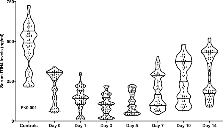 |
Figure 1 Longitudinal changes in serum levels of inter-alpha-trypsin inhibitor heavy chain 4 after acute intracerebral hemorrhage. Serum inter-alpha-trypsin inhibitor heavy chain 4 levels substantially reduced abruptly after acute stroke and remained low until day 14. Compared to controls, patients had significantly decreased serum inter-alpha-trypsin inhibitor heavy chain 4 levels 14 days after acute brain hemorrhage (P<0.001). Abbreviation: ITIH4, inter-alpha-trypsin inhibitor heavy chain 4. |
Admission Serum ITIH4 Levels in Relation to Stroke Severity and Systemic Inflammatory Response After ICH
As listed in Table 1, serum ITIH4 levels at admission were highly correlated with the presence of diabetes mellitus, extension of hematoma into the intraventricular space, NIHSS scores, hematoma size, blood leukocyte count, blood glucose levels, and serum C-reactive protein levels (all P<0.05). Subsequently, a multivariate linear regression model was constructed in which the seven significantly correlated variables were forced, and serum C-reactive protein levels (β, −6.436; 95% CI, −8.705--4.168; VIF, 2.382; P<0.001), NIHSS scores (β, −5.207; 95% CI, −8.716--1.697; VIF, 2.350; P=0.004), and hematoma size (β, −3.714; 95% CI, −5.354--2.074; VIF, 2.843; P<0.001) were retained as three independent factors, which were correlated with serum ITIH4 levels. As shown in Figure 2, there was a significant inverse correlation between serum ITIH4 levels and NIHSS scores (P<0.001), serum ITIH4 levels and hematoma size (P<0.001), and serum ITIH4 levels and serum C-reactive protein levels (P<0.001).
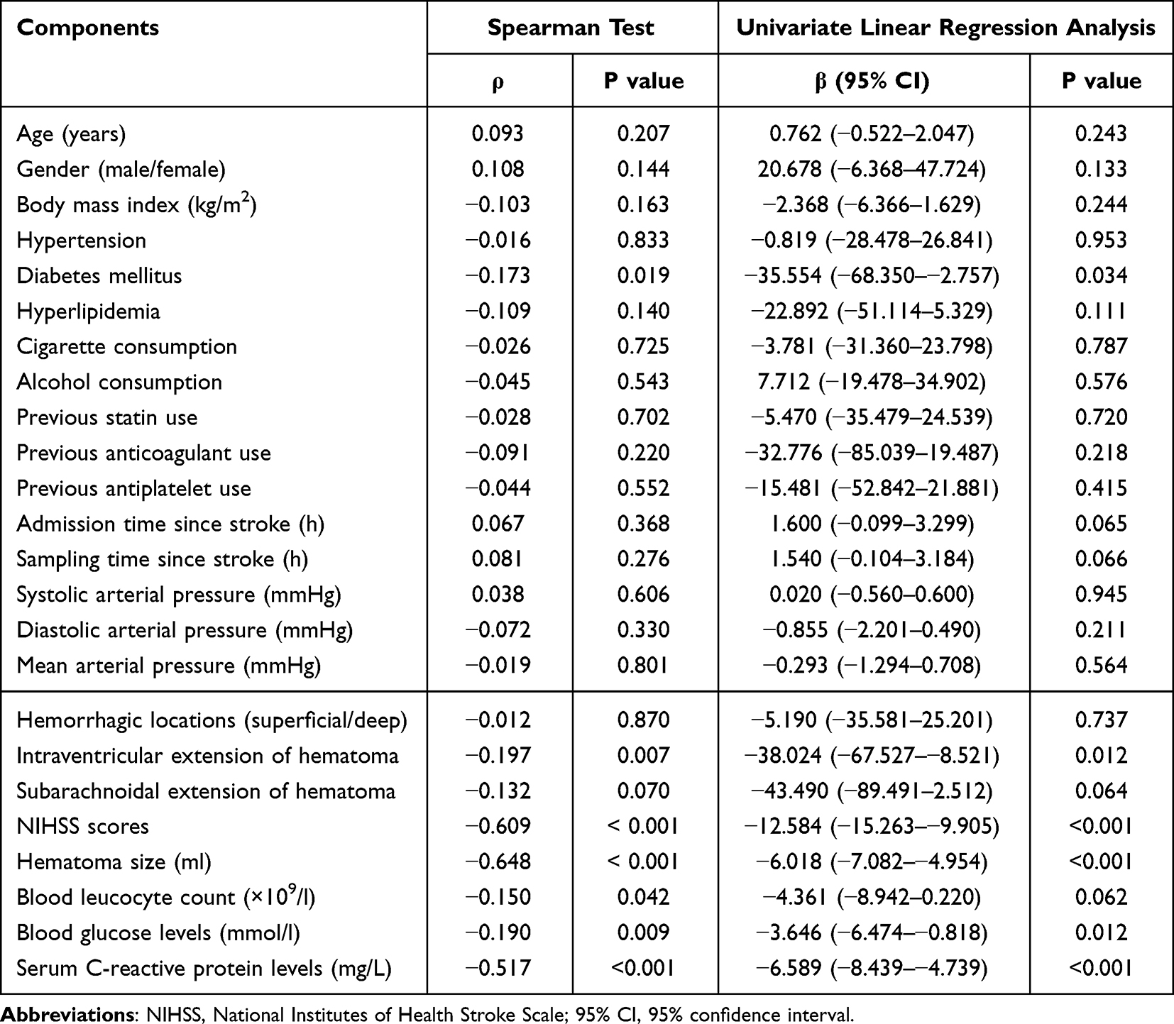 |
Table 1 Factors Related to Serum Inter-Alpha-Trypsin Inhibitor Heavy Chain 4 Levels After Acute Intracerebral Hemorrhage |
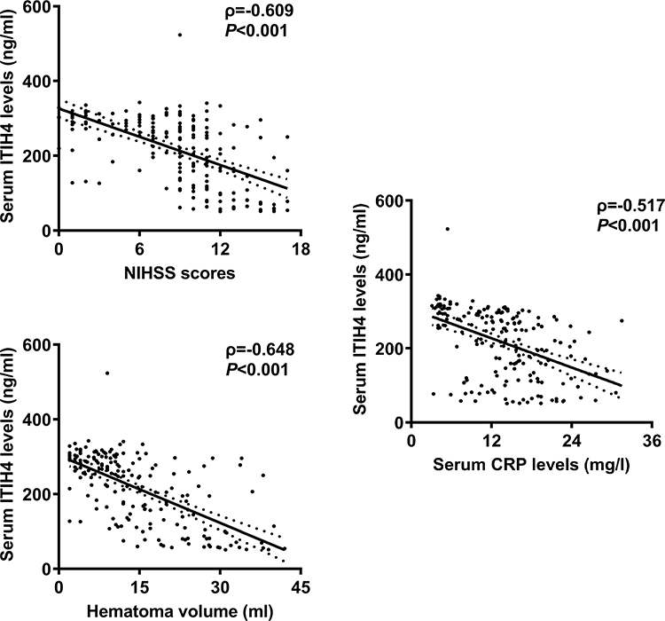 |
Figure 2 Relation of serum inter-alpha-trypsin inhibitor heavy chain 4 levels to stroke severity and systemic inflammation of acute intracerebral hemorrhage. Serum inter-alpha-trypsin inhibitor heavy chain 4 levels were significantly inversely correlated with serum C-reactive protein levels (P<0.001), National Institutes of Health Stroke Scale scores (P<0.001), and hematoma volume (P<0.001) in acute intracerebral patients. Abbreviations: ITIH4, inter-alpha-trypsin inhibitor heavy chain 4; NIHSS, National Institutes of Health Stroke Scale; CRP, C-reactive protein. |
Admission Serum ITIH4 Levels in Correlation with mRS Scores at Six Months After ICH
At post-stroke 6 months, mRS scores varying from 0 to 6 were observed in 20, 23, 52, 31, 28, 14, and 17 patients, respectively, with a median value of 2 (lower-upper quartiles, 2–4). As shown in Table 2, the variables that were closely correlated with six-month mRS scores on univariate analysis were diabetes mellitus, hematoma extending into the intraventricular space, extension of hematoma into the subarachnoid cavity, NIHSS scores, hematoma volume, blood glucose levels, serum C-reactive protein levels, and serum ITIH4 levels (all P<0.05). The above-mentioned significantly correlative variables were entered into multivariate model. Multivariate analysis revealed that serum ITIH4 levels (β, −0.004; 95% CI, −0.006--0.001; VIF, 2.084; P=0.021), NIHSS score (β, 0.119; 95% CI, 0.046–0.191; VIF, 2.475; P=0.001), and hematoma size (β, 0.036; 95% CI, 0.001–0.071; VIF, 3.139; P=0.046) were independently correlated with mRS scores 6 months after hemorrhagic stroke. As shown in Figure 3, admission serum ITIH4 levels were significantly higher in patients with an mRS score of 0, followed by scores ranging from 1 to 5, and were substantially lower in those with an mRS score of 6. Admission serum ITIH4 levels were closely correlated with mRS scores at six months after ICH.
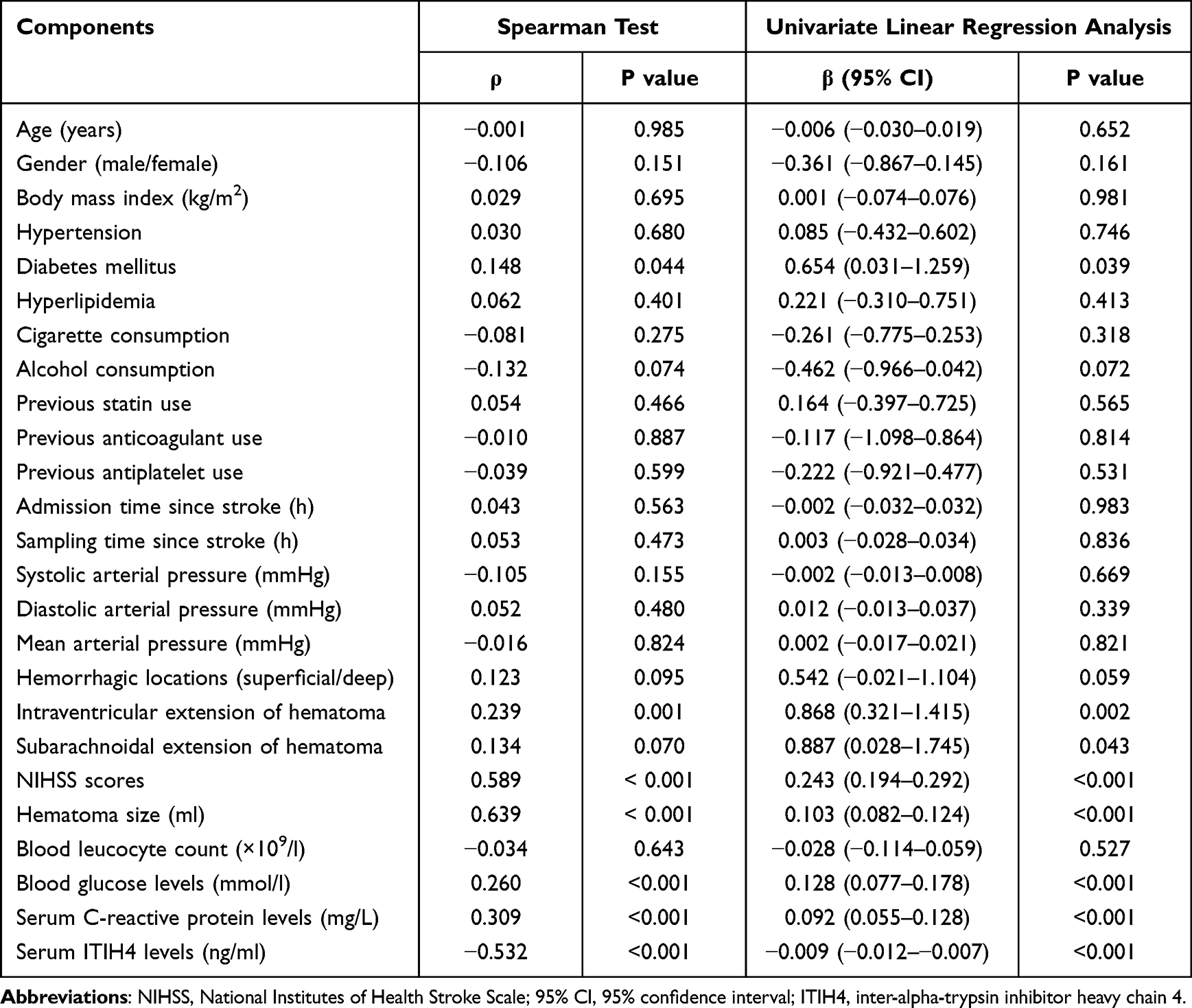 |
Table 2 Factors Related to Modified Rankin Scale Scores at Six Months After Acute Intracerebral Hemorrhage |
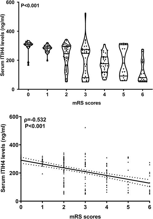 |
Figure 3 Elationship between serum inter-alpha-trypsin inhibitor heavy chain 4 levels and modified Rankin Scale scores at six months after acute intracerebral hemorrhage.Serum inter-alpha-trypsin inhibitor heavy chain 4 levels were highly correlated with modified Rankin Scale scores (P<0.001) and were markedly reduced in the order of modified Rankin Scale scores from 0 to 6 in patients with ICH (P<0.001). Abbreviations: ITIH4, inter-alpha-trypsin inhibitor heavy chain 4; mRS, modified Rankin Scale. |
Admission Serum ITIH4 Levels Associated with Risk of END After ICH
A total of 49 patients experienced END in this cohort of ICH patients. As shown in Figure 4, patients with END had significantly lower serum ITIH levels at admission than those who did not experience END (P<0.001); admission serum ITIH4 levels substantially distinguished the risk of END (P<0.001); and serum ITIH4 levels less than 221.0 ng/ml predicted END with 73.5% sensitivity, 65.4% specificity, Youden index of 0.389, positive predictive value of 43.4% and negative predictive value of 87.3%. To investigate the relationship between serum ITIH4 levels at admission and the development of END, serum ITIH4 was transformed into a categorical variable using a cutoff value (namely, 221.0 ng/ml), which was selected in accordance with the Youden method. As shown in Table 3, as opposed to patients without END development, those presenting with END exhibited substantially increased percentages of diabetes mellitus, intraventricular extension of hematoma, admission serum ITIH4 levels below 221.0 ng/ml, and dramatically increased NIHSS scores, hematoma size, serum C-reactive protein levels, and blood glucose levels (all P<0.05). The preceding significant variables on univariate analysis were forced into the multivariate model. Using binary logistic regression analysis, NIHSS scores (odds ratio, 1.228;95% CI, 1.053–1.432; P=0.009), hematoma size (odds ratio, 1.074;95% CI, 1.031–1.120; P=0.001), and serum ITIH4 levels < 221.0 ng/ml (odds ratio, 2.356; 95% CI, 1.036–5.359; P=0.047) were independent predictors of END. As plotted in Figure 5, a nomogram in which NIHSS scores, hematoma size, and admission serum ITIH4 levels were incorporated visually represented the END prediction model. Moreover, the model had comparative stability under the calibration curve (Figure 6) and showed relative clinical effectiveness in the decision curve analysis (Figure 7). However, the model did not exhibit a significantly higher predictive ability than either the NIHSS score or hematoma size (both P>0.05) under the ROC curve (Figure 8).
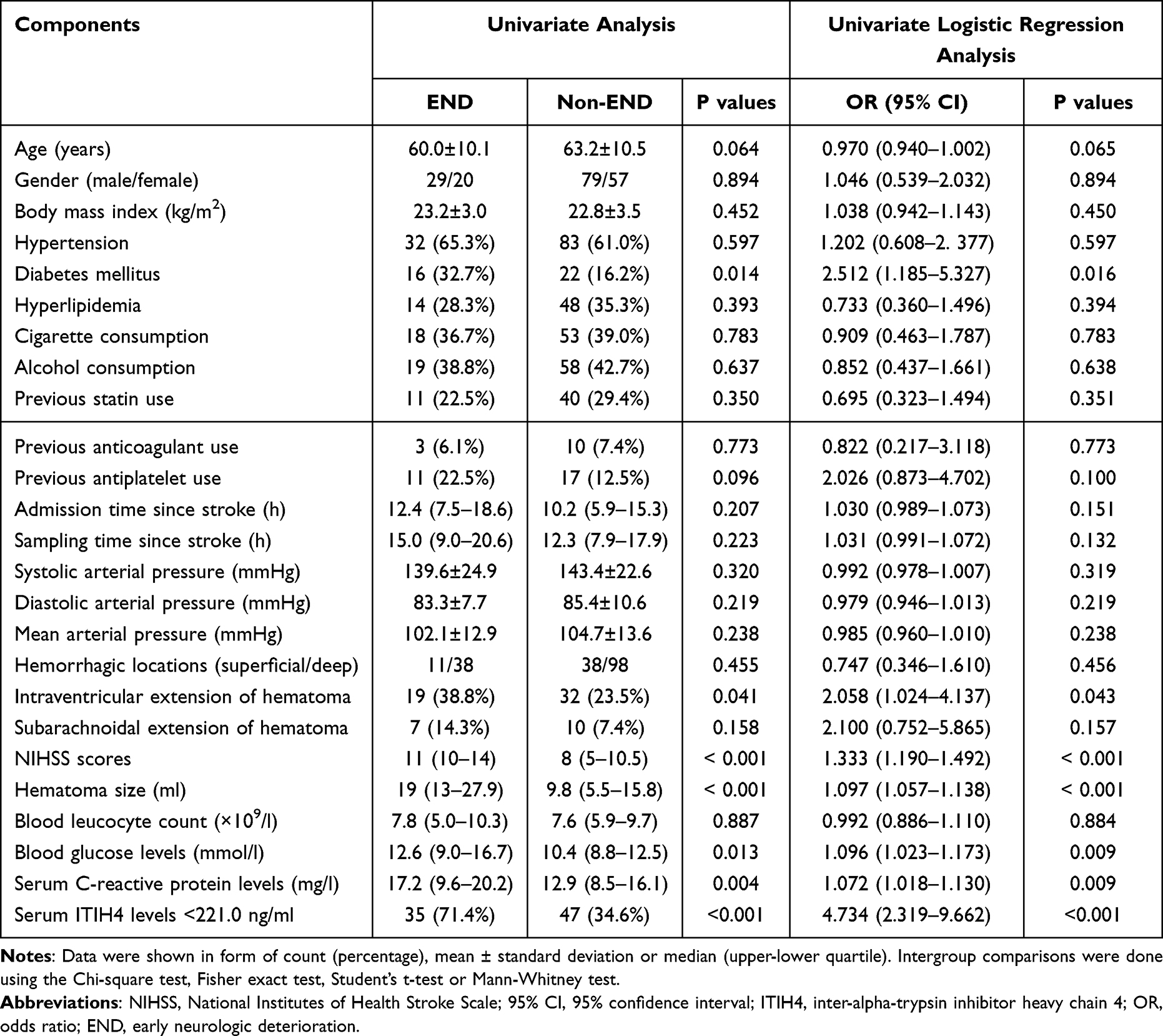 |
Table 3 Factors Related to Early Neurologic Deterioration After Acute Intracerebral Hemorrhage |
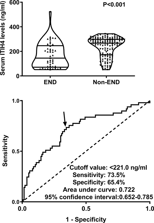 |
Figure 4 Relationship between serum inter-alpha-trypsin inhibitor heavy chain 4 levels and early neurologic deterioration after acute intracerebral hemorrhage. Serum inter-alpha-trypsin inhibitor heavy chain 4 levels were dramatically lower in patients with early neurological deterioration than in those without (P<0.001). Under the receiver operating characteristic curve, serum levels effectively predicted early neurological deterioration after an acute stroke (P<0.001). Arrow indicates cutoff value of serum inter-alpha-trypsin inhibitor heavy chain 4 levels. Abbreviations: ITIH4, inter-alpha-trypsin inhibitor heavy chain 4; END, early neurologic deterioration. |
 |
Figure 5 Nomogram displaying combination model for predicting early neurologic deterioration following acute intracerebral hemorrhage. The model was composed of serum inter-alpha-trypsin inhibitor heavy chain 4, National Institutes of Health Stroke Scale scores, and hematoma volume and was able to efficiently discriminate patients at risk of early neurologic deterioration after acute intracerebral hemorrhage. Abbreviations: ITIH4, inter-alpha-trypsin inhibitor heavy chain 4; NIHSS, National Institutes of Health Stroke Scale. |
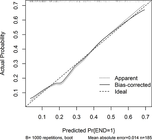 |
Figure 6 Calibration curve showing model stability for predicting early neurologic deterioration after acute intracerebral hemorrhage. The prediction model showed good stability for predicting early neurological deterioration after acute intracerebral the calibration curve. Abbreviation: END, early neurologic deterioration. |
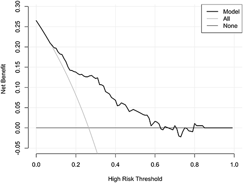 |
Figure 7 Decision curve verifying clinical benefit of model for predicting early neurologic deterioration after acute intracerebral hemorrhage. The prediction model had a medium-high clinical benefit for predicting early neurological deterioration after acute ICH, according to the decision curve. |
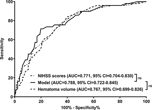 |
Figure 8 Receiver operating characteristic curve with respect to the predictive value of combination model for early neurologic deterioration after acute intracerebral hemorrhage. The predictive ability of the model was similar to that of the National Institutes of Health Stroke Scale scores and hematoma volume (both P>0.05). Abbreviations: NIHSS, National Institutes of Health Stroke Scale; AUC, area under the curve; 95% CI, 95% confidence interval; ns, non-significant. |
Admission Serum ITIH4 Levels Related to Poor Prognosis at Six Months After ICH
Ninety patients had a poor prognosis 6 months after ICH. As shown in Figure 9, admission serum ITIH levels were significantly diminished in patients with poor prognosis compared to those with good prognosis (P<0.001), whereas admission serum ITIH4 levels efficiently discriminated patients at risk of poor prognosis (P<0.001). And serum ITIH4 levels less than 279.0 ng/ml predicted END with 87.8% sensitivity, 54.7% specificity, Youden index of 0.425, positive predictive value of 64.8% and negative predictive value of 82.5%. Furthermore, to perform a prognosis analysis, serum ITIH4 was identified as a qualitative variable using a cut-off value (namely, 279.0 ng/ml), which was chosen based on the Youden method. As shown in Table 4, in contrast to patients presenting with a good prognosis, those with a poor prognosis displayed markedly elevated proportions of diabetes mellitus, intraventricular extension of hematoma, and admission serum ITIH4 levels less than 279.0 ng/ml, as well as markedly enhanced NIHSS scores, hematoma size, serum C-reactive protein levels, and blood glucose levels (all P<0.05). A binary logistic regression model was constructed, in which the preceding seven significant variables were entered, and it was verified that predictors, which were independently associated with poor prognosis, were NIHSS scores (odds ratio, 1.225;95% CI, 1.061–1.415; P=0.006), hematoma size (odds ratio, 1.069; 95% CI, 1.009–1.133; P=0.023), and admission serum ITIH4 levels below 279.0 ng/ml (odds ratio, 2.949; 95% CI, 1.270–6.846; P=0.012). As shown in Figure 10, a nomogram composed of NIHSS scores, hematoma size, and admission serum ITIH4 levels was built to delineate the prognosis prediction model. Using calibration curve analysis, the model remained stable (Figure 11), and using decision curve analysis, the model was clinically effective. (Figure 12). Intriguingly, using ROC curve analysis (Figure 13), the predictive efficiency of the model was significantly higher than that of NIHSS scores or hematoma size (both P<0.05).
 |
Table 4 Factors Related to Poor Prognosis at Six Months After Acute Intracerebral Hemorrhage |
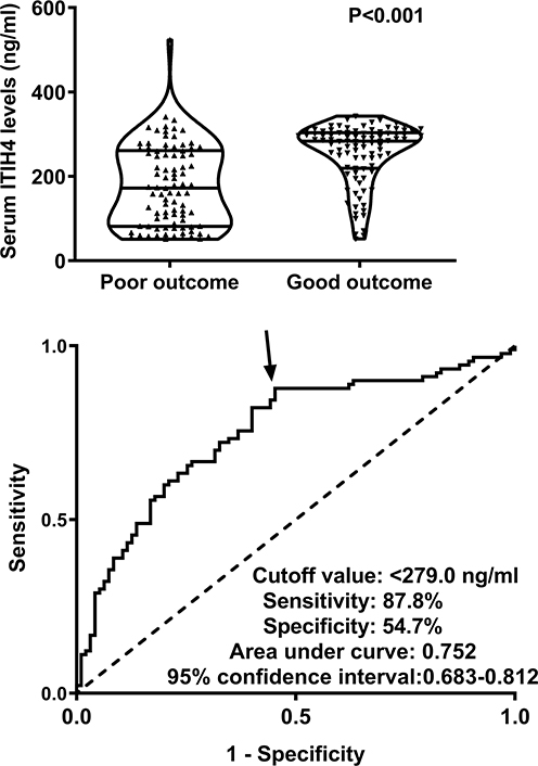 |
Figure 9 Relationship between serum inter-alpha-trypsin inhibitor heavy chain 4 levels and poor neurologic functional outcome six months after acute intracerebral hemorrhage. Serum inter-alpha-trypsin inhibitor heavy chain 4 levels were significantly lower in patients with poor prognosis than in those with good prognosis (P<0.001). Under the receiver operating characteristic curve, these levels efficiently distinguished patients at risk of poor prognosis after acute cerebral hemorrhage (P<0.001). Arrow indicates cutoff value of serum inter-alpha-trypsin inhibitor heavy chain 4 levels. Abbreviation: ITIH4, inter-alpha-trypsin inhibitor heavy chain 4. |
 |
Figure 10 Nomogram describing six-month prognosis prediction model in acute intracerebral hemorrhage. Serum inter-alpha-trypsin inhibitor heavy chain 4, National Institutes of Health Stroke Scale scores, and hematoma volume were integrated into the combination model, which efficiently predicted a poor prognosis after acute intracerebral hemorrhage. Abbreviations: ITIH4, inter-alpha-trypsin inhibitor heavy chain 4; NIHSS, National Institutes of Health Stroke Scale. |
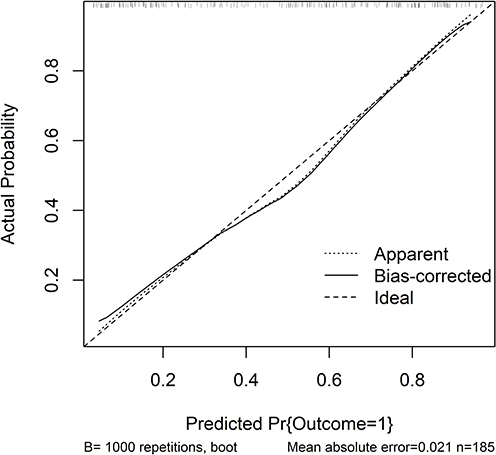 |
Figure 11 Calibration curve demonstrating stability of six-month prognosis prediction model in acute intracerebral hemorrhage. The prognosis prediction model was rather stable in acute intracerebral hemorrhage under calibration curve. |
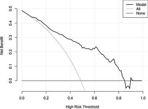 |
Figure 12 Decision curve showing clinical benefit of six-month prognosis prediction model in acute intracerebral hemorrhage. The prognosis prediction model had a comparative clinical benefit in acute intracerebral hemorrhage under the decision curve. |
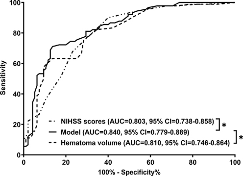 |
Figure 13 Receiver operating characteristic curve for analyzing the prognostic predictive performance of the combination model in acute intracerebral hemorrhage. The prognostic ability of the prediction model was significantly higher than that of the National Institutes of Health Stroke Scale scores and the hematoma volume alone (both P<0.05). Abbreviations: NIHSS, National Institutes of Health Stroke Scale; AUC, area under the curve; 95% CI, 95 confidence interval; * P<0.05. |
Discussion
Clinical data regarding ischemic stroke have shown that circulating ITIH4 levels may diminish after acute brain injury.27,28 Nevertheless, serum ITIH4 levels of in humans after acute ICH remain unclear. We undertook this clinical investigation to discern the time course of serum ITIH4 levels following acute ICH, and subsequently found that serum ITIH4 levels were abruptly reduced after stroke, with a profoundly lowest level at days 3 and 5, and then gradually increased until day 14, as well as noteworthily, serum ITIH4 levels of patients during fourteen days were pronouncedly lower than those of controls. Using univariate and sequential multivariate analyses, we confirmed that serum ITIH4 levels were independently correlated with 6-month mRS scores, NIHSS scores, hematoma volume, and serum C-reactive protein levels. In addition, serum ITIH4 level emerged as an independent predictor of END and six-month poor prognosis. Our most intriguing finding was that prediction models of poor prognosis, but not END, in which NIHSS scores, hematoma volume, and serum ITIH4 levels were incorporated, may be of great clinical value under the ROC, calibration, and decision curves. Therefore, serum ITIH4, acting as an inflammation-related factor, may serve as a promising biomarker for severity stratification and prognostic prediction, thereby facilitating the clinical work of prognostication in ICH.
An increasing body of data strongly supports the assumption that ITIH4 exerts a protective effect on injured cells, tissues, and organs by regulating JAK-STAT signaling32 and nuclear factor kappa-B signaling.33 Notably, circulating ITIH4 has potential as an anti-inflammatory biomarker for severity assessment and prognosis prediction in numerous diseases such as chronic obstructive pulmonary disease, coronary heart disease, rheumatoid arthritis, sepsis, and hepatitis C.22–26 In five patients with acute ischemic stroke, there was a dominant downregulation of blood ITIH4 levels within 72 h after injury, which significantly increased when patients recovered.27 In another group of acute ischemic stroke patients, serum ITIH4 levels immediately decreased at admission, gradually decreased until day 3, and then increased until day 30.28 In our study, there was an obvious trend in which serum ITIH4 levels significantly declined in the initial phase, and thereafter substantially increased until 14 days after ICH. Taken together, these results suggest that ITIH4 may be partially depleted following acute brain injury. In other words, such a finding is strongly supportive of the notion that ITIH4 may be a protective factor during acute brain injury. In addition, serum ITIH4 levels are inversely correlated with serum TNF-α, interleukin-1beta, interleukin-6 and interleukin-17A in humans with acute ischemic stroke.28 C-reactive protein (CRP) is an acute-phase reaction protein, and its levels can reflect the extent of the systemic inflammatory response in ICH.34 Our study showed that serum C-reactive protein levels were independently inversely correlated with serum ITIH4 levels in the cohort of patients with ICH. These data suggest that ITIH4 may be an anti-inflammatory factor with protective functions in acute brain injury.
In a previous study of acute ischemic stroke patients, significantly reduced serum ITIH4 levels were highly correlated with infarct volume, as determined using computed tomography.27 Another study showed that serum ITIH4 levels, which substantially diminished after acute ischemic stroke, are intimately associated with disease recurrence and mortality.28 Notably, we continued to perform multivariate analysis and subsequently found some interesting results that serum ITIH4 levels were independently associated with END and poor prognosis six months after ICH. Moreover, prediction models for END and poor prognosis were built, and their stability, efficiency, and clinical benefits were verified using a series of statistical methods. Interestingly, the prognosis prediction model showed higher discriminatory effectiveness than the NIHSS scores and hematoma size alone, but the END prediction model did not. Thus, serum ITIH4 levels may represent a promising prognostic biomarker for ICH.
This study has several strengths and weaknesses. The strengths of this study are as follows: (1) serum ITIH4 levels after ICH were measured to investigate their dynamic changes, (2) relationships between serum ITIH4 levels and the severity and prognosis of ICH were analyzed using multivariate analysis, and (3) only patients with supratentorial bleeding were included in this study, and homogeneity was guaranteed. The weaknesses of this study are that (1) a poor prognosis (mRS scores of 3–6) is easily accepted as an outcome parameter of ICH.31 In this group of patients, 17 cases had mRS score 6, meaning that 17 patients died within six months after ICH. Thus, six-month mortality was 9.2% (17/185). This was an all-cause death rate. Because of a very small sample size in the deceased, it does not fit requirements of statistical analysis. Moreover, death (mRS score 6) is a component of mRS, and some relevant statistical methods have been currently employed for demonstrating relationship between serum ITIH4 levels and mRS scores. In other words, serum ITIH4 may be a prognostic biomarker of ICH, which has been verified using univariate and multivariate analysis in this study. Admittedly, it is better that more patients should be enrolled in future and thereafter a relation of serum ITIH4 levels to mortality would be investigated. (2) although we reported the longitudinal change of serum ITIH4 levels after ICH, the prognostic predictive ability of serum ITIH4 levels was not compared among multiple time-points because of a small sample size in such a group of patients who provided consent for blood-collections at multiple time-points. Hence, it may be clinically valuable to further investigate prognostic predictive performance of serum ITIH4 levels at multiple time-points after ICH. (3) the conclusions of this study should be validated in a larger cohort study because the current study was performed at a single center.
Conclusions
Serum ITIH4 levels immediately diminished upon admission after ICH and continuously declined until day 14. The levels, which were independently correlated with systemic inflammation and stroke severity, were independently associated with END and 6-month neurologic functional outcome. Notably, the prediction model, which was composed of serum ITIH4, NIHSS scores, and hematoma volume, displayed a high discriminatory efficiency. Our data offer sufficient evidence to support the hypothesis that serum ITIH4, which can act as an anti-inflammatory factor and possess neuroprotective effects, may serve as a useful prognostic biomarker in human ICH.
Data Sharing Statement
The datasets generated and/or analyzed during the current study are not publicly available because they are personal data, but are available from the corresponding author upon reasonable request.
Ethics approval and consent to participate
The current study was conducted in compliance with the tenets of the Declaration of Helsinki, and the protocol was approved by the ethics committee of Jinhua People’s Hospital (No. JPH2018014). Signed informed consent was obtained from patients’ relatives or controls.
Acknowledgments
We gratefully thank all study participants, their relatives, and the staff at the recruitment centers for their invaluable contributions.
Funding
The authors received no financial support for the research.
Disclosure
The authors declare no potential conflicts of interest in this work.
References
1. Qureshi AI, Tuhrim S, Broderick JP, Batjer HH, Hondo H, Hanley DF. Spontaneous intracerebral hemorrhage. N Engl J Med. 2001;344(19):1450–1460. doi:10.1056/NEJM200105103441907
2. Hostettler IC, Seiffge DJ, Werring DJ. Intracerebral hemorrhage: an update on diagnosis and treatment. Expert Rev Neurother. 2019;19(7):679–694. doi:10.1080/14737175.2019.1623671
3. Sondag L, Schreuder FHBM, Boogaarts HD, et al. Neurosurgical intervention for supratentorial intracerebral hemorrhage. Ann Neurol. 2020;88(2):239–250. doi:10.1002/ana.25732
4. Kuohn LR, Witsch J, Steiner T, et al. Early deterioration, hematoma expansion, and outcomes in deep versus lobar intracerebral hemorrhage: the FAST trial. Stroke. 2022;53(8):2441–2448. doi:10.1161/STROKEAHA.121.037974
5. Kirshner H, Schrag M. Management of intracerebral hemorrhage: update and future therapies. Curr Neurol Neurosci Rep. 2021;21(10):57. doi:10.1007/s11910-021-01144-9
6. Mendiola JMF, Arboix A, García-Eroles L, Sánchez-López MJ. Acute spontaneous lobar cerebral hemorrhages present a different clinical profile and a more severe early prognosis than deep subcortical intracerebral hemorrhages-A Hospital-Based Stroke Registry Study. Biomedicines. 2023;11(1):223. doi:10.3390/biomedicines11010223
7. Zhu H, Wang Z, Yu J, et al. Role and mechanisms of cytokines in the secondary brain injury after intracerebral hemorrhage. Prog Neurobiol. 2019;178:101610. doi:10.1016/j.pneurobio.2019.03.003
8. Nobleza COS. Intracerebral Hemorrhage. Continuum. 2021;27(5):1246–1277. doi:10.1212/CON.0000000000001018
9. Leira R, Dávalos A, Silva Y, et al. Early neurologic deterioration in intracerebral hemorrhage: predictors and associated factors. Neurology. 2004;63(3):461–467. 81153.ac. doi:10.1212/01.wnl.0000133204
10. Han C, Khan NI, Mady LJ. Prognosis. Otolaryngol Clin North Am. 2023;56(2):389–402. doi:10.1016/j.otc.2022.12.005
11. Capizzi A, Woo J, Verduzco-Gutierrez M. Traumatic brain injury: an overview of epidemiology, pathophysiology, and medical management. Med Clin North Am. 2020;104(2):213–238. doi:10.1016/j.mcna.2019.11.001
12. Rabinstein AA. Update on treatment of acute ischemic stroke. Continuum. 2020;26(2):268–286. doi:10.1212/CON.0000000000000840
13. Macdonald RL, Schweizer TA. Spontaneous subarachnoid haemorrhage. Lancet. 2017;389(10069):655–666. doi:10.1016/S0140-6736(16)30668-7
14. Kwah LK, Diong J. National Institutes of Health Stroke Scale (NIHSS). J Physiother. 2014;60(1):61. doi:10.1016/j.jphys.2013.12.012
15. LoPresti MA, Bruce SS, Camacho E, et al. Hematoma volume as the major determinant of outcomes after intracerebral hemorrhage. J Neurol Sci. 2014;345(1–2):3–7. doi:10.1016/j.jns.2014.06.057
16. Bader ER, Pana TA, Barlas RS, Metcalf AK, Potter JF, Myint PK. Elevated inflammatory biomarkers and poor outcomes in intracerebral hemorrhage. J Neurol. 2022;269(12):6330–6341. doi:10.1007/s00415-022-11284-8
17. Guo H, Zhang Y, Hu Z, Wang L, Du H. Screening and identification of biomarkers associated with the immune infiltration of intracerebral hemorrhage. J Clin Lab Anal. 2022;36(5):e24361. doi:10.1002/jcla.24361
18. Yang Z, Wang R, Chen X, Zhao D. Identification of potential biomarkers and small‑molecule compounds related to intracerebral hemorrhage with bioinformatics analysis. Acta Neurobiol Exp (Wars). 2022;82(2):187–196. doi:10.55782/ane-2022-017
19. Zhuo L, Kimata K. Structure and function of inter-alpha-trypsin inhibitor heavy chains. Connect Tissue Res. 2008;49(5):311–320. doi:10.1080/03008200802325458
20. Kashyap RS, Nayak AR, Deshpande PS, et al. Inter-alpha-trypsin inhibitor heavy chain 4 is a novel marker of acute ischemic stroke. Clin Chim Acta. 2009;402(1–2):160–163. doi:10.1016/j.cca.2009.01.009
21. Shi X, Ohta Y, Liu X, et al. Acute anti-inflammatory markers ITIH4 and AHSG in mice brain of a novel Alzheimer’s disease model. J Alzheimers Dis. 2019;68(4):1667–1675. doi:10.3233/JAD-181218
22. Lee KY, Feng PH, Ho SC, et al. Inter-alpha-trypsin inhibitor heavy chain 4: a novel biomarker for environmental exposure to particulate air pollution in patients with chronic obstructive pulmonary disease. Int J Chron Obstruct Pulmon Dis. 2015;10:831–841. doi:10.2147/COPD.S81611
23. Huo Y, Lai Y, Feng Q, Wang Q, Li J. Serum ITIH4 in coronary heart disease: a potential anti-inflammatory biomarker related to stenosis degree and risk of major adverse cardiovascular events. Biomark Med. 2022;16(18):1279–1288. doi:10.2217/bmm-2022-0673
24. He K, He S, Su M. Inter-alpha-trypsin inhibitor heavy chain 4: a serologic marker relating to disease risk, activity, and treatment outcomes of rheumatoid arthritis. J Clin Lab Anal. 2022;36(3):e24231. doi:10.1002/jcla.24231
25. Sira MM, Behairy BE, Abd-Elaziz AM, Abd Elnaby SA, Eltahan EE. Serum inter-alpha-trypsin inhibitor heavy chain 4 (ITIH4) in children with chronic hepatitis C: relation to liver fibrosis and viremia. Hepat Res Treat. 2014;2014:307942. doi:10.1155/2014/307942
26. Zhao X, Guo Y, Li L, Li Y. Longitudinal change of serum inter-alpha-trypsin inhibitor heavy chain H4, and its correlation with inflammation, multiorgan injury, and death risk in sepsis. J Clin Lab Anal. 2023;37(3):e24834. doi:10.1002/jcla.24834
27. Nayak AR, Kashyap RS, Kabra D, Purohit HJ, Taori GM, Daginawala HF. Time course of inflammatory cytokines in acute ischemic stroke patients and their relation to inter-alfa trypsin inhibitor heavy chain 4 and outcome. Ann Indian Acad Neurol. 2012;15(3):181–185. doi:10.4103/0972-2327.99707
28. Zhang J, Hu J, Zhao W. Longitudinal change of serum inter-α-trypsin inhibitor heavy chain H4 and its relation with inflammation, disease recurrence, and mortality in acute ischemic stroke patients. Tohoku J Exp Med. 2023;259(3):221–227. doi:10.1620/tjem.2022.J116
29. Kothari RU, Brott T, Broderick JP, et al. The ABCs of measuring intracerebral hemorrhage volumes. Stroke. 1996;27(8):1304–1305. doi:10.1161/01.str.27.8.1304
30. You S, Zheng D, Delcourt C, et al. Determinants of early versus delayed neurological deterioration in intracerebral hemorrhage. Stroke. 2019;50(6):1409–1414. doi:10.1161/STROKEAHA.118.024403
31. Hao Y, Avadhani R, Caron JL, et al. Efficacy and safety of minimally invasive surgery with thrombolysis in intracerebral haemorrhage evacuation (MISTIE III): a randomised, controlled, open-label, blinded endpoint phase 3 trial. Lancet. 2019;393(10175):1021–1032. doi:10.1016/S0140-6736(19)30195-3
32. Ma Y, Li R, Wang J, et al. ITIH4, as an inflammation biomarker, mainly increases in bacterial bloodstream infection. Cytokine. 2021;138:155377. doi:10.1016/j.cyto.2020.155377
33. Stober VP, Lim YP, Opal S, Zhuo L, Kimata K, Garantziotis S. Inter-α-inhibitor ameliorates endothelial inflammation in sepsis. Lung. 2019;197(3):361–369. doi:10.1007/s00408-019-00228-1
34. Wang D, Wang J, Li Z, et al. C-reaction protein and the severity of intracerebral hemorrhage: a study from Chinese Stroke Center Alliance. Neurol Res. 2022r;44(4):285–290. doi:10.1080/01616412.2021.1980842
 © 2023 The Author(s). This work is published and licensed by Dove Medical Press Limited. The
full terms of this license are available at https://www.dovepress.com/terms
and incorporate the Creative Commons Attribution
- Non Commercial (unported, 3.0) License.
By accessing the work you hereby accept the Terms. Non-commercial uses of the work are permitted
without any further permission from Dove Medical Press Limited, provided the work is properly
attributed. For permission for commercial use of this work, please see paragraphs 4.2 and 5 of our Terms.
© 2023 The Author(s). This work is published and licensed by Dove Medical Press Limited. The
full terms of this license are available at https://www.dovepress.com/terms
and incorporate the Creative Commons Attribution
- Non Commercial (unported, 3.0) License.
By accessing the work you hereby accept the Terms. Non-commercial uses of the work are permitted
without any further permission from Dove Medical Press Limited, provided the work is properly
attributed. For permission for commercial use of this work, please see paragraphs 4.2 and 5 of our Terms.
Recommended articles
Plasma SIRT3 as a Biomarker of Severity and Prognosis After Acute Intracerebral Hemorrhage: A Prospective Cohort Study
Yan T, Wang ZF, Wu XY, Du Q, Yu WH, Hu W, Zheng YK, Wang KY, Dong XQ
Neuropsychiatric Disease and Treatment 2022, 18:2199-2210
Published Date: 26 September 2022
Usability of Serum Stanniocalcin-1 as a Prognostic Biochemical Marker of Acute Supratentorial Intracerebral Hemorrhage: A Prospective Cohort Study
Gao CF, Zhang GH, Ye ZH, Xu YY, Li Z
International Journal of General Medicine 2023, 16:2791-2803
Published Date: 3 July 2023
Serum Secreted Protein Acidic and Rich in Cysteine-Like 1 as a Biochemical Predictor for Prognosticating Clinical Outcomes After Acute Supratentorial Intracerebral Hemorrhage: A Prospective Cohort Study
Huang J, Shao F, Chen B, Zheng G, Shen J, Qiu S
Neuropsychiatric Disease and Treatment 2023, 19:2709-2728
Published Date: 5 December 2023
Prognostic Relevance of ACSL4 as a Serological Marker and Its Mediating Roles in Acute Supratentorial Intracerebral Hemorrhage: An Observational Analytical Study
Li M, Zheng H, Fu Z, Yan X, Mao D, Yu G
International Journal of General Medicine 2026, 19:580561
Published Date: 24 February 2026
Serum Peroxiredoxin 6 Levels and Clinical Outcomes After Acute Intracerebral Hemorrhage in Elderly Patients: A Multicenter Observational Analytical Study
Lu T, Zheng B, Wang D, Liao W, Su C, Wu X, Zhong X, Chen X, Ying G, Cai Y, Du Q, Dong X
Clinical Interventions in Aging 2026, 21:582268
Published Date: 1 May 2026