
Back to Journals » Journal of Inflammation Research » Volume 18
Yuyang Decoction Regulates Macrophage Polarization and Repairs the Intestinal Mucosal Barrier via the TLRs/Tollip Signaling Pathway
Authors Lian J, Jin G  , Li J, Zhang W, Chang Y, Li W, Zhi H, Tian J , Liang C, Su J
Received 6 January 2025
Accepted for publication 19 July 2025
Published 4 August 2025 Volume 2025:18 Pages 10499—10518
DOI https://doi.org/10.2147/JIR.S515902
Checked for plagiarism Yes
Review by Single anonymous peer review
Peer reviewer comments 2
Editor who approved publication: Professor Ning Quan
Jie Lian, Guichun Jin, Jing Li, Weijuan Zhang, Yongtai Chang, Wenwu Li, Hua Zhi, Jiao Tian, Chen Liang, Juanping Su
Shanxi Traditional Chinese Medical Hospital, Taiyuan, People’s Republic of China
Correspondence: Juanping Su, Shanxi Traditional Chinese Medical Hospital, Taiyuan, People’s Republic of China, Email [email protected]
Background: Ulcerative colitis (UC) is a chronic inflammatory bowel disease characterized by relapsing inflammation and impaired intestinal epithelial barrier. Current treatments have limitations, prompting interest in traditional Chinese medicine as potential alternatives. Yuyang Decoction (YYD) is a traditional formula with observed gastrointestinal protective effects, but its mechanisms remain unclear.
Objective: To evaluate the therapeutic effects and underlying mechanisms of YYD on intestinal mucosal barrier repair in a UC mouse model.
Methods: UC was induced in male C57/BL6 mice using 3% dextran sulfate sodium (DSS). Mice were randomly assigned to six groups: control, DSS model, low/medium/high-dose YYD, and mesalazine (positive control). Disease activity index (DAI), colon length, histopathology, cytokine levels, and tight junction protein expression were assessed. Tollip and IKK-β levels were detected by Western blot and immunofluorescence. Active components in YYD-containing serum were identified by UPLC-Q-Tof-MS/MS. In vitro, Tollip knockdown in THP-1 macrophages examined YYD’s effect on macrophage polarization and Caco-2 cell barrier integrity.
Results: YYD significantly reduced DAI scores, reversed colon shortening, and alleviated histological damage. Expression of tight junction proteins (occludin, claudins, ZO-1) and Tollip was upregulated. YYD-containing serum suppressed LPS-induced M1 macrophage polarization and protected epithelial tight junctions in vitro. These effects were diminished upon Tollip silencing.
Conclusion: YYD alleviates DSS-induced colitis by enhancing intestinal barrier repair and modulating immune responses. Its therapeutic effect may involve activation of the Tollip signaling pathway, which inhibits pro-inflammatory M1-like macrophage differentiation and reduces epithelial damage.
Keywords: Yuyang decoction, ulcerative colitis, occlusal protein, closure protein, TLRs/Tollip signaling pathway
Ulcerative colitis (UC) is an inflammatory bowel disease (IBD) of unknown etiology, clinically characterized by abdominal pain, diarrhea, and mucopus-bloody stool.1 It is noted for its prolonged course, complex pathology, and resistance to cure. Given that the mechanisms underlying the initiation and progression of UC remain elusive, a definitive cure is currently unattainable, necessitating prolonged, and even lifelong, treatment. At present, the incidence of UC is increasing year by year, and the trend of youthfulness.2 Consequently, UC poses not only a significant medical challenge but also a societal issue. 5-aminosalicylic acid (5-ASA) is widely used in the treatment of UC. However, the 5-ASA response rate is only 30 to 60%.3 The “multi-component-multi-target” advantage of traditional Chinese medicine (TCM) provides a therapeutic strategy different from single-target western medicine by addressing the interaction of immune barriers at multiple regulatory nodes. TCM grounded in the holistic perspective, employs syndrome differentiation and treatment, addressing both the overall condition and local manifestations, thereby enhancing our understanding of the pathogenesis of UC.4
In our previous research, the research team developed a unique two-step enema therapy for the treatment of ulcerative colitis (UC), which combines the use of Yuyang Decoction (YYD) with saline enemas. This therapy exhibits distinct therapeutic efficacy and minimal toxic and side effects. The present experimental study applies the principles of Traditional Chinese Medicine (TCM) in treating UC through this innovative two-step enema approach. Specifically, the colon mucosa is first cleansed with a saline enema, followed by a retention enema with YYD, allowing the medicinal liquid to directly interact with the mucosal surface. Over the years, this therapy has demonstrated reliable clinical outcomes, offering unique therapeutic benefits and minimal toxicity and side effects, thus presenting significant advantages and potential. Current research suggests that the TLRs/Tollip signaling pathway maintains the intestinal mucosal immune system, exhibiting immune tolerance to normal intestinal microbiota and immune surveillance against pathogenic microorganisms. This pathway not only eliminates pathogenic microbial infections but also avoids excessive immune responses.5 Therefore, investigating the mechanism by which Yuyang Decoction regulates Tollip in the context of intestinal mucosal barrier dysfunction, and thereby deepening our understanding of the pathogenesis of UC, can provide a solid theoretical basis for further exploring novel therapeutic targets for UC.
It is currently recognized that an important factor in the etiology and pathogenesis of ulcerative colitis (UC) is the abnormal immune response of the intestinal mucosa’s innate and adaptive immune response systems to microbial antigens within the gut, leading to the initiation and progression of intestinal mucosal inflammation6 Studies have shown that the TLRs/Tollip signaling pathway maintains the immune tolerance of the intestinal mucosal immune system to normal intestinal microbiota and immune surveillance against pathogenic microorganisms, enabling the clearance of pathogenic microbial infections while avoiding excessive immune responses.7 Tollip is involved in the process of monocyte autophagy, and Tollip participates in the process of monocyte/macrophage autophagy by mediating the fusion of lysosomes with endosomes/autolysosomes.7 A large number of macrophages can be detected in many patients with colitis,8 playing a crucial role in the initiation and resolution of inflammation. M1 macrophages, induced by LPS, produce a series of pro-inflammatory cytokines such as IL-6 and iNOS, while M2 macrophages, induced by IL-4, release various anti-inflammatory cytokines.9 Normally, TLRs in the intestinal mucosal innate immune system are constitutively low-expressed, while Tollip is constitutively high-expressed,10 maintaining the tolerance of the intestinal mucosal innate immune system to continuous LPS stimulation. Upon pathogen recognition or tissue damage signals, TLRs recruit adaptor proteins including MyD88, forming a complex that activates interleukin-1 receptor-associated kinase 1 (IRAK1).11 This kinase undergoes autophosphorylation, a pivotal step for downstream NF-κB and MAPK pathway activation.12,13 However, persistent stimulation by certain pathogenic factors (such as highly pathogenic intestinal microorganisms) can alter intestinal mucosal immune function,14 resulting in decreased Tollip expression and/or overexpression and activation of TLRs, disrupting the immune balance of the intestinal mucosa. Upregulation of Tollip can inhibit M1 polarization, further alleviating intestinal mucosal damage, promoting intestinal mucosal repair and mucosal barrier reconstruction, and thus achieving therapeutic effects on intestinal inflammation.
Therefore, investigating the mechanism by which Yuyang Decoction regulates Tollip in intestinal mucosal barrier dysfunction can deepen our understanding of the pathogenesis of UC and provide a solid theoretical basis for further exploring novel therapeutic targets for UC.
Methods and Materials
Yuyang Soup Source
YYD a self-formulated prescription by Professor Feng Wujin from the Shanxi Provincial Institute of TCM, comprises the following herbs: Frankincense (6g), Myrrh (6g), Chinese Gall (3g), Pollen Typhae Carbonisatus (10g), Sophora Flavescens (10g), Radix Sanguisorbae Carbonisatus (10g), Colla Corii Asini (6g), Tripterygium Wilfordii Hook F. (10g), totaling 61g. Table 1. The plant medicinal materials contained in YYD are listed in the Chinese Pharmacopoeia, and no additional approval is required for research on the use of plant materials. The plant medicinal materials were purchased from the sales organization of Chinese plant medicinal materials. The source plants were pretreated according to the requirements of the Chinese Pharmacopoeia before clinical use, and could not be purchased for clinical use until the licensed physician issued a prescription. Commercial source: Shanxi Ruixing Pharmaceutical Co., LTD. China.
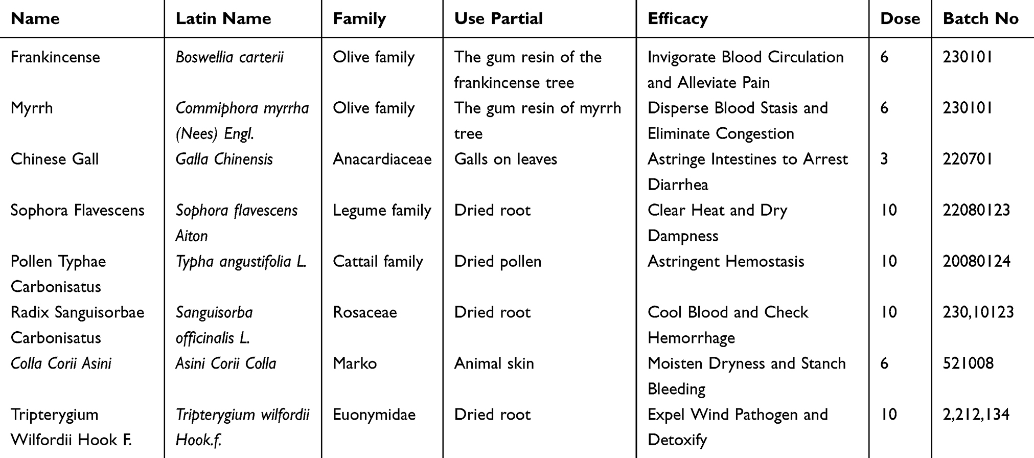 |
Table 1 Plant Material Composition Table |
Animal Source and Breeding Environment
Thirty-six male C57/BL6 mice, aged 4 weeks and weighing approximately 20g (ensuring that the initial average weight of mice in each group was similar), were randomly divided into eight groups using a random number table: control group, model group, high, medium, and low-dose Yuyang Decoction (YYD) groups, and positive drug group (Mesalazine suppository). The approval obtained from the Medical Ethics Committee of Shanxi Institute of Traditional Chinese Medicine (No.: SZYLY2023KY-0406), we hereby supplement the name of the guidelines followed for the welfare of the laboratory animals. This study adhered to the “Regulations on the Administration of Laboratory Animals” and internationally recognized principles of animal welfare to ensure proper care of the experimental animals during feeding, handling, and experimentation.
Modeling and Drug Administration
The DSS group (Dextran Sodium Sulfate) and DSS+YYD group consumed 3% DSS solution ad libitum for 7 days to induce the ulcerative colitis (UC) model, while the positive drug group also consumed 3% DSS solution. After successful modeling by intragastric administration for 7 days, corresponding treatments were given to each group on the 8th day (Figure 1). The dosage for enema administration was calculated based on the guidelines outlined in “Experimental Animals and Animal Experimental Techniques”, using the equivalent dose ratio of body surface area between humans and mice. Boil 1000 mL of distilled water until only 61 mL of the original liquid remains, which is now the concentrate containing 1 g of crude drug per mL. Subsequently, dilute this concentrate according to the required dosage. Each mouse received an enema of 0.2mL, with the low, medium, and high doses of the herbal medicine containing 4.62, 9.25 and 18.50 g/kg/day of crude drug, respectively. The positive drug group received an enema of 1.33g/kg of Mesalazine (suppository), while the blank and model groups received enemas of saline solution. All groups were administered 2 mL per dose, once daily, for two consecutive weeks, with all groups having access to water and food ad libitum. Then six mice per group (n=6) were finally selected for detection. Blood samples were collected uniformly on the second day after the modeling period ended, and mice were euthanized by cervical dislocation for sample collection.
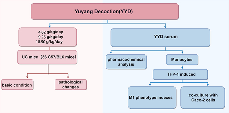 |
Figure 1 Diagram of model replication and drug administration. |
Disease Activity Index (DAI) Score
Throughout the entire experimental period, the activity levels and fecal characteristics of the mice were observed daily. Fecal occult blood tests were conducted using occult blood test strips, and the body weights of the mice were recorded. The DAI was calculated as follows: DAI = (score for weight loss + score for fecal characteristics + score for rectal bleeding) / 3. Twenty days after modeling, the mice were euthanized to observe the pathological changes in their colonic tissues. Intestinal samples were collected for subsequent research.
Hematoxylin and Eosin (H&E) Staining
The colonic tissues of the mice were fixed in 4% paraformaldehyde, embedded in paraffin, and sectioned at a thickness of 5 μm. These sections were then stained with hematoxylin and eosin (HE). Pathological changes were observed and scored under a 400x light microscope (Pannoramic 250, Danjier).
Enzyme-Linked Immunosorbent Assay
ELISA kits were utilized to detect the expression levels of inflammatory cytokines, including tumor necrosis factor-α (TNF-α), interleukin-1β (IL-1β), IL-6, and IL-10, in the mucosal lamina propria. Specifically, the target antibodies were coated onto 48-well microplates to serve as solid-phase carriers. Samples were then added to the wells, along with the respective antibodies TNF-α (ZC-39024), IL-1β (ZC-37974), IL-6 (ZC-37988), and IL-10 (ZC-37962), all purchased from Shanghai Zhuocai Biotechnology Co., Ltd. China. Subsequently, horseradish peroxidase-labeled antibodies were added, followed by the addition of TMB substrate for color development. The absorbance (OD value) was measured at a wavelength of 450 nm using a microplate reader, and the sample concentrations were calculated accordingly.
Western Blot (WB) Assay
WB was employed in animal experiments to detect the expression of tight junction proteins claudin-1, claudin-2, Zonula occludens-1 (ZO-1), and Occludin in intestinal epithelial cells, as well as the expression of pathway proteins IKK-β and Tollip. Total protein was extracted from colon tissue samples, and 20 μL of a 25 mg/mL protein standard solution was used as the loading volume. After electrophoresis, membrane transfer, and blocking, the membranes were incubated with primary antibodies against claudin-1 (1:5000, A21971), claudin-2 (1:500, A14085), Occludin (1:5000, A2601), IKK-β (1:5000, A2087), Tollip (1:5000, A23780), ZO-1 (1:2000, AF5145), and β-actin (1:50000, AC026), all purchased from Abclonal Biotechnology Co., Ltd. China. The membranes were then incubated with secondary antibody Goat Anti-Rabbit IgG (H+L) HRP (S0001, 1:5000, Affbiotech) at room temperature for 2 hours.
In cellular experiments, protein samples were extracted, and the membranes were incubated overnight at 4°C with primary antibodies against MyD88 (1:1000, GB111554, Servicebio), P65 (1:2000, bs-0465R, Bioss), p-P65 (1:500, AP0123, Abclonal), Tollip (1:1000, A23780, Abclonal), and β-actin (1:50000, AC026, Abclonal). Secondary antibodies were then added. After washing the membranes, color development was performed in the dark. The bands were exposed using Tanon fluorescence image analysis software V2.0, and the integrated optical density (IOD) of the target proteins was used for quantification.
Immunofluorescent Staining
Paraffin sections were dewaxed to water and then washed in distilled water with xylene and anhydrous ethanol gradient solution in turn. The sections were immersed in citrate buffer 6.0) for antigen repair. The slices were placed in 3% hydrogen peroxide and incubated at room temperature away from light for 25 min. The slides were placed in PBS (PH7.4) and washed three times on a decolorizing shaking table for 5min each time. Bovine serum albumin at room temperature for more than 30 minutes. Primary antibodies against IKK-β (1:100, BS-0465R, Beijing Biosynthesis Biotechnology Co., Ltd.) and Tollip (1:100, GB113109, Wuhan Servicebio Technology Co., Ltd.) were applied. Secondary antibodies included HRP-labeled goat anti-rabbit IgG (1:100, GB23301, Servicebio) and CY3-labeled goat anti-rabbit IgG (1:100, GB21303, Servicebio). DAPI was added for nuclear staining, and the sections were mounted using an anti-fade mounting medium. Fifteen coverslips with human Caco-2 cells were placed in staining jars and rinsed. The cells were treated with permeabilization solution (G1204, 0.5%, Servicebio) and incubated at room temperature for 10 minutes. After serum blocking, primary antibodies against claudin-1 (1:1000, 13,050-1-ap, Proteintech) and ZO-1 (1:1000, 21,773-1-ap, Proteintech) were applied, and the cells were incubated at 4°C overnight. Secondary antibody (FITC-labeled goat anti-rabbit IgG, GB22303, 1:100, Servicebio) was added, and the cells were incubated at 37°C for 30 minutes. The cell nuclei were counterstained with DAPI, and the coverslips were mounted. The mean fluorescence intensity of each image was measured using the Image-J image analysis system.
Preparation of YYD Extract Solution
25 mL of the original solution was treated with distilled water through reflux and filtration processes. A 300 μL aliquot of the combined filtrate was mixed with 900 μL of acetonitrile in a new 1.5 mL Eppendorf (EP) tube. The mixture was sonicated for 30 minutes and then centrifuged at 12,000 rpm for 10 minutes at 4°C. The supernatant was collected for subsequent UPLC-Q-TOF-MS/MS analysis.
Liquid Phase Mass Spectrometry
Chromatographic Conditions: Waters HSS T3 1.7μm 2.1*100mm was used with a column temperature set at 40°C. Mobile phase A consisted of 0.1% formic acid, while mobile phase B was 100% methanol. The flow rate was maintained at 0.3 mL/min, and the analysis time for each component was 16 min. Mass Spectrometry Conditions: MS1 parameters, Resolution: 70,000; AGCtarget: 1e6; MaximumIT: 50ms; Scanrange: 150to1500m/z. For MS2 parameters, Resolution: 17,500; AGCtarget: 1e5; MaximumIT: 50ms; TopN: 10; NCE/steppedNCE: 10, 30, 55.
Database Search
The mass spectrometry files were preprocessed using MS-DIAL 4.70 (MS-DIAL: data-independent MS/MS deconvolution for comprehensive metabolome analysis. Nature Methods, 12, 523–526, 2015, which included peak extraction, noise removal, deconvolution, and peak alignment. A three-dimensional data matrix (raw data matrix) in CSV format was exported. The extracted peak information was compared with databases, and a comprehensive search was conducted across three databases: MassBank, Respect, and GNPS (totaling 14,951 records). This three-dimensional matrix contained information on sample details, retention times, mass-to-charge ratios, and mass spectrometry response intensities (peak areas).
Preparation of Medicated Serum
After administering the YYD by gavage for 7 days, the mice were anesthetized 1 hour after the last administration. Blood was collected from the abdominal aorta and allowed to stand at room temperature for 1 hours. The blood samples were then centrifuged at 3000 rpm for 10 minutes to obtain the supernatants, which were mixed uniformly within the same group. Subsequently, the supernatants were inactivated by a 56°C water bath for 30 minutes and sterile-filtered through a 0.22μm microporous filter.
Cell Culture
Culture of Human Colonic Adenocarcinoma Cells (Caco-2, iCell-h032, iCell): The cells were cultured in D10 medium, which is Dulbecco’s Modified Eagle’s Medium (DMEM) supplemented with 10% v/v fetal bovine serum, 2 mM L-glutamine, 100 U/mL penicillin, and 100 μg/mL streptomycin.
Culture of Human Monocytic Leukemia Cells (THP-1, iCell-h123, iCell): The cells were maintained in RPMI 1640 medium containing 10% v/v fetal bovine serum and 1% v/v penicillin-streptomycin, with the use of THP-1 cell-specific medium (iCell-h123-001b, iCell).
Differentiation of THP-1 Monocytes into Macrophages (M0 Phenotype): When the cells were in good growth condition, log-phase cells were harvested and plated at a density of 1×10⁶ cells/mL. These cells were then stimulated with 100 mg/mL PMA for 24 hours to induce differentiation into macrophages. The differentiation process was carried out in a 37°C incubator with 5% CO2. M1 Phenotype Differentiation: Following PMA stimulation, the M0 cells were further induced to differentiate into M1 macrophages by stimulating them with 20 ng/mL IFN-γ and 100 ng/mL LPS for 24 hours. The IFN-gamma used was of human origin (HY-P7025, MCE, Cell).
siRNA Transfection
To prepare the riboFECT™CP transfection complex, 15 μL of siRNA was diluted in 360 μL of 1X riboFECT™CP Buffer. Subsequently, 36 μL of riboFECT™CP Reagent was added, and the mixture was incubated at room temperature for 15 minutes to achieve a final siRNA concentration of 50 nM. This transfection complex was then ready for use. Logarithmically growing THP-1 cells were selected, and the riboFECT™CP transfection complex (siRNA-NC or siRNA-Tollip, A10003, Genepharma) was added to an appropriate volume of antibiotic-free complete medium. The mixture was thoroughly mixed and added to the well plate for incubation at 37°C with 5% CO2. After transfection, cell precipitates were obtained according to the grouping for WB validation. Following transfection with siRNA-NC or siRNA-Tollip, THP-1 cells were stimulated with 100 mg/mL PMA for 24 hours to induce differentiation into M0 cells. Subsequently, 20 ng/mL IFN-γ and 100 ng/mL LPS were added for an additional 24 hours of stimulation to induce differentiation and M1 polarization. During the M1 polarization induction, fetal bovine serum was replaced with 10% serum-free medium or medium containing drug-containing serum. Simultaneously, siRNA-NC THP-1 and siRNA-Tollip THP-1 cells were also prepared for comparison. The siRNA-Tollip was purchased from GenePharma.
Co-Culture System
Caco-2 cells were seeded in D10 medium for 21 days to achieve complete differentiation into intestinal epithelial cells. On the last day prior to the experiment, D10 medium was removed from the Caco-2 cells and replaced with RPMI-1640 (PM150110, Procell). TPH-1-derived M1 macrophages were then introduced into the outer compartment of the tissue culture insert at a density of 1×10⁶ cells/mL in R10 medium. Caco-2 cells and M1 macrophages were cocultured, and corresponding blank serum and drug-containing serum were added to the groups as per the experimental design. Following a 48-hour incubation period, detection was performed.
Counting Kit-8 (CCK-8)
Cell viability assessment using the CCK-8 across various groups. Caco-2 cells, previously cultured in R10 medium, were co-seeded in the upper chambers of Transwell inserts with TPH-1-induced M1 macrophages. Corresponding blank serum and drug-containing serum were added according to the experimental protocol, with four replicates per group. Following a 48-hour co-incubation period, cell viability was assessed. CCK-8 reagent (BS350B, Biosharp) was diluted 1:10 in serum-free medium and 200 μL of the diluted CCK-8 working solution was added to each well and the insert chamber. The culture plate was gently agitated several times and then further incubated at 37 °C with 5% CO2 for an additional 2 hours. Absorbance values of each well were measured using a microplate reader at a wavelength of 450 nm.
Flow Cytometry (FCM)
THP-1 cells were seeded into 6-well plates and stimulated with 100 mg/mL PMA for 24 hours to induce their differentiation into M0 cells. Subsequently, the cells were further stimulated with 20 ng/mL IFN-γ and 100 ng/mL LPS for an additional 24 hours to induce their polarization into M1 macrophages. During the M1 polarization induction, the fetal bovine serum was replaced with 10% blank serum or drug-containing serum according to the experimental groups. Cells were harvested by centrifuging at 250 g for 5 minutes. After resuspension in 100 μL of PBS, the cells were stained with CD11b (FITC anti-human CD11b, 301330, Biolegend) and CD86 (PE anti-human CD86, 305405, Biolegend) antibodies for 30 minutes at 4 °C in the dark. The cells were then washed once with PBS and resuspended in 500 μL of PBS for flow cytometry analysis. The stained cells were analyzed using a flow cytometer to assess the expression levels of CD11b and CD86 on the surface of the M1 macrophages.
Real-Time Quantitative PCR (RT-qPCR)
Total RNA was extracted using the Molpure® Cell/Tissue Total RNA Kit (19221ES50, YEASEN). The cDNA template was synthesized by reverse transcription using a PCR amplification system and the PrimeScript RT reagent Kit (RR047A, Takara Bio Inc). RT-PCR was performed using a QuantStudio™ 3 Real-Time PCR System (ThermoFisher, USA). The reaction protocol was as follows: initial denaturation at 95°C for 30 seconds, followed by 45 cycles of denaturation at 95°C for 10 seconds, annealing at 55°C for 30 seconds, and extension at 72°C with fluorescence collection for 30 seconds. GAPDH was used as the internal control gene, and the experiment was repeated three times. The relative mRNA expression levels of the target genes were analyzed using the 2−ΔΔCt method. The primer sequences were synthesized by Sangon Biotech (China) and are listed in Table 2.
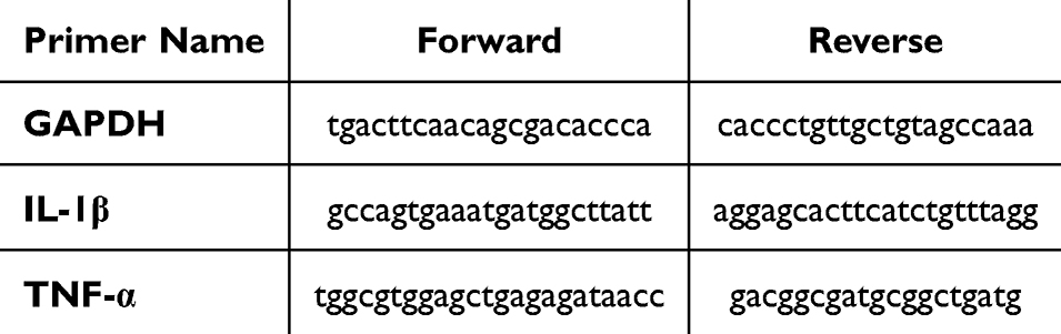 |
Table 2 Primer Sequences and Base Pairs |
Statistical Methods
The experimental data were analyzed by SPSS 26.0 software. Pairwise statistical comparisons were performed using one way ANOVA. GraphPad Prism 8.3 software was used for measurement data analysis. Data conforming to the normal distribution are expressed as mean ± SD.
Result
YYD Serum Pharmacochemical Analysis
The chemical components in YYD are complex and diverse. In order to identify the components as much as possible, the chemical components of YYD were fully described by positive and negative ESI ionization modes in this study. The specific base peak ion flow chromatography is shown in Figure 2. Through database comparison, 840 chemical components of YYD were obtained, and the top 20 were selected for display, as shown in Table 3.
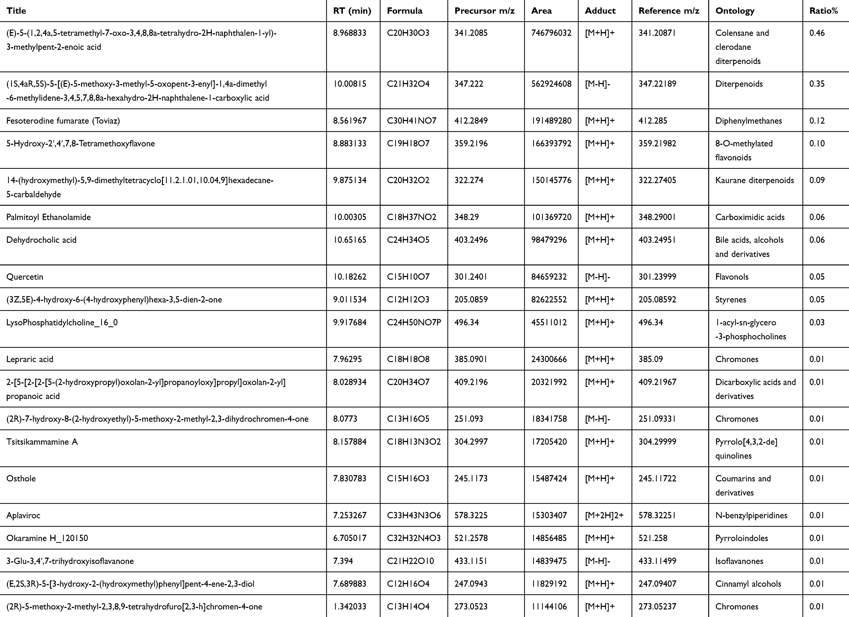 |
Table 3 YYD Chemical Composition Analysis and Identification Results |
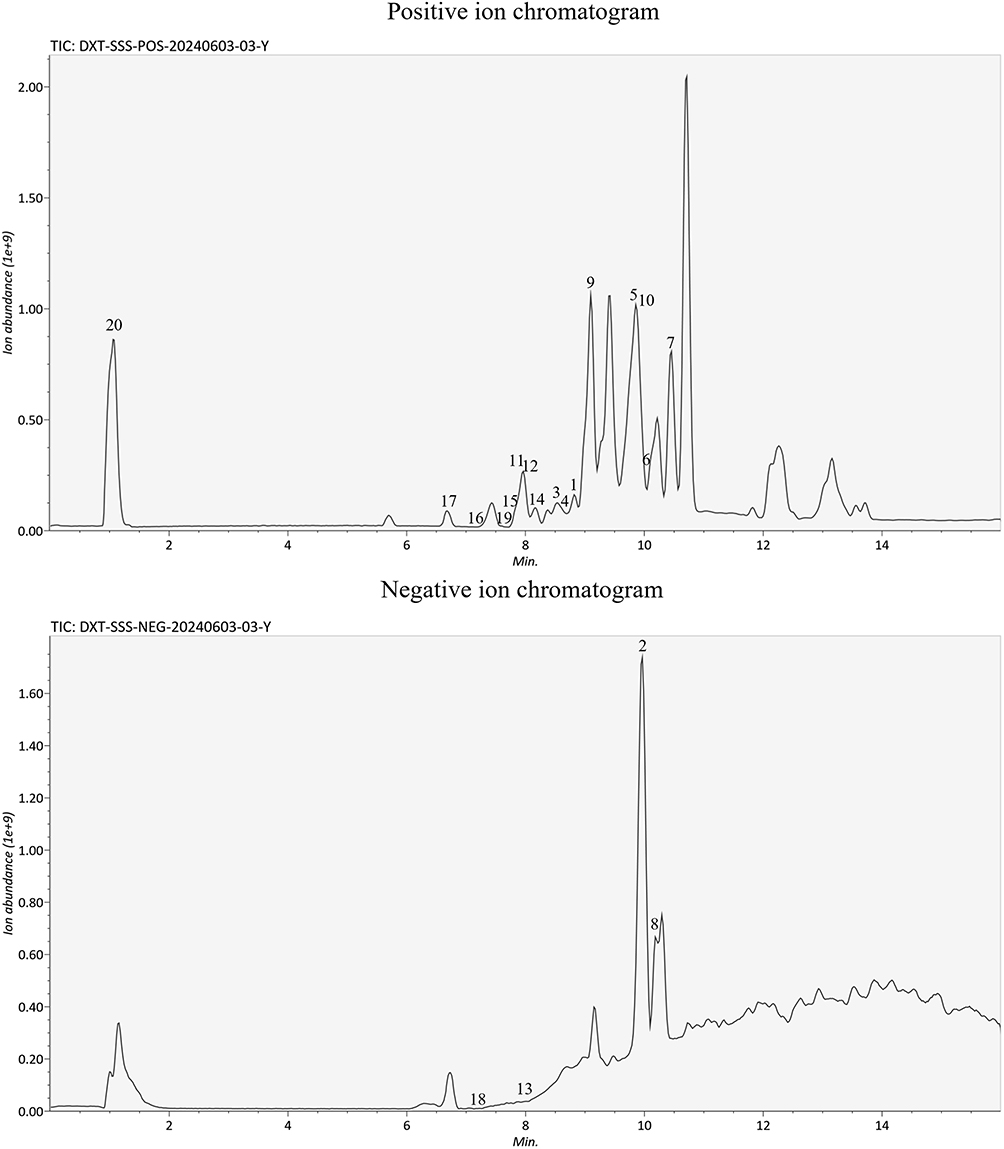 |
Figure 2 UPLC-Q-TOF-MS/ MS-based peak ion flow chromatography of YYD extract. |
Basic Condition Assessment of Mice with Colonic Mucosal Barrier Injury Treated with YYD
The anti-colitis properties of YYD were evaluated using a 3% DSS-induced colitis model for 21 days (Figure 3a). Changes in the body weight of mice were monitored during the experiment, and on day 21, model group showed a reduction in body weight compared to the control group. The DAI (Disease Activity Index) scores were assessed based on observations of body weight, fecal consistency, and the degree of fecal occult blood. Compared to the control group, the model group mice exhibited gross hematochezia. Following YYD and positive drug interventions, the pathological symptoms of fecal consistency, diarrhea, and fecal occult blood were alleviated in mice from each group, resulting in a significant reduction in DAI scores (Figure 3b). Figure 3c show that the colon length was shortened and edema occurred due to DSS. Moreover, colon length shortening and edema were improved in all YYD and positive drug groups.
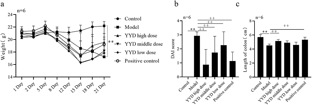 |
Figure 3 Basic condition assessment of mice with colonic mucosal barrier injury treated with YYD. (a and b) The body weight and disease activity index (DAl) of these mice during the experimental period were depicted. (c) Representative images of colon lengths from a macroscopic perspective and quantitative measurement of colon length. *P <0.05, **P <0.01 vs Control; +P <0.05, ++P <0.01 vs Model, n=6. The same below. |
YYD Improves Intestinal Mucosal Barrier Damage in UC Mice
As shown in Figure 4a, the HE pathological changes of colonic tissue injury in model group mice are mainly manifested as destruction of the inherent structure at the colonic ulcer site, accompanied by crypt expansion and destruction. Within the ulcerated lesions, there is visible infiltration of inflammatory cells, including neutrophils and lymphocytes, leading to severe damage to the tissue architecture. YYD and positive drug treatment were able to reduce these pathological signs compared to model mice.
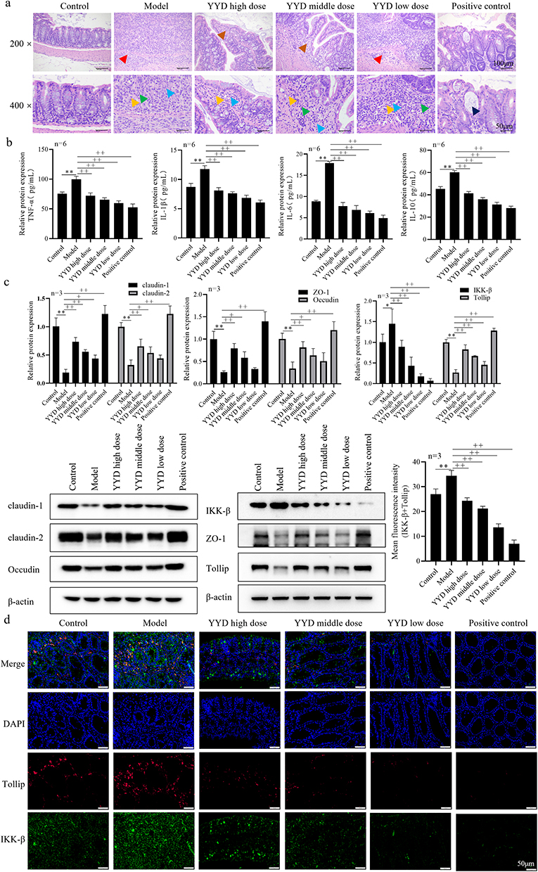 |
Figure 4 Pathological Changes in Mice with Colonic Mucosal Barrier Injury Treated with YYD. (a) HE staining was used to describe the pathological features of colitis (Amplification: 200×, 400×, Scale: 50μm, 100 μm). Full-thickness mucosal necrosis with submucosa involvement (Red arrow); Neutrophils (Green arrow); Fibroblasts (Yellow arrow); Necrotic cell debris (Blue arrow); Dilatation of intestinal glands (Navy blue arrow); Necrosis of Lamina propria (Orange arrow). (b) ELISA was used to detect the expression of TNF-α, IL-1β, IL-6 and IL-10 in lamina propria mucosa. (c) WB was used to detect related proteins (claudin-1, claudin-2, ZO-1, Occudin, IKK-β, Tollip). (d) The expression levels of IKK-β and Tollip in intestinal wall were detected by immunofluorescent antibody double staining. |
From Figure 4b, compared with the control group, the expression of TNF-α, IL-1β, IL-6 and IL-10 in the colon tissue of model mice increased. Compared with the model group, YYD can effectively inhibit the expression of these four inflammatory factors. Figure 4c show that compared with the control group, the expression of claudin-1, claudin-2, ZO-1, Occludin, and Tollip proteins in the colonic tissues were decreased in model group. Meanwhile, the expression of the pathway protein IKK-β was increased. In contrast, the expression of those proteins in YYD and the positive groups were increased compared to the model group, while the expression of IKK-β protein was decreased. When compared with the positive group, the expression of claudin-1, claudin-2, ZO-1, Occludin, and Tollip proteins in the YYD high, medium, and low dose groups was significantly reduced, while the expression of IKK-β protein was increased. Next, immunofluorescence was used to detect intestinal barrier marker proteins IKK-β and Tollip (Figure 4d). Compared with the control group, the fluorescence intensity of Tollip and IKK-β in the colon tissue of the model group mice decreased. Compared with the model group, the fluorescence intensity of Tollip in positive control group, as well as the high, medium, and low-dose groups, increased.
Effect of M1 Macrophage Proliferation Activity in LPS-Stimulated Macrophages by YYD
M1+ blank serum 0% decreased compared with M0+ blank serum 0%. Compared with M1+ blank serum 0%, M1 drug serum 2.5% decreased. However, at a high concentration of M1+ drug serum (10%), there was a decrease in proliferative activity (Figure 5), indicating a possible cytotoxic or saturation effect of the drug at high concentrations.
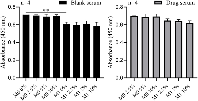 |
Figure 5 The proliferative activity was detected by CCK8. **P <0.01 vs M0 Black serum 0%. |
Expression of M1 Activating Factor in LPS-Stimulated Macrophages by YYD
Compared with M0 blank serum 10% group, the expression of TNF-α and IL-1β mRNA in human cells of M1+ blank serum 10% group was increased. Compared with M1 drug serum 10% group, the expression of TNF-α and IL-1β mRNA in M1 blank serum 10% group, and the M1 drug serum 2.5% group and M1 drug serum 5% group were increased. The expression levels were lowest in the M0 + 10% blank serum group, as the cells were not induced to differentiate into M1 macrophages. In the M1 + 10% blank serum group, the expression levels of TNF-α and IL-1β were the highest, indicating that the cells had been induced to differentiate into M1 macrophages and were releasing typical inflammatory cytokines (Figure 6a). The expression levels of TNF-α and IL-1β varied with different drug concentrations, suggesting that YYD affects the expression of TNF-α and IL-1β by inhibiting the activation of M1 macrophages.
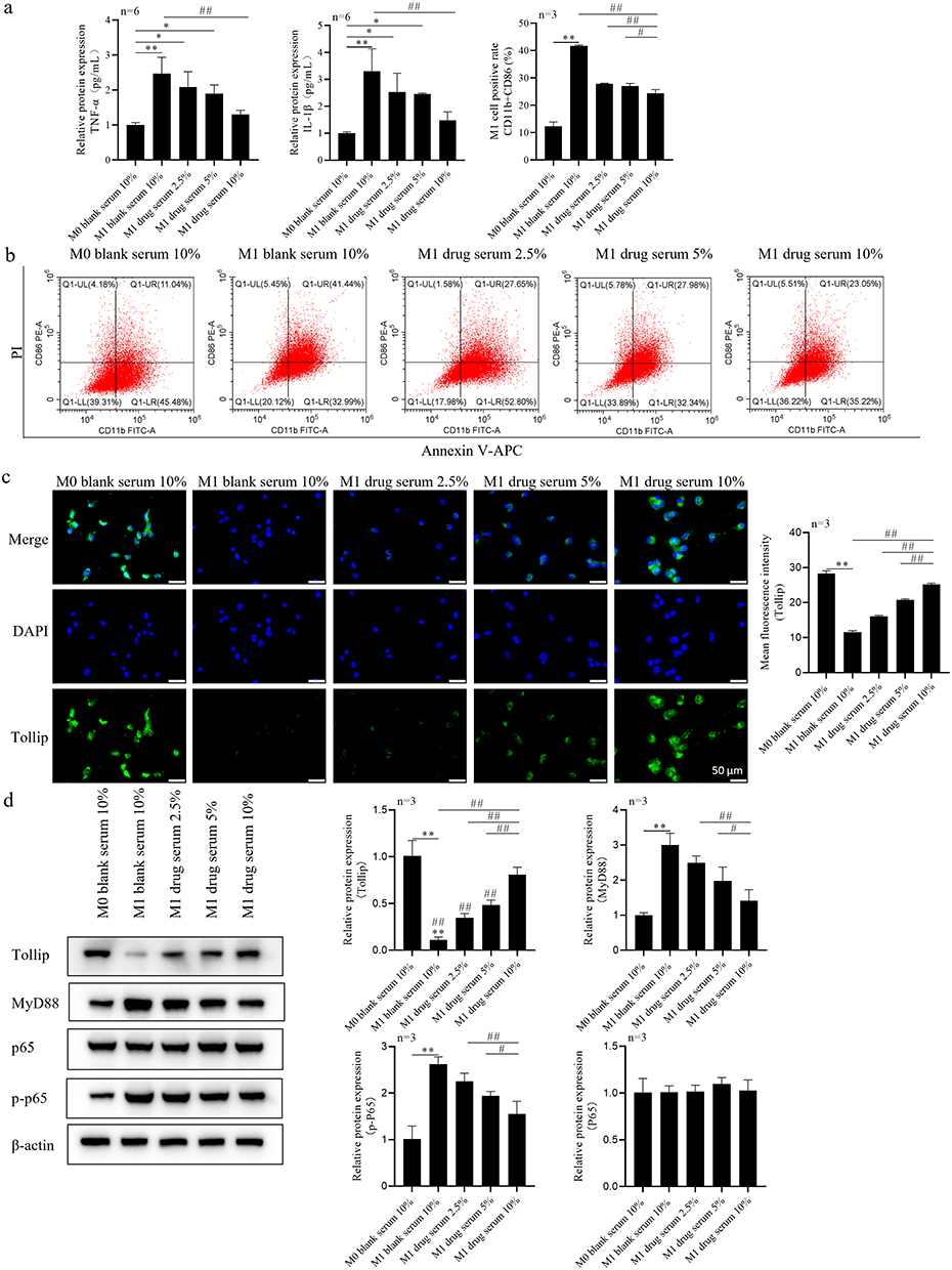 |
Figure 6 Regulation of M1 polarization of LPS-stimulated macrophages by YYD and regulation of TLRs/Tollip signaling expression. (a) Serum levels of inflammatory factors (TNF-α, IL-1β) were detected by RT-PCR. (b) The proportion of M1 macrophages CD11b+CD86 was determined by flow cytometry. (c) Tollip expression was observed by immunofluorescence staining. (d) TLRs/Tollip signaling pathway proteins in macrophages were detected by WB. *P<0.05, **P <0.01 vs M0 blank serum 10%; #P<0.05, ##P <0.01 vs M1 drug serum 10%. |
YYD Regulates the Proportion of Macrophages Stimulated by LPS
The proportion of M1 macrophages (CD11b+CD86) in each group was detected by flow cytometry. Compared with the M0 blank serum 10% group, the proportion of CD11b+CD86 double positive cells in the M1+ blank serum 10% group increased, indicating that the cells were successfully differentiated into M1 macrophages under the condition of M1 polarization, and the proportion remained at a certain level without drug intervention. Compared with the M1 drug serum 10% group, the proportion of M1 2.5 and 5 drug serum CD11b+CD86 double positive cells increased (Figure 6b), indicating that YYD effectively inhibited the differentiation of M1 macrophages.
YYD Regulates LPS-Stimulated Tollip Expression in Macrophages
Compared to the M0+10% blank serum group, the human macrophage Tollip fluorescence intensity in the M1+10% blank serum group decreased, indicating that M1 polarization conditions may inhibit Tollip expression. When compared to the M1+10% drug serum group, as the drug concentration increased, the Tollip expression level gradually rose in a concentration-dependent manner. At a 10% drug concentration, Tollip expression reached its peak, primarily distributed in the cytoplasm (Figure 6c), suggesting that the drug can effectively promote Tollip expression.
YYD Regulates Proteins of TLRs/Tollip Signaling Pathway in LPS-Stimulated Macrophages
Consistent with the immunofluorescence staining results, Tollip expression decreased after M1 polarization compared to the M0+10% blank serum group. After YYD intervention, Tollip expression levels gradually increased with increasing concentrations. The protein expression of MyD88, p65 and p-p65 in each group was further detected. The expression levels of p65 and p-p65 reflect the activation status of NF-κB, which plays a key role in M1 polarization. Following M1 polarization, MyD88 and p-p65 expression increased. After drug intervention, as the concentration increased, MyD88 and p-p65 expression decreased (Figure 6d), suggesting that YYD may exert its effects by inhibiting the TLRs/Tollip signaling pathway.
YYD Activated Tollip to Inhibit the Expression of M1 Macrophage Activating Factor
To further investigate whether YYD mediates the Tollip pathway to regulate the polarization of M1 macrophages, siRNA technology was used to silence Tollip in cells. Compared to the siRNA-NC M1 10% blank serum group, the expression of IL-1β and TNF-α mRNA in human cells of the siRNA-NC M1 10% drug serum group were decreased, indicating that the drug serum had a notable inhibitory effect on the expression of IL-1β and TNF-α. In the siRNA-Tollip M1 blank serum 10% group, the expression of IL-1β and TNF-α increased, suggesting that downregulation of the Tollip gene may lead to enhanced IL-1β and TNF-α expression. Compared to the siRNA-Tollip M1 drug serum 10% group, the expression of IL-1β and TNF-α mRNA of the siRNA-NC M1 drug serum 10% group were decreased, indicating that downregulation of the Tollip may result in increased expression of IL-1β and TNF-α (Figure 7a). The elevation of IL-1β and TNF-α mRNA expression of the siRNA-Tollip M1 10% blank serum group further supports the inhibitory effect of drug serum on IL-1β and TNF-α expression and emphasizes the importance of Tollip in regulating IL-1β and TNF-α expression.
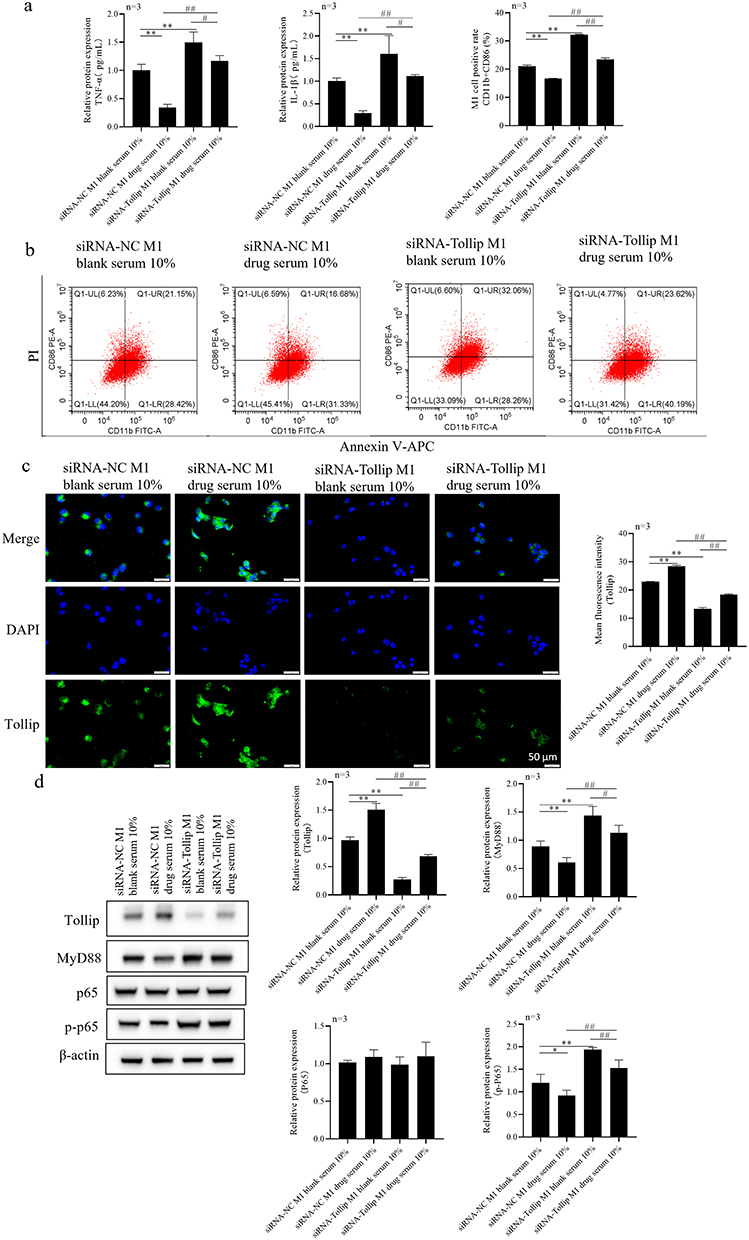 |
Figure 7 YYD inhibits macrophage M1 polarization by activating Tollip. (a) RT-PCR was used to detect the expression of TNF-α and IL-1β in the cells. (b) The proportion of M1 macrophages CD11b+CD86 was determined by flow cytometry. (c) Tollip expression was observed by immunofluorescence staining. (d) TLRs/Tollip signaling pathway proteins in macrophages were detected by WB. *P<0.05, **P <0.01 vs siRNA-NC M1 10% blank serum; #P<0.05, ##P <0.01 vs siRNA-NC M1 10% drug serum. |
YYD Inhibits M1 Macrophage Polarization by Activating Tollip
Compared to the siRNA-NC M1 blank serum 10%, the expression of CD11b+CD86 in the siRNA-NC M1 drug serum 10% was reduced, while the expression of CD11b+CD86 in the siRNA-Tollip M1 group blank serum 10% was increased, indicating that the drug serum inhibited the M1 polarization process. When compared to the siRNA-Tollip M1 drug serum 10%, the expression of CD11b+CD86 was decreased in the siRNA-NC M1 group drug serum 10%, and increased in the siRNA-Tollip M1 blank serum 10% (Figure 7b). The active ingredients in the drug serum may influence the M1 polarization process by modulating intracellular signaling pathways or gene expression. However, under the influence of siRNA-Tollip, the effects of these active ingredients are inhibited.
YYD Inhibits M1 Macrophage Polarization by Activating Tollip Expression
Compared to the siRNA-NC M1 group with 10% blank serum, the cells in the siRNA-NC M1 group with 10% drug serum exhibited increased Tollip expression, while the human cells in the siRNA-Tollip M1 group with 10% blank serum showed decreased Tollip expression. When compared to the siRNA-NC M1 group with 10% drug serum, the siRNA-Tollip M1 group with 10% drug serum demonstrated elevated Tollip expression, whereas the siRNA-Tollip M1 group with 10% blank serum exhibited decreased expression. These findings indicate that the downregulation effect of siRNA-Tollip on Tollip remains effective under conditions with drug serum. Compared with siRNA-NC M1 blank serum 10% group, the expression of Tollip in siRNA-NC M1 drug serum 10% group was increased, and the expression of Tollip in siRNA-Tollip M1 blank serum 10% group was decreased. Compared with siRNA-Tollip M1+ 10% drug serum group, the expression of Tollip was increased in siRNA-NC M1+ 10% drug serum group, and decreased in siRNA-Tollip M1+ 10% blank serum group (Figure 7c). These results indicated that the downregulation of Tollip by siRNA-Tollip was still effective under the condition of drug serum.
YYD Inhibits M1 Macrophage Polarization by Activating TLRs/Tollip Signaling Pathway Proteins
WB verified the protein expression level of Tollip in each group, which was consistent with the results of immunofluorescence staining. The protein expression of Tollip was down-regulated in the siRNA-Tollip transfected group. Compared with siRNA-NC M1 blank serum 10% group, the expression of MyD88 and p-p65 in siRNA-NC M1 drug serum 10% group was decreased, and the expression of MyD88 and p-p65 in siRNA-Tollip M1 blank serum 10% group was increased. Compared with siRNA-Tollip M1 drug serum group 10%, the expression of MyD88 and p-p65 in siRNA-NC M1 drug serum group 10% was decreased, and the expression in siRNA-Tollip M1 blank serum 10% group was increased. Knockdown of Tollip may lead to enhanced activation of TLRs/Tollip, thereby inhibiting the M1 polarization process (Figure 7d).
YYD Inhibits Caco-2 Cell Viability by Activating Tollip Signaling Pathway
To further investigate the mechanism by which YYD improves inflammation in Caco-2 cells by mediating the Tollip pathway to regulate M1 macrophage polarization, co-culture experiments were conducted with macrophages and Caco-2 cells. Compared to the Caco-2 group, the cell viability of the Caco-2 + M1 group was decreased. Compared to the Caco-2 + M1 group, the cell viability of both the Caco-2 + M1 + drug serum 10% group and the Caco-2 + siRNA NC M1 drug serum 10% group were increased. Compared to the Caco-2 + siRNA NC M1 drug serum 10% group, the cell viability of the Caco-2 + siRNA Tollip M1 drug serum 10% group decreased (Figure 8a). This suggests that the knockdown of the Tollip gene may have affected the function of M1 cells, which in turn influenced the viability of Caco-2 cells.
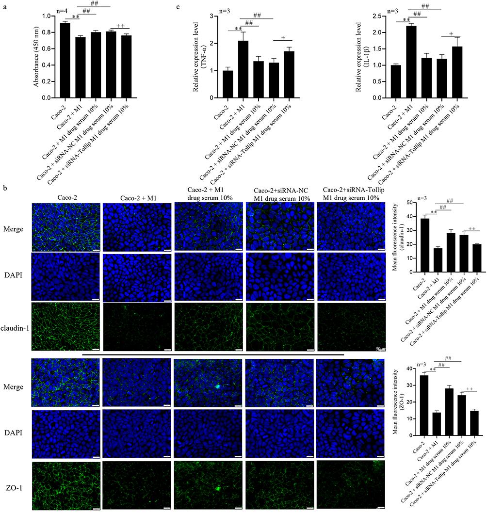 |
Figure 8 YYD activates the tollip signaling pathway to inhibit lps-induced M1-like differentiation of macrophages. (a) CCK8 Assay to Detect Caco-2 Cell Viability. (b) Immunofluorescence Staining to Observe Tight Junction Proteins (Claudin-1, ZO-1) in Caco-2 Cells. (c) PCR to Detect Expression of Inflammatory Cytokines (TNF-α, IL-1β) in Caco-2 Cells. *P<0.05, **P <0.01 vs Caco-2; #P<0.05, ##P <0.01 vs Caco-2 + M1; +P<0.05, ++P <0.01 vs Caco-2 + siRNA-NC M1 drug serum 10%. |
YYD Activates Tollip Signaling Pathway To Inhibit M1 Polarization and Reduce Inflammation of Intestinal Epithelial Cells
Compared to the Caco-2 group, the fluorescence intensity of the Caco-2 + M1 group was decreased. Compared to the Caco-2 + M1 group, the fluorescence intensity of both the Caco-2 + M1 + drug serum 10% group and the Caco-2 + siRNA NC M1 drug serum 10% group were increased. Compared to the Caco-2 + siRNA NC M1 drug serum 10% group, the fluorescence intensity of the Caco-2 + siRNA Tollip M1 drug serum 10% group increased. Compared to the siRNA NC treatment group, the siRNA Tollip treatment group exhibited a lower degree of recovery in the expression of tight junction proteins, suggesting that the knockdown of the Tollip may further impair the barrier function of Caco-2 cells (Figure 8b).
YYD Activates Tollip Signaling Pathway To Inhibit M1 Polarization and Alleviate Intestinal Epithelial Cell Barrier Damage
Compared to the Caco-2 group, the expression of TNF-α, IL-1β in the Caco-2 + M1 group were increased. Compared to the Caco-2 + M1 group, Caco-2 + M1 + drug serum 10% group and the Caco-2 + siRNA NC M1 drug serum 10% group were decreased. Compared to the Caco-2 + siRNA NC M1 drug serum 10% group, Caco-2 + siRNA Tollip M1 drug serum 10% group decreased. With the addition of drug-containing serum and the treatment with siRNA, the expression levels of inflammatory cytokines decreased (Figure 8c). This indicates that the knockdown of the Tollip gene may contribute to alleviating the inflammatory response induced by M1 cells.
Discussion
The pathogenesis of UC remains unclear to date.1 Currently, it is believed that an important factor in the etiology and pathogenesis of UC is the abnormal immune response of the intestinal mucosal innate and adaptive immune response systems to intestinal microbial antigens, leading to the initiation and progression of intestinal mucosal inflammation.15 Recently, the exploration of traditional Chinese medicine (TCM) for the treatment of UC has become a research hotspot in the field of digestive diseases.16 The enema method of TCM administration for UC has a long history. When administered intestinally, the medication is dissolved in intestinal secretions, selectively absorbed by the large intestinal wall, and directly enters the systemic circulation through a semi-permeable membrane to produce its effects.16,17 The intestinal barrier plays a crucial role in maintaining the balance of the intestinal microbiota. In this study, a mouse model of UC was established, and a unique two-step enema method combining YYD with normal saline enema was used to treat UC. After drug treatment, the body weight of the mice increased, and pathological manifestations such as hematochezia, diarrhea, colonic mucosal inflammation, and ulcers were alleviated.
Toll-interacting protein (Tollip), serves as a crucial negative regulator in the series of signaling pathways that initiate immune responses against microbial antigens via TLRs (Toll-like receptors) in the innate immune system.18 It functions to inhibit the production of excessive inflammatory mediators by suppressing TLR-mediated inflammatory signals.19 Immune cells and epithelial cells are intricately involved in the complex process of intestinal mucosal repair.20 Studies have shown that the expression of Tollip in the intestinal mucosa of patients with inflammatory bowel disease (IBD) is significantly reduced. By upregulating Tollip expression across cellular barriers, it is possible to target intestinal macrophages to suppress inflammatory responses and alleviate IBD symptoms.21 Notably, Tollip-knockout mice exhibit aggravated inflammatory damage in the intestinal mucosa and delayed repair, suggesting that inactivation of the Tollip signaling pathway is a significant contributor to the development and progression of IBD.5 Furthermore, Tollip can bind to the TLR4/MyD88/IL-1 receptor-associated kinase (IRAK) complex, thereby inhibiting the phosphorylation and activation of IRAK in response to LPS stimulation. This interaction subsequently reduces the transcription of NF-κB and suppresses inflammatory responses.18
Under normal physiological conditions, the intestinal mucosal innate immune system maintains a state of tolerance to continuous LPS stimulation through the sustained low expression of TLRs (Toll-like receptors) and high expression of Tollip.22 However, persistent stimulation by certain pathogenic factors, such as highly pathogenic intestinal microorganisms, can alter the immune function of the intestinal mucosa, leading to decreased Tollip expression and/or overexpression and activation of TLRs. This disruption of the intestinal mucosal immune balance contributes to the occurrence and progression of intestinal immune-related diseases, including UC.23 Tight junction proteins are crucial structures for maintaining the barrier function of intestinal epithelial cells.24 Among them, Claudin, ZO-1, and Occludin play pivotal roles in preserving intestinal barrier function, preventing the entry of harmful substances into the systemic circulation, and facilitating intestinal mucosal repair.25 The findings of this study demonstrate that YYD inhibits DSS-induced colitis inflammation in vivo by upregulating the expression of junctional proteins (Claudin, ZO-1, and Occludin), thereby repairing the epithelial barrier in mice. Similarly, YYD has been shown to upregulate the expression of junctional and tight junction proteins in the colonic epithelial barrier, reducing intestinal mucosal permeability and restoring barrier integrity.
Previous studies have shown that the expression of Tollip is suppressed in intestinal tissues of patients with inflammatory bowel disease (IBD), suggesting an association between Tollip and macrophage activation.26 Macrophages play a crucial role in intestinal immunity, and their polarization state directly influences intestinal inflammation and repair processes.27,28 Lipopolysaccharide (LPS), the major pathogenic component of Gram-negative bacteria, can stimulate the differentiation of macrophages towards the M1 phenotype, triggering a robust inflammatory response.29 The present study found that YYD can activate the Tollip signaling pathway and inhibit the differentiation of macrophages towards the M1 phenotype, thereby alleviating epithelial cell inflammation and barrier damage. During the identification of the active components in the serum containing YYD, we employed a series of advanced separation and identification techniques to successfully identify multiple active ingredients with anti-inflammatory, antioxidant, and immunomodulatory effects. These components may act synergistically on the intestinal immune system, exerting a cooperative therapeutic effect.
This study employs traditional Chinese medicine methodologies to delve into the specific mechanisms underlying the occurrence and progression of ulcerative colitis (UC), revealing the complex interplay between TLRs/Tollip signaling and macrophage polarization in the context of intestinal inflammation.
Conclusion
YYD reverses the shortening of colon length, mitigates histological damage, and improves intestinal epithelial barrier dysfunction. By inhibiting LPS-induced M1-like differentiation of macrophages through the TLRs/Tollip signaling pathway, YYD alleviates epithelial cell inflammation and barrier damage.
Data Sharing Statement
The datasets used and/or analysed during the current study are available from the corresponding author on reasonable request. The date is not publicly available due to privacy or ethical restrictions.
Ethics Approval
The experimental research and clinical sampling were examined by the Medical Ethics Committee of Shanxi Institute of Traditional Chinese Medicine, which met the requirements of medical ethics. (SZYLY2023KY-0406.)
Consent to Publish
Informed consent was obtained from all individual participants included in the study.
Author Contributions
All authors made a significant contribution to the work reported, whether that is in the conception, study design, execution, acquisition of data, analysis and interpretation, or in all these areas; took part in drafting, revising or critically reviewing the article; gave final approval of the version to be published; have agreed on the journal to which the article has been submitted; and agree to be accountable for all aspects of the work.
Funding
The study was supported by Shanxi Provincial Administration of Traditional Chinese Medicine (20232YYC016).
Disclosure
The authors report no conflicts of interest in this work.
References
1. Huang X, Li L, Zheng C, Li J, Chen G, Chen Y. Xuanbi Yuyang decoction ameliorates DSS-induced colitis by inhibiting pyroptosis via blocking of IL-17 pathway activation. J Inflamm Res. 2024;17:5235–5249. doi:10.2147/JIR.S472812
2. Buie MJ, Quan J, Windsor JW, et al. Global hospitalization trends for Crohn’s disease and ulcerative colitis in the 21st century: a systematic review with temporal analyses. Clin Gastroenterol Hepatol. 2023;21(9):2211–2221. doi:10.1016/j.cgh.2022.06.030
3. Gros B, Kaplan GG. Ulcerative colitis in adults: a review. JAMA. 2023;330(10):951–965. doi:10.1001/jama.2023.15389
4. Liu Y, Li BG, Su YH, et al. Potential activity of traditional Chinese medicine against ulcerative colitis: a review. J Ethnopharmacol. 2022;289:115084. doi:10.1016/j.jep.2022.115084
5. Begka C, Pattaroni C, Mooser C, et al. Toll-interacting protein regulates immune cell infiltration and promotes colitis-associated cancer. iScience. 2020;23(3):100891. doi:10.1016/j.isci.2020.100891
6. Le Berre C, Honap S, Peyrin-Biroulet L. Ulcerative colitis. Lancet. 2023;402(10401):571–584. doi:10.1016/S0140-6736(23)00966-2
7. Fernandes P, MacSharry J, Darby T, et al. Differential expression of key regulators of Toll-like receptors in ulcerative colitis and Crohn’s disease: a role for Tollip and peroxisome proliferator-activated receptor gamma? Clin Exp Immunol. 2016;183(3):358–368. doi:10.1111/cei.12732
8. Zhang M, Li X, Zhang Q, Yang J, Liu G. Roles of macrophages on ulcerative colitis and colitis-associated colorectal cancer. Front Immunol. 2023;14:1103617. doi:10.3389/fimmu.2023.1103617
9. Ishida K, Nagatake T, Saika A, et al. Induction of unique macrophage subset by simultaneous stimulation with LPS and IL-4. Front Immunol. 2023;14:1111729. doi:10.3389/fimmu.2023.1111729
10. Harris G, KuoLee R, Chen W. Role of toll-like receptors in health and diseases of gastrointestinal tract. World J Gastroenterol. 2006;12(14):2149–2160. doi:10.3748/wjg.v12.i14.2149
11. Li WW, Fan XX, Xu ZS, et al. BLK positively regulates TLR/IL-1R signaling by catalyzing TOLLIP phosphorylation. J Cell Biol. 2024;223(2). doi:10.1083/jcb.202302081
12. Shen X, Gu M, Zhan F, et al. Porcine beta defensin 2 attenuates inflammatory responses in IPEC-J2 cells against Escherichia coli via TLRs-NF-κB/MAPK signaling pathway. BMC Vet Res. 2024;20(1):357. doi:10.1186/s12917-024-04220-7
13. Wang J, Li X, Bello BK, et al. Activation of TLR2 heterodimers-mediated NF-κB, MAPK, AKT signaling pathways is responsible for vibrio alginolyticus triggered inflammatory response in vitro. Microb Pathogenesis. 2022;162:105219. doi:10.1016/j.micpath.2021.105219
14. Lavelle EC, Murphy C, O’Neill LA, Creagh EM. The role of TLRs, NLRs, and RLRs in mucosal innate immunity and homeostasis. Mucosal Immunol. 2010;3(1):17–28. doi:10.1038/mi.2009.124
15. Du L, Ha C. Epidemiology and Pathogenesis of Ulcerative Colitis. Gastroenterol Clin North Am. 2020;49(4):643–654. doi:10.1016/j.gtc.2020.07.005
16. Yan ZX, Liu YM, Ma T, et al. Efficacy and safety of retention enema with traditional Chinese medicine for ulcerative colitis: a meta-analysis of randomized controlled trials. Complement Ther Clin Pract. 2021;42:101278. doi:10.1016/j.ctcp.2020.101278
17. Sun YX, Wang X, Liao X, et al. An evidence mapping of systematic reviews and meta-analysis on traditional Chinese medicine for ulcerative colitis. BMC Complement Med Therap. 2021;21(1):228. doi:10.1186/s12906-021-03387-y
18. Kaplan GG. The global burden of IBD: from 2015 to 2025. Nat Rev Gastroenterol Hepatol. 2015;12(12):720–727. doi:10.1038/nrgastro.2015.150
19. Kawai T, Ikegawa M, Ori D, Akira S. Decoding Toll-like receptors: recent insights and perspectives in innate immunity. Immunity. 2024;57(4):649–673. doi:10.1016/j.immuni.2024.03.004
20. Li MX, Li MY, Lei JX, et al. Huangqin decoction ameliorates DSS-induced ulcerative colitis: role of gut microbiota and amino acid metabolism, mTOR pathway and intestinal epithelial barrier. Phytomedicine. 2022;100:154052. doi:10.1016/j.phymed.2022.154052
21. Liu X, Ren X, Zhou L, et al. Tollip orchestrates macrophage polarization to alleviate intestinal mucosal inflammation. J Crohn’s Colitis. 2022;16(7):1151–1167. doi:10.1093/ecco-jcc/jjac019
22. Maillard MH, Bega H, Uhlig HH, et al. Toll-interacting protein modulates colitis susceptibility in mice. Inflamm Bowel Dis. 2014;20(4):660–670. doi:10.1097/MIB.0000000000000006
23. Diao N, Zhang Y, Chen K, et al. Deficiency in toll-interacting protein (Tollip) skews inflamed yet incompetent innate leukocytes in vivo during DSS-induced septic colitis. Sci Rep. 2016;6:34672. doi:10.1038/srep34672
24. Kuo WT, Odenwald MA, Turner JR, Zuo L. Tight junction proteins occludin and ZO-1 as regulators of epithelial proliferation and survival. Ann N Y Acad Sci. 2022;1514(1):21–33. doi:10.1111/nyas.14798
25. Kuo WT, Zuo L, Odenwald MA, et al. The tight junction protein ZO-1 is dispensable for barrier function but critical for effective mucosal repair. Gastroenterology. 2021;161(6):1924–1939. doi:10.1053/j.gastro.2021.08.047
26. Kim WS, Kim K, Byun EB, et al. RM, a novel resveratrol derivative, attenuates inflammatory responses induced by lipopolysaccharide via selectively increasing the Tollip protein in macrophages: a partial mechanism with therapeutic potential in an inflammatory setting. Int Immunopharmacol. 2020;78:106072. doi:10.1016/j.intimp.2019.106072
27. Spalinger MR, Sayoc-Becerra A, Santos AN, et al. PTPN2 regulates interactions between macrophages and intestinal epithelial cells to promote intestinal barrier function. Gastroenterology. 2020;159(5):1763–77.e14. doi:10.1053/j.gastro.2020.07.004
28. Dharmasiri S, Garrido-Martin EM, Harris RJ, et al. Human intestinal macrophages are involved in the pathology of both ulcerative colitis and Crohn disease. Inflamm Bowel Dis. 2021;27(10):1641–1652. doi:10.1093/ibd/izab029
29. Wu MM, Wang QM, Huang BY, et al. Dioscin ameliorates murine ulcerative colitis by regulating macrophage polarization. Pharmacol Res. 2021;172:105796. doi:10.1016/j.phrs.2021.105796
 © 2025 The Author(s). This work is published and licensed by Dove Medical Press Limited. The
full terms of this license are available at https://www.dovepress.com/terms
and incorporate the Creative Commons Attribution
- Non Commercial (unported, 4.0) License.
By accessing the work you hereby accept the Terms. Non-commercial uses of the work are permitted
without any further permission from Dove Medical Press Limited, provided the work is properly
attributed. For permission for commercial use of this work, please see paragraphs 4.2 and 5 of our Terms.
© 2025 The Author(s). This work is published and licensed by Dove Medical Press Limited. The
full terms of this license are available at https://www.dovepress.com/terms
and incorporate the Creative Commons Attribution
- Non Commercial (unported, 4.0) License.
By accessing the work you hereby accept the Terms. Non-commercial uses of the work are permitted
without any further permission from Dove Medical Press Limited, provided the work is properly
attributed. For permission for commercial use of this work, please see paragraphs 4.2 and 5 of our Terms.