Back to Journals » Clinical and Experimental Gastroenterology » Volume 16
Wandering Spleen and Acute Gastric Volvulus in an Elderly Woman with Acute Abdomen: A Case Report
Authors Basu S, Pratap A, Bhartiya SK, Shukla VK
Received 26 July 2023
Accepted for publication 20 October 2023
Published 25 October 2023 Volume 2023:16 Pages 181—185
DOI https://doi.org/10.2147/CEG.S428679
Checked for plagiarism Yes
Review by Single anonymous peer review
Peer reviewer comments 2
Editor who approved publication: Dr Santosh Shenoy
Somprakas Basu,1 Arvind Pratap,2 Satyanam Kumar Bhartiya,2 Vijay Kumar Shukla2
1Department of General Surgery, All India Institute of Medical Sciences, Rishikesh, India; 2Department of General Surgery, Institute of Medical Sciences, Banaras Hindu University, Varanasi, India
Correspondence: Somprakas Basu, Department of General Surgery, All India Institute of Medical Sciences, Rishikesh, 249203, India, Email [email protected]
Abstract: Gastric volvulus is an uncommon clinical condition with the potentially life-threatening complication of acute gastric necrosis. A wandering spleen may also be associated with gastric volvulus and can produce a diagnostic dilemma as the cause of an acute abdomen. We present a case of an elderly woman who presented with acute abdominal symptoms. She did not have the classical Borchardt triad to diagnose gastric volvulus and had a coexisting wandering spleen. Although torsion and ischemia of the wandering spleen were initially thought to be the cause of acute abdomen, a subsequent contrast-enhanced CT (CECT) scan confirmed a coexistent mesenteric-axial gastric volvulus with gangrenous changes. We present this case to highlight a rare combination of pathologies, either of which can confuse the diagnosis or cause a delay in management. Early diagnosis with CECT is emphasized, and segmental resection is feasible when the rest of the viscus can be preserved.
Keywords: gastric volvulus, gastric gangrene, wandering spleen, acute abdomen
Introduction
A wandering spleen is a rare clinical entity. It occurs because of laxity of the splenic ligaments, leading to complications such as torsion and infarction due to excessive splenic mobility.1 It mostly presents as an abdominal lump. However, complicated associations with acute pancreatitis, intestinal obstruction, internal herniation, and gastric volvulus have been reported.2 Gastric gangrene is an uncommon surgical emergency with a grave prognosis.3 Rotation of the stomach by more than 180 degrees leads to closed-loop obstruction. Depending on the axis of rotation, three types have been described.4 In the organo-axial type (60% of cases), the stomach rotates around its long axis, while in the mesenteric-axial type, the rotation takes place around the axis of the gastrohepatic mesentery, thus bringing the gastroesophageal and pyloric ends very close to each other. The third variety is a combination of the two varieties. It can cause life-threatening complications such as gastric gangrene, perforation, and death in 30–50% of cases.2 Interestingly, both the wandering spleen and gastric volvulus are associated with laxity of the peritoneal ligaments, although they do not frequently coexist.
Here, we report a case of gastric volvulus with gastric gangrene associated with coexisting wandering spleen. At presentation, the wandering spleen was thought to be an abdominal lump, and the cause of the acute abdomen was thought to be torsion. However, on exploration, gastric volvulus and gangrene were confirmed as the main culprits of the acute presentation, while the spleen was found to be spared. A coexisting gastric volvulus may be missed in the presence of a wandering spleen, leading to a delay in diagnosis and the resultant morbidity.
Written informed consent was obtained from the patient for publication of this report. Institutional Ethics Committee approval was taken to publish this case.
Case Report
A 65-year-old woman presented to the emergency department with acute central abdominal pain for 24 hours. The pain became severe and was associated with retching, sweating, and constipation. She denied any history of abdominal trauma, gastroenteritis, caustic ingestion, or drug abuse. There was a history of vague pain twice in the past year, which was medically managed at a nearby health facility. On examination, she was dehydrated and had tachycardia (117/min), tachypnoea (32/min), had a blood pressure of 92/56 mm Hg, epigastric fullness, generalized abdominal tenderness, and no bowel sounds. A tender mobile lump measuring 10 × 15 cm in the epigastric region was palpated, and digital rectal examination was unremarkable. Her urine output was low (240 mL in the last 12 hours). Investigations revealed anemia (Hb 10.5 gm %), leukocytosis (TLC, 16,600; polymorphs, 86%), and mild azotemia (urea, 58 mg/dL). She was resuscitated with intravenous crystalloids and moist oxygen using a nasal prong. A nasogastric tube was passed with difficulty, which drained approximately three liters of dirty, foul-smelling fluid. After a bedside ultrasonographic (USG) examination was done, the abdominal lump was diagnosed as a wandering spleen. However, the spleen showed normal vascularity on the duplex scan and no twisting in the pedicle. Moderate ascites, gastric dilatation, and collapsed bowel loops were reported. There was no evidence of pneumoperitoneum, and the rest of the viscera was unremarkable.
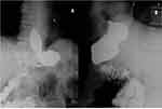
Within six hours of admission, despite adequate resuscitative measures, her condition further deteriorated, and a contrast-enhanced CT (CECT) scan showed a twist in the stomach on its mesenteric axis. A wandering spleen was found to adhere to the anterior gastric wall (Figure 1). It did not show any vascular compromise. Multiple gas foci in the stomach wall, suggestive of gangrenous changes, were also observed. Emergency laparotomy revealed a distended stomach with mesenteric-axial torsion of approximately 180°. After detorsion and decompression of the stomach, multiple gangrenous patches were observed only in the body, whereas the fundus and antrum appeared healthy. The spleen was congested, enlarged, and had a long pedicle, without a twist. Moderate hemorrhagic ascites was present, and the rest of the viscera was unremarkable. Segmental resection of the gangrenous gastric body was performed, followed by complete mobilization of the duodenum and end-to-end tension-free anastomosis between the fundus and antrum. Pyloroplasty was performed as the drainage procedure. A splenectomy had to be performed due to its intimate relation to the anterior wall of the stomach, and also from the concern that retaining the spleen may lead to recurrence of volvulus. The patient’s postoperative course was uneventful, except for superficial abdominal wound dehiscence. The nasogastric tube was removed on the second postoperative day, and the patient was discharged on the eighth day. A follow-up gastrografin study after three months documented a well-formed but smaller stomach and free flow of dye from the esophagus through the stomach to the duodenum (Figure 2).
 |
Figure 2 Postoperative upper gastrointestinal contrast (gastrograffin) study. (A) Right anterior oblique and (B) lateral view revealed a smaller stomach with the absence of gastric body. |
Discussion
In 1904, Borchardt described the classical triad of epigastric pain, retching, and inability to pass a nasogastric tube in acute organoaxial torsion of the stomach.4 Since then, Borchardt’s triad has become the diagnostic triad of gastric volvulus, to the extent that the ability to pass a nasogastric tube in the stomach may rule out gastric volvulus. While Borchardt described the triad based on organoaxial torsion, interestingly, a nasogastric tube can be passed at times with difficulty in mesenteric-axial gastric volvulus. Therefore, in the present case, when a nasogastric tube was inserted and a large volume of dirty gastric fluid was drained, gastric volvulus was not considered clinically. We consider it to be a potential cause of the delay in diagnosis.
Acute cholecystitis, acute pancreatitis, and peptic ulcer perforation are common causes of acute upper abdominal pain in elderly patients. Acute gastric volvulus and gastric gangrene are rare and may not be considered the first-line diagnosis of acute abdomen. Although abdominal USG screening is the first-line investigation for acute abdomen, incomplete gastric volvulus may be missed. In the present case, a CECT scan was used for the diagnostic investigation. Therefore, an early CECT scan can be more informative and diagnostic for gastric volvulus as well as a wandering spleen as its precipitating cause.
Abdominal radiography has often been described as a modality for evaluating gastric volvulus.5 It may show a large distended gastric bubble with or without elevation of the left hemidiaphragm. Gas shadows of the small bowel are sparse or absent in patients with complete obstruction. Two distinct air-fluid levels can be observed.5 It may also reveal multiple linear intramural radiolucent shadows, suggesting intramural gas arising from gangrenous changes. However, the absence of these features does not rule out the possibility of a gastric volvulus or gangrene. In contrast, a CECT scan is a more sensitive investigation for identifying intramural gas as a sign of gangrene.6,7 Thus, the concomitant presence of a wandering spleen identified on abdominal USG screening may falsely indicate splenic ischemia as the cause of abdominal pain. Therefore, CECT remains the diagnostic modality of choice for confirming both conditions, and the present case teaches us that an early CECT scan is more informative of the predisposing cause and complications, especially when both conditions coexist.
Surgical treatment options for gastric volvulus include gastropexy and subtotal or total gastrectomy.8 Subtotal gastrectomy or total gastrectomy followed by gastrojejunostomy or esophagojejunal anastomosis are usually performed in the presence of extensive gangrene. In the present case, the stomach was not extensively gangrenous to consider either subtotal or total resection, as the fundus and antral parts were healthy and vascular. The enlarged stomach with a mesenteric-axial rotation and the lax ligaments had already freed a greater part of the viscus and brought the fundus and antrum close enough for a tension-free anastomosis. This procedure has not previously been described for incomplete gastric gangrenous changes. We believe that it is feasible and can be performed in selected cases of mesenteric-axial torsion when a good part of the viscus can be preserved, although a strict decision should be made in the presence of a dilemma and suspicion of an unhealthy residual viscus that may threaten anastomosis safety. However, segmental resection, if possible, leads to organ and function preservation, and can spare the patient from more extensive and time-consuming resection procedures in an emergency setting.
The wandering spleen may be managed by splenopexy or splenectomy. In the event of splenic ischemia, a splenectomy is preferred. In cases where the spleen is adherent to the gangrenous stomach, a splenectomy needs to be done if the spleen cannot be mobilized for resection of the stomach or its vascularity is doubtful. In pediatric populations where the spleen is smaller and gastric gangrene has not set in, laparoscopic splenopexy and gastropexy have been reported with successful results.9,10 However, with the preservation of the spleen, the concern for recurrence of gastric volvulus in adults is a possibility, but long-term data is not available.
Conclusion
The present case is hereby reported for the rarity of the coexistence of two uncommon conditions, and also to highlight the fact that the clinical diagnosis was delayed because the wandering spleen was considered the primary cause of the acute abdomen. Unless the spleen is gangrenous, in such cases, one would tend to buy time for optimization rather than surgery as the first-line treatment. Individually, either of these conditions is rare, and their combinations are rarer. We also emphasize the diagnostic role of early CECT. The presence of a wandering spleen diagnosed on USG should not necessarily inhibit the performance of a CECT scan, as a more ominous pathology may coexist. Although the presence of extensive gangrene of the stomach demands subtotal or total gastrectomy, in selected cases with patchy gangrene of the body, segmental resection may be considered for organ and function preservation.
Funding
There is no funding to report.
Disclosure
The authors report no conflicts of interest in this work.
References
1. Andley M, Basu S, Chibber P, Ravi B, Kumar A. Internal herniation of wandering spleen--A rare cause of recurrent abdominal pain. Int Surg. 2000;85(4):322–324.
2. Cherukupalli C, Khaneja S, Bankulla P, Schein M. CT diagnosis of acute gastric volvulus. Dig Surg. 2003;20(6):497–499. doi:10.1159/000073536
3. Srivastava V, Basu S, Ansari M, Gupta S, Kumar A. Massive gangrene of the stomach due to primary antiphospholipid syndrome: report of two cases. Surg Today. 2010;40(2):167–170. doi:10.1007/s00595-009-4015-8
4. Singham S, Sounness B. Mesenteroaxial volvulus in an adult: time is of the essence in acute presentation. Biomed Imaging Interv J. 2009;5(3):e18. doi:10.2349/biij.5.3.e18
5. Federle MP, Guliani-Chabra S. Gastric volvulus. In: Fedrele MP, Jeffrey RB, Dresser TS, Anne VS, Eraso A, editors. Diagnostic Imaging Abdomen.
6. Liu HT, Lau KK. Wandering spleen: an unusual association with gastric volvulus. AJR Am J Roentgenol. 2007;188(4):W328–W330. doi:10.2214/AJR.05.0672
7. Mazaheri P, Ballard DH, Neal KA, et al. CT of gastric volvulus: interobserver reliability, radiologists’ accuracy, and imaging findings. AJR Am J Roentgenol. 2019;212(1):103–108. doi:10.2214/AJR.18.20033
8. Lal Meena M, Jenav RK, Mandia R. Gangrenous stomach in mesentroaxial volvulus. Indian J Surg. 2011;73(1):78–79. doi:10.1007/s12262-010-0140-2
9. François-Fiquet C, Belouadah M, Chauvet P, Lefebvre F, Lefort G, Poli-Merol ML. Laparoscopic gastropexy for the treatment of gastric volvulus associated with wandering spleen. J Laparoendosc Adv Surg Tech A. 2009;19(Suppl 1):S137–S179. doi:10.1089/lap.2008.0091.supp
10. Nakagawa Y, Uchida H, Amano H, et al. Patients with gastric volvulus recurrence have high incidence of wandering spleen requiring laparoscopic gastropexy and splenopexy. Pediatr Surg Int. 2022;38(6):875–881. doi:10.1007/s00383-022-05125-y
 © 2023 The Author(s). This work is published and licensed by Dove Medical Press Limited. The full terms of this license are available at https://www.dovepress.com/terms.php and incorporate the Creative Commons Attribution - Non Commercial (unported, v3.0) License.
By accessing the work you hereby accept the Terms. Non-commercial uses of the work are permitted without any further permission from Dove Medical Press Limited, provided the work is properly attributed. For permission for commercial use of this work, please see paragraphs 4.2 and 5 of our Terms.

