Back to Journals » Clinical Ophthalmology » Volume 16
Vitreous Opacity Vitrectomy (VOV): Safest Possible Removal of “Floaters”
Authors Morris RE ![]()
Received 1 March 2022
Accepted for publication 11 May 2022
Published 1 June 2022 Volume 2022:16 Pages 1653—1663
DOI https://doi.org/10.2147/OPTH.S361557
Checked for plagiarism Yes
Review by Single anonymous peer review
Peer reviewer comments 2
Editor who approved publication: Dr Scott Fraser
Video abstract presented by Robert E Morris.
Views: 2285
Robert E Morris1,2
1Helen Keller Foundation for Research and Education, Birmingham, Alabama, USA; 2Retina Specialists of Alabama, LLC, Birmingham, Alabama, USA
Correspondence: Robert E Morris, Helen Keller Foundation for Research and Education, 2208 University Boulevard, Suite 101, Birmingham, Alabama, 35233, USA, Tel +1 205 936-0704, Fax +1 205 558-2567, Email [email protected]
Purpose: Primary opacities that develop in the aging vitreous, commonly termed “floaters,” were once considered merely a nuisance, not justifying any risk of surgical removal. However, vitreoretinal specialists are increasingly recognizing that extensive symptomatic vitreous opacities (SVO) that substantially interfere with activities that critically depend on vision (daily visual activities, DVA), constituting degenerative vitreous syndrome (DVS, see http://floaterstories.com), warrant removal albeit with minimal risk - but no description of how to reduce vitrectomy risks to least possible has been forthcoming. We here describe such a method.
Patients and Methods: The safest possible removal of extensive SVO as described herein was attained by an operation specifically designed for DVS treatment (vitreous opacity vitrectomy, VOV), rather than as only a means of achieving subsequent retinal surgery in the same procedure, as is usually the case. We retrospectively reviewed the outcomes of 100 consecutive VOV operations (in 81 patients, average age 66) performed with ultra-high speed, 27-gauge vitrectomy probes.
Results: All eyes rapidly achieved continuously clear vision, and no eye developed a clinically significant complication during a year of follow-up. Three small, existent retinal breaks were discovered prior to peripheral vitrectomy and one apparently iatrogenic retinal tear was found at VOV completion, when each was treated. In the eyes that were not pseudophakic, postoperative nuclear sclerosis progression was successfully managed by subsequent cataract extraction.
Conclusion: The goals of VOV for DVS are to safely restore continuously clear vision by performing tractionless vitreous removal with respect to the retina and to reduce the lifetime risk of retinal detachment, both by such vitreous removal and by microscopic examination of the peripheral retina under anesthesia (MEPRUA), guiding appropriate prophylactic retinopexy. The otherwise healthy DVS eyes so treated warrant this specific form of vitrectomy, continually focused on achieving least possible risk, to maintain an acceptable risk/benefit ratio.
Keywords: floaters, vitrectomy, symptomatic vitreous opacities, daily visual activities, degenerative vitreous syndrome, vitreous opacity vitrectomy, retinal tear
Plain Language Summary
Lens opacities developing in the aging eye are called cataracts. Cataracts are commonly removed with minimal risk and excellent results, replaced with a clear, artificial lens. Behind the lens, the “vitreous” gel also develops opacities that seem to “float” (floaters), appearing as false objects in the field of vision as the gel becomes more mobile with advancing age.
While floaters are usually only an occasional nuisance, some eyes develop many symptomatic vitreous opacities that substantially interfere with reading, driving, safely using stairs, and other daily visual activities (see http://floaterstories.com). We call this “Degenerative Vitreous Syndrome,” or DVS.
In this article, we describe how to diagnose DVS; and how to remove DVS vitreous opacities with the least possible surgical risk, similar to that of cataract extraction. We call this operation “Vitreous Opacity Vitrectomy”, or VOV. It immediately restores continuously clear vision for DVS sufferers, as the eye quickly and permanently replaces the removed DVS gel with clear aqueous fluid.
Introduction
The disability associated with extensive “floating” opacities developing in the aging vitreous (floaters) has commonly been underestimated. A likely reason is that visual disability is assessed primarily by loss of visual acuity that is measured by the untimed reading of single letters, during moments of clarity. The visual acuity so measured is little affected by floating, symptomatic vitreous opacities (SVO).1
Moreover, the risks and morbidity of vitrectomy to remove these opacities have only recently been reduced to an acceptable level, similar to the removal of cataract opacities, and this has not been widely recognized.2 Thus, no reports of elective vitrectomy for floaters appear in a PubMed search covering 29 years from the advent of vitrectomy in 1971 until the first report (on five cases) in 2000.3 Only seven reports occurred in the next eight years, while during the same period, approximately 864 reports occurred in PubMed on elective vitrectomy for macular pucker.4–10
In 2007, we introduced the term Degenerative Vitreous Syndrome (the spontaneous occurrence in the aging vitreous of opacities that substantially interfere with activities of daily living, ADL) in a video submission that received the Buckler Award at the Annual Meeting of the American Society of Retina Specialists (Supplement 1- Video).11 This award marked the first time any ophthalmologic society honored a presentation depicting vitrectomy removal of symptomatic vitreous opacities in the aging vitreous, long dismissed as merely “floaters,” not justifying treatment, no matter how extensive.
DVS terminology helps distinguish the important but relatively rare condition of extensive SVO (see http://floaterstories.com) from common nuisance floaters that can be annoying but that do not substantially impair vision. We also subsequently developed the term “daily visual activities” (DVA, activities that critically depend on vision) as more appropriate than ADL, a time-honored term more accurately describing general disability.12
Although vitreoretinal surgeons are increasingly recognizing that removal of such primary opacities is justified if complications can be minimized (Retina Today Supplement, November 2021), no report heretofore describes in detail how to perform the safest possible vitrectomy. In this report, we meticulously describe Vitreous Opacity Vitrectomy (VOV), a procedure that employs a series of specific safeguards to achieve least possible risk. We review technical issues that distinguish VOV, in which primary vitreous opacity removal in an otherwise healthy eye is the key element of surgery, from more common vitrectomy in which vitreous removal is only a prelude to treatment of retinal disease as the key element. We then briefly review a case series so treated.
Patients and Methods
Patients seeking treatment for “floaters” were evaluated by a complete vision history and ocular examination. The diagnosis of DVS was established by a detailed discussion of symptoms, a dynamic examination of the vitreous, and a review of patient symptom statements as described in the discussion below. VOV was scheduled after we had diagnosed DVS not improving over a period of at least three months,13 and after adequate counseling resulting in informed consent had been concluded, usually following a second visit or a follow-up interview. The few patients without confirmed posterior vitreous detachment (PVD) were informed that cortical vitreous might be retained.
We analyzed a consecutive series of 100 eyes treated by the author for DVS by VOV, using 27-gauge (G) 7500 cuts per minute (cpm) probes, between 2015 and 2019. Anesthesia choices were local/sedation (subtenons irrigation, occasionally topical) or general via laryngeal mask. We avoided retrobulbar block and endotracheal general anesthesia in the interest of risk/morbidity reduction. Our preferred treatment techniques are described below.
The VOV method begins with mindset. The NASA moon landing program famously adopted a “zero defects” quality control theme based on the concept that loss of even one astronaut was unacceptable.14 Similarly, “zero errors,” producing a complication rate as close to zero as possible, is the conscious operative goal in each step of a VOV operation.
Speed is important during extensive vitrectomy to treat complicated vitreoretinal conditions, but it is inevitably associated with increasing risk. Slower, low risk vitrectomy is appropriate in VOV, and safety should transcend speed as the surgeon’s uppermost thought during vitreous removal. Moreover, meticulous opening and closing should also be regarded as critical components, and the responsible surgeon should perform the entire operation as a key risk reduction measure. Although VOV is relatively simple technically, there is no role for surgeons in training.1
Beyond topical antibiotic prophylaxis, immediate preoperative prophylaxis with a single dose of systemic moxifloxacin is a reasonable additional risk-reduction measure in VOV.15,16 Once the eye is prepped and draped, the scrub treats the conjunctiva with povidone iodine-soaked cotton tips. The surgeon then irrigates the “browned” conjunctiva prior to beginning VOV.
After the microscope is positioned, we carefully enter the eye through avascular areas of the conjunctiva overlying the pars plana and bordering the nasal and temporal horizontal meridians. The conjunctiva is displaced and held with a dry cotton tipped applicator to avoid incising it directly overlying the sclerotomy wound. Sclerotomies are made 3 to 4 mm posterior to the limbus by first indenting the conjunctiva and sclera with the flat surface of a trocar blade and slightly angling into the sclera before moving forward, turning radial for the final entry. The infusion cannula placement is confirmed and announced with “lights off” fiber-optic illumination before vitrectomy infusion is begun. The infusion line is positioned by the surgeon and taped by the scrub, both near the eye and at the stopcock, and infusion is ordered on at 30 mm hg.
Once cannulation and infusion are established, the endoilluminator and vitrectomy probe are emplaced, and the posterior segment is examined with the wide-field microscope. After noting the vitreous opacities and confirming retinal integrity, the vitrectomy is cautiously begun while confirming adequate infusion. Preventing retinal traction during vitreous removal is a conscious intraoperative goal. Since no prolonged retinal treatment is needed, vitreous may be removed relatively slowly while still retaining a reasonably brief operative time. This argues for employing the least suction necessary, continually adjusted to the rate of vitreous movement; high speed cutting (preferably 20,000 cpm); and a small aspiration port (27G).17
After vitreous opacities have been removed centrally, intravitreal triamcinolone marking (optional) can guide the safest and most efficient removal of remaining vitreous. It helps confirm PVD seen in most cases, or aids in gently creating one if necessary and if indicated. In the interest of risk reduction for the few cases without PVD, excessive efforts to attain vitreous detachment are contraindicated,13 and clear cortical vitreous left in place typically detaches gradually postoperatively without sequelae. The disc and macula are then examined, and the absence of any treatable traction maculopathy is confirmed.18
After removal of central vitreous, vitreous is removed around the infusion cannula to assure a vented eye for optimal scleral depression.19 Prior to peripheral vitrectomy, the peripheral retina is examined with surgeon scleral depression, under microscopic viewing and endoillumination, to detect existent defects that may have escaped notice preoperatively. The vitreous “skirt” is then reduced using maximum cutting speed, at 400 mm hg maximum suction, without scleral depression, avoiding the vitreous base. Particular attention is given to reducing the superior vitreous skirt to prevent gravitational extension of retained vitreous towards the visual axis in the upright position postoperatively. Anterior vitreous and capsular opacity are removed immediately behind an implant lens, but vitreous is left in place retrolental in phakic eyes.
After vitreous removal is completed, the peripheral retina is again carefully examined by the VOV team (using smooth on-and-off scleral depression by the surgeon, with only the endoilluminator inserted) to rule out new retinal tears. The search should be especially vigilant in the few eyes undergoing intraoperative creation of PVD.20,21 All defects and lattice degeneration are then treated with focal endolaser retinopexy. Placement of encircling laser prophylaxis is rarely employed for a high-risk eye, based on the patient’s and surgeon’s preference.22,23
Achieving watertight sclerotomy closure is especially important to avoid even transient postoperative hypotony,24 and to reduce susceptibility to postoperative endophthalmitis (risk less than 0.08%).25 Our wound creation technique sets the stage for a self-sealing result, but we also use a bipolar cautery tip to exert point pressure at the sclerotomy site as we slowly withdraw each cannula under a pressure of 20 mm hg, to avoid vitreous wicking and to create a slight “weld” of the conjunctival wound. If sutureless wound integrity is in doubt, we employ a 9–0 absorbable suture. As watertight sclerotomies are being confirmed, we irrigate the ocular surface with 1% povidone iodine as a final infection precaution.26,27 The operating time for a typical VOV is 20–25 minutes, with additional time for laser retinopexy if needed.
Postoperative supervision during recovery should be especially meticulous, emphasizing to the patient that treatment is not over until the at-risk period is concluded. On the postoperative day one examination, we remind the patient to promptly report any significant deterioration in visual acuity, comfort, or appearance of the eye, as risk of infection occurs primarily in the early postoperative period. On a two-week postoperative visit, the patient is reminded of the symptoms of retinal detachment and the need to periodically examine the visual field by monocular testing. At a two-month postoperative visit, we again perform a detailed examination of the peripheral retina to rule out retinal breaks, and this step concludes postoperative supervision.
A summary of the VOV method is enclosed as Supplement 2.
Results
We analyzed 100 consecutive eyes of 81 DVS patients treated solely by the author between 2015 and 2019, using the 27G VOV techniques described. The mean patient age was 66 years (range 38–83, median 67). Ninety-five eyes had preoperative PVD with Weiss ring and/or imaging confirmation, and four eyes had PVD carefully created intraoperatively without sequelae. Seventy-one of 81 patients were referred from other eye care providers, and the remaining 10 were self-referred. All patients presented with chief complaints related to SVO and most of these patients provided written descriptions of their DVS symptoms preoperatively at my request.
All patients had a minimum of six months follow-up postoperatively, with a mean follow-up of 37 months. Every patient had the restoration of continuously clear vision (CCV) in the affected eye(s) by their own assessment early postoperatively. At final follow-up, all patients reported being satisfied with the result of VOV surgery, usually “very satisfied.” Two patients reported occasionally noticing a single residual, peripheral “floater.”
Seventy eyes were pseudophakic at the time of VOV and 15 eyes had concurrent cataract extraction,14 with an implanted lens. All postoperatively pseudophakic patients reported stable or improved visual acuity at final follow-up. One eye that was originally 21 diopters myopic had lens removal and was left aphakic with unchanged corrected visual acuity.
One eye with a completely clear lens in a 44-year-old male remained clear three years after VOV with 20/20 corrected visual acuity. Thirteen of 14 eyes with early nuclear sclerosis cataract and no concomitant lens procedure underwent uncomplicated cataract extraction within two years postoperatively and achieved final visual acuity within one line of preoperative VOV visual acuity. One eye developed staphylococcus coagulase negative endophthalmitis after cataract surgery 11 months after VOV. This eye subsequently achieved 20/25 visual acuity.
At surgery, three retinal breaks were found in three eyes prior to initiating peripheral vitreous removal, using endo-illumination, scleral depression, and wide-angle microscope viewing, thereby illustrating the value of microscopic examination of the peripheral retinal under anesthesia (MEPRUA) in VOV. During reexamination after removal of the vitreous skirt, one (presumably new) peripheral tear without hemorrhage or subretinal fluid was found in a fourth eye. All four breaks in the four eyes were treated with laser retinopexy without sequelae.
One patient was lost to follow-up after two weeks, skipping the two months postoperative examination. He returned 14 months postoperatively with a retinal detachment that was repaired with 20/50 final visual acuity. There were no infections, no macular pucker or cystoid macular edema development, and no other complications intraoperatively or postoperatively.
Discussion
Symptoms
DVS produces visual disability due to focal vitreous opacities causing frequent momentary obscurations in the line of sight and false object movements in the peripheral visual field that randomly and relentlessly command attention (Supplement 1- Video). A patient once described this by saying, “I feel like I’m seeing between my floaters.” Concomitant diffuse vitreous opacification causes glare in bright light environments and reduced contrast sensitivity. Although these are rarely patients’ primary complaints, glare from headlights can become a safety problem, leading to night driving avoidance.
Occasionally a patient has diffuse vitreous opacification (Figure 1) sufficient to constitute DVS, without discrete, symptomatic opacities. Even less frequently a single large opacity (Supplement 3 - Video), or a smaller opacity with a central “null point” (Figure 2) will justify VOV. This can often be seen as shadowing on optical coherence tomography (OCT, Figure 3A and B). A full discussion of DVS symptomatology and 28 examples of patient statements can be seen at http://floaterstories.com
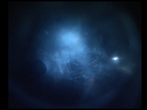 |
Figure 1 Diffuse vitreous opacification illuminated intraoperatively. Image courtesy of Retina Specialists of Alabama LLC (RSA). |
 |
Figure 2 Fundus photograph showing a single, focal SVO in the central, posterior vitreous that was highly symptomatic and non-resolving. Image courtesy of RSA. |
 |
Figure 3 (A) Preoperative OCT image showing shadowing of the macula by the same SVO seen in Figure 1, mimicking the recurrent SVO scotoma seen by the DVS patient. (B) Postoperative OCT image after VOV showing absent shadowing and a high-quality image through a clear visual axis. Images courtesy of RSA. |
Especially vexing to some DVS patients is the unremitting nature of SVO distractions, and the likelihood that, having found no treatment or having been rejected for treatment, they may never find relief. DVS sufferers have sometimes been characterized as being psychologically abnormal (Supplement 4),1 or as having unrealistic expectations.2 But while an occasional patient has complaints out of proportion to their vitreous opacities, the vast majority of DVS sufferers are realistic and know well what they are seeking - a continuously clear view of the world that they once had and still remember.
DVS entoptic phenomena can give rise to a sense of isolation as patients feel they are trapped in a form of “virtual reality” - frequently leading to elation upon VOV release. As an example:
I am continually seeing things that are not there. I move the hair that isn’t in front of my face. I search for the insect or small animal that never existed. I turn my head to speak to the person who didn’t approach me. And I stop to allow the mythical car to pass before I walk across the street.
MJK, DVS sufferer.
The leader of a large corporation once told me,
In meetings of our Board of Directors, I often sense that I am separated from all the others who are not seeing what I am seeing. My interactions are profoundly affected, sometimes at critical moments when I should be at the top of my game.
He added postoperatively that without relief it would have soon been necessary to resign his position.
It is finally being recognized that extensive vitreous opacities can arguably be even more debilitating than the constant blur of moderate cataracts that, enabled by technological improvements, are safely treated today at an earlier stage than in the past with excellent results. Similar improvements in technology detailed below now enable safe vitrectomy for substantial, symptomatic vitreous opacities.
Diagnosis
While the concept of VOV treatment of DVS is straightforward, identification and counseling of DVS patients can be complex.1,13 DVS is best diagnosed clinically by dynamic slit lamp examination of the anterior and posterior vitreous, noting focal and diffuse vitreous opacities and their vertical and horizontal movements and after-movements. These findings are correlated with a patient’s detailed description of visual dysfunction during daily visual activities (eg, reading, driving, etc.). Because some patients’ DVA are particularly demanding, their symptoms may be proportionally worse for a given amount of vitreous opacities.
When the patient and the physician agree that vitreous opacities substantially interfere with their DVA and have been present for several months without improvement, the diagnosis of DVS is made, and VOV is an appropriate treatment. However, once this conclusion is reached, patients are counseled that the opacities pose no threat to their ocular health, so the choice of VOV treatment or periodic observation is elective and is completely theirs.
In the evaluation process we find patients’ written statements more revealing than generic quality of life (QoL) scores as they more accurately describe and document each patient’s visual problems attributable to DVS. Patient statements also reinforce informed consent since we usually ask patients to “write your story” as the initial visit concludes. A decision regarding VOV is then made in a second phone or in-person discussion after their review, ensuring the patient’s assimilation of information exchanged during the initial examination. If the patient is coming from a distance, these items can be started in advance to conclude a definitive initial visit.
DVS is an inherently qualitative disease, and the bottom line in the decision to treat is not a visual acuity, quantitative ultrasound, contrast sensitivity, QoL, or utility value number.1,28–30 These serve an ancillary documentation purpose, but only a patient’s substantial symptoms (correlated with vitreous physical examination) are sufficient, indispensable, and treatment actionable.31 For example, while cautioning regarding vitrectomy risks, Henry et al posed the rhetorical question “Is a reduction in contrast sensitivity enough of an indication to support vitrectomy, when a patient has excellent visual acuity?”32 But after having read statements by DVS sufferers, one would quickly understand that their problems go well beyond either a contrast sensitivity or visual acuity number.
Treatment
We infrequently treat vitreous opacities in patients under 50 years of age, as their formed vitreous and strong retinal attachments add significant risk. When doing so, however, the vitrectomy is typically subtotal for safety purposes, and patients are counseled that cortical vitreous will likely remain, rendering them still susceptible to a future tractional event or new floaters when posterior vitreous detachment eventually occurs naturally. But retinal tears and substantial SVO recurrences are still unlikely.
DVS is much more commonly associated with vitreous syneresis and PVD in patients over 60 years of age,13 who can be completely cured by a VOV procedure continually focused on minimizing treatment risk. PVD initiates symptoms in most DVS patients (excepting high myopes), as in this series with PVD present in 95% of patients having a mean age of 66 years.
This same age group presents with either existent pseudophakia or a high rate of nuclear sclerosis cataract that will likely accelerate after vitrectomy. The high presenting rate of pseudophakia in this series may reflect referring doctors’ efforts to assuage symptoms through cataract extraction prior to referral, but with incomplete success due to concomitant DVS. Alternatively, PVD might occur soon after pseudophakia, leading to the onset of DVS symptoms.
In phakic eyes, depending on the current lens status, the patient’s preference, and the preference of the referring eye care provider, an early cataract may be left in place during VOV, or a combined procedure may be planned. In the latter instance, care must be taken to ensure a stable anterior chamber and scleral depression must be especially cautious. The cataract surgeon should be apprised to remove viscoelastic fluid behind the implanted lens before placing a suture in the primary wound. The posterior capsule must then be located with endoillumination (as it can be considerably posterior to the concomitantly implanted lens) and carefully avoided during anterior vitreous removal.
After the vitreoretinal surgeon’s examination confirms persistent, extensive vitreous opacities that reasonably account for a patient’s detailed and substantial complaints, the most important number is a careful estimate of surgical risk. But one must also understand that, without vitrectomy, DVS patients also experience risks to their safety in using stairs, crossing streets, and in driving – forcing them to adopt considerable avoidance strategies to reduce their chance of injury.
Treatment Risks
Endophthalmitis is an important but rare complication (0.08%) of vitrectomy.25 The endophthalmitis risk in VOV is likely even lower as it involves especially small wounds and short duration surgery with few instrument reentry events. Moreover, VOV includes every possible infection preventive measure.
The major risk of vitrectomy has historically been retinal detachment, currently reported to be 2.5%.33 In this regard, the first goal of VOV is to remove vitreous gel without tearing the retina (possibly leading to retinal detachment). The defining characteristic of VOV is its aggregate emphasis on safety throughout each step of vitrectomy, resulting in its potential to actually reduce the lifetime risk of retinal detachment in DVS eyes.
The iatrogenic retinal tear rate during vitrectomy has decreased from as high as 37% in the 1970ʹs,34 to 2–7% today.33,35,36 Using the technological innovations of small gauge, cannulated entry,37,38 and the ultra-high-speed suction/cutters recently developed by outstanding corporate partners in the vitreoretinal surgery industry, my experience is that the iatrogenic tear rate can now be further reduced to less than 1%, using VOV techniques.
A second goal of VOV is to find and treat existent lesions so as to achieve a net reduction of retinal detachment risk in post vitrectomy eyes.20 Thus, a paradigm shift may now be possible using VOV techniques, from regarding elective vitrectomy as an additional risk factor for retinal detachment to regarding it as a risk-reduction procedure, as an ancillary benefit to that for which the procedure was chosen.
We explain to DVS patients that while there are no risk-free procedures, the risk of significant loss of vision from VOV performed as described above is less than 1% in our experience (DVS patients in a study by Wagle were willing to accept a 7% risk of blindness to be relieved of floater symptoms).39 Thus, the risk of removing symptomatic vitreous opacities in DVS with VOV now approaches the operative risk of cataract extraction for lens opacity, because properly performed VOV is continually focused on pursuing tractionless vitrectomy with respect to the retina, seeking to make iatrogenic retinal tear a “never” event in this form of elective vitrectomy.
While traction on the vitreous gel is inevitable during its removal, our goal is to have all traction end in vitreous tissue, well away from the peripheral retina. We achieve this by using ultra-high-speed cutting, least possible suction, 27G probes with small aspiration ports, vitreous staining when needed, rarely and very cautiously producing intraoperative PVD, and by avoiding the vitreous base.17 Our new tear rate was 1% in this series, greatly reduced relative to the iatrogenic retinal tear rate during the era of big probes, slow cutters, and large aspiration ports,17 when microscopic examination of the peripheral retina to find and treat such tears was also exceedingly difficult.
At the conclusion of vitrectomy, wide angle attachments now easily enable microscopic examination of the peripheral retina under anesthesia with endoillumination and dynamic scleral depression, in a vented and thus malleable eye.19 Therefore, VOV minimizes the chance that a new tear would occur or escape notice (and definitive endolaser treatment), forestalling development of postoperative retinal detachment. Such an intraoperative examination under anesthesia is considerably better than an examination performed preoperatively in a clinic with an indirect ophthalmoscope. Thus, we detected (prior to peripheral vitrectomy) and treated three existent, undiscovered retinal defects in this series. As a result, in pseudophakia eyes, the long-term risk of retinal detachment after VOV is arguably less than the long-term risk of retinal detachment from persistent vitreous traction in non-vitrectomized eyes, which is approximately ten times that of the general population.40
Conflicting reports in the literature discuss a risk of post vitrectomy eyes eventually developing ocular hypertension or glaucoma, for no apparent reason. This potential long-term risk has not commonly been part of counseling in other forms of elective vitrectomy, and unless it is confirmed and quantified, it is not currently a decisive factor.41 It is obvious that post-surgical eyes in general should be regularly examined for a lifetime.
Conclusion
VOV techniques can potentially reduce risk to least possible in elective vitrectomy performed for any indication. In a properly diagnosed DVS patient, the VOV benefits of promptly, completely, and permanently restoring continuously clear vision, and ensuring retinal integrity by microscopic examination of the peripheral retina under anesthesia, are usually deemed to outweigh the risks. This coincides with the recent finding that vitrectomy for DVS produces greater quality of life improvement per cost of treatment than either cataract extraction or retinal detachment repair.30
In DVS treatment, the modest but systematic precautions here described turn our attention from the relatively speedy removal of vitreous as a prelude to retinal treatment, to the safest possible vitreous removal by a single, experienced vitreoretinal surgeon,1 as the key component of VOV. The risk/benefit equation is thus kept in balance to enable definitive treatment of symptomatic vitreous opacities in degenerative vitreous syndrome, achieving the patient’s goal of restoring continuously clear vision.
Abbreviations
CCV, continuously clear vision; DVA, daily visual activities; DVS, degenerative vitreous syndrome; MEPRUA, microscopic examination of the peripheral retina under anesthesia; OCT, optical coherence tomography; PVD, posterior vitreous detachment; QoL, quality of life: SVO, symptomatic vitreous opacities; VOV, vitreous opacity vitrectomy.
Author’s Note
This is a single author article and Vitreous Opacity Vitrectomy is ideally a single surgeon operation, as discussed in the methods section. Moreover, despite many years of reporting in the literature, DVS recognition and treatment remain developmental and controversial,2 although encouragingly less so during the last year (Retina Today Supplement, November 2021). Therefore, to an unusual extent, each surgeon must establish individualized techniques for identifying, counseling, and treating DVS patients. The VOV described herein is my technique, and these results were congruous with my experience of 28 years treating DVS with extreme care, so as to achieve least possible risk. Other vitreoretinal surgeons will establish their own unique methods of identifying DVS patients, and their own version of VOV for this completely curative but completely elective procedure in otherwise healthy eyes. Now that it is documented that DVS is comparatively debilitating to cataract,30 there will come a time when treatment of properly selected DVS patients is no more controversial than treatment of moderate cataract opacities has become since enabled by technological progress.
Ethics Approval and Informed Consent
This study received approval from the Western Institutional Review Board, and it was conducted in accordance with the ethical standards of the 1964 Declaration of Helsinki and its later amendments. Written informed consent was obtained from all patients from whom protected health information was obtained and used for the purposes of this study. As well, written informed consent for the publication of individual case details was provided by the patients described in this manuscript.
Acknowledgments
The author thanks the many patients whose statements have helped define degenerative vitreous syndrome. As in many prior articles, the author is grateful for excellent research, clinical, and clerical support by Ferenc Kuhn, Christina Sullivan, Jessica Haynes, Dewayne Conn, Clotil Lebeau, Claire Buha, Ashley Baker, and Laura Beckwith. The author also appreciates the supportive environment provided by the staff and physicians of Retina Specialists of Alabama and the UAB Callahan Eye Hospital.
Supplementary Materials
Supplement 1 Video - ASRS 2007 Buckler Award.
Supplement 2 A Summary of the VOV Method for Treatment of Degenerative Vitreous Syndrome.
Supplement 3 Video - Large ring opacity, an unusual form of focal DVS.
Supplement 4 Psychological aspects of DVS.
Funding
Support for this research was provided by the Helen Keller Foundation for Research and Education, through grants by the Hanna Charitable Trust of Birmingham, Alabama and by the Kent Companies, Midland, Texas.
Disclosure
The author reports no conflicts of interest with respect to this study. Dr Robert E Morris reports personal fees from Retina Today, outside the submitted work. Subsequent to the study, the author became a consultant to Alcon, Inc.
References
1. Hoerauf H. Vitrectomy against floaters. In: Kirchhof B, Wang D, editors. Essentials in Ophthalmology; Vitreoretinal Surgery. New York, USA: Springer; 2007:115–124.
2. Cohen MN, Rahimy E, Ho AC, Garg SJ. Management of symptomatic floaters: current attitudes, beliefs, and practices among vitreoretinal surgeons. Ophthalmic Surg Lasers Imaging Retina. 2015;46(8):859–865. doi:10.3928/23258160-20150909-11
3. Schiff WM, Chang S, Mandava N, Barile GR. Pars plana vitrectomy for persistent, visually significant vitreous opacities. Retina. 2000;20(6):591–596. doi:10.1097/00006982-200011000-00001
4. Hong PH, Han DP, Burke JM, Wirostko WJ. Vitrectomy for large vitreous opacity in retinitis pigmentosa. Am J Ophthalmol. 2001;131(1):133–134. doi:10.1016/S0002-9394(00)00713-3
5. Mossa F, Delaney YM, Rosen PH, Rahman R. Floaterectomy: combined phacoemulsification and deep anterior vitrectomy. J Cataract Refract Surg. 2002;28(4):589–592. doi:10.1016/S0886-3350(01)01104-X
6. Delaney YM, Oyinloye A, Benjamin L. Nd: YAG vitreolysis and pars plana vitrectomy: surgical treatment for vitreous floaters. Eye. 2002;16(1):21–26. doi:10.1038/sj.eye.6700026
7. Hoerauf H, Müller M, Laqua H. [Vitreous body floaters and vitrectomy with full visual activity]. Ophthalmologe. 2003;100(8):639–643. German. doi:10.1007/s00347-002-0766-y
8. Quintyn JC, Brassuer G. [Vitrectomy for floaters]. J Fr Ophtalmol. 2004;27(5):491–495. French. doi:10.1016/S0181-5512(04)96169-4
9. Roth M, Trittibach P, Koerner F, Sarra G. [Pars plana vitrectomy for idiopathic vitreous floaters]. Klin Monbl Augenheilkd. 2005;222(9):728–732. German. doi:10.1055/s-2005-858497
10. Roufail ED, Polkinghorne P. Vitreous Floaters. Compr Ophthalmol Update. 2006;7(4):171–177.
11. Morris RE, Witherspoon CD, Kimble J, Kuhn F, Roberts D, Sapp MR. Vitreous Opacity Vitrectomy (VOV) for Degenerative Vitreous Syndrome (DVS) – let’s talk about floaters. Video submission,
12. Chappelow J. Activities of Daily Living (ADL). Wohlner R. Ed. Investopedia; 2020. Available from: https://www.investopedia.com/terms/a/adl.asp.
13. Ivanova T, Jalil A, Antoniou Y, et al. Vitrectomy for primary symptomatic vitreous opacities: an evidence-based review. Eye. 2016;30(5):645–655. doi:10.1038/eye.2016.30
14. Weiss HM. NASA quality requirements & cost control. quality assurance division, office of reliability & quality assurance, Washington, DC, USA: NASA Headquarters; 1965. Available from: https://ntrs.nasa.gov/archive/nasa/casi.ntrs.nasa.gov/19650008885.pdf.
15. Bratzier DW, Houck BM. Antimicrobial prophylaxis for surgery: an advisory statement from the national surgical infection prevention project. Surgical infection prevention guideline writers workgroup. Am J Surg. 2005;189(4):395–404. doi:10.1016/j.amjsurg.2005.01.015
16. Lott MN, Fuller JJ, Hancock HA, Singh J, Singh H. McGwin G
17. Teixeira A, Chong LP, Matsuoka N, et al. Vitreoretinal traction created by conventional cutters during vitrectomy. Ophthalmol. 2010;117(7):1387–2010. doi:10.1016/j.ophtha.2009.11.004
18. Morris R, Kuhn F, Witherspoon CD. Hemorrhagic macular cysts. Ophthalmology. 1994;101(1):1. doi:10.1016/S0161-6420(13)31237-8
19. Witherspoon CD, Morris RE, Goggans WE
20. Tan S, Mura M, Oberstein SYL, Bijl HM. Safety of vitrectomy for floaters. Am J Ophthalmol. 2011;151(6):995–998. doi:10.1016/j.ajo.2011.01.005
21. Tan HS, Mura M, de Smet MD. Iatrogenic retinal breaks in 25-gauge macular surgery. Am J Ophthalmol. 2009;148(3):427–430. doi:10.1016/j.ajo.2009.04.002
22. Koh HJ, Cheng L, Kosobucki B, Freeman WR. Prophylactic intraoperative 360 degrees laser retinopexy for prevention of retinal detachment. Retina. 2007;27(6):744–749. doi:10.1097/IAE.0b013e318030ebd7
23. Morris R, Shere J, Witherspoon CD, et al. Intraoperative retinal detachment prophylaxis in vitrectomy for retained cataract fragments. J Cataract Refract Surg. 2009;35(3):491–495. doi:10.1016/j.jcrs.2008.11.037
24. Hsu J, Chen E, Gupta O, Fineman MS, Garg SJ, Regillo CD. Hypotony after 25-gauge vitrectomy using oblique versus direct cannula insertions in fluid-filled eyes. Retina. 2008;28(7):937–940. doi:10.1097/IAE.0b013e31816c6855
25. Oshima Y, Kadonosono K, Yamaji H, et al; Japan Microincision Vitrectomy Surgery Study Group. Multicenter survey with a systematic overview of acute-onset endophthalmitis after transconjunctival microincision vitrectomy surgery. Am J Ophthalmol. 2010;150(5):716–725.e1. doi:10.1016/j.ajo.2010.06.002
26. Apt L, Isenberg SJ, Yoshimori R, et al. The effect of povidone-iodine solution applied at the conclusion of ophthalmic surgery. Am J Ophthalmol. 1995;119(6):701–705. doi:10.1016/S0002-9394(14)72773-4
27. Koerner JC, George MJ, Meyer DR, Rosco MG, Habib MM. Povidone-iodine concentration and dosing in cataract surgery. Surv Ophthalmol. 2018;63(6):862–868. PMID: 29778494. doi:10.1016/j.survophthal.2018.05.002
28. Mamou J, Wa CA, Yee KM, et al. Ultrasound-based quantification of vitreous floaters correlates with contrast sensitivity and quality of life. Invest Ophthalmol Vis Sci. 2015;56(3):1611–1617. PMID: 25613948; PMCID: PMC4554261. doi:10.1167/iovs.14-15414
29. Garcia GA, Khoshnevis M, Yee KMP, Nguyen-Cuu J, Nguyen JH, Sebag J. Degradation of contrast sensitivity function following posterior vitreous detachment. Am J Ophthalmol. 2016;172(7–12):7–12. PMID: 27633841. doi:10.1016/j.ajo.2016.09.005
30. Rostami B, Nguyen-Cuu J, Brown G, Brown M, Sadun AA, Sebag J. Cost-effectiveness of limited vitrectomy for vision-degrading myodesopsia. Am J Ophthalmol. 2019;204:1–6. PMID: 30849342. doi:10.1016/j.ajo.2019.02.032
31. Ryan EH. Current treatment strategies for symptomatic vitreous opacities. Curr Opin Ophthalmol. 2021;32(3):198–202. PMID: 33710011. doi:10.1097/ICU.0000000000000752
32. Henry CR, Smiddy WE, Flynn HW
33. Zeydanli EO, Parolini B, Ozdek S, et al.; EVRS Floaters Study Group. Management of vitreous floaters: an international survey the European VitreoRetinal Society Floaters study report. Eye. 2020;34(5):825–834. PMID: 32313173; PMCID: PMC7182575. doi:10.1038/s41433-020-0825-0
34. Michels RG, Ryan SJ
35. Saleh OA, Al-Dwairi RA, Mohidat H, et al. International multi-center study of iatrogenic retinal tears in pars plana vitrectomy. Int J Ophthalmol. 2019;12(6):996–1000. PMID: 31236359; PMCID: PMC6580211. doi:10.18240/ijo.2019.06.19
36. Yu Y, Qi B, Liang X, Wang Z, Wang J, Liu W. Intraoperative iatrogenic retinal breaks in 23-gauge vitrectomy for stage 3 and stage 4 idiopathic macular holes. Br J Ophthalmol. 2021;105(1):93–96. PMID: 32217539. doi:10.1136/bjophthalmol-2019-315579
37. Fujii GY, De Juan E Jr, Humayun MS, et al. A new 25-gauge instrument system for transconjunctival sutureless vitrectomy surgery. Ophthalmology. 2002;109(10):
38. Oshima Y, Wakabayashi T, Sato T, Ohji M, Tano Y. A 27-gauge instrument system for transconjunctival sutureless microincision vitrectomy surgery. Ophthalmology. 2010;117(1):93–102.e2. PMID: 19880185. doi:10.1016/j.ophtha.2009.06.043
39. Wagle AM, Lim WY, Yap TP, et al. Utility values associated with vitreous floaters. Am J Ophthalmol. 2011;152(1):60–65.e1. doi:10.1016/j.ajo.2011.01.026
40. Qureshi MH, Steel DHW. Retinal detachment following cataract phacoemulsification-a review of the literature. Eye. 2020;34(4):616–631. PMID: 31576027; PMCID: PMC7093479. doi:10.1038/s41433-019-0575-z
41. Thompson JT. Does vitrectomy increase the risk of glaucoma? Retina. 2011;31(6):1007–1008. doi:10.1097/IAE.0b013e31820d4019
 © 2022 The Author(s). This work is published and licensed by Dove Medical Press Limited. The
full terms of this license are available at https://www.dovepress.com/terms
and incorporate the Creative Commons Attribution
- Non Commercial (unported, 3.0) License.
By accessing the work you hereby accept the Terms. Non-commercial uses of the work are permitted
without any further permission from Dove Medical Press Limited, provided the work is properly
attributed. For permission for commercial use of this work, please see paragraphs 4.2 and 5 of our Terms.
© 2022 The Author(s). This work is published and licensed by Dove Medical Press Limited. The
full terms of this license are available at https://www.dovepress.com/terms
and incorporate the Creative Commons Attribution
- Non Commercial (unported, 3.0) License.
By accessing the work you hereby accept the Terms. Non-commercial uses of the work are permitted
without any further permission from Dove Medical Press Limited, provided the work is properly
attributed. For permission for commercial use of this work, please see paragraphs 4.2 and 5 of our Terms.