Back to Journals » Diabetes, Metabolic Syndrome and Obesity » Volume 15
ViphyllinTM, a Standardized Black Pepper Extract Exerts Antihyperglycemic Effect and Improves Sciatic Nerve Conduction in High Fat Diet/Streptozotocin-Induced Diabetic Model Rats
Authors Ramanaiah I, Sudeep HV ![]() , Shyamprasad K
, Shyamprasad K
Received 22 March 2022
Accepted for publication 25 May 2022
Published 15 June 2022 Volume 2022:15 Pages 1819—1829
DOI https://doi.org/10.2147/DMSO.S366609
Checked for plagiarism Yes
Review by Single anonymous peer review
Peer reviewer comments 2
Editor who approved publication: Professor Ming-Hui Zou
Illuri Ramanaiah, Heggar Venkataramana Sudeep, Kodimule Shyamprasad
Department of Preclinical Studies, R&D Center for Excellence, Vidya Herbs Pvt Ltd, Bangalore, Karnataka, 560 105, India
Correspondence: Heggar Venkataramana Sudeep, Department of Preclinical Studies, R&D Center for Excellence, Vidya Herbs Pvt Ltd, Bangalore, Karnataka, 560 105, India, Tel +91 80-42094158, Email [email protected]
Purpose: Research on plant-based formulations has drawn considerable attention in the management of diabetic neuropathy (DN) for having lesser side effects than the synthetic counterparts. Here, we have investigated for the first time the therapeutic effects of a standardized Piper nigrum L., (black pepper) seed extract, ViphyllinTM in mitigating hyperglycemia and neuropathic pain of type 2 diabetes model rats.
Methods: Type 2 diabetes was induced in male Wistar rats using high fat diet and a single dose of streptozotocin (60 mg/kg i.p.). The diabetic rats were orally administered with Viphyllin containing not less than 30% β-caryophyllene (BCP), at 25 mg, 50 mg and 100 mg/kg/day doses for 6 weeks. Changes in body weight, fasting blood glucose (FBG), glucose tolerance, and blood biochemical parameters were measured. The nociceptive response to thermal stimulus (tail flick test) and sciatic nerve conduction velocity (NCV) were recorded at the end of study.
Results: Viphyllin treatment markedly improved the body weight and glucose tolerance in diabetic rats. Also, the extract could significantly reduce the diabetes-induced elevation in FBG, liver and kidney indices. Further, Viphyllin dose-dependently increased the nociception latency in tail flick test compared to untreated diabetic rats (p< 0.05). Viphyllin at 100 mg/kg significantly increased the NCV (44.12± 1.91*** m/s vs diabetic control 25.80± 1.88 m/s). The antioxidant enzyme activities in sciatic nerve tissue were considerably increased in Viphyllin-treated groups compared to diabetic control. A 6-week treatment with Viphyllin markedly reversed the pathological manifestations of diabetes in vital organs such as liver, kidney and pancreas.
Conclusion: The study concludes that Viphyllin exerts antidiabetic effects and improves nerve conduction to mitigate neuropathic pain.
Keywords: diabetic neuropathy, thermal hyperalgesia, peripheral nerves, black pepper
Introduction
Diabetes mellitus (DM) is a serious health complication prevalent in the modern world.1 It is known from the epidemiologic studies that diabetics are prone to the peripheral and central nervous system changes.2 One-half of diabetic population experiences the pain associated with diabetic neuropathy (DN).3–5 Clinical manifestations of DN include a compromised peripheral nervous system with reduced nerve conduction velocity (NCV) resulting in the occurrence of symptoms such as sensory loss and/or spontaneous pain in the feet.6,7 Neuropathic pain often lead to anxiety and depressive behavior, deteriorating the quality of life.8
There is a lacuna in the treatment for DN and hence its prevention is the foremost important aspect in diabetes care.9 The approved drugs for pain in DN include Pregabalin, Duloxetine and gabapentin which exert the analgesic effect via activation of α2δ channel.10 However, these medications are associated with reported side effects such as dizziness, peripheral edema, and somnolence.11,12 In contrast to modern medicine, plant-based formulations are effective with fewer side effects.13 Plant-based natural extracts have been well-studied for potential beneficial effects in preclinical models of diabetes.14 Shah et al have demonstrated the antihyperglycemic effect of Rhinacanthus nasutus leaf extract using in vitro and in vivo experimental approaches.15,16 A rhinacanthins-rich R. nasutus extract was reported to effectively ameliorate the complications of diabetic nephropathy by mitigating the oxidative stress and inflammation in streptozotocin (STZ)‐nicotinamide‐induced model rats.17 In another study, rosemary extract was demonstrated to exert antihyperalgesic and neuroprotective effects in STZ-induced diabetic model rats.18
Black pepper from Piper nigrum L., is one of the major spices valued across the globe for its characteristic flavor.19 Black pepper fruits are a rich source of pharmacologically active terpenes, alkaloids, flavones and lignans.20 The black pepper oil contains β-caryophyllene, a CB2 (cannabinoid type 2 receptor) agonist with potential pharmacological activities reported including neuroprotection, anti-inflammation and antinociception.21–24 BCP is reported to have beneficial effects in ameliorating dyslipidemia and hyperglycemia.25 In a recent study from Geddo et al,26 the black pepper extract containing higher content of BCP was reported to have antiadipogenic activity. The extract could substantially reduce the lipid accumulation in 3T3-L1 cells. In the present study we have used a black pepper seed extract (ViphyllinTM) with standardized content of BCP. Previously, we have investigated the neuroprotective and cognitive health benefits of Viphyllin.27 Further in preclinical pain models we have demonstrated the antinociceptive activity of Viphyllin.28 To the best of our knowledge, the present study for the first time reports the antihyperglycemic effect of a black pepper seed extract standardized to the content of BCP, and its impact on nerve conduction velocity in diabetic model rats.
Materials and Methods
Plant Extract
ViphyllinTM is a proprietary herbal extract from Vidya Herbs Pvt Ltd., Bangalore, India. The extract was prepared from black pepper seeds of Indian origin, using supercritical fluid extraction and standardized to the content of β-caryophyllene not less than 30% w/w (GCMS analysis).27
Chemicals and Reagents
Rodent high fat diet (D12492) was procured from Research Diets, Inc. USA. Streptozotocin (STZ) (Cat No. 14653) was purchased from Sisco Research Laboratories Pvt. Ltd. (SRL), India. Glucose meter and glucose strips were procured from Accu-Chek Extra Care Roche Diabetes Care India Pvt. Ltd. Biochemical kits for estimation of total cholesterol, triglycerides and LDL-cholesterol test kits were purchased from Randox Laboratories India Pvt Ltd. Creatinine, uric acid, and urea test kits were purchased from Robonik India Pvt Ltd. Superoxide dismutase (SOD, E-BC-K020-M), catalase (CAT, E-BC-K031-M), and glutathione peroxidase (GPx, E-BC-K096-M) kits were purchased from Elabscience Biotechnology Inc, USA.
Animals
Fifty healthy male Wistar albino rats weighing between 180–200 g were provided by Biogen laboratory Pvt Ltd animal house in Bangalore, India. All the animals were kept in polypropylene cages with a 12/12 h light/dark cycle and free access to drinking water and standard laboratory diet at 23±2 °C in rooms with a relative humidity of 45–55%. Before the experiments, the rats were acclimatised to laboratory conditions for seven days. The experimental protocol was reviewed and approved by the Institutional Animal Ethics Committee (IAEC) of Vidya Herbs Pvt Ltd, Bangalore, India (VHPL/PCL/IAEC/15/2020). The animal experimental protocol was carried out in compliance with The Committee for the Purpose of Control and Supervision of Experiments on Animals (CPCSEA), Govt of India.
Induction of Diabetes and Treatment Schedule
The rats were divided into two dietary regimens: fed with either regular chow or HFD (D12492, Research Diets Inc. USA) for 4 weeks. Later, the overnight fasted HFD rats were administered with a single dose of STZ (60 mg/kg i.p., dissolved in 100 mM citrate buffer, pH 4.5) while the control rats were given the vehicle alone. The fasting blood glucose (FBG) was measured using an Accu-chek instant glucometer after one week of induction by collecting a few drops of blood from the retro-orbital plexus under gaseous anaesthesia; rats with FBG greater than 200 mg/dL were considered as diabetic and used in the experiment.29
The experimental rats were allocated to five groups (n=6). Normal control group was fed with chow diet and received vehicle. Model group/diabetic control (HFD + STZ) group rats were continued with HFD and received vehicle. The three treatment groups received 25, 50 and 100 mg/kg/day oral doses of Viphyllin respectively along with HFD for 6 weeks. The body weight of animals was recorded on a weekly basis. Figure 1 shows the experimental design and treatment schedule.
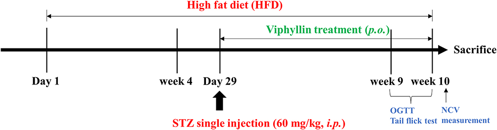 |
Figure 1 Experimental design and treatment schedule. |
Oral Glucose Tolerance Test (OGTT)
At the end of treatment, OGTT was performed in overnight fasted rats. The animals received a single dose of glucose 10% solution (1g/kg) by oral gavage. Few drops of blood samples were withdrawn from the retro-orbital plexus before (0 min) and after the glucose challenge (30, 60, 90, and 120 minutes) and glucose levels were measured using glucometer.
Tail Immersion Test
Thermal hyperalgesia in experimental rats was assessed using tail immersion test. Briefly, a temperature regulated hot water bath was used to obtain warm water at a temperature of 52 ± 0.5 °C. The rats were gently restrained using a towel, and a 5 cm-long tail was marked for exposure to warm water. The pre-marked length of the tail was swiftly submerged in warm water, and the duration of the tail immersion and the onset of the tail flick reflex were recorded using a stopwatch. To avoid tissue injury, the cut-off duration was set to 30 seconds. The trial was performed three times per rat, with a 5-minute experimental gap between trials.30
Nerve Conduction Velocity Measurement
The nerve conduction velocity (NCV) was recorded at the end of a six-week treatment period as described previously. Briefly, the rats were anaesthetized using a single intraperitoneal injection of Ketamine/xylazine (87 mg/13 mg/kg b.w.). The sciatic nerve in the right leg was exposed, and electrodes (AD Instruments, PowerLab, Bella Vista, NSW, Australia) for stimulation and recording were implanted in its proximity. The action potential at the distal end of the sciatic nerve after it was stimulated at the proximal end with square-wave pulses (duration: 0.001ms, intensity: 200mV) delivered through bipolar recording electrodes was recorded. A spacing of 5 mm was kept between the stimulation sites. The distance between the stimulation and recording electrodes, as well as the sciatic nerve’s action potential delay, were evaluated to determine the nerve conduction velocity using the following formula: NCV (m/s) is equal to distance(D)/ potential latency(L).31
Biochemical Analysis
At the end of experiment, blood was collected to separate the serum by centrifuging the samples at 1500 g for 10 minutes. The serum samples were immediately stored at −80 °C until further analysis. The samples were subsequently analyzed for total triglyceride (TG), total cholesterol (TC), low-density lipoprotein cholesterol (LDL-C), urea, blood urea nitrogen (BUN), and uric acid concentrations in a clinical chemistry analyzer (Randox RX Imola) using commercial kits.
Organ Indices
The liver and both kidneys were rapidly dissected from the visceral cavity of the rat following euthanasia and weighed to determine the organ index (mg/g).
Determination of Enzymatic Antioxidants in Sciatic Nerve Tissue
The sciatic nerve tissue samples were homogenized in phosphate buffered saline (pH 7.4) and centrifuged at 10,000 g for 15 minutes in a refrigerated centrifuge. The supernatants were used for the determination of antioxidant enzyme activities. The assays were performed using commercial kits following the manufacturer’s instructions.
Histopathology
The liver, kidney and pancreas tissue samples were fixed in 10% formalin solution. Later the paraffin-embedded sections (5 µm) were stained using hematoxylin and eosin (H&E).
Statistical Analysis
The data were statistically analyzed by one way analysis of variance (ANOVA) followed by Tukey’s test, using GraphPad Prism 9.0, and presented as mean±SD. p<0.05 were considered statistically significant.
Results
Effect of Viphyllin on Body Weight and Organ Indices in Diabetic Rats
Figure 2A shows the weekly body weight measurement of the experimental rats. The HFD-fed rats showed a considerably higher body weight gains from week 0–4 compared to normal diet fed control rats. However, the changes were not significant. Following STZ induction, the diabetic control rats showed significant decrease in the body weight from week 6 till the end of study, compared to control group (p<0.001). Viphyllin at 25, 50 and 100 mg/kg markedly improved the body weight of diabetic rats compared to the untreated diabetic control (p<0.05).
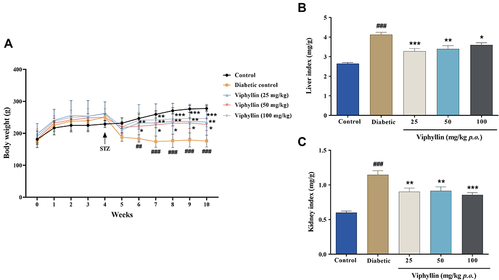 |
Figure 2 Effect of Viphyllin on body weight and organ indices in diabetic rats. (A) Weekly measurement of body weight. The data were analyzed by two-way ANOVA followed by Tukey’s test. (B and C) Changes in liver and kidney indices. The data were analyzed by one-way ANOVA followed by Tukey’s test. Values are expressed as mean±SD (n=5). #p<0.05, ##p<0.01 and ###p<0.001 vs control; *p<0.05, **p<0.01 and ***p<0.001 vs diabetic control. Abbreviations: ANOVA, analysis of variance; SD, standard deviation. |
Diabetic control rats showed a significantly higher liver (55.85%) and kidney indices (90.81%) than control group (p<0.001). However, treatment with Viphyllin at different doses markedly restored the organ indices in diabetic rats (Figure 2B). Viphyllin at 25, 50 and 100 mg/kg showed a reduction of 20.51% (p<0.001), 17.63% (p<0.01) and 12.83% (p<0.05) in the liver indices respectively compared to the untreated diabetic rats. The Viphyllin-treated groups further reduced the kidney indices to 21.20% (p<0.01), 20.14% (p<0.01) and 25.36% (p<0.001) as compared to the diabetic control rats (Figure 2C).
Effect of Viphyllin on Oral Glucose Tolerance and Fasting Blood Glucose (FBG) in Diabetic Rats
The blood glucose level (BGL) was considerably higher in the diabetic control group at time 0 and elevated to a significant extent upon administration of glucose (1 g/kg, p.o.) upto 120 min as compared to control rats (p<0.001). Viphyllin treatment at different doses reduced the BGL of diabetic rats for 120 min. Viphyllin at 50 (p<0.05) and 100 mg/kg (p<0.001) significantly reduced the BGL at 60 min of glucose administration in diabetic rats compared to the untreated diabetic control group (Figure 3A). AUC analysis revealed that Viphyllin treatment at 25, 50 and 100 mg/kg showed 5.42%, 11.77% (p<0.05) and 12.11% (p<0.05) decrease in BGL levels respectively compared to diabetic control rats (Figure 3B). As expected, there was a significantly higher FBG level in diabetic control rats at the end of study compared to control (p<0.001). Viphyllin at 100 mg/kg markedly reduced the FBG in comparison with untreated diabetic rats (p<0.05) (Figure 3C).
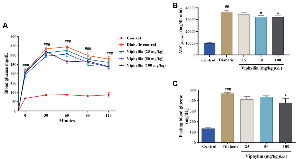 |
Figure 3 Effect of Viphyllin on glucose tolerance in diabetic rats. (A) Blood glucose concentrations measured at time 0 and after oral administration of (1 g/kg body weight) glucose. (B) Area under the curve (AUCglucose) from 0–120 min after the oral administration of glucose. (C) Fasting blood glucose at the end of study. The data were analyzed by one-way ANOVA followed by Tukey’s test. Values are expressed as mean±SD (n=5). ##p<0.05 and ###p<0.001 vs control; *p<0.05 and ***p<0.001 vs diabetic control. Abbreviations: ANOVA, analysis of variance; SD, standard deviation. |
Effect of Viphyllin on Thermal Hyperalgesia in Diabetic Model Rats
As shown in Figure 4, there was an obvious reduction in tail flick latency time observed in the diabetic rats compared to control group (p<0.001), indicating thermal hyperalgesia. However, treatment with Viphyllin at 25, 50 and 100 mg/kg dose-dependently increased the latency time compared to diabetic control group (p<0.05).
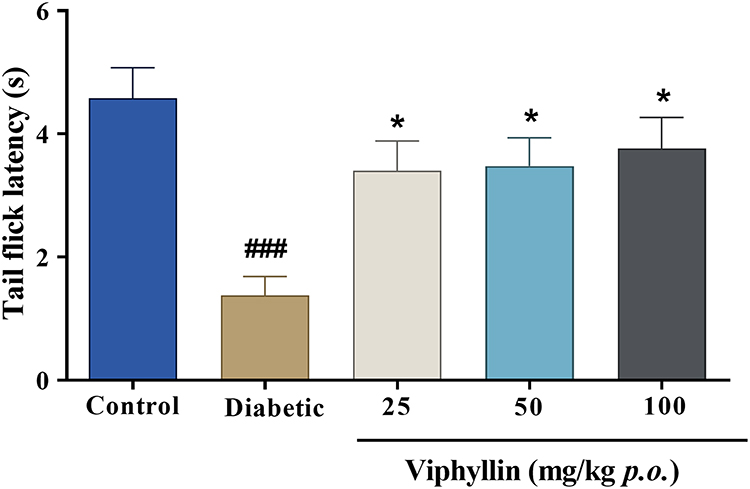 |
Figure 4 Effect of Viphyllin on tail flick latency in diabetic rats. The data were analyzed by one-way ANOVA followed by Tukey’s test. Values are expressed as mean±SD (n=5). ###p<0.001 vs control; *p<0.05 vs diabetic control. Abbreviations: ANOVA, analysis of variance; SD, standard deviation. |
Effect of Viphyllin on Nerve Conduction Velocity and Antioxidant Enzyme Activity in Sciatic Nerve Tissue
The diabetic rats showed significantly (p<0.001) reduced NCV values (25.80±1.88 m/s) compared to the control animals (55. ±6.46 m/s). A 6-week treatment with Viphyllin resulted in 12.95%, 30.70% and 71.01% increase in the NCV at 25, 50 (p<0.05) and 100 mg/kg (p<0.001) doses respectively, compared to diabetic control (Figure 5A).
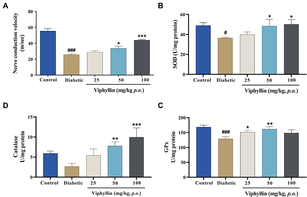 |
Figure 5 Effect of Viphyllin on nerve conduction velocity (NCV) (A) and antioxidant status (B–D) in sciatic nerve tissue. The data were analyzed by one-way ANOVA followed by Tukey’s test. Values are expressed as mean±SD (n=5). #p<0.05 and ###p<0.001 vs control; *p<0.05, **p<0.01 and ***p<0.001 vs diabetic control. Abbreviations: ANOVA, analysis of variance; SD, Standard deviation. |
The diabetic model rats showed a noticeable decrease in the SOD (p<0.05), CAT and GPx (p<0.001) enzyme levels in the sciatic nerve tissue as compared to control rats (Figure 5B–D). Viphyllin markedly restored these enzyme activities in diabetic rats. At 100 mg/kg, Viphyllin exhibited 1.37-fold (p<0.05), 3.73-fold (p<0.001) and 1.15-fold increase in SOD, CAT and GPx levels compared to untreated diabetic rats.
Effect of Viphyllin Administration on Serum Biochemical Markers in Diabetic Rats
As shown in Figure 6, there was an obvious increase in the TC (p<0.05), TG (p<0.001) and LDL-c levels of the diabetic rats compared to control group. However, Viphyllin dose-dependently reduced the TC and TG levels of diabetic rats. TC was reduced to 22.86% (p<0.05) and 31.31% (p<0.001) in 50 mg and 100 mg/kg Viphyllin treatment groups compared to diabetic control. The extract at 50 mg and 100 mg/kg significantly decreased the TG level (33.86% and 40.69% respectively) as compared to diabetic rats (p<0.001). Viphyllin treatments also reduced the serum LDL-c levels. However, the data were not significant compared to the untreated diabetic rats.
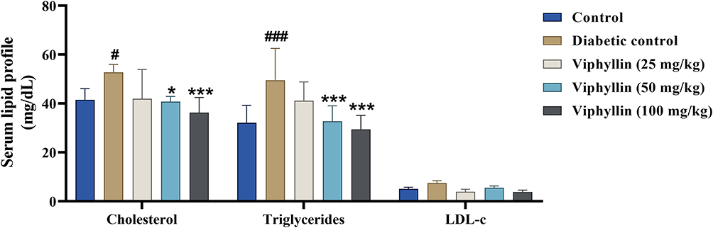 |
Figure 6 Effect of Viphyllin on serum lipid profile of diabetic rats. The data were analyzed by two-way ANOVA and presented as mean±SD (n=5). Values are mean±SD (n=5). #p<0.05 and ###p<0.001 vs control; *p<0.05 and ***p<0.001 vs diabetic control. Abbreviations: ANOVA, analysis of variance; SD, standard deviation. |
The untreated diabetic rats showed significantly higher levels of creatinine (p<0.001) and uric acid (p<0.05) compared to control rats indicating renal damage whereas Viphyllin treatment considerably reduced the renal health markers in diabetic rats (Figure 7A and B). At 100 mg/kg, Viphyllin showed 38.24% and 58.16% reduction in the serum levels of creatinine and uric acid respectively relative to untreated diabetic rats (p<0.01). Further, Viphyllin insignificantly lowered the BUN levels in diabetic rats (Figure 7C).
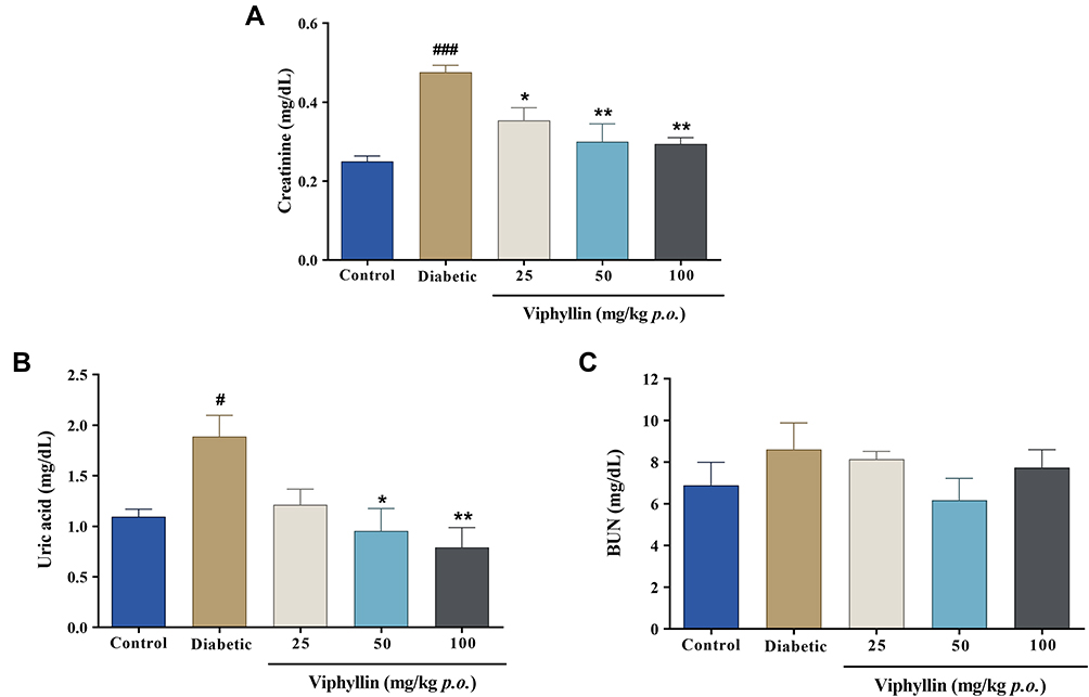 |
Figure 7 Effect of Viphyllin on serum levels of renal health markers in diabetic rats. (A) Creatinine, (B) uric acid, and (C) blood urea nitrogen. The data were analyzed by one-way ANOVA followed by Tukey’s test. Values are mean±SD (n=5). #p<0.05 and ###p<0.001 vs control; *p<0.05 and **p<0.01 vs diabetic control. Abbreviations: ANOVA, analysis of variance; SD, standard deviation. |
Effect of Viphyllin on Histopathological Changes in Liver, Kidney, and Pancreas
Liver histopathological examination revealed normal hepatocytes in control group while in diabetic control group characteristic pathological changes viz., inflammation, focal and fatty vacuolations, and biliary hyperplasia were evident. Viphyllin at 25, 50 and 100 mg/kg markedly improved the liver architecture in diabetic rats (Figure 8A). Diabetic rats showed STZ-induced changes such as glomerular atrophy, arterial fibrosis, inflammation and fatty vacuolation. On the contrary, Viphyllin treated diabetic rats showed reversal of these pathological changes to restore the normal histomorphology (Figure 8B). The control group rats showed normal architecture of pancreas with typical islets of Langerhans and acinar cells whereas the diabetic control rats exhibited pathological manifestations including swollen acinar cells, islets emptied, atrophy of islets, degenerative hyperplasia, and congestion. Interestingly, Viphyllin-treated rats showed normal architecture of pancreas (Figure 8C).
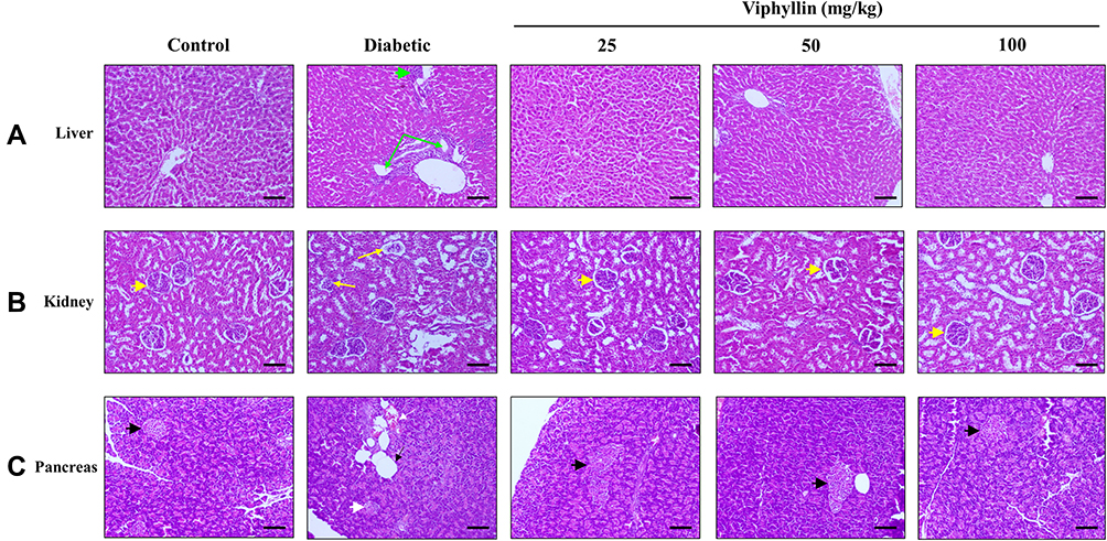 |
Figure 8 Effect of Viphyllin on histopathology of vital organs in diabetic rats. Representative images (H&E staining) showing histological changes in liver (A), kidney (B) and pancreas (C). Green arrowhead: inflammation; green arrows: biliary hyperplasia; yellow arrowhead: normal glomeruli; yellow arrows: glomerular atrophy; black arrowhead: normal islets of Langerhans; white arrowhead: atrophy of islets of Langerhans; black arrow: islets emptied; white arrow: congestion. (Magnification: ×100; scale bar: 100 µm). |
Discussion
The present study was performed to investigate the therapeutic effects of Viphyllin in an experimental model of diabetes. HFD associated with single dose of STZ has been reported previously to establish type 2 DM in experimental animals.32,33 HFD contributes to the insulin resistance while STZ induces impaired insulin secretion.34 In the present study, male Wistar rats were fed a HFD for 4 weeks followed by intraperitoneal injection of STZ at 60 mg/kg. The HFD-fed rats showed an insignificant increase in the body weight during the first 4 weeks of study while the subsequent STZ treatment resulted in weight loss and an increase in BGL in the animals compared to normal rats. However, a 6-week oral administration of Viphyllin markedly improved the body weight, and FBG in diabetic rats. Viphyllin treatment further reduced the STZ-induced increase in liver and kidney indices.
OGTT is a measure of impaired insulin sensitivity.35 In the present study, the diabetic rats exhibited significantly higher BGL for 120 min of glucose load compared to control, indicating the insulin resistance. The AUC of glucose was however reduced significantly in Viphyllin treated rats compared to the untreated diabetic control. These data clearly indicate the antihyperglycemic effects of Viphyllin.
DM associated nerve conduction deficits and neuropathic pain leads to alterations in the sensory responses.36 The change in sensation, and neuropathic pain results in hyperalgesia and allodynia.37 Several studies have pointed out the ability of plant extracts in modulating the pain threshold of neuropathic pain models.38,39 Behavioral assessment of altered nociceptive response to external stimulus is demonstrative of neuropathic changes in diabetes. In the present study, the diabetic control rats showed a decrease in latency to thermal stimulus (tail flick test) indicating hyperalgesia. On the contrary, Viphyllin treatment notably increased the tail flick latency of diabetic rats to a significant extent. The nerve conduction impairment associated with neuropathy were clear in the diabetic rats. The NCV was significantly reduced in the untreated diabetic rats compared to control whereas the Viphyllin treated rats showed improvement in the sciatic nerve conduction. The outcome of present study strongly align with the earlier reports. Previously, Laddha et al demonstrated the correlation between the antinociceptive activity of Bauhinia variegata extracts as a function of improved NCV in diabetic rats.40 In another study, treatment with an ayurvedic formulation, Triphala churna significantly increased the nociceptive response latency in diabetic model rats. Further, the authors showed that Triphala markedly increased the NCV in rats.41
Biochemically, the mammalian nerve tissues contain the phospholipids and high mitochondrial number which can generate enough free radicals to cause oxidative damage.42 Previous studies have demonstrated that a compromised antioxidant defense in the nerve tissues could contribute to the neuropathic pain in animal models.43 In line with these observations, the diabetic rats showed a reduced activity of antioxidant enzymes in the sciatic nerve compared to normal rats. Viphyllin substantially restored the antioxidant defense in the sciatic nerve tissue of rats. Thus Viphyllin-mediated increase of NCV and antioxidant activity in the sciatic nerve could attribute to its modulatory effect on peripheral sensory functions.
Diabetic complications progressively lead to renal diseases and dyslipidemia.44,45 In the present study, Viphyllin treatment improved the serum lipid profile and renal parameters in diabetic rats. The histopathological changes in the vital organs such as liver, kidney and pancreas of diabetic rats were reversed to a good extent by the treatment with Viphyllin. These data further support the antidiabetic effect of the extract.
A large body of evidence from experimental studies support the therapeutic potential of BCP, the characterized active constituent in Viphyllin.46 Previously, Suijin et al have reported that treatment of pancreatic β cells with BCP increased the glucose-stimulated insulin secretion.47 BCP as a principal component of several plant extracts has been demonstrated to exert hypoglycemic activity in diabetic rats.48 Recently, Aguilar-Ávila et al studied the effect of BCP on neuropathic pain in diabetic rats. BCP could markedly reduce the BGL while increasing the insulin levels in STZ-induced diabetic rats.49 These data strongly support the possible involvement of BCP in Viphyllin-mediated therapeutic effect in the present study.
Conclusion
Oral administration of Viphyllin significantly improved the sciatic nerve conduction and sensory responses in HFD/STZ-induced diabetic rats. In addition, the extract demonstrated antihyperglycemic effects diabetic rats. In conclusion, the present study findings suggest protective effects of Viphyllin in diabetic neuropathy.
Acknowledgment
The authors thank Dr. Muralidhar for technical assistance in the histopathological examination of tissues.
Disclosure
All the authors are employed by Vidya Herbs Pvt Ltd., and hence declare potential conflicts of interest; the authors report no other potential conflicts of interest in relation to this work.
References
1. International Diabetes Federation. IDF Diabetes Atlas.
2. Vinik AI, Nevoret M-L, Casellini C, et al. Diabetic neuropathy. Endocrinol Metab Clin North Am. 2013;42:747–787. doi:10.1016/j.ecl.2013.06.001
3. Várkonyi T, Körei A, Putz Z, et al. Advances in the management of diabetic neuropathy. Minerva Med. 2017;108:419–437. doi:10.23736/S0026-4806.17.05257-0
4. Callaghan BC, Price RS, Chen KS, et al. The importance of rare subtypes in diagnosis and treatment of peripheral neuropathy: a review. JAMA Neurol. 2015;72:1510–1518. doi:10.1001/jamaneurol.2015.2347
5. Abbott CA, Malik RA, van Ross ER, et al. Prevalence and characteristics of painful diabetic neuropathy in a large community-based diabetic population in the U.K. Diabetes Care. 2011;34:2220–2224. doi:10.2337/dc11-1108
6. Tesfaye S, Boulton AJ, Dyck PJ, et al. Diabetic neuropathies: update on definitions, diagnostic criteria, estimation of severity, and treatments [published correction appears in Diabetes Care. Diabetes Care. 2010;33:2285–2293. doi:10.2337/dc10-1303
7. von Hehn CA, Baron R, Woolf CJ. Deconstructing the neuropathic pain phenotype to reveal neural mechanisms. Neuron. 2012;73:638–652. doi:10.1016/j.neuron.2012.02.008
8. Kioskli K, Scott W, Winkley K, et al. Psychosocial factors in painful diabetic neuropathy: a systematic review of treatment trials and survey studies. Pain Med. 2019;20:1756–1773. doi:10.1093/pm/pnz071
9. Pop-Busui R, Boulton AJ, Feldman EL, et al. Diabetic neuropathy: a position statement by the American diabetes association. Diabetes Care. 2017;40:136–154. doi:10.2337/dc16-2042
10. Javed S, Alam U, Malik RA. Mirogabalin and emerging therapies for diabetic neuropathy. J Pain Res. 2018;11:1559–1566. doi:10.2147/JPR.S145999
11. Freeman R, Durso-Decruz E, Emir B. Efficacy, safety, and tolerability of pregabalin treatment for painful diabetic peripheral neuropathy: findings from seven randomized, controlled trials across a range of doses. Diabetes Care. 2008;31:1448–1454. doi:10.2337/dc07-2105
12. Wiffen PJ, Derry S, Bell RF, et al. Gabapentin for chronic neuropathic pain in adults. Cochrane Database Syst Rev. 2017;6:Cd007938. doi:10.1002/14651858.CD007938.pub4
13. Tiwari R, Siddiqui MH, Mahmood T, et al. Herbal remedies: a boon for diabetic neuropathy. J Diet Suppl. 2019;16:470–490. doi:10.1080/19390211.2018.1441203
14. Lee J, Noh S, Lim S, et al. Plant extracts for type 2 diabetes: from traditional medicine to modern drug discovery. Antioxidants. 2021;10(1):81. doi:10.3390/antiox10010081
15. Shah MA, Reanmongkol W, Radenahmad N, et al. Anti-hyperglycemic and anti-hyperlipidemic effects of rhinacanthins-rich extract from Rhinacanthus nasutus leaves in nicotinamide-streptozotocin induced diabetic rats. Biomed Pharmacother. 2019;113:108702. doi:10.1016/j.biopha.2019.108702
16. Shah MA, Khalil R, Ul-Haq Z, et al. α-Glucosidase inhibitory effect of rhinacanthins-rich extract from Rhinacanthus nasutus leaf and synergistic effect in combination with acarbose. J Funct Foods. 2017;36:325–331. doi:10.1016/j.jff.2017.07.021
17. Zhao LL, Makinde EA, Shah MA, Olatunji OJ, Panichayupakaranant P. Rhinacanthins-rich extract and rhinacanthin C ameliorate oxidative stress and inflammation in streptozotocin-nicotinamide-induced diabetic nephropathy. J Food Biochem. 2019;43(4):e12812. doi:10.1111/jfbc.12812
18. Rasoulian B, Hajializadeh Z, Esmaeili-Mahani S, et al. Neuroprotective and antinociceptive effects of rosemary (Rosmarinus officinalis L.) extract in rats with painful diabetic neuropathy. J Physiol Sci. 2019;69:57–64. doi:10.1007/s12576-018-0620-x
19. Salehi B, Zakaria ZA, Gyawali R, et al. Piper species: a comprehensive review on their phytochemistry, biological activities and applications. Molecules. 2019;24:1364. doi:10.3390/molecules24071364
20. Musenga A, Mandrioli R, Ferranti A, et al. Analysis of aromatic and terpenic constituents of pepper extracts by capillary electrochromatography. J Sep Sci. 2007;30:612–619. doi:10.1002/jssc.200600456
21. Viveros-Paredes JM, Gonzalez-Castaneda RE, Gertsch J, et al. Neuroprotective effects of beta-caryophyllene against dopaminergic neuron injury in a murine model of Parkinson’s disease induced by MPTP. Pharmaceuticals. 2017;10:60. doi:10.3390/ph10030060
22. Guo K, Mou X, Huang J, et al. Trans-caryophyllene suppresses hypoxia-induced neuroinflammatory responses by inhibiting NF-kappa B activation in microglia. J Mol Neurosci. 2014;54:41–48. doi:10.1007/s12031-014-0243-5
23. Ojha S, Javed H, Azimullah S, et al. beta-caryophyllene, a phytocannabinoid attenuates oxidative stress, neuroinflammation, glial activation, and salvages dopaminergic neurons in a rat model of Parkinson disease. Mol Cell Biochem. 2016;418:59–70. doi:10.1007/s11010-016-2733-y
24. Klauke AL, Racz I, Pradier B, et al. The cannabinoid CB2 receptor-selective phytocannabinoid beta-caryophyllene exerts analgesic effects in mouse models of inflammatory and neuropathic pain. Eur Neuropsychopharmacol. 2014;24:608–620. doi:10.1016/j.euroneuro.2013.10.008
25. Kumawat VS, Kaur G. Therapeutic potential of cannabinoid receptor 2 in the treatment of diabetes mellitus and its complications. Eur J Pharmacol. 2019;862:172628. doi:10.1016/j.ejphar.2019.172628
26. Geddo F, Scandiffio R, Antoniotti S, et al. PipeNig®-FL, a fluid extract of black pepper (Piper nigrum L.) with a high standardized content of Trans-β-caryophyllene, reduces lipid accumulation in 3T3-L1 preadipocytes and improves glucose uptake in C2C12 myotubes. Nutrients. 2019;11:2788. doi:10.3390/nu11112788
27. Sudeep HV, Venkatakrishna K, Gouthamchandra K, et al. A standardized black pepper seed extract containing β-caryophyllene improves cognitive function in scopolamine-induced amnesia model mice via regulation of brain-derived neurotrophic factor and MAPK proteins. J Food Biochem. 2021;45:e13994. doi:10.1111/jfbc.13994
28. Venkatakrishna K, Sundeep K, Sudeep HV, et al. Viphyllin, a standardized black pepper seed extract exerts antinociceptive effects in murine pain models via activation of cannabinoid receptor CB2, peroxisome proliferator-activated receptor-alpha and TRPV1 ion channels. J Pain Res. 2022;15:355–366. doi:10.2147/JPR.S351513
29. Zhang M, Lv XY, Li J, et al. The characterization of high-fat diet and multiple low-dose streptozotocin induced type 2 diabetes rat model. Exp Diabetes Res. 2008;2008:704045. doi:10.1155/2008/704045
30. Leng J, Li X, Tian H, et al. Neuroprotective effect of diosgenin in a mouse model of diabetic peripheral neuropathy involves the Nrf2/HO-1 pathway. BMC Complement Med Ther. 2020;20:126. doi:10.1186/s12906-020-02930-7
31. Wang X, Huan Y, Li C, et al. Diphenyl diselenide alleviates diabetic peripheral neuropathy in rats with streptozotocin-induced diabetes by modulating oxidative stress. Biochem Pharmacol. 2020;182:114221. doi:10.1016/j.bcp.2020.114221
32. Atanasovska E, Tasic V, Slaninka-miceska M, et al. Six-week follow-up of metabolic effects induced by a high-fat diet and streptozotocin in a rodent model of type 2 diabetes mellitus. Contributions Sec Med Sci. 2014;35:169–179.
33. Veerapur VP, Prabhakar KR, Kandadi MR, et al. Antidiabetic effect of Dodonaea viscosa aerial parts in high fat diet and low dose streptozotocin-induced type 2 diabetic rats: a mechanistic approach. Pharm Biol. 2010;48:1137–1148. doi:10.3109/13880200903527736
34. Srinivasan K, Viswanad B, Asrat L, et al. Combination of high-fat diet-fed and low-dose streptozotocin-treated rat: a model for type 2 diabetes and pharmacological screening. Pharmacol Res. 2005;52:313–320. doi:10.1016/j.phrs.2005.05.004
35. Shaham O, Wei R, Wang TJ, et al. Metabolic profiling of the human response to a glucose challenge reveals distinct axes of insulin sensitivity. Mol Syst Biol. 2008;4:214. doi:10.1038/msb.2008.50
36. Tesfaye S, Boulton AJ, Dickenson AH. Mechanisms and management of diabetic painful distal symmetrical polyneuropathy. Diabetes Care. 2013;36:2456–2465. doi:10.2337/dc12-1964
37. Didangelos T, Doupis J, Veves A. Painful diabetic neuropathy: clinical aspects. Handbook Clin Neurol. 2014;126:53–61.
38. Wang P, Wen C, Olatunji OJ. Anti-inflammatory and antinociceptive effects of Boesenbergia rotunda polyphenol extract in diabetic peripheral neuropathic rats. J Pain Res. 2022;15:779–788. doi:10.2147/JPR.S359766
39. Nawaz NUA, Saeed M, Rauf K, et al. Antinociceptive effectiveness of Tithonia tubaeformis in a vincristine model of chemotherapy induced painful neuropathy in mice. Biomed Pharmacother. 2018;103:1043–1051. doi:10.1016/j.biopha.2018.04.115
40. Laddha AP, Garud MS, Kulkarni YA. Neuroprotective effect of Bauhinia variegata Linn. leaf extracts in streptozotocin induced diabetes in Sprague Dawley rats. J Diabetes Metab Disord. 2021;20(2):1639–1645. doi:10.1007/s40200-021-00915-y
41. Suryavanshi SV, Barve K, Addepalli V, et al. Triphala churna-a traditional formulation in ayurveda mitigates diabetic neuropathy in rats. Front Pharmacol. 2021;12:662000. doi:10.3389/fphar.2021.662000
42. Areti A, Yerra VG, Naidu V, et al. Oxidative stress and nerve damage: role in chemotherapy induced peripheral neuropathy. Redox Biol. 2014;2:289–295. doi:10.1016/j.redox.2014.01.006
43. Kawaguchi M, Satoh Y, Otsubo Y, et al. Molecular hydrogen attenuates neuropathic pain in mice. PLoS One. 2014;9:e100352. doi:10.1371/journal.pone.0100352
44. Yokota K, Fukuda M, Matsui Y, et al. Visit-to-visit variability of blood pressure and renal function decline in patients with diabetic chronic kidney disease. J Clin Hypertens. 2014;16:362–366. doi:10.1111/jch.12293
45. Bhowmik B, Siddiquee T, Mujumder A, et al. Serum lipid profile and its association with diabetes and prediabetes in a Rural Bangladeshi population. Int J Environ Res Public Health. 2018;15(9):1944. doi:10.3390/ijerph15091944
46. Hashiesh HM, Meeran MFN, Sharma C, Sadek B, Kaabi JA, Ojha SK. Therapeutic potential of β-caryophyllene: a dietary cannabinoid in diabetes and associated complications. Nutrients. 2020;12:2963. doi:10.3390/nu12102963
47. Suijun W, Zhen Y, Ying G, et al. A role for trans-caryophyllene in the moderation of insulin secretion. Biochem Biophys Res Commun. 2014;444:451–454. doi:10.1016/j.bbrc.2013.11.136
48. Uddin N, Hasan MR, Hossain MM, et al. In vitro α–amylase inhibitory activity and in vivo hypoglycemic effect of methanol extract of Citrus macroptera Montr. fruit. Asian Pac J Trop Biomed. 2014;4:473–479. doi:10.12980/APJTB.4.2014C1173
49. Aguilar-Ávila D, Flores Soto M, Tapia-Vázquez C, et al. β -caryophyllene, a natural sesquiterpene, attenuates neuropathic pain and depressive-like behavior in experimental diabetic mice. J Med Food. 2019;22:460–468. doi:10.1089/jmf.2018.0157
 © 2022 The Author(s). This work is published and licensed by Dove Medical Press Limited. The
full terms of this license are available at https://www.dovepress.com/terms
and incorporate the Creative Commons Attribution
- Non Commercial (unported, 3.0) License.
By accessing the work you hereby accept the Terms. Non-commercial uses of the work are permitted
without any further permission from Dove Medical Press Limited, provided the work is properly
attributed. For permission for commercial use of this work, please see paragraphs 4.2 and 5 of our Terms.
© 2022 The Author(s). This work is published and licensed by Dove Medical Press Limited. The
full terms of this license are available at https://www.dovepress.com/terms
and incorporate the Creative Commons Attribution
- Non Commercial (unported, 3.0) License.
By accessing the work you hereby accept the Terms. Non-commercial uses of the work are permitted
without any further permission from Dove Medical Press Limited, provided the work is properly
attributed. For permission for commercial use of this work, please see paragraphs 4.2 and 5 of our Terms.