Back to Journals » Risk Management and Healthcare Policy » Volume 13
Validation of Cariogram in Caries Prediction in Women and Their Children 4 Years After Pregnancy – Longitudinal Study
Authors Dolic O ![]() , Obradovic M, Kojic Z, Trtic N, Sukara S, Knezevic N, Veselinovic V
, Obradovic M, Kojic Z, Trtic N, Sukara S, Knezevic N, Veselinovic V
Received 7 January 2020
Accepted for publication 12 May 2020
Published 9 June 2020 Volume 2020:13 Pages 549—557
DOI https://doi.org/10.2147/RMHP.S243907
Checked for plagiarism Yes
Review by Single anonymous peer review
Peer reviewer comments 2
Editor who approved publication: Dr Kent Rondeau
Olivera Dolic,1 Marija Obradovic,1 Zeljka Kojic,2 Natasa Trtic,2 Slava Sukara,1 Natasa Knezevic,3 Valentina Veselinovic4
1Department of Preventive and Pediatric Dentistry, Faculty of Medicine, University of Banja Luka, Banja Luka, Republic of Srpska, Bosnia and Herzegovina; 2Department of Periodontics and Oral Medicine, Faculty of Medicine, University of Banja Luka, Banja Luka, Republic of Srpska, Bosnia and Herzegovina; 3Department of Endodontics and Restorative Dentistry, Faculty of Medicine, University of Banja Luka, Banja Luka, Republic of Srpska, Bosnia and Herzegovina; 4Department of Prosthodontics, Faculty of Medicine, University of Banja Luka, Banja Luka, Republic of Srpska, Bosnia and Herzegovina
Correspondence: Olivera Dolic
Faculty of Medicine, University of Banja Luka, Bulevar Vojvode Petra Bojovića 1A, Banja Luka 78000, Bosnia and Herzegovina
Tel +387 51 348 131
Email [email protected]
Background: Cariogram®, an algorithm-based software model, for predicting caries risk has been used to assess the caries risk profile of many different groups. The aims of the study were to evaluate Cariogram caries risk assessment during pregnancy with DMFT/dmft incidence in mothers and their children 4 years after pregnancy and to check if there is an association between children’s caries risk profiles using Cariogram and caries risk profiles (by Cariogram) of their mothers during pregnancy.
Methods: The study population consisted of 96 pregnant women (average age 27.4± 7.2 years at baseline) who completed clinical baseline examination and salivary tests. The follow-up study was initiated 4 years later and the 80 pairs of mother and children (from that pregnancy) were re-examined using the same procedure at baseline. An individual caries risk profile and DMFT/dmft incidence were made for each woman and child. The prediction of the Cariogram was compared to the actual dental experience in 4 years. Sensitivity, specificity, positive predictive value (PPV) and negative predictive value (NPV) for two cut-offs were calculated to express the outcome.
Results: The results showed a strong association between the risk categories of pregnant women and their offspring as well as between caries development in offspring and the Cariogram risk categories of pregnant women. Sensitivity and PPV for new DMFT (ΔDMFT> 0) 4 years after for women were high (> 80%) for those participants assessed with 0– 60% “chance to avoid caries”, as well as diagnostic accuracy (74.00%). High specificity (91.00%), very high PPV (95.00%) and clinically useful values according to Youden’s index (0.53) were obtained for moderate-risk and two lowest-risk groups for dmft in children.
Conclusion: Cariogram was valid in the authors’ sample only and highly predictive in caries risk assessment in investigated children based on caries risk assessment of their mothers in pregnancy.
Keywords: specificity, early childhood caries, Cariogram, pregnant women, sensitivity, longitudinal study
Background
Caries is a multifactorial disease, and it is determined by the interaction of different microbial, genetic, immunological, behavioral, as well as environmental factors.1 The most common chronic disease in childhood is early childhood caries (EEC) that occur in children aged below 71 months. Still, it remains a huge problem both in developed and in developing countries.2,3 Data on the prevalence of Severe Early Childhood Caries (S-ECC) in Banja Luka municipality was 34.9%.3 Many studies have demonstrated the association of MS (mutans streptococci) with ECC.4–7 The systematic reviews confirmed that maternal factors influence bacterial acquisition, whereas colonization was mostly mediated by oral health and feeding habits8,9 during the first year of life. Cross-sectional10 and longitudinal11 studies have corroborated the association between parental dental status and offspring caries.
Identifying children at the greatest risk of caries to design appropriate preventive activity has been a goal of the dental profession for many years. It is expected that multiple factors, combined on an appropriate scale and accounted for possible interactions, may improve the prediction of caries risk. The challenge is to develop a really effective model for predicting caries risk. Cariogram®, an algorithm-based software, assesses an individual’s caries risk profile and contains many “if” conditions – it can operate with 5 million combinations of caries-related factors.12 The program offers recommendations for targeted preventive measures that should be implemented to avoid the formation of new caries lesions.13,14
Cariogram has been used to assess the caries risk profile of schoolchildren,15,16 teenagers, young adults,17–21 orthodontic patients22 and elderly patients.23 Some studies used Cariogram to assess the caries risk profile of preschool children.24–29 In a previous study, we presented that Cariogram can be a useful tool in caries risk assessment and identification of significant caries risk factors in a group of pregnant women,30 but to our knowledge, there are no studies that used Cariogram of pregnant women in offspring risk profile prediction.
Therefore, the aim of this study was to evaluate baseline caries risk assessment according to Cariogram during pregnancy with the DMFT/dmft development in mothers 4 years after pregnancy and their children.
The null hypothesis was that there is no association between children’s caries risk profiles by Cariogram and caries risk profiles (by Cariogram) of their mothers during pregnancy.
Methods
Study Group
This longitudinal study was performed as a follow-up of the study previously presented by Dolic et al.30 The baseline survey was carried out in 2007 (January to April) and follow-up was in 2011 (January to April). The study population consisted of 96 pregnant women who were between the ages of 20 and 42 years (average age 27, 4±7.2 years) at the start of the study. They lived in different socio-economic areas of Banja Luka, the second largest city in Bosnia and Herzegovina. Banja Luka had a total population of 170,000 in 2006. There is no official data about the number of pregnant women as well as prevalence of caries in pregnant women in that area. Pregnant women were recruited at the Pregnancy counseling center at Public Hospital Banja Luka, the only counseling center in the city. The sample size was calculated by simple random sample selection, with confidence level 95% and confidence interval 10. There is no official data on pregnant women, but 1452 children were born in 2006 in Banja Luka. Among the 213 pregnant women who were initially eligible, 96 fulfilled the inclusion criteria and were included in the study. The study included only pregnant women a) who were in the last trimester of a normal, single fetus pregnancy, b) who did not have high-risk pregnancies, c) who did not have previous medical conditions, pregnancy complications, or pregnancy-related issues that required hospitalization, d) who did not have any chronic disease, e) who had not taken antibiotics or other drugs during pregnancy, and f) who gave their written consent to participate in the research.
The follow-up study was initiated 4 years later and the mothers and their children (from that pregnancy) were re-examined by the same examiner, using the same procedure as that followed at baseline. The examiner was previously trained in using the dmft/DMFT in a group of ten 4-year-old and ten adult subjects with a wide range of levels of caries not included in the final sample. The examiner then examined a group of twenty-five 4-year-old and twenty-five adult subjects not included in the final sample twice, on successive days. Duplicate examinations performed on twenty-five 4-year-old and twenty-five adult subjects at the start of the study, about half-way through and at the end of the study. Kappa statistics were used to test the intra-investigator reliability. The kappa value for the intra-consistency of the field work investigator was >0.87, obtained by repeat examination. In order to reduce subjective bias, the researcher did not have data of the names of the patients and did not know previous caries risk by Cariogram at follow-up. The follow-up sample included 80 mother–child pairs (83.33% recall rate of women from the baseline survey), and 16 participants dropped out – 10 did not want to continue the study and 6 had moved from the area.
In Banja Luka, pregnant women and children younger than 15 years have access to free dental care at public dental clinics. At baseline and follow-up, the women were given detailed verbal and written information on the outline of the study, and they signed informed consent for themselves and for their children. During the study period, the participants were not informed of any result and they received regular dental care. Pregnant women were referred to their dentists to for necessary dental care concerning individual needs. All the participants used low natural fluoride drinking water (<0.30 mg/l).
The research has been conducted in full accordance with the 1964 Helsinki declaration, and its later amendments or comparable ethical standards and approval for the study were obtained from the Research Committee of Faculty of Medicine, University of Banja Luka (code 0602-350/07).
Clinical Examination
The study consisted of five steps: a questionnaire, interview, clinical examination, saliva sampling, and assessment of caries risk using Cariogram. Interview/questionnaire was taken only to assess the personal data needed for calculating one’s caries risk by Cariogram.30 The questionnaire contained questions on general health and medication, diet and oral hygiene habits including the use of fluoride supplements. The guardians were responsible for completing the child’s questionnaire.
All examinations were performed by one trained and calibrated examiner in the morning, between meals and at least 1 hour after a meal and after brushing the teeth to avoid possible contaminations during saliva sampling, especially for microbial tests. A plane mirror, World Health Organization Community Periodontal Index (WHO-CPI) probe, and air syringe were used in the clinical visual–tactile examination following WHO criteria.26 Clearly visible lesions with cavities on tooth surfaces were classified as dental caries (ie, d3-level cavities), whereas changes in transparency, initial enamel demineralization with intact surfaces and no cavitation were noted as intact teeth.26 The teeth were not professionally cleaned and no radiographs were taken. Caries prevalence was registered at the Decayed-Missed-Filled-Teeth (DMFT) level. Oral hygiene and amount of plaque were estimated using a mirror and periodontal probe following Silness and Löe plaque index.31
Saliva sampling consisted of measurement of the saliva secretion rate, measurements of saliva buffer capacity using Dentobuff® Strip test, measurement of mutans streptococci in the saliva using Dentocult® SM Strip mutans test and measurement of lactobacilli in the saliva using Dentocult® LB test, performed according to the the manufacturer’s instructions. Paraffin-stimulated whole saliva was collected for 5 minutes. The secretion rate was expressed as mL/min. Buffer capacity was categorized as high, medium or low. The density of colonies (Streptococcus mutans and Lactobacillus) was compared with a chart provided by the manufacturer (Orion Diagnostica, Finland).
Data for the computerized caries risk assessment were incorporated into the Cariogram to obtain caries risk profile for all participants. The following five Cariogram categories were used: “very high risk” = 0–20% “chance to avoid caries”; “high risk” = 21–40% “chance to avoid caries”; “moderate risk” = 41–60% “chance to avoid caries”; “low risk” = 61–80% “chance to avoid caries”; and “very low risk” =81–100% “chance to avoid caries”.
The patient and the patient’s ordinary dental team were not informed of the calculated Cariogram risk category. The patient’s regular dental team was responsible for restorative dental care during the entire study period.
Statistical methods
All data were processed by SPSS software (version 16.0, Chicago Ill., USA). χ2 test of contingency was used to compare the difference between groups. Kruskal–Wallis ANOVA was used for differences between baseline and follow-up DMFT/dmft between different Cariogram groups. Student’s t-test for independent samples (if the difference in the variance of the observed characteristics was not statistically significant) and nonparametric Mann–Whitney U test (if the difference in the variance of the observed characteristics was statistically significant) were used to compare the mean values of the characteristics. Likelihood ratio and Youden’s index were used for measuring diagnostic accuracy. Youden’s index was used for the evaluation of the overall discriminative power of a diagnostic procedure.
Sensitivity, specificity, positive predictive value (PPV) and negative predictive value (NPV) were calculated by two-by-two tables. Statistically significant were P values less than 0.05.
Results
Risk categories for children in regard to risk categories of their mothers during pregnancy are shown in Figure 1. The results showed that 85.00% (n = 68) of the children were in the same risk category as their mother during pregnancy (at baseline). A 10% (n=8) of the children were in higher-risk category in comparison to their mothers during pregnancy. A 5% (n=4) of the children (P < 0.001) were in decreased risk category in comparison to their mothers during pregnancy.
 |
Figure 1 Risk categories for children in regard to risk categories of their mother during pregnancy. |
The distribution of women with new DMFT in relation to the baseline Cariogram risk category, mean DMFT at baseline and 4-year follow-up, as well as mean DMFT increment are shown in Table 1. The positive relationship was statistically significant and the women in very-high-risk and high-risk categories had more DMFT than those in very-low-risk group (p<0.05). The difference in mean DMFT at the baseline and at follow-up for a very-low-risk group was not statistically significant.
 |
Table 1 Crosstabulation of DMFT Incidence at 4-Year Follow Up with Overall Caries Risk Level Predicted by Cariogram at Baseline |
Sensitivity, specificity and predictive values for Cariogram 4 years after for women are displayed in Table 2. Sensitivity (Se) and specificity (Sp) of the Cariogram were compared at two cut-off points. The moderate-risk group, low-risk and very-low-groups were taken as cut-off point 1. The moderate-, high- and very-high-risk groups were taken as cut-off point 2. When the cut-off point 2 was used as referred, the sensitivity and PPV was high (>80%) for those assessed with 0–60% “chance to avoid caries”, as well as diagnostic accuracy (74.00%). When the moderate risk and the two lowest risk groups were used as a cut-off level (point 1), sensitivity, diagnostic accuracy and NPV were lower than 60%.
 |
Table 2 Sensitivity, Specificity and Predictive Values for Cariogram Over 4 Years for Women |
The distribution of children with dmft in relation to the Cariogram risk category of mothers during pregnancy and mean dmft of children is shown in Table 3. The percentage of “∆dmft=0 and ∆dmft >0” was calculated from the total number of pregnant women in each risk category. The children of high- and very high-risk mothers during pregnancy had more dmft than those in very-low-risk group (p<0.05).
 |
Table 3 Distribution of Children with dmft in Relation to the Cariogram Risk Category of Their Mother in Pregnancy |
Sensitivity, specificity and predictive values for dmft (Δdmft >0) in children are displayed in Table 4. Sensitivity (Se) and specificity (Sp) of the baseline Cariogram (pregnant women) were compared at two cut-off points in the same way as for women. The moderate-risk group, low-risk, and very-low-groups were taken as cut-off point 1. The moderate-, high- and very-high-risk groups were taken as cut-off point 2. When the moderate risk and the two lowest risk groups were used as a cut-off level (point 1), sensitivity and PPV were higher than 82%.
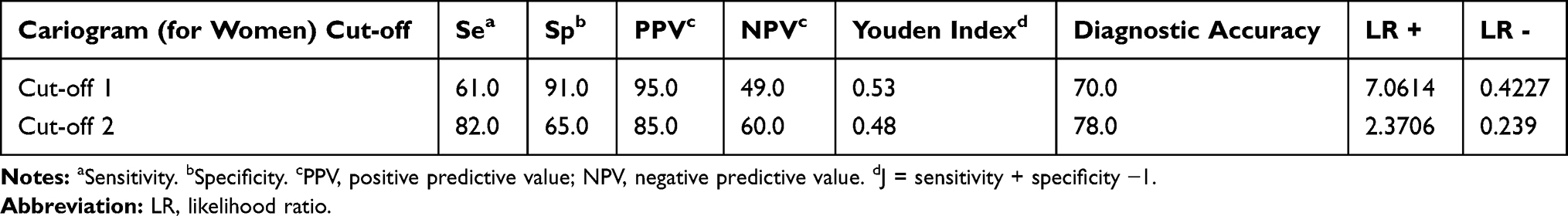 |
Table 4 Sensitivity, Specificity and Predictive Values for Cariogram in Children Compared to Baseline Mothers Risk Level |
Discussion
Oral health has been an important aspect in the well-being and quality of life in adults and in children. Untreated dental caries combined with discomfort/pain can affect weight gain, normal growth and child’s development; therefore, preventive and therapeutic measures must be based on the most current scientific and clinical knowledge available.3 Although the Health Insurance Fund of Bosnia and Herzegovina fully covers dental care for children up to the age of 15 years, the prevalence of early childhood caries is extremely high, and most of the lesions are untreated.3 In countries like Bosnia and Herzegovina, with high caries prevalence, caries risk assessment is very important, but only data on caries risk assessment in 12-year-old children19 and pregnant women is available.30
In the present study, the majority of participants (women and children) were at high risk of caries according to Cariogram. It was found that 22.50% of 4-year-old children are in a very-high-risk group and 26.25% of 4-year-old children are in high-riska group. Garg et al also showed that highest percentage of 5-year-old Indian children (66.2%) developed new caries lesions in the category of high-risk group by Cariogram.29 Stecksen-Blicks et al investigated the existing caries risk factors in 2-year-old Swedish children and showed that 51% of the sample had a low chance (or very high risk) of avoiding caries in the future.24 Study concerning Greek (2–6 years old) preschool children reported the highest number of patients in a moderate caries risk group (65%) and only 29% in a high-risk group by Cariogram.26 The study conducted in Macedonia among 4- to 5-year-old children showed that 55.10% children had a moderate dental caries risk and 40.82% children had a high dental caries risk according to Cariogram.28
It is very difficult to compare the results of Cariogram for pregnant women as no studies were conducted in pregnant women and their offspring. Study performed in children and adult populations in Turkey showed that the majority of their participants had a high risk for caries, while Celik et al reported that majority of the Turkish adults (20–21 years old) had medium and low caries risk by Cariogram (33% and 24%, respectively).32,33 In the study by Sonbul et al, the prevalence rates of caries risk in adults by Cariogram with several dental restorations in Saudi Arabia were high.34 On the other hand, there are many studies with different models for caries risk assessment focusing mother’s caries risk during pregnancy and subsequent risk of their children in the future. There have been numerous studies that have linked the presence of caries in mothers with the incidence of caries in children. The influence of maternal caries status on the same in preschool children was also confirmed in studies in Turkey,35 Thailand36 and New York,37 while it was not significant in the Japanese study.38
Factors such as low socioeconomic status, low maternal education, and unemployed mother are significant in many studies.5,39-41 In the review by Kirthiga M. et al in 2019, the important risk factors (OR>1) in high-income countries were low maternal education, smoking during pregnancy, maternal age younger than 25 years and negative parental attitudes.42 A study by Kateeb and Momany in 2018 found that older mothers and mothers who had more than one child had higher scores on DMFT as well as mothers’ beliefs in oral hygiene (OH) during pregnancy were the most important factors in their high caries experience.43
In the early caries risk assessment by Cariogram studies, researchers identified and measured various caries-related factors and correlated them to the current caries status of the individual, ie, after 1–3 years, in longitudinal studies. Data obtained from these studies often used a simple correlation for analyses. Stamm et al state that useful risk assessment program should be characterised by high simplicity, sensitivity, and specificity.44 Often, it may be very impractical to be achieved both simplicity and accuracy. But in more recent years, sensitivity/specificity, predictive powers, and Youden’s index have been applied for validation in Cariogram studies.45–47
The principle of sensitivity/specificity is to apply a cut-off value for the factor under study and to define a specific outcome of the test. In the present study, the relatively high negative predictive values are found when we used cut-off point 1 and cut-off point 2 for verification of both aims: (1) to validate baseline Cariogram caries risk with the current caries development over a 4-year period in a group of women; (2) validate caries risk classification of pregnant women according to Cariogram with the offspring actual caries development after 4 years. Similar results are found in Holgerson’s longitudinal study in Sweden, which showed that validation of a modified Cariogram in 2-year-old preschool children had a high sensitivity for future caries 5 years later, but the method was not precise and not accurate.25
The predictive ability to use Cariogram for pregnant women at both cut-off points had specificity lower than 70%, which may be categorized in the high-risk group some individuals with actual low caries risk and unnecessary preventive measures may be taken. On the other hand, according to PPV (>78%) in both cut-offs, it can be seen that a higher percentage of patients will develop caries. According to PPV (95% cut off 1, 85% cut off 2) for a possibility of caries developing in offspring if pregnant women are at a higher risk of caries according to Cariogram.
In recent studies in Hong Kong, results showed that Cariogram for preschool children generally exhibited a higher accuracy.27,48 In the present study, for validation of caries experience in children using Cariogram model of their mothers during pregnancy, accuracy was higher than 70% for both cut-offs. Also, the way of validation of Cariogram used in this study obtained clinically useful values according to Youden’s index, when moderate and two lowest risk groups have combined.
The limitations of the study are the relatively small sample size, which is a consequence of the factors for inclusion in the study and the small number of pregnant women in the region of Banja Luka. Early childhood caries is associated with many other factors not included in the Cariogram program, so this would also be the limitation of the current study. Although Cariogram requires laboratory tests, it is easily applicable, and according to research, it was valid for certain population groups.15,23,25,27,29,45,47 The main reason for use of Cariogram in this study and the strength of the study was that an important aspect is a benefit to the children, ie, early preventive measures which would be taken for children based on high risk of caries of their mothers in pregnancy.
Conclusions
With the limits of this study, Cariogram model can be a useful tool for caries prediction in both women and their children based on caries risk assessment during pregnancy. Cariogram was valid and highly predictive in caries risk assessment in a group of investigated children based on caries risk assessment of their mothers during pregnancy.
Abbreviations
EEC, early childhood caries; WHO-CPI probe, World Health Organization Community Periodontal Index; DMFT, decayed-missed-filled-teeth; Se, sensitivity; Sp, specificity; PPV, positive predictive value; NPV, negative predictive value.
Data Sharing Statement
This study is a part of the Ph.D. thesis and all data and materials (ethical approval, consent to participate and publish) can be found in official documents at the University of Banja Luka archive on the native language.
Ethics Approval and Consent to Participate
The research has been conducted in full accordance with the 1964 Helsinki declaration and its later amendments or comparable ethical standards and approval for the study was obtained from the Research Committee of Faculty of Medicine, University of Banja Luka (code 0602-350/07). All participants gave their written consent to participate in research. The guardians were responsible for the child’s written consent to participate in research.
Acknowledgments
We are grateful to Bojan Stanković for statistical analysis.
Author Contributions
All authors made substantial contributions to conception and design, acquisition of data, or analysis and interpretation of data; took part in drafting the article or revising it critically for important intellectual content; gave final approval of the version to be published; and agree to be accountable for all aspects of the work.
Disclosure
The authors declare that they have no competing interests. The authors alone are responsible for the content and writing of the paper. The authors do not hold any stocks or shares in an organization that may in any way gain or lose financially from the publication of this manuscript. The authors as well do not hold or currently applying for any patents relating to the content of the manuscript, and never had received fundings from an organization that holds or has applied for patents relating to the content of the manuscript. There are no nonfinancial competing interests (political, personal, religious, academic, ideological, intellectual, commercial or any other).
References
1. Harris R, Nicoll AD, Adair PM, Pine CM. Risk factors for dental caries in young children: a systematic review of the literature. Community Dent Health. 2004;21(1):71–85.
2. Çolak H, Dülgergil ÇT, Dalli M, Hamidi MM. Early childhood caries update: a review of causes, diagnoses, and treatments. J Nat Sci Biol Med. 2013;4:29–38.
3. Obradovic M, Dolic O, Sukara S. Caries prevalence among 24 to 71-month old children from Banja Luka. Balk J Dent Med. 2016;20(3):168–171. doi:10.1515/bjdm-2016-0027
4. Ramamurthy PH, Swamy HS, Bennete F, Rohini M, Nagarathnamma T. Relationship between severe-early childhood caries, salivary mutans streptococci, and lactobacilli in preschool children of low socioeconomic status in Bengaluru city. J Indian Soc Pedod Prev Dent. 2014;32(1):44–47. doi:10.4103/0970-4388.127054
5. Seow WK, Clifford H, Battistutta D, Morawska A, Holcombe T. Case-control study of early childhood caries in Australia. Caries Res. 2009;43(1):25–35. doi:10.1159/000189704
6. Tankkunnasombut S, Youcharoen K, Wisuttisak W, Vichayanrat S, Tiranathanagul S. Early colonization of mutans streptococci in 2- to 36-month-old Thai children. Pediatr Dent. 2009;31(1):47–51.
7. Parisotto TM, Steiner-Oliveira C, Duque C, Peres RCR, Rodrigues LKA, Nobre-dos-Santos M. Relationship among microbiological composition and presence of dental plaque, sugar exposure, social factors and different stages of early childhood caries. Arch Oral Biol. 2010;55(5):365–373. doi:10.1016/j.archoralbio.2010.03.005
8. Leong PM, Gussy MG, Barrow SWL, de Silva-sanigorski A, Waters E. A systematic review of risk factors during first year of life for early childhood caries. Int J Paediatric Dent. 2013;23(4):235–250. doi:10.1111/j.1365-263X.2012.01260.x
9. Obradović M, Dolic O, Vojinovic J, Sukara S. Association between feeding habits and severe - early childhood caries in children up to 24 month old. Serb Dent J. 2016;63(3):117–124. doi:10.1515/sdj-2016-0012
10. Dye BA, Vargas CM, Lee JJ, Magder L, Tinanoff N. Assessing the relationship between children’s oral health status and that of their mothers. J Am Dent Assoc. 2011;142(2):173–183. doi:10.14219/jada.archive.2011.0061
11. Shearer DM, Thomson WM, Caspi A, Moffitt TE, Broadbent JM, Poulton R. Family history and oral health: findings from the Dunedin Study. Community Dent Oral Epidemiol. 2012;40(2):105–115. doi:10.1111/j.1600-0528.2011.00641.x
12. Petersson GH, Twetman S, Bratthall D. Evaluation of a computer program for caries risk assessment in schoolchildren. Caries Res. 2002;36(5):327–340. doi:10.1159/000065963
13. Petersson GH. Assessing caries risk using the Cariogram model. Swe Dent J Supp. 2003;158:1–65.
14. Twetman S, Petersson GH, Bratthall D. Caries risk assessment as a predictor of metabolic control in young Type I diabetics. Diabet Med. 2005;22(3):312–315. doi:10.1111/j.1464-5491.2005.01419.x
15. Campus G, Cagetti MG, Sale S, Carta G, Lingstrom P. Cariogram validity in schoolchildren: a two-year follow-up study. Caries Res. 2012;46(1):16–22. doi:10.1159/000334932
16. Petersson GH, Isberg PE, Twetman S. Caries risk profiles in schoolchildren over 2 years assessed by Cariogram. Int J Paediatr Dent. 2010;20(5):341–346. doi:10.1111/j.1365-263X.2010.01064.x
17. Cabral RN, Hilgert LA, Faber J, Leal SC. Caries risk assessment in schoolchildren - a form based on Cariogram® software. J Appl Oral Sci. 2014;22(5):397–402. doi:10.1590/1678-775720130689
18. Hebbal M, Ankola A, Metgud S. Caries risk profile of 12 year old school children in an Indian city using Cariogram. Med Oral Patol Oral Cir Bucal. 2012;17(6):e1054–e1061. doi:10.4317/medoral.17880
19. Zukanovic A, Kobaslija S, Ganibegovic M. Caries risk assessment in Bosnian children using Cariogram computer model. Int Dent J. 2007;57(3):177–183. doi:10.1111/j.1875-595X.2007.tb00122.x
20. Campus G, Cagetti MG, Sacco G, Benedetti G, Strohmenger L, Lingström P. Caries risk profiles in Sardinian schoolchildren using Cariogram. Acta Odontol Scand. 2009;67(3):146–152. doi:10.1080/00016350902740498
21. Petersson GH, Ericson E, Isberg PE, Twetman S. Caries risk assessment in young adults using Public Dental Service guidelines and the Cariogram- a comparative study. Acta Odontol Scand. 2013;71(3–4):534–540. doi:10.3109/00016357.2012.696696
22. Al Mulla A, Kharsa S, Kjellberg H, Birkhed D. The use of Cariogram to evaluate caries-risk profiles in orthodontic patients. World J Orthod. 2010;11(2):160–167.
23. Petersson GH, Fure S, Bratthall D. Evaluation of a computer-based caries risk assessment program in an elderly group of individuals. Acta Odontol Scand. 2003;61:165–170.
24. Stecksen-Blicks C, Holgerson PL, Twetman S. Caries risk profiles in two-year-old children from northern Sweden. Oral Health Prev Dent. 2007;5(3):215–221.
25. Holgerson PL, Twetman S, Stecksèn-Blicks C. Validation of an age-modified caries risk assessment program (Cariogram) in preschool children. Acta Odontol Scand. 2009;67(2):106–112. doi:10.1080/00016350802714734
26. Kavvadia K, Agouropoulos A, Gizani S, Papagiannouli L, Twetman S. Caries risk profiles in 2- to 6-year-old Greek children using the Cariogram. Eur J Dent. 2012;6(4):415–421. doi:10.1055/s-0039-1698981
27. Gao X, Di Wu I, Lo EC, Chu CH, Hsu CY, Wong MC. Validity of caries risk assessment programmes in preschool children. J Dent. 2013;41(9):787–795. doi:10.1016/j.jdent.2013.06.005
28. Jakupi JA, Iljovska S, Naskova S, Pavlevska M, Nuhii N. Assessing the caries risk factor among children at age prom 4-5 using the cariogram program. Int J Sci Eng Res. 2015;6(11):554–562.
29. Garg A, Madan M, Dua P, et al. Validating the Usage of Cariogram in 5-and 12-year-old school-going children in paonta sahib, Himachal Pradesh, India: a 12-month prospective study. Int J Clin Pediatric Dent. 2018;11(2):110. doi:10.5005/jp-journals-10005-1495
30. Dolic O, Obradovic M, Kojic Z, Sukara S. Caries risk assessment in pregnant women using cariogram. Srp Arh Celok Lek. 2017;145(3–4):178–183. doi:10.2298/SARH160209044D
31. Loe H. The gingival index, the plaque index and the retention index systems. J Periodontol. 1967;38(6):610–616. doi:10.1902/jop.1967.38.6_part2.610
32. Gökalp SG, Dogan BG, Tekçiçek MT, Berberoglu A, Unlüer S. National survey of oral health status of children and adults in Turkey. Community Dent Health. 2010;27(1):12–17.
33. Celik EU, Gokay N, Ates M. Efficiency of caries risk assessment in young adults using Cariogram. Eur J Dent. 2012;6(03):270–279. doi:10.1055/s-0039-1698961
34. Sonbul H, Al-Otaibi M, Birkhed D. Risk profile of adults with several dental restorations using the Cariogram model. Acta Odontol Scand. 2008;66(6):351–357. doi:10.1080/00016350802325853
35. Ersin NK, Eronat N, Cogulu D, Uzel A, Aksit S. Association of maternal-child characteristics as a factor in early childhood caries and salivary bacterial counts. J Dent Child. 2006;73:105–111.
36. Thitasomakul S, Piwat S, Thearmontree A, Chankanka O, Pithpornchaiyakul W, Madyusoh S. Risks for early childhood caries analyzed by negative binomial models. J Dent Res. 2009;88(2):137–141. doi:10.1177/0022034508328629
37. Smith RE, Badner VM, Morse DE, Freeman K. Maternal risk indicators for childhood caries in an inner city population. Community Dent Oral Epidemiol. 2002;30(3):176–181. doi:10.1034/j.1600-0528.2002.300303.x
38. Kawashita Y, Fukuda H, Kawasaki K, et al. Dental caries in 3-year-old children is associated more with child-rearing behaviors than mother-related health behaviors. J Public Health Dent. 2009;69(2):104–110. doi:10.1111/j.1752-7325.2008.00107.x
39. Ostberg AL, Skeie MS, Skaare AB, Espelid I. Caries increment in young children in Skaraborg, Sweden: associations with parental sociodemography, health habits, and attitudes. Int J Paediatr Dent. 2017;27(1):47–55. doi:10.1111/ipd.12225
40. Peltzer K, Mongkolchati A, Satchaiyan G, Rajchagool S, Pimpak T. Sociobehavioral factors associated with caries increment: a longitudinal study from 24 to 36 months old children in Thailand. Int J Environ Res Public Health. 2014;11(10):10838–10850. doi:10.3390/ijerph111010838
41. Mahesh R, Muthu MS, Rodrigues SJ. Risk factors for early childhood caries: a case control study. Eur Arch Paediatr Dent. 2013;14(5):331–337. doi:10.1007/s40368-013-0089-5
42. Kirthiga M, Murugan M, Saikia A, Kirubakaran R. Risk factors for early childhood caries: a systematic review and meta-analysis of case control and cohort studies. Pediatr Dent. 2019;41(2):95–112.
43. Kateeb E, Momany E. Dental caries experience and associated risk indicators among Palestinian pregnant women in the Jerusalem area: a cross-sectional study. BMC Oral Health. 2018;18(1):170. doi:10.1186/s12903-018-0628-x
44. Stamm JW, Stewart PW, Bohannan HM, Disney JA, Graves RC, Abernathy JR. Risk assessment for oral diseases. Adv Dent Res. 1991;5(1):4–17. doi:10.1177/08959374910050010401
45. Dou L, Luo J, Fu X, Tang Y, Gao J, Yang D. The validity of caries risk assessment in young adults with past caries experience using a screening Cariogram model without saliva tests. Int Dent J. 2018;68(4):221–226. doi:10.1111/idj.12378
46. Petersson GH, Twetman S. Caries risk assessment in young adults: a 3 year validation of the Cariogram model. BMC Oral Health. 2015;15(1):17. doi:10.1186/1472-6831-15-17
47. Sudhir KM, Kanupuru KK, Nusrath F, Embeti S, Chaitra NT. Validation of cariogram as a tool for caries risk prediction among 12-year-old institutionalized children-a longitudinal follow-up study. OHDM. 2017;16(4):
48. Gao X, Hsu CY, Loh T, Hwarng B, Koh D. Role of microbiological factors in predicting early childhood caries. Pediatr Dent. 2014;36(4):348–354.
 © 2020 The Author(s). This work is published and licensed by Dove Medical Press Limited. The
full terms of this license are available at https://www.dovepress.com/terms
and incorporate the Creative Commons Attribution
- Non Commercial (unported, 3.0) License.
By accessing the work you hereby accept the Terms. Non-commercial uses of the work are permitted
without any further permission from Dove Medical Press Limited, provided the work is properly
attributed. For permission for commercial use of this work, please see paragraphs 4.2 and 5 of our Terms.
© 2020 The Author(s). This work is published and licensed by Dove Medical Press Limited. The
full terms of this license are available at https://www.dovepress.com/terms
and incorporate the Creative Commons Attribution
- Non Commercial (unported, 3.0) License.
By accessing the work you hereby accept the Terms. Non-commercial uses of the work are permitted
without any further permission from Dove Medical Press Limited, provided the work is properly
attributed. For permission for commercial use of this work, please see paragraphs 4.2 and 5 of our Terms.