Back to Journals » International Journal of Women's Health » Volume 9
Uterine sarcoma – current perspectives
Received 7 April 2017
Accepted for publication 21 July 2017
Published 31 August 2017 Volume 2017:9 Pages 597—606
DOI https://doi.org/10.2147/IJWH.S117754
Checked for plagiarism Yes
Review by Single anonymous peer review
Peer reviewer comments 2
Editor who approved publication: Professor Elie Al-Chaer
Charlotte Benson,1 Aisha B Miah1,2
1Sarcoma Unit, Royal Marsden Hospital, 2Department of Radiotherapy and Imaging, The Institute of Cancer Research, London, UK
Abstract: Uterine sarcomas comprise a group of rare tumors with differing tumor biology, natural history and response to treatment. Diagnosis is often made following surgery for presumed benign disease. Currently, preoperative imaging does not reliably distinguish between benign leiomyomas and other malignant pathology. Uterine leiomyosarcoma is the most common sarcoma, but other subtypes include endometrial stromal sarcoma (low grade and high grade), undifferentiated uterine sarcoma and adenosarcoma. Clinical trials have shown no definite survival benefit of adjuvant radiotherapy or chemotherapy and have been hampered by the rarity and heterogeneity of these disease types. There is a role of adjuvant treatment in carefully selected cases following multidisciplinary discussion at sarcoma reference centers. In patients with metastatic disease, systemic chemotherapy can then be considered. There is activity of a number of agents, including doxorubicin, trabectedin, gemcitabine-based chemotherapy, eribulin and pazopanib. Patients should be considered for clinical trial entry where possible. Close international collaboration is important to allow progress in this group of diseases.
Keywords: sarcoma, leiomyosarcoma, endometrial stromal sarcoma, undifferentiated uterine sarcoma, leiomyoma
Introduction
Soft tissue sarcomas arising from the uterus are a rare and varied group of neoplasms, all of mesenchymal origin. They can occur at any anatomical site and exhibit a wide range of behaviors, which largely depend on the histologic subtype and associated tumor grade. The incidence of uterine sarcoma is ~3%–7% of all uterine malignancies and is associated with a poor prognosis when compared to endometrial carcinoma.1 Recent results from a Surveillance Epidemiology and End Results (SEER) database analysis have shown a higher incidence rate for those aged ≥50 years compared to younger patients and twice the incidence in women of Afro-Caribbean descent compared to Caucasian women.2 The underlying etiology of this group of tumors is poorly understood, although there are putative links with raised or unopposed estrogen levels, treatment with tamoxifen, obesity and diabetes.3–5
The aims and objectives of this review were to summarize and critically appraise recently published literature concerning the clinical medical management of uterine sarcomas. To this end, a literature search has been performed using relevant search terms.
Background and histopathology
There are several distinct histopathological uterine sarcoma subtypes that can be described, and it is critical to distinguish these when considering optimal treatment pathways. Carcinosarcomas or mixed Müllerian tumors are tumors of epithelial rather than mesenchymal origin and are no longer considered as uterine sarcomas. They are treated along a carcinoma paradigm and so will not be included in this review. Smooth muscle tumors such as uterine leiomyosarcoma (ULMS) is the most common, followed by low-grade endometrial stromal sarcoma (ESS), high-grade ESS (HGESS), undifferentiated uterine sarcoma (UUS) and adenosarcoma. Other rarer soft tissue sarcoma subtypes may also arise in the uterus, including rhabdomyosarcoma, which is treated along specific pediatric rhabdomyosarcoma protocols, but are not included in this review.
Uterine sarcomas have been classified into two categories: non-epithelial and mixed non-epithelial/epithelial malignancy, dependent on the tissue of origin. The distinction between uterine sarcomas and other uterine tumors is clinically difficult and is reliant on histological and imaging features.6,7 Uterine tumors are rare mesenchymal tumors: the commonest are leiomyomas and their variants usually occur in women between the ages of 40 and 50 years. Distinction between leiomyoma and leiomyosarcoma is made with conventional morphological criteria (mitosis, atypia and necrosis). However, in certain circumstances, the criteria can be effected by hormonal status or other treatments; hence, a diagnosis of smooth muscle tumor of uncertain malignant potential (STUMP) is made when morphologically there are equivocal changes. ESSs are mesenchymal neoplasms composed of cells that morphologically resemble proliferative-phase endometrial stroma. They are classified into low-grade ESS and HGESS; a separate subtype of aggressive uterine sarcomas is classified as UUSs. High-grade uterine sarcomas (HGUSs) currently include UUS and HGESS. Uterine adenosarcomas have a benign epithelial component, whereas the stromal component is typically a low-grade sarcoma. The classification of uterine sarcomas has been further refined with the addition of immunohistochemical markers alongside morphological features to determine histological subtype (Table 1).
 | Table 1 Histological characteristics |
Diagnostic pathway
Preoperative diagnosis of uterine sarcomas remains very challenging. Presenting symptoms can be vague and nonspecific and may comprise lower abdominal or pelvic pain, abdominal distension and, most commonly, abnormal vaginal bleeding. A recent cohort study of women from Norway diagnosed with ULMS between 2000 and 2012 showed that in 52.4% patients, malignancy was not suspected at the time of surgery.8 Increasing age is a risk factor for ULMS with an incidence of diagnosis of occult ULMS of 9.8/10,000 women in the 25- to 39-year age group increasing to 33.4/10,000 women in the 50- to 64-year age group in a group of US women having laparoscopic hysterectomy for presumed fibroids.9
Common imaging modalities such as ultrasound or magnetic resonance imaging (MRI) cannot yet accurately and reliably distinguish between benign leiomyoma and malignant pathology although progress is being made.10,11 Preoperative diagnosis is essential for characterization of uterine tumors to determine the safest therapeutic strategy. Minimally invasive techniques, including laparoscopic intervention, morcellation, myomectomy and uterine artery embolization, have been developed for the treatment of uterine leiomyoma. However, preoperative differentiation between atypical leiomyoma and LMS is critical with respect to both pathology and imaging as uterine sarcoma requires a specific surgical technique to prevent dissemination. Furthermore, given the high prevalence of benign uterine pathology, the cost of performing MRI scans on all women with likely benign leiomyoma makes such screening prohibitively expensive. Unlike ovarian cancer, there are no available tumor markers or blood tests to arouse suspicion. However, work has been published looking at the use of conventional and dynamic MRI scan and serum lactate dehydrogenase, which when used in combination does help to distinguish between degenerated leiomyoma and LMS with an increase in positive predictive and negative values from 93.3% and 83.3%, respectively, to 100% and 100% in a series of 227 women.12 Most often the diagnosis is made postoperatively following histopathology review. There are certain clinical features that should raise suspicion, for instance, a rapidly growing “fibroid” in a peri- or postmenopausal woman, which would not be expected given the usual decrease in circulating estrogen levels at this time. Furthermore, where a patient has a large and heterogenous uterine mass seen on MRI scan preoperatively, then a core needle biopsy to determine pathology should be considered before planned surgery.13
Management of early stage disease
Total abdominal hysterectomy and bilateral salpingo-oophorectomy are the standards of care in the management of early stage uterine sarcomas. The risk of lymph node metastases and omental metastases is negligible, therefore avoiding the morbidity of bilateral pelvic lymphadenectomy and omentectomy. With the advent of minimally invasive techniques to remove benign uterine tumors, it is critical to ensure that these techniques are not used to remove a uterine sarcoma. Inadvertent myomectomy and/or morcellation have been reported to be a poor prognostic factor for survival.14,15 Clear risk stratification is required to minimize the risk of pelvic and peritoneal dissemination of a uterine sarcoma. Brohl et al16 reported that the incidence of sarcomatous features within a fibroid increases with age, particularly in the perimenopausal state. Preoperative diagnosis is strongly recommended in women with a rapidly enlarging fibroid in the post- or perimenopausal state with other suspicious features such as postmenopausal bleeding.
Following the diagnosis of a uterine sarcoma, management and surveillance should be undertaken in collaboration with a sarcoma reference center. Review of the operative findings, pre- and post-imaging, pathology review confirming histological subtype and endocrine status all aid the discussion to confirm staging and discuss the role of adjuvant therapy. International Federation of Gynecology and Obstetrics (FIGO) staging for uterine sarcomas has been adapted to account for specific histological subtype (Tables 2 and 3). To date, there is no evidence to support the role of adjuvant chemotherapy or adjuvant radiotherapy (RT) in all uterine sarcoma subtypes.
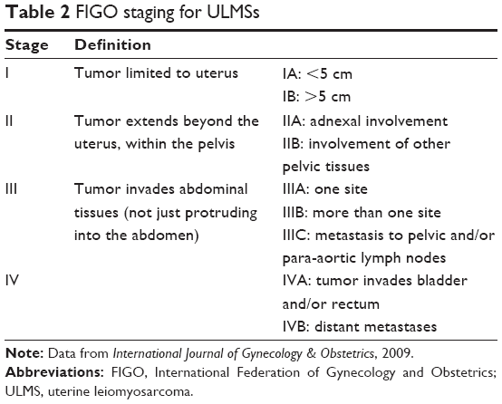 | Table 2 FIGO staging for ULMSs |
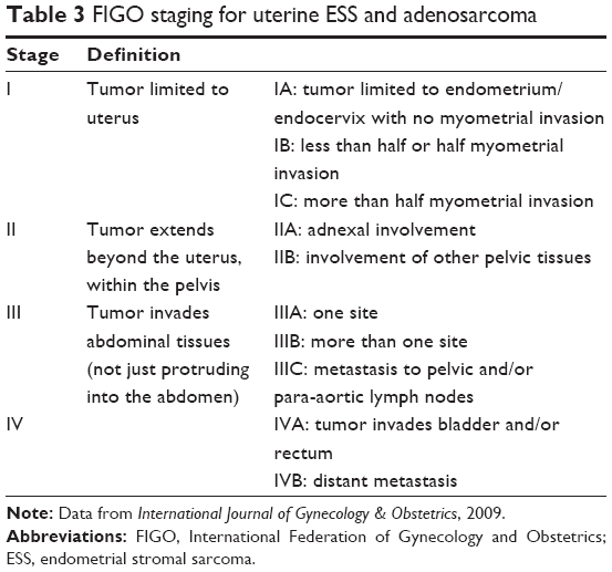 | Table 3 FIGO staging for uterine ESS and adenosarcoma |
ULMS
As described, total abdominal hysterectomy and bilateral salpingo-oophorectomy are the standards of care for early stage ULMS. The risk of lymph node metastases has been reported to be between 3% and 11%; thus, routine pelvic lymphadenectomy is not routinely indicated. The role of oophorectomy in premenopausal women is unclear. The risk of ovarian metastases has been reported at 4%, and hence, ovarian conservation may be considered on a case-by-case basis in those with endocrine receptor-negative ULMS without compromising survival outcome.17,18
An European Organisation for Research and Treatment of Cancer/Gynaecologic Cancer Group (EORTC/CGC) randomized Phase III study of RT in uterine sarcomas, comparing external beam pelvic RT with observation in FIGO stage I and II ULMS, ESS and carcinosarcoma. The study was able to report on the ULMS cohort.19 There was no benefit for adjuvant RT in this group. Isolated local recurrences occurred in 4% and 24% of patients who had RT or were observed, respectively, but any local recurrence occurred in 20% and 24% of patients, respectively. However, of those who received RT, 54% developed metastases compared with 33% in the observation group. Sampath et al reported on a cohort of 920 ULMS patients who had adjuvant RT (n=230). The addition of RT resulted in the improvement of the 5-year locoregional disease-free survival rate from 84% to 98% (P<0.01) in stage II and above disease. In a disease where there is a high risk of metastases, adjuvant pelvic RT does not have a survival benefit in LMS but may reduce local pelvic recurrences, possibly for disease that has spread beyond the uterus (FIGO stages II–IV).20 Adjuvant RT could therefore be considered in selected cases where there may be a higher risk of local recurrence.
The risk of disease recurrence following resection of organ confined that ULMS has been reported at 50%–70% at 2 years. The Gynecologic Oncology Group (GOG) conducted a randomized Phase III trial of doxorubicin compared with observation for ULMS or carcinosarcoma. Adjuvant pelvic radiation was also permitted at the clinician’s discretion. In the ULMS subgroup, a nonsignificant reduction in recurrence was reported with chemotherapy (44% in the doxorubicin arm vs 61% in the observation arm).21
A prospective study of resected ULMS evaluated the role of four cycles of adjuvant gemcitabine plus docetaxel; 45% remained disease-free at 2 years. Among the 18 women with uterus-limited disease, 59% were progression free at 2 years. Median progression-free survival (PFS) exceeded 3 years. Because of the efficacy of doxorubicin in advanced LMS, the subsequent study was designed to offer four cycles of gemcitabine plus docetaxel, followed by four cycles of doxorubicin. A total of 47 women with uterus-limited disease participated in the study. With median follow-up of 27.4 months, 78% of women remained progression free at 2 years, and median PFS was 39.3 months.22 This led to a joint GOG and EORTC Phase III study of four cycles of gemcitabine and docetaxel, followed by four cycles of doxorubicin versus observation, in resected uterus-limited LMS. Unfortunately, the study has closed early due to poor accrual (National Clinical Trial identifier NCT01533207).
ESS
Total abdominal hysterectomy and bilateral salpingo-oophorectomy are the standards of care. The risk of lymph node metastases from an ESS is reported to be <10%, again advocating the avoidance of lymphadenectomy unless suspicious lymphadenopathy is noted on preoperative imaging. An SEER database analysis of 1,010 women with ESS showed that addition of lymphadenectomy to hysterectomy did not improve either cause-specific survival or overall survival as compared with hysterectomy alone, either for “low”- or for “high”-grade disease.23
Low-grade ESS has an indolent behavior with high overall survival with episodes of recurrences, which may be amenable to resection and/or endocrine therapy. Unfortunately, because of the rarity of the disease, there are no randomized data that have evaluated the role of adjuvant RT and where retrospective series that have reported on local therapy, HGESS or HGUS and low-grade ESS have not been separated. The aforementioned SEER database study also reported that adjuvant radiation conferred no survival benefit. However, other reports have suggested an improvement in local control with adjuvant RT; Gadducci et al reported on 66 women with ESS, 26 having low-grade tumors and 40 with high-grade tumors. In the low-grade group, 31% receiving surgery alone relapsed locally, whereas none of the three women who had adjuvant RT had a local failure.24 Sampath et al showed a significant decrease in 5-year local–regional recurrence rate (8% vs 2%; P<0.05), with adjuvant RT compared with surgery alone in a retrospective analysis of 376 women with ESS; however, the data were not reported by grade.25 For early stage, low-grade tumor, the National Comprehensive Cancer Network (NCCN) consensus guidelines recommend observation alone.26 It remains uncertain whether the toxicity and long-term risks from RT can be justified by a small relative gain in local control. The decision whether to use adjuvant RT should be determined on a case-by-case basis.
Low-grade ESS has generally low response rates to conventional cytotoxic chemotherapy, and there is no evidence to support its use in the adjuvant setting. However, almost 80% of ESSs express estrogen receptor (ER) alpha and progesterone receptor (PgR), providing an opportunity for adjuvant endocrine therapy. Two small reports have demonstrated a reduction in recurrence with adjuvant endocrine therapy. Chu et al27 reported on 22 patients: recurrence rate was 31% with progestins versus 67% with observation alone. Leath et al reported on 30 patients with low-grade ESS. Median overall survival was 97 months with endocrine therapy versus 72 months with observation alone (P=0.07).28 Both these small studies suggest a possible benefit, and this provides some evidence to consider adjuvant endocrine therapy in cases at a high risk of relapse. However, there is uncertainty as to the optimal duration of endocrine therapy, and it is important to consider the potential impact on patients’ quality of life.
UUS and HGESS
UUS and high-grade endometrial sarcoma have also been reclassified by the World Health Organization (WHO) as HGUS. However, despite the different subtypes within this classification, HGUS and particularly HGESS have more aggressive behavior than low-grade ESS. The management of early stage HGUS usually includes total abdominal hysterectomy and bilateral salpingo-oophorectomy. The value of lymphadenectomy and debulking of gross extra-uterine disease remain unclear.24,28 No prospective studies of adjuvant treatments have been conducted, and where few studies have been reported, they have included both low-grade ESS and HGUS in the analysis and have provided very limited information on the value of adjuvant RT or adjuvant chemotherapy.
HGUS represents an aggressive subtype with poor outcomes regardless of the stage at presentation. A single institution series of 21 patients reported in 11 of 18 patients with complete gross resection at primary surgery presented with abdominal disease progression by the time they had undergone postoperative staging investigations.29 The patterns of relapse into the abdominal cavity and distant metastases highlight the limited role of adjuvant RT to the pelvis and the greater unmet need evaluating the role of systemic therapy.
Adenosarcoma
Adenosarcomas without sarcomatous overgrowth are indolent in behavior. In these cancers, a benign epithelial component exists together with a malignant stromal component that resembles low-grade ESS. As they are classified as low-grade malignancies, cytotoxic chemotherapy is unlikely to be beneficial, particularly in the adjuvant setting. There are no studies advocating the role of radiation treatment in this cohort of patients. Endocrine therapy also has a limited role.
Advanced disease
Owing to the often aggressive disease biology of uterine sarcomas, patients may often present late with metastatic disease in situ. Even in those with resected early stage disease, the risk of distant metastatic relapse is high, and therefore, such patients should be placed under regular radiologic surveillance.
Systemic therapy in patients with locally advanced, recurrent or metastatic uterine sarcoma is an option for those with symptomatic disease progression. Disappointingly, response rates have not improved significantly over time, nor is there any definite overall survival benefit with chemotherapy, although this view may be challenged by publication of recent clinical trial data. As with all soft tissue sarcomas, choice of treatment is histology guided and the common uterine sarcoma histologic subtypes have varied response rates to systemic treatment.30 Where entry into clinical trials is possible, this should be encouraged in order to further gain knowledge of this disease group.
Other treatment options for metastatic uterine sarcoma include judicious use of surgery, RT, radiofrequency ablation and other interventional radiology techniques. Ablative treatments can be considered for those with oligometastatic disease, ideally following a period of active surveillance and focused on those with long disease-free interval and relatively indolent disease biology. However, it is worth noting that there are little prospective data to support this approach and retrospective data have inherent selection bias. Palliative RT may be a helpful treatment option for those with symptomatic or localized disease. Decisions around treatment options should be considered by the multidisciplinary sarcoma team and involve a patient-centered approach.
ULMS
Systemic chemotherapy
ULMS is commonly viewed as one of the more chemosensitive soft tissue sarcoma subtypes. The choice of chemotherapy is along a soft tissue sarcoma paradigm, and optimal sequencing of agents remains under evaluation. Single-agent doxorubicin is standard first-line chemotherapy with response rates in the order of 25% in patients with uterine sarcoma.31 The role of single-agent anthracycline-based chemotherapy versus combination was investigated by Judson et al32 in a large randomized EORTC trial. This showed that while the response rate was higher for a combination of doxorubicin and ifosfamide (60 [26%] of 227 patients on ifosfamide/doxorubicin vs 31 [14%] of 228 patients on doxorubicin; P<0.0006), there was no overall survival benefit (median overall survival 12.8 months [95% CI 10.5–14.3] in the doxorubicin group vs 14.3 months [95% CI 12.5–16.5] in the doxorubicin and ifosfamide group; hazard ratio [HR] 0.83 [95% CI 0.67–1.03], stratified log-rank test P=0.076). Although quality of life was not incorporated to this study, rates of hospital admission with febrile neutropenia were significantly higher in the combination group. This trial has helped to elucidate the use of doublet chemotherapy, which can be considered in patients where an increased response rate is required or when symptom burden is high.
Interestingly, ifosfamide as a single agent is less widely used in ULMS following retrospective data, also from pooled EORTC trials of first-line ifosfamide, showing that those with LMS benefited less in terms of overall survival.33 A Phase II trial of single-agent ifosfamide in patients with uterine LMS showed objective response rates of 17%.34 These studies when taken into consideration with the toxicity profile of ifosfamide, the need for inpatient administration and also the number of other systemic treatment options has led to a decline in its use.
Subsequently, drug development has focused on improving on the long-held standard of single-agent doxorubicin. Following the failure of large Phase III trials of agents, including doxorubicin and palifosfamide and doxorubicin and evofosfamide to demonstrate any clinically meaningful benefit, there has been considerable interest in a novel agent olaratumab, a monoclonal antibody to platelet-derived growth factor receptor α (PDGFRα). An open-label Phase I and randomized Phase II study of olaratumab in combination with doxorubicin included a relatively high proportion of patients with LMS (uterine and non-uterine; 36% in the intervention arm and 40% in the control arm) and met its predefined end point for improvement in PFS. Median PFS in the Phase II study was 6.6 months (95% CI 4.1–8.3) with olaratumab plus doxorubicin and 4.1 months (95% CI 2.8–5.4) with doxorubicin alone (stratified HR 0.67, 0.44–1.02, P=0.0615) and also showed an unexpectedly large improvement in overall survival (median overall survival was 26.5 months [20.9–31.7 months]) with olaratumab plus doxorubicin and 14.7 months (9.2–17.1 months) with doxorubicin (stratified HR 0.46, 0.30–0.71, P=0.0003).35 The exact mechanism of action of olaratumab and how it exerts its effects in concert with doxorubicin remains undefined and is an area of considerable interest. Interestingly, PDGFR has been found to be overexpressed in ~60% of patients with uterine LMS.36 In the trial, the exploratory assay used for PDGFRα showed that while 33% and 34% of patients in the intervention and control arms, respectively, were positive for PDGFRα expression, the interaction effect between expression and treatment was not significant for overall survival or PFS. The randomized, double-blind, Phase III trial of doxorubicin ± olaratumab (ANNOUNCE) with stratification for LMS has now completed accrual, and its outcome is awaited with interest.
The antimetabolite gemcitabine is also active in ULMS, either as a single agent or in combination. A Phase II GOG trial of single-agent gemcitabine showed an overall response rate (ORR) of 20.5% with median duration of response of 4.9 months.37 The combination of gemcitabine and docetaxel is particularly active with ORRs quoted in the order of 25%–53%.38,39 However, this regimen does have significant toxicity, namely, alopecia, myelosuppression requiring growth factor support and fatigue. The combination of gemcitabine and docetaxel has recently been compared in the first-line setting in the UK GEDDIS trial for patients with metastatic/locally advanced soft tissue sarcoma where it was found to be non-inferior to doxorubicin but more toxic and was given with more dose delays.40 The addition of the vascular-targeted agent bevacizumab to gemcitabine docetaxel failed to add any clinical benefit.41 Gemcitabine in combination with dacarbazine is also an active combination and may be utilized where high-dose steroids need to be avoided or where hair loss is preferred to be avoided.42 Dacarbazine as a single agent also has modest activity in ULMS and may be used beyond second line but toxicities such as nausea, fatigue and myelosuppression can be problematic.
Trabectedin is a cytotoxic agent derived from the marine tunicate Ecteinascidia turbinata. Its mechanism of action is complex but in the main is thought to be through binding to the minor groove of DNA, leading to inhibition of transcription factors, which in turn results in cell-cycle arrest. Trabectedin is also thought to have immunomodulatory effects both on the tumor microenvironment and on tumor-associated macrophages.43 A prospective Phase II GOG study of previously untreated ULMS patients on trabectedin showed an ORR of 10%, disease control rate (DCR) of 60% and a median PFS of 5.8 months. One of the advantages of trabectedin is that it is well tolerated with little cumulative toxicity – adverse effects include neutropenia, fatigue and transient alterations of liver functions. Subsequently, a Phase II study of trabectedin and doxorubicin by the French Sarcoma Group in patients with LMS of gynecologic or soft tissue origin showed response rate of 59.6% and 27% SD with a DCR of 87.2% in the ULMS group.44 A retrospective pooled analysis of five Phase II trials showed ORR 16%, DCR 51% and median PFS of 3.3 months.45 These results have been confirmed in a recently reported large Phase III study of trabectedin compared with dacarbazine in patients with LMS and liposarcoma.46 In clinical practice, the possibility of prolonged disease stability and a generally good toxicity profile makes trabectedin a good second line option for many patients with ULMS.
Targeted agents
Currently, the only licensed targeted agent is the oral multi-targeted tyrosine kinase inhibitor pazopanib, which has activity against VEGF-1–3 and PDGFRα. The Phase III, placebo-controlled, double-blind PALETTE trial in patients with non-adipocytic soft tissue sarcoma following doxorubicin showed a significant increase in PFS (4.6 months vs 1.6 months; P<0.001); ORR was 6%, but there was no statistically significant improvement in OS.47 A subsequent analysis of patients with uterine sarcoma in the PALETTE trial showed similar response rates to the overall population.48
Trials of other oral targeted agents have thus far proven disappointing, for example the multi-targeted drug sunitinib that failed to achieve stabilization or objective responses in ULMS and sorafenib with response rates too low for further investigation.49,50 More recently, there have been early reports of clinical benefit to the oral CDK4/6 inhibitor palbociclib in a patient with ULMS.51
Hormonal therapies
Immunohistochemical expression of ER and PgR in ULMS is variable and may have prognostic significance with those patients with a high level of hormonal expression reported to have disease that behaves in a more indolent manner.52 This expression may be targeted for therapeutic benefit along a similar paradigm to breast cancer therapy. However, it is important to note that tamoxifen is not advised in this setting due to partial agonist activity. Retrospective data from our own institution of aromatase inhibitors used in the first- and second-line setting showed a clinical benefit rate of 62.5% in the first line and 50% in the second line, with patients with low-grade disease faring better when compared to those with high grade.53 There is only one prospective Phase II study of aromatase inhibitors in ULMS with primary end point of PFS at 12 weeks.54 While no objective responses were seen, stable disease was recorded in 14 out of 27 patients and PFS at 12 weeks of 50%, and median duration of treatment was only 2.2 months. This therapeutic approach has the benefit of being relatively well tolerated, especially when compared to chemotherapy and is recommended in patients with more indolent disease patterns and lower burden of disease.
Immunotherapy
Immunotherapy is an area of considerable area of interest in sarcoma, given significant gains seen in other tumor types such as malignant melanoma and renal cell cancer. Treatment strategies include manipulation of immune checkpoints such as CTLA-4, PD-1 and PDL-1 vaccination and adoptive immunotherapy. It is also interesting to combining immunotherapy and RT, thus harnessing the abscopal effect. PD-1 and PD-L1 expressions have been reported in soft tissue sarcoma in 105 patients with a variety of sarcoma subtypes including LMS.55 Some early data have shown activity of both nivolumab and pembrolizumab in LMS and have spawned a number of trials as monotherapy and in combination with metronomic chemotherapy.56,57
Low-grade ESS
This disease type often has very indolent disease biology and relapses are often late. Those patients with a low volume of metastatic disease may be offered the option of radiologic surveillance as a first option. Patients with oligometastatic disease can be considered for localized approaches such as surgery, RT or radiofrequency ablation. The hormonal receptors ER and PgR are frequently expressed and may be exploited for therapeutic benefit with response rates to aromatase inhibitors with a clinical benefit rate of 92.4% and a 2-year progression free rate of 88.9% in our own institution.58 Following radiologic and symptomatic disease progression on first-line hormonal treatment, then a switch to steroidal aromatase inhibitors such as exemestane or to progestogens is advised. The development of endocrine resistance involves the mammalian target of rapamycin (mTOR) pathway, and adding an mTOR inhibitor such as sirolimus to hormone treatment may reverse hormonal resistance.59 Systemic chemotherapy may be considered on failure of hormonal treatment, although response rates are low in this subtype. There is some evidence to support the use of trabectedin.60
HGUS
This is an aggressive disease subtype (HGESS and UUS) with universally poor outcomes. These tumors are often undifferentiated neoplasms with low response rates to conventional chemotherapy that is offered along a soft tissue sarcoma paradigm as outline earlier. These patients where fit should be considered for clinical trials of novel agents. A recent EORTC trial is investigating the use of maintenance with cabozantinib, an oral agent with activity against VEGFR and MET, for those patients with HGUS who have responded systemic chemotherapy (NCT01979393).
Adenosarcoma
In patients with recurrent or metastatic disease and sarcomatous overgrowth, treatment is generally along a sarcoma paradigm, although supportive evidence is sparse. Where there is hormone receptor expression, then a hormonal approach may be considered. There are published reports of the activity of trabectedin in this subtype.61
Conclusion and future perspectives
It is clear that in this challenging group of diseases, early recognition and diagnosis of uterine sarcoma are critical in order to improve patient outcomes. This will involve ongoing education and sharing of knowledge between colleagues working in the sarcoma field, primary care and the gynecological community. Patients should be referred to sarcoma centers, ideally before planned surgery so that multimodal measures may be considered as well as entry into appropriate clinical trials. So far, surgery remains the only option for cure, and it is important that the correct approach is made, avoiding morcellation and unnecessary procedures such as lymphadenectomy and omentectomy. Thus far, adjuvant therapies are not of proven benefit, and the most recent clinical trial between EORTC and GOG closed early due to lack of accrual. It is important to include patient groups in devising future trials that are acceptable to all participants.
For those with metastatic disease, there remains a challenge of drug development in rare tumor types. This requires close cooperation between industry and also multicenter groups and careful choice of clinically meaningful trial end points, consideration of quality of life measures and appropriate imaging modalities. There remains hope that further progress in understanding of tumor biology and associated pathways, continued development of targeted novel therapies and immunotherapy will improve outcomes of patients with this group of rare diseases.
Acknowledgments
We acknowledge Dr Khin Thway, consultant histopathologist, Sarcoma Unit, The Royal Marsden Hospital and The Institute of Cancer Research, London, for providing summary Table 1. We acknowledge National Health Service funding to the NIHR Biomedical Research Centre.
Disclosure
The authors report no conflicts of interest in this work.
References
D’Angelo E, Prat J. Uterine sarcomas: a review. J Gynecol Oncol. 2010;116(1):131–139. | ||
Hosh M, Antar S, Nazzal A, Warda M, Gibreel A, Refky B. Uterine sarcoma: analysis of 13,089 cases based on surveillance, epidemiology, and end results database. Int J Gynecol Cancer. 2016;26(6):1098–1104. | ||
Schwartz SM, Weiss NS, Daling JR, et al. Exogenous sex hormone use, correlates of endogenous hormone levels, and the incidence of histologic types of sarcoma of the uterus. Cancer. 1996;77(4):717–724. | ||
Arenas M, Rovirosa A, Hernández V, et al. Uterine sarcomas in breast cancer patients treated with tamoxifen. Int J Gynecol Cancer. 2006;16(2):861–865. | ||
Felix AS, Cook LS, Gaudet MM, et al. The etiology of uterine sarcomas: a pooled analysis of the epidemiology of endometrial cancer consortium. Br J Cancer. 2013;108(3):727–734. | ||
Hanley KZ, Birdsong GG, Mosunjac MB. Recent developments in surgical pathology of the uterine corpus. Arch Pathol Lab Med. 2017;141(4):528–541. | ||
Santos P, Cunha TM. Uterine sarcomas: clinical presentation and MRI features. Diagn Interv Radiol. 2015;21(1):4–9. | ||
Skorstad M, Kent A, Lieng M. Preoperative evaluation in women with uterine leiomyosarcoma. A nationwide cohort study. Acta Obstet Gynecol Scand. 2016;95(11):1228–1234. | ||
Rodriguez AM, Asoglu MR, Sak ME, Tan A, Borahay MA, Kilic GS. Incidence of occult leiomyosarcoma in presumed morcellation cases: a database study. Eur J Obstet Gynecol Reprod Biol. 2016;197:31–35. | ||
Cornfeld D, Israel G, Martel M, Weinreb J, Schwartz P, McCarthy S. MRI appearance of mesenchymal tumors of the uterus. Eur J Radiol. 2010;74(1):241–249. | ||
Thomassin-Naggara I, Dechoux S, Bonneau C, et al. How to differentiate benign from malignant myometrial tumours using MR imaging. Eur Radiol. 2013;23(8):2306–2314. | ||
Goto A, Takeuchi S, Sugimura K, Maruo T. Usefulness of Gd-DTPA contrast-enhanced dynamic MRI and serum determination of LDH and its isozymes in the differential diagnosis of leiomyosarcoma from degenerated leiomyoma of the uterus. Int J Gynecol Cancer. 2002;12(4):354–361. | ||
Wilkinson MJ, Martin JL, Khan AA, Hayes AJ, Thomas JM, Strauss DC. Percutaneous core needle biopsy in retroperitoneal sarcomas does not influence local recurrence or overall survival. Ann Surg Oncol. 2015;22(3):853–858. | ||
Skorstad M, Kent A, Lieng M. Uterine leiomyosarcoma – incidence, treatment, and the impact of morcellation. A nationwide cohort study. Acta Obstet Gynecol Scand. 2016;95(9):984–990. | ||
Lieng M, Berner E, Busund B. Risk of morcellation of uterine leiomyosarcomas in laparoscopic supracervical hysterectomy and laparoscopic myomectomy, a retrospective trial including 4791 women. J Minim Invasive Gynecol. 2015;22(3):410–414. | ||
Brohl AS, Li L, Andikyan V, et al. Age-stratified risk of unexpected uterine sarcoma following surgery for presumed benign leiomyoma. Oncologist. 2015;20(4):433–439. | ||
Major FJ, Blessing JA, Silverberg SG, et al. Prognostic factors in early-stage uterine sarcoma. A gynecologic oncology group study. Cancer. 1993;71(4 suppl):1702–1709. | ||
Kapp DS, Shin JY, Chan JK. Prognostic factors and survival in 1396 patients with uterine leiomyosarcomas: emphasis on impact of lymphadenectomy and oophorectomy. Cancer. 2008;112(4):820–830. | ||
Reed NS, Mangioni C, Malmstrom H, et al; European Organisation for Research and Treatment of Cancer Gynaecological Cancer Group. Phase III randomised study to evaluate the role of adjuvant pelvic radiotherapy in the treatment of uterine sarcomas stages I and II: an European organisation for research and treatment of cancer gynaecological cancer group study (protocol 55874). Eur J Cancer. 2008;44(6):808–818. | ||
Sampath S, Schultheiss TE, Ryu JK, Wong JY. The role of adjuvant radiation in uterine sarcomas. Int J Radiat Oncol Biol Phys. 2010;76(3):728–734. | ||
Omura GA, Blessing JA, Major F, et al. A randomized clinical trial of adjuvant adriamycin in uterine sarcomas: a gynecologic oncology group study. J Clin Oncol. 1985;3(9):1240. | ||
Hensley ML, Wathen JK, Maki RG, et al. Adjuvant therapy for high-grade, uterus-limited leiomyosarcoma: results of a phase 2 trial (SARC 005). Cancer. 2013;119(8):1555–1561. | ||
Barney B, Tward JD, Skidmore T, Gaffney DK. Does radiotherapy or lymphadenectomy improve survival in endometrial stromal sarcoma? Int J Gynecol Cancer. 2009;19(7):1232–1238. | ||
Gadducci A, Landoni F, Sartori E, et al. Uterine leiomyosarcoma: analysis of treatment failures and survival. Gynecol Oncol. 1996;62(1):25–32. | ||
Sampath S, Hitchcock YJ, Shrieve DC, Randall RL, Schultheiss TE, Wong JY. Radiotherapy and extent of surgical resection in retroperitoneal soft-tissue sarcoma: multi-institutional analysis of 261 patients. Surg Oncol. 2010;101(5):345–350. | ||
National Comprehensive Cancer Network (NCCN) [webpage on the Internet]. NCCN Clinical Practice Guidelines in Oncology. 2017. Available from: http://www.nccn.org/professionals/physician_gls/f_guidelines.asp. Accessed July 21, 2017. | ||
Chu MC, Mor G, Lim C, Zheng W, Parkash V, Schwartz PE. Low-grade endometrial stromal sarcoma: hormonal aspects. Gynecol Oncol. 2003;90:170–176. | ||
Leath CA 3rd, Huh WK, Hyde J Jr, et al. A multi-institutional review of outcomes of endometrial stromal sarcoma. Gynecol Oncol. 2007;105(3):630–634. | ||
Tanner EJ, Garg K, Leitao MM Jr, Soslow RA, Hensley ML. High grade undifferentiated uterine sarcoma: surgery, treatment, and survival outcomes. Gynecol Oncol. 2012;127(1):27–31. | ||
Ray-Coquard I, Rizzo E, Blay JY, et al. Impact of chemotherapy in uterine sarcoma (UtS): review of 13 clinical trials from the EORTC soft tissue and bone sarcoma group (STBSG) involving advanced/metastatic UtS compared to other soft tissue sarcoma (STS) patients treated with first line chemotherapy. Gynecol Oncol. 2016;142(1):95–101. | ||
Omura GA, Major FJ, Blessing JA, et al. A randomized study of adriamycin with and without dimethyl triazenoimidazole carboxamide in advanced uterine sarcomas. Cancer. 1983;52(4):626–632. | ||
Judson I, Verweij J, Gelderblom H, et al; European Organisation and Treatment of Cancer Soft Tissue and Bone Sarcoma Group. Doxorubicin alone versus intensified doxorubicin plus ifosfamide for first-line treatment of advanced or metastatic soft-tissue sarcoma: a randomised controlled phase 3 trial. Lancet Oncol. 2014;15(4):415–423. | ||
Sleijfer S, Ouali M, van Glabbeke M, et al. Prognostic and predictive factors for outcome to first-line ifosfamide-containing chemotherapy for adult patients with advanced soft tissue sarcomas: an exploratory, retrospective analysis on large series from the European organization for research and treatment of cancer-soft tissue and bone sarcoma group (EORTC-STBSG). Eur J Cancer. 2010;46(1):72–83. | ||
Sutton GP, Blessing JA, Barrett RJ, McGehee R. Phase II trial of ifosfamide and mesna in leiomyosarcoma of the uterus: a gynecologic oncology group study. Am J Obstet Gynecol. 1992;166(2):556–559. | ||
Tap WD, Jones RL, Van Tine BA, et al. Olaratumab and doxorubicin versus doxorubicin alone for treatment of soft-tissue sarcoma: an open-label phase 1b and randomised phase 2 trial. Lancet. 2016;388(10043):488–497. | ||
Anderson SE, Nonaka D, Chuai S, et al. p53, epidermal growth factor, and platelet-derived growth factor in uterine leiomyosarcoma and leiomyomas. Int J Gynecol Cancer. 2006;16(2):849–853. | ||
Look KY, Sandler A, Blessing JA, Lucci JA 3rd, Rose PG. Phase II trial of gemcitabine as second-line chemotherapy of uterine leiomyosarcoma: a gynecologic oncology group (GOG) study. Gynecol Oncol. 2004;92(2):644–647. | ||
Hensley ML, Blessing JA, Mannel R, Rose PG. Fixed-dose rate gemcitabine plus docetaxel as first-line therapy for metastatic uterine leiomyosarcoma: a gynecologic oncology group phase II trial. Gynecol Oncol. 2008;109(3):329–334. | ||
Maki RG, Wathen JK, Patel SR, et al. Randomized phase II study of gemcitabine and docetaxel compared with gemcitabine alone in patients with metastatic soft tissue sarcomas: results of sarcoma alliance for research through collaboration study 002. J Clin Oncol. 2007;25(19):2755–2763. | ||
Seddon BM, Whelan J, Strauss SJ, et al. GeDDiS: a prospective randomised controlled phase III trial of gemcitabine and docetaxel compared with doxorubicin as first-line treatment in previously untreated advanced unresectable or metastatic soft tissue sarcomas (EudraCT 2009-014907-29). J Clin Oncol. 2015;33(15_suppl):10500. | ||
Hensley ML, Miller A, O’Malley DM, et al. Randomized phase III trial of gemcitabine plus docetaxel plus bevacizumab or placebo as first-line treatment for metastatic uterine leiomyosarcoma: an NRG oncology/gynecologic oncology group study. J Clin Oncol. 2015;33(10):1180–1185. | ||
García-Del-Muro X, López-Pousa A, Maurel J, et al; Spanish Group for Research on Sarcomas. Randomized phase II study comparing gemcitabine plus dacarbazine versus dacarbazine alone in patients with previously treated soft tissue sarcoma: a Spanish group for research on sarcomas study. J Clin Oncol. 2011;29(18):2528–2533. | ||
D’Incalci M, Galmarini CM. A review of trabectedin (ET-743): a unique mechanism of action. Mol Cancer Ther. 2010;9(8):2157–2163. | ||
Pautier P, Floquet A, Chevreau C, et al; French Sarcoma Group. Trabectedin in combination with doxorubicin for first-line treatment of advanced uterine or soft-tissue leiomyosarcoma (LMS-02): a non-randomised, multicentre, phase 2 trial. Lancet Oncol. 2015;16(4):457–464. | ||
Cesne AL, Judson I, Maki R, et al. Trabectedin is a feasible treatment for soft tissue sarcoma patients regardless of patient age: a retrospective pooled analysis of five phase II trials. Br J Cancer. 2013;109(7):1717–1724. | ||
Demetri GD, von Mehren M, Jones RL, et al. Efficacy and safety of trabectedin or dacarbazine for metastatic liposarcoma or leiomyosarcoma after failure of conventional chemotherapy: results of a phase III randomized multicenter clinical trial. J Clin Oncol. 2016;34(8):786–793. | ||
van der Graaf WT, Blay JY, Chawla SP, et al. Pazopanib for metastatic soft-tissue sarcoma (PALETTE): a randomised, double-blind, placebo-controlled phase 3 trial. Lancet. 2012;379(9829):1879–1886. | ||
Benson C, Ray-Coquard I, Sleijfer S, et al. Outcome of uterine sarcoma patients treated with pazopanib: a retrospective analysis based on two European Organisation for Research and Treatment of Cancer (EORTC) soft tissue and bone sarcoma group (STBSG) clinical trials 62043 and 62072. Gynecol Oncol. 2016;142(1):89–94. | ||
Hensley ML, Sill MW, Scribner DR Jr, et al. Sunitinib malate in the treatment of recurrent or persistent uterine leiomyosarcoma: a Gynecologic Oncology Group phase II study. Gynecol Oncol. 2009;115(3):460–465. | ||
Maki RG, D’Adamo DR, Keohan ML, et al. Phase II study of sorafenib in patients with metastatic or recurrent sarcomas. J Clin Oncol. 2009;27(19):3133–3140. | ||
Elvin JA, Gay LM, Ort R, et al. Clinical benefit in response to palbociclib treatment in refractory uterine leiomyosarcomas with a common CDKN2A alteration. Oncologist. 2017;22(4):416–421. | ||
Ioffe YJ, Li AJ, Walsh CS, et al. Hormone receptor expression in uterine sarcomas: prognostic and therapeutic roles. Gynecol Oncol. 2009;115(3):466–471. | ||
Thanopoulou E, Thway K, Khabra K, Judson I. Treatment of hormone positive uterine leiomyosarcoma with aromatase inhibitors. Clin Sarcoma Res. 2014;4:5. | ||
George S, Feng Y, Manola J, et al. Phase 2 trial of aromatase inhibition with letrozole in patients with uterine leiomyosarcomas expressing estrogen and/or progesterone receptors. Cancer. 2014;120(5):738–743. | ||
Kim JR, Moon YJ, Kwon KS, et al. Tumor infiltrating PD1-positive lymphocytes and the expression of PD-L1 predict poor prognosis of soft tissue sarcomas. PLoS One. 2013;8(12):e82870. | ||
Paoluzzi L, Cacavio A, Ghesani M, et al. Response to anti-PD1 therapy with nivolumab in metastatic sarcomas. Clin Sarcoma Res. 2016;6:24. | ||
George S, Miao D, Demetri GD, et al. Loss of PTEN is associated with resistance to anti-PD-1 checkpoint blockade therapy in metastatic uterine leiomyosarcoma. Immunity. 2017;46(2):197–204. | ||
Thanopoulou E, Aleksic A, Thway K, Khabra K, Judson I. Hormonal treatments in metastatic endometrial stromal sarcomas: the 10-year experience of the sarcoma unit of Royal Marsden Hospital. Clin Sarcoma Res. 2015;5:8. | ||
Martin-Liberal J, Benson C, Messiou C, Fisher C, Judson I. Reversion of hormone treatment resistance with the addition of an mTOR inhibitor in endometrial stromal sarcoma. Case Rep Med. 2014;2014:612496. | ||
Le Cesne A, Cresta S, Maki RG, et al. A retrospective analysis of antitumour activity with trabectedin in translocation-related sarcomas. Eur J Cancer. 2012;48(16):3036–3044. | ||
Schroeder BA, Rodler ET, Loggers ET, Pollack SM, Jones RL. Clinical benefit of trabectedin in uterine adenosarcoma. Med Oncol. 2013;30(2):501. |
 © 2017 The Author(s). This work is published and licensed by Dove Medical Press Limited. The
full terms of this license are available at https://www.dovepress.com/terms
and incorporate the Creative Commons Attribution
- Non Commercial (unported, 3.0) License.
By accessing the work you hereby accept the Terms. Non-commercial uses of the work are permitted
without any further permission from Dove Medical Press Limited, provided the work is properly
attributed. For permission for commercial use of this work, please see paragraphs 4.2 and 5 of our Terms.
© 2017 The Author(s). This work is published and licensed by Dove Medical Press Limited. The
full terms of this license are available at https://www.dovepress.com/terms
and incorporate the Creative Commons Attribution
- Non Commercial (unported, 3.0) License.
By accessing the work you hereby accept the Terms. Non-commercial uses of the work are permitted
without any further permission from Dove Medical Press Limited, provided the work is properly
attributed. For permission for commercial use of this work, please see paragraphs 4.2 and 5 of our Terms.