
Back to Journals » International Journal of Women's Health » Volume 17
Unlocking Success: The Optimal Gonadotrophin Starting Point for Luteal-Phase Stimulation in IVF Patients with Normal Responses
Authors Nie X  , Wang Q, Xu M, Dai R, Pang B , Hu C
Received 14 May 2025
Accepted for publication 12 September 2025
Published 19 September 2025 Volume 2025:17 Pages 3119—3131
DOI https://doi.org/10.2147/IJWH.S540342
Checked for plagiarism Yes
Review by Single anonymous peer review
Peer reviewer comments 2
Editor who approved publication: Dr Matteo Frigerio
Xinyu Nie,1,2 Qi Wang,1,3 Mengmeng Xu,4 Rulin Dai,1 Bo Pang,5 Cong Hu1
1Reproductive Medicine Center, Prenatal Diagnosis Center, First Hospital of Jilin University, Changchun, Jilin, People’s Republic of China; 2Department of Gynecology, Chongming Hospital Affiliated to Shanghai University of Medicine and Health Sciences, Shanghai, People’s Republic of China; 3Obstetrics and Gynecology Center, First Hospital of Jilin University, Changchun, Jilin, People’s Republic of China; 4Department of Rehabilitation medicine, First Hospital of Jilin University, Changchun, Jilin, People’s Republic of China; 5Department of Cardiology, The First Hospital of Jilin University, Changchun, Jilin, People’s Republic of China
Correspondence: Bo Pang, Department of Cardiology, The First Hospital of Jilin University, Changchun, Jilin Province, 130000, People’s Republic of China, Email [email protected] Cong Hu, Reproductive Medicine Center, Prenatal Diagnosis Center, First Hospital of Jilin University, Changchun, Jilin Province, 130000, People’s Republic of China, Email [email protected]
Purpose: To analyze and discover the optimal gonadotrophin (Gn) starting point for luteal-phase stimulation (LPS) in in vitro fertilization (IVF) Patients with normal ovarian response (NOR).
Patients and Methods: A total of 199 IVF or intracytoplasmic sperm injection (ICSI) cycles performed at the Reproductive Center of the First Hospital of Jilin University from January 1, 2021, to December 31, 2023, were recruited in the study. Of these, 121 cycles followed the follicular phase ovarian stimulation (FPS) (119 cases of adding antagonists), while 78 cycles adhered to the LPS protocol. The LPS group was divided into three subgroups based on the timing of Gn initiation: early, mid-, and late LPS. Comparisons were made between the LPS and FPS groups, as well as among the three subgroups, regarding Gn duration, total Gn dose, number of oocytes retrieved, number of embryos, rate of good-quality embryos, blastocyst formation rate, clinical pregnancy rate, cumulative pregnancy rate, live birth rate and cumulative live birth rate.
Results: The rate of high-quality embryos in the late luteal phase was significantly higher than that in the early luteal group and slightly higher than that in the mid-luteal group. The clinical pregnancy rate and live birth rate of the late luteal group were slightly higher than those of the early and mid-luteal group, and were not significantly different from those of the FPS group.
Conclusion: Initiating Gn stimulation during the late luteal-phase appears to be the optimal timing in NOR patients.
Keywords: luteal phase ovarian stimulation, follicular phase ovarian stimulation, normal ovarian response, timing of gonadotropin initiation, pregnancy outcome
Introduction
Approximately one in six couples of reproductive age were diagnosed with infertility globally.1 Intense societal pressures have normalized late marriage and delayed childbearing. Factors such as advanced age, environmental pollution, and unhealthy lifestyle habits jeopardize human reproductive health, leading to an increasing number of infertile couples each year.2 With the growing use of in vitro fertilization-embryo transfer (IVF-ET) and the ongoing refinement of controlled ovarian stimulation (COS) protocols, luteal phase support strategies and laboratory techniques, the cumulative live-birth rate (CLBR) following IVF has improved significantly.3 Traditional COS protocols include the gonadotropin-releasing hormone agonist (GnRH-a), microstimulation/mild stimulation, and gonadotropin-releasing hormone antagonist (GnRH-A) protocols. Recent research has increasingly focused on progestin-primed ovarian stimulation (PPOS) protocols, such as luteal phase ovarian stimulation (LPS) and its variants—follicular phase PPOS, flexible PPOS (fPPOS), and double stimulation protocols.
The conventional theory is follicles recruit from the late luteal phase of the preceding menstrual cycle to the early follicular phase of the current cycle.4 To promote the synchronized development of multiple follicles, supraphysiological doses of gonadotropins (Gn) are typically administered during the early follicular phase. This approach aims to minimize the need for repeated ovarian stimulation and surgical oocyte retrieval, resulting in a higher yield of usable oocytes. Reproductive gynecologists have been refining ovulation protocols for nearly 50 years due to the risk that high estrogen levels can trigger an early luteinizing hormone (LH) peak during ovarian stimulation, which can cause premature ovulation or affect oocyte quality. The classic long protocol addresses this issue by using GnRH-a to prevent early LH peaks, called controlled ovarian stimulation (COS). GnRH-a works by initially causing a brief “flare effect” before desensitizing the pituitary gland.5 However, this medication has a delayed onset and can lead to significant discomfort for patients due to severe downregulation, increased initial doses, and prolonged ovulation duration, resulting in both economic and emotional burdens. The GnRH-A protocol, which blocks the GnRH receptor on the pituitary gland to rapidly inhibit LH release during the late stage of ovulation, is effective but costly. Therefore, there is a pressing need for new ovulation protocols that better mimic physiological conditions and effectively prevent early LH peaks. In 2003, Baerwald et al introduced the follicular wave theory, which discovered that 2–3 waves of follicle development can occur within a single menstrual cycle in 50 women of reproductive age.6 Based on this theory, the LPS protocol was first successfully implemented for cancer patients who needed to preserve their fertility before undergoing chemotherapy. There was no significant difference in the number of oocytes retrieved, oocyte viability, or fertilization rate between the LPS protocol and follicular phase ovarian stimulation (FPS).7,8 In 2015, Kuang et al proposed the follicular phase PPOS protocol, in which exogenous progesterone (P) was administered to inhibit LH secretion during the follicular phase, with simultaneous Gn stimulation.9 Exogenous progesterone administration during the follicular phase or high endogenous progesterone levels during the luteal phase could inhibit the formation of early-onset LH peaks through negative feedback regulation, and there were no significant differences in the numbers of oocytes retrieved and good-quality embryos compared with those of the FPS group.10,11 While both PPOS and LPS protocols are effective in preventing early-onset LH peaks in non-downregulated COS protocols, a drawback is that they necessitate frozen-thawed embryo transfers (FET). This requirement arises because the endogenous or exogenous progesterone used in these protocols can adversely affect the endometrium, causing it to develop out of sync with the embryos.
In recent years, with the pandemic of novel coronavirus pneumonia and poor patient mobility, PPOS protocols have been used in many reproductive centers due to their convenient oral administration, low cost, and high flexibility. However, compared with mainstream protocols, such as agonist and antagonist protocols, the PPOS protocol focused more on the treatment of patients with poor ovarian responses and less on patients with normal ovarian responses. Available evidence has shown that, in patients with normal ovarian reserve function, although the number of oocytes retrieved was significantly lower with the follicular phase PPOS protocol than with the GnRH-a protocol, there was no significant difference in clinical pregnancy or miscarriage rates between the two protocols.12 The rates of preterm births, abnormal newborn birth weights, and congenital malformations were similar between the two protocols.13 Perinatal and neonatal outcomes after the transplantation of euploid blastocysts obtained using the LPS protocol were not significantly different from those obtained using FPS.14 Compared with the short and antagonist protocols, there were no statistically significant differences in the rates of preterm births, low birth weights, neonatal mortalities, congenital anomalies, or physical development and health at three years of life in the high P status ovulation protocol.15,16 Based on these studies, the safety of the PPOS protocol is considered comparable to that of traditional protocols. However, larger prospective clinical trials are needed to further validate its long-term safety and efficacy. Additionally, the PPOS protocol should be optimized and tailored to specific patient characteristics and application strategies to enhance its clinical value and effectiveness in assisted reproductive therapies.
In this study, we aimed to provide reliable evidence to guide the selection of an optimal COS protocol for patients with NOR by comparing clinical and laboratory parameters and pregnancy outcomes after the application of FPS and LPS protocols in a population with NOR. Although the LPS protocol has become a viable alternative to traditional FPS, current research has only explored the optimal timing for Gn administration in the LPS protocol for patients with poor ovarian response (POR).17 However, there is currently no clear definition of the optimal Gn administration strategy for the LPS protocol in patients with NOR. This study is the first to compare three subgroups according to the different initiation times of Gn in the LPS protocol, in order to seeks to identify the most effective time window for Gn initiation within the LPS protocol, with a view to improving the efficiency of ovulation induction and optimizing clinical outcomes.
Materials and Methods
Study Design
Women who underwent IVF or ICSI at the Reproductive Center of the First Hospital of Jilin University from January 1, 2021, to December 31, 2023, were retrospectively analyzed. Patients were categorized into FPS group and LPS group Gn initiation according to their menstrual cycle at the time of visit, with the LPS group further divided into three subgroups based on Gn initiation timing: early, mid-, and late luteal.
Inclusion criteria for the study were: 1) couples undergoing their first IVF or ICSI cycle at the center during the specified period; 2) ovulation induction using either the FPS (flexible antagonist protocol) or LPS protocol; 3) age between 20 and 45 years; 4) transfer of 2 cleavage stage embryos or 1 blastocyst stage embryo.
Patients who meet any of the following criteria were excluded: 1) chromosomal abnormalities in one or both spouses; 2) endocrine system disorders, including diabetes mellitus, pituitary microadenomas, thyroid dysfunction, or hyperprolactinemia; 3) patients with tuberculosis of the reproductive system, tubal effusion/pus, or other systemic underlying diseases; 4) patients with recurrent pregnancy loss;18 5) abnormalities of the uterine cavity, including congenital uterine abnormalities, uterine adhesions, submucous fibroids, thin endometrium, or chronic endometritis; 6) diminished ovarian reserve (AFC < 5, AMH< 1.2 ng/mL), ≤3 or> 20 oocytes retrieved in the COS cycle (those not meeting these criteria are defined as having normal ovarian response); and 7) not the first FET cycle.
Treatment Protocols
The luteal phase was confirmed by transvaginal ultrasound examination of the dominant follicle after ovulation or by monitoring elevated serum P levels and initiation of ovulation stimulation with menotrophin (Ledebao, Livzon, China), recombinant human follicle-stimulating hormone (Gonal-F, Merck, Italy), or urofollitropin (Lishenbao, Livzon, China) at a dosage range of 150–300 IU/day. For those who underwent ovulation monitoring, the dominant follicle discharge was regarded as D0; D0–D4 (or ≥11 days to the next menstrual period, with P levels of 1–3 ng/mL) was defined as the early luteal group; D5–D9 (6–10 days to the next menstrual period, with P levels of 5–25 ng/mL) was defined as the mid-luteal group; and D10–D16 (0–5 days to the next menstrual period, with P levels of <5 ng/mL) was defined as the late luteal group. When LH levels reached basal values, 0.25 mg per day of cetrorelix acetate (Cetrotide, Fareva Pau, Germany) was subcutaneously administered until the trigger day.
The FPS protocol was initiated using the same standards. Subcutaneous injection of cetrorelix acetate once per day until the trigger day was initiated when ultrasound monitoring revealed a maximal follicle diameter of >12 mm, LH levels were elevated to more than 1.5 times the baseline value, or E2 level >600 pg/mL.
The dosage of Gn and the timing of the trigger were determined based on the patient’s hormone levels, follicle size, and duration of Gn use. When one of the dominant follicles reached 18 mm or three reached 17 mm in diameter, 250 μg recombinant human chorionic gonadotropin (r-HCG, Ovidrel, Merck Serono S.p.A, Italy) or 0.1 mg GnRH-a (Diphereline, Ipsen Pharma Biotech, France) with 250 μg r-HCG was administered subcutaneously, and the oocytes were retrieved under ultrasonographic guidance 35–37 h later.
Embryo Culture, Preservation, and Resuscitation
IVF or ICSI was selected based on the semen quality. The size, number, evenness and fragmentation rate of cleavage balls were observed on days 1–6. FET was performed after whole-embryo freezing or ≥1 month following follicle retrieving cycle. The embryos were thawed using a vitrification-thawing solution kit (Kato, Japan, VT102), and 2 cleavage stage embryos or one blastocysts were transferred per FET cycle.
Embryo Transfer and Luteal Support
Patients with regular or irregular menstruation underwent a natural or hormone replacement cycle, respectively. Progesterone (Huangtitong, Zhejiang Xianju, China) at 40 mg/day or progesterone sustained-release vaginal gel (Crinone, Merck, Italy) was used for endometrial transformation. Human chorionic gonadotrophin test was performed on the 14th day after transfer. If results were positive, the original dose of estrogen and progesterone was maintained, and the dose was gradually reduced until 7–8 weeks of gestation and stopped at 11–12 weeks of gestation.
Statistical Analysis
The evaluation of the data utilized Student’s t-test for continuous variables that followed a normal distribution, while the Mann–Whitney U-test was applied to those with a non-normal distribution. Results are reported as the mean with standard deviation (SD). For categorical variables, Pearson’s chi-square test or Fisher’s exact test was employed as appropriate, and these results are expressed as percentages. Statistical analyses were conducted using the Statistical Package for the Social Sciences (version 27, SPSS Inc). A P-value of less than 0.05 was deemed statistically significant.
Results
Originally, based on the inclusion criteria, 357 cycles were enrolled (220 cycles were assigned to the FPS group, and 137 cycles were assigned to the LPS group). Following the exclusion criteria, 99 cycles were excluded from the FPS group and 59 cycles were excluded from the LPS group. Ultimately, 199 cycles met the inclusion and exclusion criteria, with 121 cycles in the FPS group and 78 cycles in the LPS group (Figure 1).
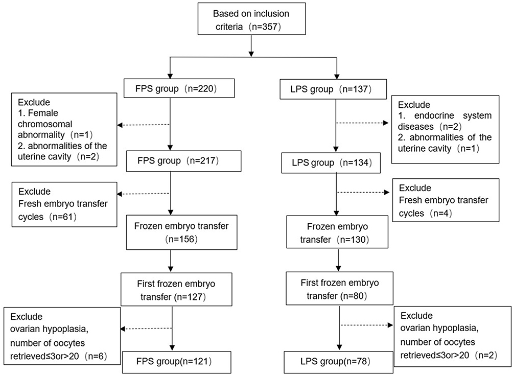 |
Figure 1 Flow chart of the study. Abbreviations: FPS, follicular phase ovarian stimulation; LPS, luteal-phase ovarian stimulation. |
LPS Group Shows Higher Gn Dose, Improved Embryo Quality, but Lower High-Quality Blastocysts Ratio Compared to FPS Group
The age, body mass index (BMI), infertility factors, years of infertility, fertilization mode, and basal levels of FSH, LH, E2, or P showed no significant differences between the FPS and LPS groups (P>0.05, Supplementary Table 1). The Gn initiation dose was similar for both protocols, but the duration and total Gn dose were significantly higher in the LPS group (P<0.001). Luteal atrophy can diminish the negative feedback regulation of LH by progesterone. In the LPS group, 45 of 78 patients received prophylactic antagonists to suppress early LH peaks during the late ovarian stimulation phase; the antagonist dose and trigger day LH and E2 levels were significantly lower compared to the FPS group (P<0.001), with no early ovulation observed in either group. The retrieved oocytes, good-quality embryos, MII oocyte rate and the rate of good-quality embryos was significantly higher in the LPS group (P<0.05). However, the FPS group had significantly more follicles >14 mm, and a ratio of good-quality blastocysts (P<0.05) (Table 1). Subgroup analysis revealed that for IVF cycles, the LPS group had a higher MII oocyte rate and the rates of good-quality embryos compared to the FPS group (P<0.05), but, good-quality blastocyst ratios were similar (P>0.05). After ICSI, there were no significant differences between the groups in terms of retrieved oocytes, MII oocytes, 2PN, and available embryos. However, the FPS group had significantly more good-quality blastocysts and a higher rate of good-quality blastocysts, whereas the rate of good-quality embryos in the LPS group was significantly higher than that in the FPS group (P<0.001) (Supplementary Table 2). Thus, it is suggested that the differences in laboratory indicators between the two protocols are not related to the fertilization method, and the LPS protocol shows advantages in several reproductive indicators, such as higher MII oocyte and good-quality embryo rates, which provides a certain reference basis for clinically selecting the optimal ovulation stimulation protocol.
 |
Table 1 Demographic Characteristics and IVF Cycle-Related Variables of the FPS and LPS Groups |
Clinical Pregnancy and Live Birth Rates Were Similar in LPS and FPS Groups
To investigate differences in pregnancy outcomes between the two protocols, we compared 121 cycles of the first frozen embryo transfer performed using the FPS protocol and 78 cycles of the first frozen embryo transfer performed using the LPS protocol. Although the ongoing pregnancy rate was lower in the LPS group than in the FPS group (P<0.05), there were no significant differences in the biochemical pregnancy rate, clinical pregnancy rate, ectopic pregnancy rate, miscarriage rate, cumulative pregnancy rate, preterm labor rate, live birth rate, cumulative live birth rate, and neonatal birth weight between the two groups (P>0.05), and there was no deliveries of congenital malformations in either group (Table 2). Therefore, the LPS protocol may offer advantages in certain reproductive indicators, such as the rates of MII oocytes and high-quality embryos. However, its overall effect on important clinical indicators, such as clinical pregnancy rate, cumulative pregnancy rate and live birth rate, did not show a significant difference compared with that of the antagonist protocol.
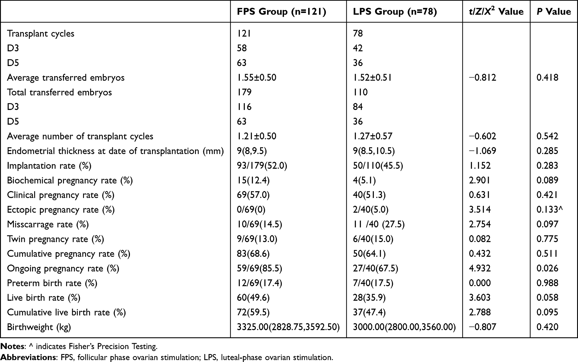 |
Table 2 Comparison of Pregnancy Outcomes Between FPS and LPS Groups |
One-way and multifactorial logistic regression analyses were performed for female age, BMI, years of infertility, infertility factors, fertilization mode, basal hormone levels (FSH, LH, E2, and P), Gn initiation dose, total Gn dose, Gn duration, number of antagonists, and ovarian stimulation protocols. Age (odds ratio [OR]=0.909, 95% confidence interval [CI]: 0.849–0.973, P<0.001) and infertility (odds ratio [OR]=0.535, 95% confidence interval [CI]: 0.288–0.996, P<0.05) were independent factors that influenced the live birth rate. After adjusting for confounding factors, there was no significant difference in live birth rates between the FPS and LPS protocols. The results indicate that the LPS protocol yields a higher rate of high-quality embryos than the FPS protocol and is comparable to the FPS protocol in terms of the live birth rate. Whether the LPS protocol can be adopted as a standard ovarian stimulation regimen requires comprehensive assessment by clinicians based on the specific circumstances of each patient.
Superior Laboratory Parameters in Late Luteal Group
To clarify whether differences in the timing of Gn initiation contributed to the differences between the LPS and FPS protocol groups, the LPS protocol group was divided into the early, mid-, and late LPS subgroups based on the timing of Gn initiation. The baseline information is shown in Supplementary Table 3. The clinical indicators were compared between the groups, and the Gn initiation dose criteria were consistent. The duration of Gn was significantly higher in the mid-luteal and late luteal groups than in the FPS group (P< 0.001). The total Gn dose was significantly higher in the mid-luteal and late luteal groups than in the early luteal and FPS groups; it was higher in the early luteal group than in the FPS group (P< 0.001); and there was no statistically significant difference in the mid-luteal compared with the late luteal group.
To determine the most favorable luteal phase induction time for patients, we compared the LH, E2, and P levels at various time points following Gn administration (Figure 2). Statistical analysis revealed that, although none of the four groups experienced premature ovulation or cycle cancellation due to early-onset LH peaks, the use of antagonists was significantly higher in the late luteal and FPS groups compared to the early and mid-luteal groups; the late luteal group also had a significantly higher usage than the FPS group (P<0.001). On days 7–9 of Gn administration, dominant follicles often had diameters ≥14 mm, necessitating the addition of antagonists in non-downregulated protocols. In this study, antagonist addition ratios for the early, mid-, and late luteal groups, and the FPS group were 15/29, 11/27, 19/22, and 119/121, respectively. We observed that LH levels on the day of HCG administration in the FPS group were significantly higher than those in the early, mid-, and late luteal groups (P<0.01), with a statistically significant difference between the FPS and mid-luteal phase groups in post-hoc comparisons. These results suggest that due to the feedback inhibitory effect of the physiological corpus luteum on LH release, only minimal amounts of antagonists are required in LPS protocols to prevent premature ovulation. Comparing E2 levels among the four groups, the mid- and late luteal phase groups had significantly lower E2 levels throughout the stimulation cycle compared to the FPS group, with E2 levels on the trigger day also significantly lower in all three LPS subgroups than in the FPS group. This indicates that, under high progesterone levels, such as during the luteal peak and post-peak periods, ovulation stimulation significantly reduces serum E2 levels throughout the stimulation cycle, potentially decreasing the incidence of OHSS and improving patients’ physiological tolerance, comfort, and compliance during the treatment.
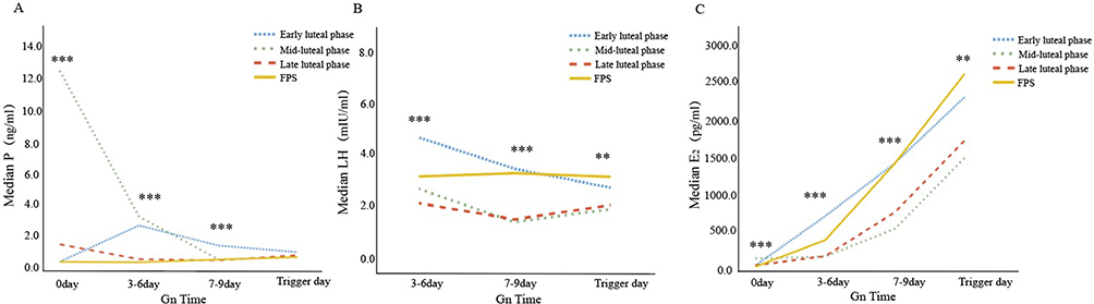 |
Figure 2 Comparison of hormone levels in Gn of different ovulation promotion programs. Abbreviations: Gn, gonadotropin; FPS, follicular phase ovarian stimulation; LH, luteinizing hormone; E2, estradiol; P, progesterone. Notes: (A) Median LH at different Gn times in early luteal, mid-luteal, late luteal and FPS groups; (B) Median E2 at different Gn times among the four groups; (C) Median P at different Gn times among the four groups. ** indicates P<0.01; *** indicates P<0.001. |
Although laboratory indicators differed between the various luteal periods of ovulation and the FPS group, the late LPS group demonstrated more favorable rates of MII oocytes and high-quality embryos (P<0.05). There was no significant difference between the late LPS group and the FPS group in terms of blastocyst formation rate and the ratio of high-quality blastocysts. Additionally, the timing of Gn administration, total Gn dose, and number of antagonists required were not significantly increased compared to the other two groups. Shown in Table 3. These findings hold substantial implications for the optimization of clinical ovulation induction protocols.
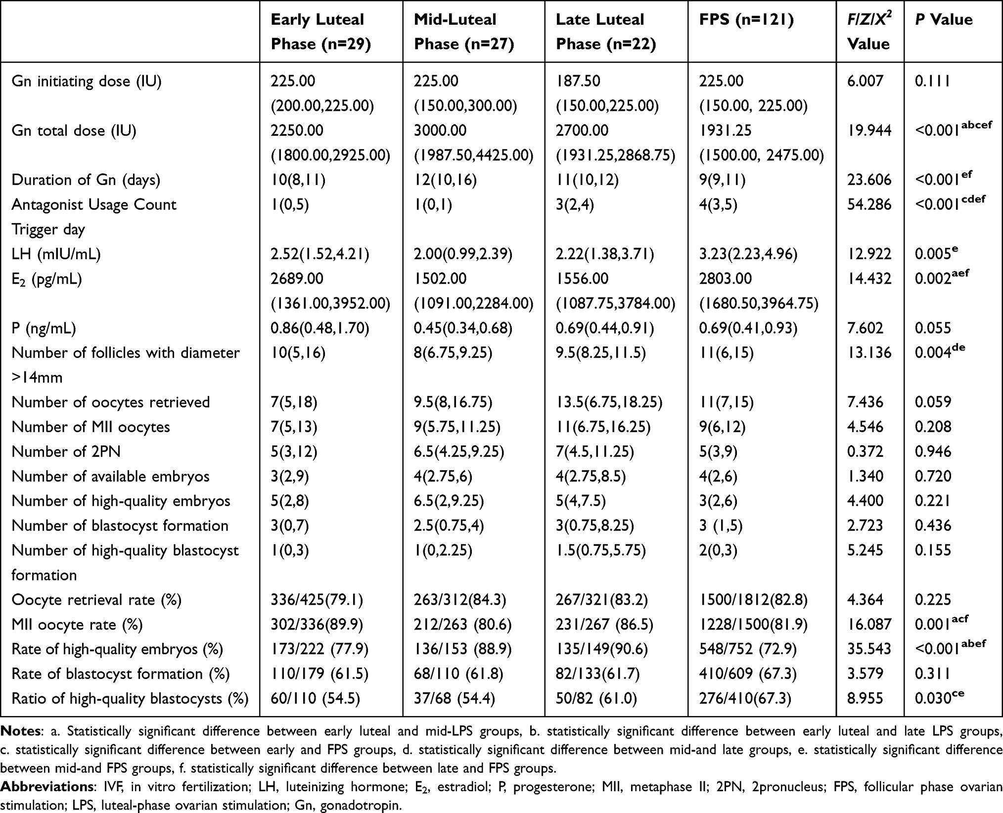 |
Table 3 Demographic Characteristics and IVF Cycle-Related Variables in Different Luteal Phase Stimulation and FPS Groups |
Late LPS Group Shows Similar Pregnancy Outcome Indicators Compared to FPS Group
Given that there were no statistical differences in the baseline data, the clinical and laboratory indicators in the late luteal group were slightly higher than those in the early and mid-luteal groups and comparable to those in the FPS group. Regarding the primary outcome measures of pregnancy, the embryo implantation rate in the FPS group was not significantly different from that in the late luteal group but was significantly higher than that in the early and mid-luteal groups (P<0.01). Although the clinical pregnancy, cumulative pregnancy, live birth and cumulative live birth rates in the late luteal group were slightly higher than those in the early and mid-luteal groups, the differences were not statistically significant (P>0.05). The biochemical pregnancy rates of the late luteal group were higher than those of the early and mid-luteal groups, and the miscarriage rate was higher in the mid-luteal group than in the other two groups, however, the differences were not statistically significant (P>0.05). There was no significant difference in the birth weight of the newborns in each group, and no neonatal developmental malformations occurred in any of the groups (Table 4). Furthermore, the initiation of Gn in the late LPS protocol is closer to the menstrual period and has a median Gn duration that is 2 days longer than that of the FPS protocol, along with lower E2 levels. This likely contributed to the successful pregnancies observed in 3 of the 4 patients undergoing late LPS fresh-cycle transfers in this study. In women with normal ovarian reserve, initiating Gn in the late luteal phase of the LPS protocol may achieve similar clinical pregnancy and live birth rates as the FPS protocol, with the additional benefits of enabling fresh embryo transfers and reducing cycle waiting time.
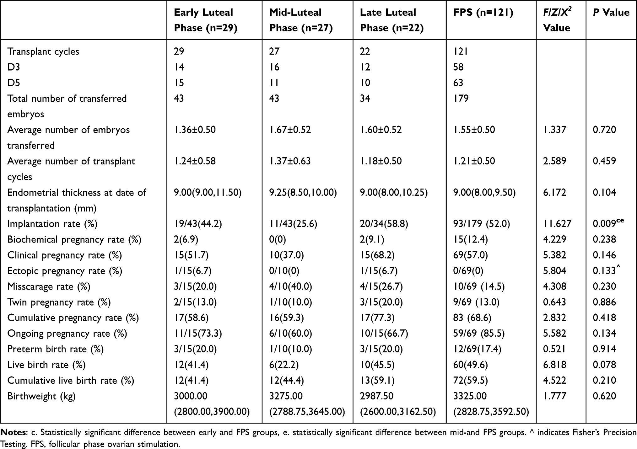 |
Table 4 Comparison of Pregnancy Outcomes in Different Luteal Phase Stimulation and FPS Groups |
Discussion
Females with NOR constitute the primary population undergoing IVF treatment; therefore, a more rational ovarian stimulation protocol is crucial. In our study, the LPS group had a longer Gn duration and a higher total Gn dose than the FPS group with the same Gn initiation dose criteria. To prevent the weakening of the negative feedback inhibition of early LH peak formation by progesterone after luteal atrophy, antagonists were added prophylactically late in the ovulation induction period in 45 of 78 ovulation induction cycles in the LPS group. Nonetheless, the number of antagonists used in the LPS group was significantly lower than that in the antagonist group. LH levels on trigger day were significantly lower in the LPS group than in the FPS group, and no early ovulation occurred in either group. This suggests that the LPS protocol requires a longer duration of ovulation and more ovulation-stimulating medicine but fewer doses of antagonists to achieve the same result as the FPS protocol in a population with NOR. Thus, from an economic perspective, there was no significant difference in overall medication costs between the two protocols. In line with our findings, a meta-analysis suggested that the LPS protocol requires a longer duration of Gn use and higher doses than both the antagonist and short stimulation protocols in populations with both poor and normal ovarian responses.19
Traditionally, ovarian stimulation initiates Gn during the menstrual phase. The introduction of the LPS protocol has expanded the options for the timing of ovarian stimulation, typically initiating luteal-phase stimulation within 0 to 5 days post-ovulation.17,20,21 To reflect real-world clinical practice, differing from previous grouping approaches, in the present study, for the first time, we subdivided the ovarian responders with normal ovarian response who received the LPS protocol into three subgroups based on the actual luteal phase at the time of Gn initiation. This approach allows for a comparison of pregnancy outcomes among subgroups with different initiation times, aiming to identify the optimal window for Gn initiation. The standard starting doses were consistent across the three groups, with no significant differences in Gn starting doses between groups. The Gn duration in the mid-luteal phase group was significantly longer than in the early phase group (P = 0.002), and the total Gn dose in the mid-luteal phase group was slightly higher than in the early and late luteal phase groups, but the differences were not statistically significant. On one hand, during the early luteal phase (days 1–4 post-ovulation), the corpus luteum is forming, and the mid-luteal phase (days 5–9 post-ovulation) is a relatively stable and mature stage of corpus luteum development. During these two phases, serum E2 and P levels gradually increase or are already at high levels, at which point the pituitary gland is inhibited, thus requiring a longer ovulation induction period. On the other hand, previous studies have found that the average number of healthy follicles per ovary in the early and late luteal phase groups was higher than in the mid-luteal phase group (1.6 vs 3.0 vs 0.3), The ratio of the sum of testosterone and androstenedione to estrogen was lower in the early and late luteal phase groups compared to the mid-luteal phase group. This suggests that granulosa cell sensitivity may be lower in the mid-luteal phase compared to the early and late luteal phases, resulting in lower sensitivity of follicles to Gn stimulation during the mid-luteal phase and thus requiring a longer duration of Gn stimulation.20 Whether ovulation promotion and physiologic hyperprogesterone during ovulation affects egg recruitment and embryo development has also been a hot issue in recent years. In in vitro experiments, the addition of progesterone to the bovine oocyte-ovocyte complex reduced the rate of blastocyst formation although it did not decrease the rate of cleavage. It may be related to the fact that progesterone inhibits the expression of the alpha subunit of ovarian thalamus cell inhibin, and inhibin-related peptides affect the final maturation of the oocyte, thus adversely impacting blastocyst formation.22 In addition, synthetic progesterone exhibits a unique affinity for the progesterone receptor and acts on other steroid receptors and exerts effects that are different from those of natural.23 Some studies have found that exposure to high progesterone levels during the follicular phase in mice decreases the number of sinus follicles and reduces ovulation.24 Although patients with elevated P levels on trigger days obtain significantly more oocytes, they have not only fewer high-quality embryos but also exhibit low cumulative live birth rates.25–27 This study was conducted post-ovulation, thereby excluding any interference from exogenous progesterone. In this study, the early luteal group was exposed to progesterone throughout the entire stimulation cycle, whereas the mid- and late luteal groups were exposed to progesterone only during the early follicular phase. The trigger day P levels were significantly higher in the early than in the mid-luteal group, and there was no statistically significant difference in the trigger day P levels between the mid-, late luteal and FPS groups. The MII oocyte rate in the early luteal group was significantly higher than those in the mid-luteal group, and the rate of good-quality embryos in the mid- and late luteal group was significantly higher than those in the early luteal group, but the rate of blastocyst formation, and the ratio of good-quality blastocysts were not significantly different among the three groups. The results suggest that exposure to high levels of progesterone during follicular development may not affect oocyte and embryo quality and that high levels of progesterone on trigger day do not adversely affect embryo quality.
The effect of serum E2 levels on embryonic developmental potential on trigger day has also been one of the topics of long-standing controversy. Previous studies have suggested that estrogen levels exceeding 2500pg/mL on trigger day may be negatively correlated with pregnancy outcomes,28,29 while others confirmed that estrogen levels do not affect the oocyte or embryo quality or reduce the rate of sustained pregnancies.30–33 In this study, the trigger day E2 levels were significantly higher in the FPS group than in the mid- and late luteal groups, E2 levels were similar in the mid- and late luteal groups. Clinical pregnancy and live birth rates in the late luteal group were similar to those of the FPS group and higher than those of the early and mid-luteal group, although the differences were not significant. Therefore, these results suggest that E2 levels on trigger day may not be the primary factor affecting pregnancy outcomes in the LPS protocol; however, this conclusion requires confirmation in a large-sample study.
The development of FET has provided more options for fertility preservation in infertile patients and the use of ovulation promotion protocols in high-progesterone states. FET does not adversely affect pregnancy outcomes and long-term fetal growth and developmental outcomes and is now a valid alternative to fresh embryo transfer.34–36 A multicenter retrospective study with a large sample size showed that the cryopreservation duration is negatively associated with pregnancy and live birth and is particularly important to rationalize the timing of the first FET.37 High levels of E2 can desynchronize embryo and endometrium development, reducing endometrial receptivity. Consequently, pregnancy rates are higher in FET cycles following the antagonist protocol compared to fresh embryo transfer cycles.38 However, the inability to perform fresh embryo transfer remains a disadvantage of the LPS protocol. This study only compared the pregnancy outcomes of the first FET between the two protocols. There were no significant differences in pregnancy outcomes such as clinical pregnancy rate, live birth rate, and cumulative live birth rate between the 121 FPS group and the 78 LPS group NOR patients who underwent their first FET in this study. Unlike previous literature that advocated early luteal ovulation and starting ovulation within 4 days, we found that ovulation at any time is feasible and the rate of good quality embryos, blastocyst formation, and the ratio of good quality blastocysts in the late luteal group was favorable. The number of follicles >14 mm was significantly higher in the late luteal phase group and the FPS group than in the mid-luteal phase group. The rate of high-quality embryos was significantly higher in the mid-luteal phase group and the late luteal phase group than in the early luteal phase group and the FPS group, with statistically significant differences (P < 0.001). The rate of high-quality blastocysts in the late luteal phase group was not significantly different from that in the FPS group, but the rates in the early and mid-luteal phase groups were significantly lower than that in the FPS group (P < 0.05). Although the clinical pregnancy rate, cumulative pregnancy rate, live birth rate, and cumulative live birth rate in the late luteal group were slightly higher than those in the early and mid-luteal group, the differences were not statistically significant. The biochemical pregnancy rate of the late luteal group was higher than that of the early and mid-luteal group, and the miscarriage rate of the mid-luteal group was higher than that of the other two groups, but the differences were not statistically significant. Due to the limited sample size in each subgroup, some laboratory parameters and pregnancy outcomes did not show statistically significant differences among the four groups. For example, the number of MII oocytes and 2PNs in the late luteal phase group was higher than in the other groups, but the differences were not statistically significant. In terms of clinical pregnancy rate, miscarriage rate, and live birth rate, the mid-luteal phase group showed a clear disadvantage, but the differences did not reach statistical significance. The potential clinical significance of these differences warrants further exploration, and future studies should be designed with more robust, prospective designs and larger sample sizes to specifically validate the advantages and disadvantages of different LPS initiation strategies. Perinatal outcomes such as gestational hypertension, diabetes mellitus, placenta praevia and premature rupture of membranes were not addressed in this study, but there were no congenital malformations and no significant difference in the birth weight of newborns between the LPS and the FPS protocols in this study. A previous multicenter study of the DuoStim protocol showed no differences in neonatal outcomes between FPS- and LPS-protocol-derived good-quality aneuploid blastocysts in the first transfer cycle for low birth weight, high birth weight, small-for-gestational-age, and large-for-gestational-age infants.14 This suggests that the LPS protocol is safe and feasible. Therefore, whether it can be used as a routine ovulation induction program requires evaluation by the clinician after considering the specific conditions of the patient.
Conclusion
In summary, in women with NOR, the LPS and the FPS protocol had similar clinical, laboratory, and pregnancy outcome indicators. Among them, the late LPS protocol was closer to the FPS protocol. These findings highlight the increased options for ovarian stimulation protocols available to patients who do not wish to undergo fresh embryo transfer, such as those undergoing preimplantation genetic testing. However, the small sample size and single-center design highlight the need for larger, multicenter trials to confirm these results and refine clinical practices, ensuring more effective and tailored approaches in reproductive medicine.
Ethics Approval and Consent to Participate
This study was approved by the Institutional Ethics Review Committee of the First Hospital of Jilin University (2023-734). The Institutional Ethics Review Committee of the First Hospital of Jilin University did not require patients to sign informed consent forms because the study posed minimal risk to participants and the results would not be used for their diagnosis. All samples and data have been de-identified to fully protect participants’ privacy rights. This study was conducted in accordance with the declaration of Helsinki.
Acknowledgments
We appreciate Professor Huanfa Yi (Central Laboratory, First Hospital of Jilin University) ‘s critical comments and invaluable suggestions on this manuscript.
Author Contributions
All authors made a significant contribution to the work reported, whether that is in the conception, study design, execution, acquisition of data, analysis and interpretation, or in all these areas; took part in drafting, revising or critically reviewing the article; gave final approval of the version to be published; have agreed on the journal to which the article has been submitted; and agree to be accountable for all aspects of the work.
Funding
Science and Technology Department of Jilin Province (20210101354JC, YDZJ202201ZYTS027, YDZJ202201ZYTS085, 20220204020YY), National Natural Science Foundation of China (82201847), Tianhua Health Public Welfare Foundation of Jilin (J2022JKJ026, J2023JKJ015, J2023JKJ025), Zhongguancun Precision Medicine Science and Technology Foundation of China (JZYX8899).
Disclosure
The authors declare that the research was conducted in the absence of any commercial or financial relationships that could be construed as a potential conflict of interest.
References
1. Shah PK, Gher JM. Human rights approaches to reducing infertility. Int J Gynaecol Obstet. 2023;162(1):368–374. doi:10.1002/ijgo.14878
2. Li Q, Yang R, Zhou Z, et al. Fertility history and intentions of married women, China. Bull World Health Organ. 2024;102(4):244–254. doi:10.2471/BLT.23.289736
3. Moragianni VA, Penzias AS. Cumulative live-birth rates after assisted reproductive technology. Curr Opin Obstet Gynecol. 2010;22(3):189–192. doi:10.1097/GCO.0b013e328338493f
4. Baerwald AR, Adams GP, Pierson RA. Ovarian antral folliculogenesis during the human menstrual cycle: a review. Hum Reprod Update. 2012;18(1):73–91. doi:10.1093/humupd/dmr039
5. Matteo M, Caroppo E, Gliozheni O, et al. Pituitary desensitization for eight weeks after the administration of two distinct gonadotrophin-releasing hormone agonists. Eur J Obstet Gynecol Reprod Biol. 2006;126(1):77–80. doi:10.1016/j.ejogrb.2005.09.018
6. Baerwald AR, Adams GP, Pierson RA. A new model for ovarian follicular development during the human menstrual cycle. Fertil Sterility. 2003;80(1):116–122. doi:10.1016/S0015-0282(03)00544-2
7. von WM, Thaler CJ, Frambach T, et al. Ovarian stimulation to cryopreserve fertilized oocytes in cancer patients can be started in the luteal phase. Fertil Sterility. 2009;92(4):1360–1365. doi:10.1016/j.fertnstert.2008.08.011
8. Bedoschi GM, de Albuquerque FO, Ferriani RA, Navarro PA. Ovarian stimulation during the luteal phase for fertility preservation of cancer patients: case reports and review of the literature. J Assist Reprod Genet. 2010;27(8):491–494. doi:10.1007/s10815-010-9429-0
9. Xie Y, Guo W, Shen X, et al. A delayed ovulation of progestin-primed ovarian stimulation (PPOS) by downregulating the LHCGR/PGR pathway. iScience. 2023;26(8):107357. doi:10.1016/j.isci.2023.107357
10. Massin N. New stimulation regimens: endogenous and exogenous progesterone use to block the LH surge during ovarian stimulation for IVF. Hum Reprod Update. 2017;23(2):211–220. doi:10.1093/humupd/dmw047
11. Xiao ZN, Peng JL, Yang J, Xu WM. Flexible GnRH Antagonist Protocol versus Progestin-primed Ovarian Stimulation (PPOS) Protocol in Patients with Polycystic Ovary Syndrome: comparison of Clinical Outcomes and Ovarian Response. Curr Med Sci. 2019;39(3):431–436. doi:10.1007/s11596-019-2055-x
12. Xu S, Wang X, Zhang Y, Han Y, Zhang C. Comparison the effects of progestin-primed ovarian stimulation (PPOS) protocol and GnRH-a long protocol in patients with normal ovarian reserve function. Gynecol Endocrinol. 2023;39(1):2217263. doi:10.1080/09513590.2023.2217263
13. Chai W, Liao M, Feng G, et al. Comparable Pregnancy Loss and Neonatal Birthweights in Frozen Embryo Transfer Cycles Using Vitrified Embryos from Progestin-Primed Ovarian Stimulation and GnRH Analogue Protocols: a Retrospective Cohort Study. J Clin Med. 2022;11(20):6151. doi:10.3390/jcm11206151
14. Vaiarelli A, Cimadomo D, Alviggi E, et al. The euploid blastocysts obtained after luteal phase stimulation show the same clinical, obstetric and perinatal outcomes as follicular phase stimulation-derived ones: a multicenter study. Hum Reprod. 2020;35(11):2598–2608. doi:10.1093/humrep/deaa203
15. Jiang S, Chen L, Cai R, Kuang Y. A follow-up study on congenital anomalies in 2208 offspring of three years old born after luteal-phase stimulation. Reprod Biomed Online. 2022;45(3):589–598. doi:10.1016/j.rbmo.2022.04.007
16. Li D, Hu Z, Chen Q, et al. Neonatal outcomes and congenital malformations in children born after progestin-primed ovarian stimulation protocol. Front Endocrinol. 2022;13:965863. doi:10.3389/fendo.2022.965863
17. Huang J, Liu L, Wu Y, et al. The earlier the initiation of gonadotropin in poor responders in luteal phase stimulation protocols, the better. Front Endocrinol. 2022;13:979934. doi:10.3389/fendo.2022.979934
18. The ESHRE Guideline Group on RPL. Bender Atik R, Christiansen OB, et al. ESHRE guideline: recurrent pregnancy loss. Human Reproduction Open. 2018;2018(2):hoy004. doi:10.1093/hropen/hoy004.
19. Lu BJ, Lin CJ, Lin BZ, Huang L, Chien LT, Chen CH. ART outcomes following ovarian stimulation in the luteal phase: a systematic review and meta-analysis. J Assist Reprod Genet. 2021;38(8):1927–1938. doi:10.1007/s10815-021-02237-7
20. Mcnatty KP, Hillier SG, Van Den Boogaard AMJ, Trimbos-Kemper TCM, Reichert LE, Van Hall EV. Follicular Development during the Luteal Phase of the Human Menstrual Cycle. J Clin Endocrinol Metab. 1983;56(5):1022–1031. doi:10.1210/jcem-56-5-1022
21. Lawrenz B, Ata B, Fatemi HM. The good, the bad and the ugly of luteal phase stimulations. Reproductive BioMedicine Online. 2024;49(6). doi:10.1016/j.rbmo.2024.104383
22. Silva CC, Knight PG. Effects of androgens, progesterone and their antagonists on the developmental competence of in vitro matured bovine oocytes. J Reprod Fertil. 2000;119(2):261–269.
23. Piette PCM. The pharmacodynamics and safety of progesterone. Best Pract Res Clin Obstet Gynaecol. 2020;69:13–29. doi:10.1016/j.bpobgyn.2020.06.002
24. Kim I, Greenwald GS. Stimulatory and inhibitory effects of progesterone on follicular development in the hypophysectomized follicle-stimulating hormone/luteinizing hormone-treated hamster. Biol Reprod. 1987;36(2):270–276. doi:10.1095/biolreprod36.2.270
25. Huang B, Ren X, Wu L, et al. Elevated Progesterone Levels on the Day of Oocyte Maturation May Affect Top Quality Embryo IVF Cycles. PLoS One. 2016;11(1):e0145895. doi:10.1371/journal.pone.0145895
26. Bu Z, Zhao F, Wang K, et al. Serum progesterone elevation adversely affects cumulative live birth rate in different ovarian responders during in vitro fertilization and embryo transfer: a large retrospective study. PLoS One. 2014;9(6):e100011. doi:10.1371/journal.pone.0100011
27. Kalakota NR, George LC, Morelli SS, Douglas NC, Babwah AV. Towards an Improved Understanding of the Effects of Elevated Progesterone Levels on Human Endometrial Receptivity and Oocyte/Embryo Quality during Assisted Reproductive Technologies. Cells. 2022;11(9):1405. doi:10.3390/cells11091405
28. Zhang W, Tian Y, Xie D, Miao Y, Liu J, Wang X. The impact of peak estradiol during controlled ovarian stimulation on the cumulative live birth rate of IVF/ICSI in non-PCOS patients. J Assist Reprod Genet. 2019;36(11):2333–2344. doi:10.1007/s10815-019-01568-w
29. M Ö, Ea T, Ö M, Ö G, V T, S E. The effects of peak and mid-luteal estradiol levels on in vitro fertilization outcome. Arch Gynecol Obstetrics. 2012;285(3). doi:10.1007/s00404-011-2090-8
30. Huang J, Lu Y, He Y, et al. The effect of peak serum estradiol level during ovarian stimulation on cumulative live birth and obstetric outcomes in freeze-all cycles. Front Endocrinol. 2023:14. doi:10.3389/fendo.2023.1130211.
31. Chen QJ, Chen QJ, Sun XX, et al. Effects of ovarian high response on implantation and pregnancy outcome during controlled ovarian hyperstimulation (with GnRH agonist and rFSH). Acta Obstetricia et Gynecologica Scandinavica. 2007;86(7):849–854. doi:10.1080/00016340701415152
32. Kyrou D, Popovic-Todorovic B, Fatemi HM, et al. Does the estradiol level on the day of human chorionic gonadotrophin administration have an impact on pregnancy rates in patients treated with rec-FSH/GnRH antagonist? Hum Reprod. 2009;24(11):2902–2909. doi:10.1093/humrep/dep290
33. B M, U U, Hf E, M A, Ea J. Elevated oestradiol concentrations are not associated with increased first trimester miscarriage rates of singleton gestations conceived by assisted conception treatment. Reproduct Biomed Online. 2006;12(1). doi:10.1016/s1472-6483(10)60977-7
34. Shavit M, Miller N, Schreiber H, et al. Twin pregnancies and perinatal outcomes: a comparison between fresh and frozen embryo transfer: a two-centre study. Reprod Biomed Online. 2019;38(2):241–248. doi:10.1016/j.rbmo.2018.11.004
35. Terho AM, Pelkonen S, Toikkanen R, et al. Childhood growth of term singletons born after frozen compared with fresh embryo transfer. Reprod Biomed Online. 2021;43(4):719–726. doi:10.1016/j.rbmo.2021.08.002
36. Ganer Herman H, Mizrachi Y, Alon AS, et al. Obstetric and Perinatal Outcomes of Pregnancies Resulting from Fresh Versus Frozen Embryo Transfer-a Sibling Cohort. Reprod Sci. 2022;29(5):1644–1650. doi:10.1007/s43032-021-00570-x
37. Zhang X, Wu S, Hao G, et al. Prolonged Cryopreservation Negatively Affects Embryo Transfer Outcomes Following the Elective Freeze-All Strategy: a Multicenter Retrospective Study. Front Endocrinol. 2021;12:709648. doi:10.3389/fendo.2021.709648
38. Fan L, Tang N, Yao C, et al. Association Between Fresh Embryo Transfers and Frozen-Thawed Embryo Transfers Regarding Live Birth Rates Among Women Undergoing Long Gonadotropin-Releasing Hormone Antagonist Protocols. Front Cell Dev Biol. 2022;10:884677. doi:10.3389/fcell.2022.884677
 © 2025 The Author(s). This work is published and licensed by Dove Medical Press Limited. The
full terms of this license are available at https://www.dovepress.com/terms
and incorporate the Creative Commons Attribution
- Non Commercial (unported, 4.0) License.
By accessing the work you hereby accept the Terms. Non-commercial uses of the work are permitted
without any further permission from Dove Medical Press Limited, provided the work is properly
attributed. For permission for commercial use of this work, please see paragraphs 4.2 and 5 of our Terms.
© 2025 The Author(s). This work is published and licensed by Dove Medical Press Limited. The
full terms of this license are available at https://www.dovepress.com/terms
and incorporate the Creative Commons Attribution
- Non Commercial (unported, 4.0) License.
By accessing the work you hereby accept the Terms. Non-commercial uses of the work are permitted
without any further permission from Dove Medical Press Limited, provided the work is properly
attributed. For permission for commercial use of this work, please see paragraphs 4.2 and 5 of our Terms.